R E S E A R C H
Open Access
Developmental expression and function of
DKKL1/Dkkl1
in humans and mice
Qiuxia Yan
1†, Xiaoping Wu
4†, Cairong Chen
1, Ruiying Diao
2, Yongqing Lai
2, Jun Huang
2, Jing Chen
2, Zhou Yu
2,
Yaoting Gui
2, Aifa Tang
3*and Zhiming Cai
3*Abstract
Background:Experiments were designed to identify the developmental expression and function of the Dickkopf-Like1 (DKKL1/Dkkl1) gene in humans and mice.
Methods:Mouse testes cDNA samples were collected at multiple postnatal times (days 4, 9, 18, 35, and 54, as well as at 6 months) and hybridized to Affymetrix mouse whole genome Genechips. To further characterize the homologous geneDKKL1in human beings, the expression profiles between human adult testis and foetal testis were compared using Affymetrix human Genechips. The characteristics ofDKKL1/Dkkl1were analysed using various cellular and molecular biotechnologies.
Results:The expression ofDkkl1was not detected in mouse testes on days 4 or 9, but was present on days 18, 35, and 54, as well as at 6 months, which was confirmed by RT-PCR and Western blot results. Examination of the tissue distribution ofDkkl1demonstrated that whileDkkl1mRNA was abundantly expressed in testes, little to no
expression ofDkkl1was observed in the epididymis or other tissues. In anin vitrofertilization assay, aDkkl1 antibody was found to significantly reduce fertilization. Human Genechips results showed that the hybridization signal intensity ofDKKL1was 405.56-fold higher in adult testis than in foetal testis. RT-PCR analysis of multiple human tissues indicated thatDKKL1mRNA was exclusively expressed in the testis. Western blot analysis also demonstrated thatDKKL1was mainly expressed in human testis with a molecular weight of approximately 34 kDa. Additionally, immunohistochemical staining showed that theDKKL1protein was predominantly located in
spermatocytes and round spermatids in human testes. An examination of the expression levels ofDKKL1in infertile male patients revealed that while noDKKL1appeared in the testes of patients with Sertoli cell only syndrome (SCOS) or cryptorchidism,DKKL1was observed with variable expression in patients with spermatogenic arrest. Conclusions:These results, together with previous studies, suggest thatDKKL1/Dkkl1may play an important role in testicular development and spermatogenesis and may be an important factor in male infertility.
Keywords:DKKL1/Dkkl1, Affymetrix Genechip, Testis, Spermatogenesis
Background
Spermatogenesis is characterized by successive periods of regulated cell proliferation, meiosis, and haploid differenti-ation. Abnormalies during any step of spermatogenesis could cause male infertility. It is estimated that approxi-mately 2,000 genes regulate the process of spermato-genesis, and most of these genes are present on the autosomes, while approximately 30 genes are found on
the Y chromosome [1]. Recent studies have shown that
Septin12[2],Fank1[3],CKT2[4],RGS22[5] andNANOS2 [6] are specifically expressed in the testis and are function-ally involved in spermatogenesis. Identification of these genes and studies on their spatial and chronological expression patterns are essential for understanding the mechanisms of spermatogenesis and male infertility [7-9].
Recently, using Affymetrix Genechips, we identified 2,058 up-regulated transcripts during the developmental period from postnatal day 4 to 6 months in mice [10] Among these transcripts were 292 testis-specific genes
[11], includingTSG23 [12],TSC21 [13],TSC24 [14] and
TSC77 [15]. In the present study, another gene,
* Correspondence:[email protected];[email protected] †Equal contributors
3
Shenzhen Second People's Hospital, The First Affiliated Hospital of Shenzhen University, Shenzhen, China
Full list of author information is available at the end of the article
Dickkopf-Like1 (DKKL1/Dkkl1) was identified using Affymetrix mouse and human Genechips.
DKKL1/Dkkl1 was identified independently as a dis-tant homologue to the Dickkopf (Dkk) family of proteins
that modulate WNT/β-catenin signalling [16]. In
con-trast to conventional Dkks, Dkkl1 does not modulate WNT/β-catenin canonical signalling [17]. Several reports
have concluded that Dkkl1 mRNA is expressed at high
levels in adult mice testis in the spermatogenic epithe-lium of the seminiferous tubules [18] and in developing
spermatocytes whereDkkl1accumulates first in
develop-ing acrosomes and then in the acrosome of mature
sperm [19]. This suggests that Dkkl1may play a role in
spermatocyte development and maturation in mice. However, little is known about the character and
func-tion of DKKL1 in human testes. Therefore, the present
study was set out to explore the spatial and chronological
expression ofDKKL1/Dkkl1in human and mouse testes
and to compare the mRNA and protein expression levels of DKKL1/Dkkl1 in fertile and infertile human testes.
A clearer understanding of the role of DKKL1/Dkkl1 in
testes may help elucidate the biological principles under-lying the increasing rate of male infertility and may pro-vide targets for the development of a male contraceptive.
Methods
Sources of samples
Male and female Balb/c mice were obtained from the Animal Laboratory Centre of South Medical University (Guangzhou, China) and maintained in a temperature and humidity-controlled room. All animals had free access to standard mouse chow and water. Male and female mice (1:3) were mated naturally, and the day of birth was designated as day 1. Testes were individually collected from Balb/c mice on days 4, 9, 18, 35, and 54, as well as at 6 months (m 6). Testis samples at postnatal days 4 (n = 30), 9 (n = 20), 18 (n = 15), 35 (n = 8), and 54 (n = 4), as well as at m 6 were collected. Other organs including the brain, heart, liver, spleen, lung, kidney, muscle, stomach, intestine, bladder and epididymis were also collected from adult mice (n = 4).
Testis biopsy material from male infertility patients aged 20–40 years with Sertoli cell only syndrome, crypt-orchidism or spermatogenic arrest were obtained from Peking University Shenzhen Hospital, Shenzhen, China. A sample of fertile human testis was obtained from an adult male patient (aged 27 yr) undergoing bilateral orchiectomy for the treatment of prostate carcinoma, and a sample of foetal testis was obtained from a natur-ally aborted embryo (aged 6 m). In addition, human tis-sues, including ovary, kidney, uterus, prostate, thyroidea, stomach and oesophagus, were also collected. All sam-ples were frozen in liquid nitrogen and then immediately
stored at −80°C. All patients signed consent forms
approved by the Committee on Human Rights in Re-search of the Ethics Committee at Peking University Shenzhen Hospital, Shenzhen, China. Animal experiments were approved by the Animal Test Centre of China.
cDNA microarray hybridization
The screen for Dkkl1 was undertaken by hybridizing
cDNA from mouse testes at six developmental stages with commercially available Affymetrix mouse Genechips, which contain 45,000 pairs of probes including 39,000 transcripts, as previously described [10]. The homologous
human gene,DKKL1, was also screened for by comparing
the expression profiles of human adult and foetal testis using Affymetrix human Genechips containing 47,000 transcripts derived from approximately 38,500 well-substantiated human genes. All of these procedures were carried out as described by Affymetrix. After hybridization, the array was washed, stained with strepta-vidin phycoerythrin using the Affymetrix Genechip Fluid-ics Workstation 400, and scanned on a Hewlett-Packard gene array scanner (Hewlett-Packard Co., Palo Alto, CA, USA). After the arrays were scanned, the signals generated were quantified and analyzed using MAS 5.0 software. Absolute and comparison analyses were also performed using MAS 5.0. After normalization of these data, experi-mental arrays were compared with baseline arrays to detect changes in the expression of transcripts across samples targeted to different arrays (see http://www.Affy-metrix.com for details on the statistics of these analyzis).
Semi-quantitative RT-PCR
Semi-quantitative RT-PCR was performed to analyse
and confirm the expression of the DKKL1/Dkkl1 genes.
Total RNA (2 μg) was reverse-transcribed into cDNA in
a reaction primed by an oligodeoxynucleotide (dT)18
pri-mer using RevertAidTMM-Mulv Reverse Transcriptase
(Fermentas, Glen Burnie, MD, USA) according to the manufacturer’s instructions. Polymerase chain reaction
(PCR) primers for DKKL1/Dkkl1, β-actin and GAPDH
were synthesized by Shanghai Bioengineering Inc. (Shanghai, China; Table 1). The PCR reaction was initiated by hot start at 94°C for 4 min, followed by 33 cycles of 94°C for 30 s, 64°C for 30 s and 72°C for 40 s, fol-lowed by extension at 72°C for 5 min. PCR products were run out on a 1% agarose gel in 0.5x TBE buffer (30 min at 100 V) and analyzed using a Rapid Agarose Gel Electro-phoresis System (Wealtec Corp.,Sparks, NE, USA).
DKKL1transcription analysis in the testes of patients with male infertility
(SCOS), cryptorchidism, and spermatogenic arrest at dif-ferent stages. Total RNA (about 2μg) was extracted using TRIzol (Invitrogen, Carlsbad, CA, USA). Reverse transcrip-tion and PCR were performed as described above.
Protein extraction and Western blot analysis
Human and mouse tissues were lysed with lysis buffer in the presence of a protease inhibitor cocktail (Merck, USA) and kept on ice for 1 h. After centrifugation at 12,000 g for 20 min at 4°C, the resulting supernatant was collected for Western blot analysis. After the protein concentration was determined by BCA protein assay (Thermo Fisher Scientific Inc., USA), supernatant frac-tions from the lysate were mixed with 6x SDS sample buffer and boiled for 10 min. Samples were reduced with 5%β-mercaptoethanol and stored at−20°C until used.
Extract samples containing approximately 30μg protein were separated by 10% SDS-polyacrylamide gel electro-phoresis (SDS-PAGE), and the extracts were then trans-ferred onto polyvinylidene difluoride (PVDF) membranes (MilliPore, Bedford, MA, USA). The membranes were
blocked in TBST (5 mmol Tris–HCl, pH 7.4; 136 mmol
NaCl; and 0.05% Tween20) containing 2% BSA overnight at 4°C. The next day, the membranes were hybridized at
room temperature for 4 h with rabbit anti-DKKL1/Dkkl1
antibody (ABGENT, USA) at a dilution of 1 μg/ml and
rabbit anti-GAPDH (Abcam) as an internal control, fol-lowed by three washes for 10 min with TBST. Next, the blots were incubated for 1 h at room temperature with HRP-conjugated goat anti-IgG rabbit antibody (1:5000; Abcam), and washed three times with TBST. Bound anti-bodies were detected by electro-chemiluminescence using SuperSignal West Dura substrates (Pierce Biotechnology Inc.) according to the manufacturer's recommendations and visualized by fluorescence detection equipment (Che-miDoc XRS, BIO-RAD, Hercules, CA, USA).
In vitro fertilization (IVF)
Female mice were superovulated, and the stage MII oocytes were collected from mice oviducts, as described
[20]. Mouse spermatozoa from cauda epididymis were cap-acitated in Human Tubal Fluid medium (HTF; In-Vitro Fertilization. Inc., USA) within 30 min, and the spermato-zoa (in drops of 50μl with a concentration of 5 × 10⁴/ml) were incubated for 30 min with either HTF or HTF con-taining anti-Dkkl1antibody at a dilution of 1μg/ml. The treated spermatozoa (50 μl) were deposited into Cleavage medium (In-Vitro Fertilization. Inc., USA) containing 30– 35 mouse oocytes and incubated for 2 h. The unbound spermatozoa were washed away. To analyze the IVF rate, two pronuclear cells were examined 6 h after fertilization. The zygote cleavages were counted at 42 h [21].
Immunohistochemistry
The specimens were fixed for 4 hr in 10% formalin and
then embedded in paraffin, sectioned at 5 μm, and
mounted on silane-coated slides. For immunohisto-chemistry, sections were dewaxed and rehydrated through descending grades of alcohol to distilled water, followed by incubation in 2% hydrogen peroxide to quench the endogenous peroxidase activity and then washed in PBS. Subsequently, nonspecific binding was blocked with goat serum (Fuzhou Maixin Biotechnology, China) for 2 h, followed by incubation with polyclonal
rabbit anti- DKKL1/Dkkl1 antibody (ABGENT, USA)
overnight at 4°C. Following three washes in PBS, the sections were incubated with horseradish peroxidase (HRP) conjugated goat anti-rabbit secondary antibody (Fuzhou Maixin Biotechnology, China) for 1 h at room temperature. Immunoreactive sites were visualized with diaminobenzidine (DAB) and mounted for bright field microscopy (DMLB; Leica Microsystems, Germany). Negative control sections were incubated with the buf-fer 1% BSA in place of the primary antibody.
Results
The expression patterns ofDKKL1/Dkkl1as shown by Genechip analysis
By hybridizing mouse testes of six different developmen-tal stages with commercially available Affymetrix mouse Table 1 Oligonucleotide sequences used in RT-PCR analysis
Transcripts Annealing
Temperature (°C)
Product size (bp)
Sequence direction (5’-3’)
DKKL1 58 299 Sense: TGCTGCTCCTCTCTACCCT Antisense: CTCTCCTGTCTTGTTGTCGG
Dkkl1 55 217 Sense: TCGTGTCCTCCTCTGCTCTCT Antisense: TTGCCCATTCTGTGCTCCT
β-actin 55 281 Sense: AACAGTCCGCCTAGAAGCAC Antisense: CGTTGACATCCGTAAAGACC
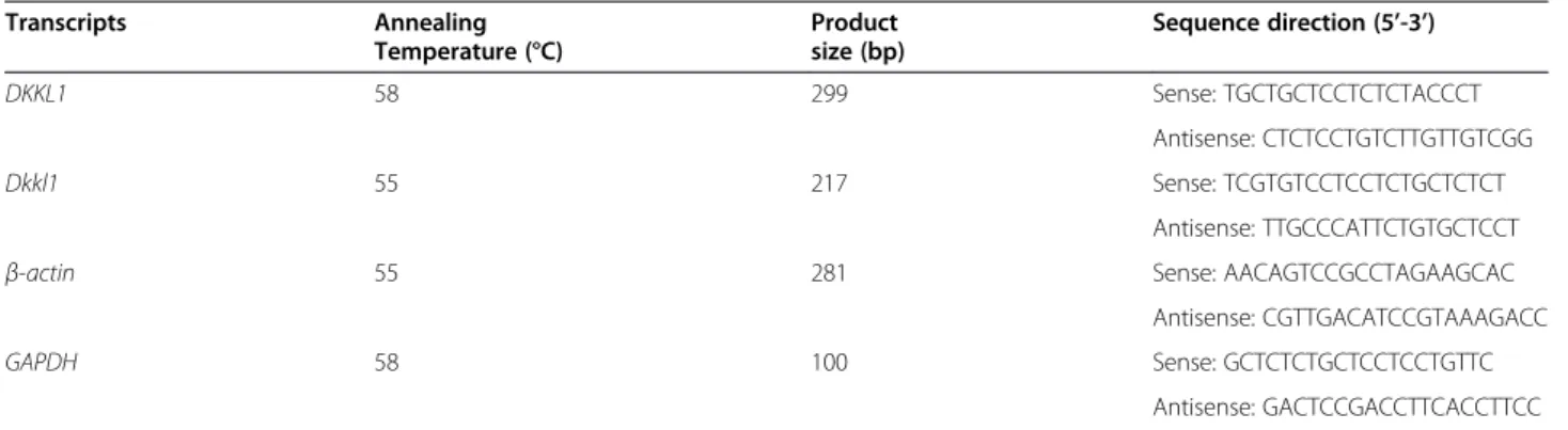
Genechips, we identified an age-dependent gene, Dkkl1 (GenBank accession number AF_177399). The hybrid-ization signal intensities from the testes of Balb/c mice on postnatal days 4, 9, 18, 35, and 54 as well as on 6 months were 1.8 (absent, or no expression on the chip, A), 9.9 (A), 1,030.5 (present, or expression on the chip, P), 2,696.8 (P), 2,987.9 (P) and 2,752.7 (P), respect-ively. That is, by Affymetrix chip analysis, the signal on days 4 and 9 was not detectable but after day 18 it gradually increased as the development of the mouse testes progressed. In comparison, the signal intensities of
β-actin were 3,688.88, 3,764.78, 3,812.9, 3,696.87, 3,679.71, and 3,757.12, respectively (Figure 1A). The
homologous human gene DKKL1 (GenBank accession
number NM_014419) was observed by hybridizing human adult or foetal testis cDNA samples with a human Affymetrix Genechip. This gene was more highly expressed in adult testis than in foetal testis. The hybridization signal intensity was 2,027.8 in adult and 5 in foetal testis, with an expression level in the adult testis approximately 405.56-fold higher than that in the foetal testis. The signal intensities ofβ-actin were 987.4 and 760.8, respectively (Figure 1B).
Expression profile ofDkkl1in mice
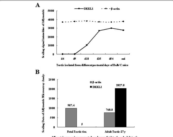
To authenticate the expression profile of Dkkl1 during
the development of the mouse testes, we performed RT-PCR and Western blot analysis using mice testes
obtained at different postnatal developmental stages.
Expression ofDkkl1was detected after day 18 and
grad-ually increased from day 18 to 54 (Figure 2A).
Further-more, he levels of Dkkl1 protein increased during
testicular development, which is consistent with the
ex-pression of Dkkl1mRNA (Figure 2C). The results from
RT-PCR and Western blot analysis were consistent with our Affymetrix chip analysis, which suggests that the ex-pression profile ofDkkl1is developmental stage specific.
The distribution of Dkkl1 was examined using
multi-tissue RT-PCR in 12 different mouse multi-tissues including brain, heart, liver, spleen, lung, kidney, muscle, stomach, intestine, bladder, testis, and epididymis. The gene was expressed at high levels in testis and at weak levels in epididymis, and was not found in the other tissues (Figure 2B).
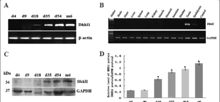
In vitro fertilization was reduced byDkkl1antibody We used in vitro fertilization assays to investigate a pos-sible role for Dkkl1 in fertilization. Successful in vitro fertilization was identified by the appearance of embryos at the 2-cell and 4-cell stages (Figure 3A). While the fertilization rate of the HTF group exceeded 57%, the
fertilization rate of the HTF+ Dkkl1 antibody group
dropped significantly to 12% (Figure 3B). This supports
the notion that Dkkl1 plays a role in fertilization and
that the Dkkl1 antibody employed in this study can
block its action.
Tissue distribution ofDKKL1mRNA and protein in humans
The expression profile of DKKL1 in various tissues was
also studied using multi-tissue RT-PCR. Of the 8 human organs tested (testis, ovary, kidney, uterus, prostate,
thyr-oidea, stomach and oesophagus),DKKL1was exclusively
expressed in the testis (Figure 4A). To examine the spe-cificity of theDKKL1antibody and confirm the results of the RT-PCR analysis, Western blot analysis was carried out on the same tissue samples. The antibody recognized a distinct band at 34 kDa, which is comparable to the
predicted molecular weight of DKKL1. The band was
only detected in human testis, suggesting thatDKKL1is
primarily expressed in human testis (Figure 4B).
Abnormal expression ofDKKL1mRNA in the testes of patients with male infertility
To investigate the contribution of DKKL1 to
spermato-genesis, we examined its expression in the testes of fer-tile and inferfer-tile men. RT-PCR results indicated that
DKKL1 was not expressed in the testes of patients with
either SCOS or cryptorchidism. DKKL1 expression in
patients with spermatogenic arrest varied. In patients
Figure 2Expression pattern ofDkkl1in mice.A: MouseDkkl1mRNA was not expressed in mouse testis on days 4 and 9 and was weakly expressed on day 18. The expression increased gradually from day 18 to 54 and remained stable after day 54.β-actinwas used as an internal loading control.B: The expression pattern ofDkkl1mRNA in 12 different mouse tissues is shown. Except for a trace amount ofDkkl1mRNA in the epididymis, expression ofDkkl1was found only in testis.GAPDHwas used as an internal control.C: Representative Western blot analyses of protein from samples obtained from testes at postnatal days 4, 9, 18, 35, and 54, as well as at 6 months. The expression ofGAPDHwas used as an internal standard for normalization. The protein level ofDkkl1increased during testicular development, which is consistent with the expression of
with arrest at the spermatogonium and primary
sperm-atocyte stages, DKKL1 expression was not detected;
however, in patients with arrest at the spermatid stage,
DKKL1 expression levels were weak or absent. In fertile
men with spermatogenic cells of every stage, theDKKL1
expression level was high. These results indicate a trend
of increasing DKKL1expression as spermatogenic cells
mature. The expression of GAPDH was comparable in the testes of all samples (Figure 5).
Expression ofDKKL1protein in the testis of infertile patients
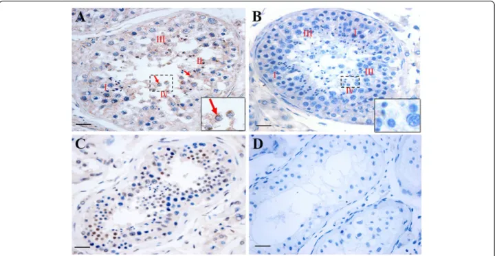
In normal testis, all stages of spermatogenic cells were found to be present in the seminiferous epithelia.
DKKL1 protein was predominantly located in the sper-matocytes and round spermatids and was not found to be located in Leydig cells or basal membranes. Following the method of Clermonts as cited in Amann’s review [22], we determined the spermatogenic stages present in
Figure 3The inhibitory effect ofDkkl1antibody onin vitrofertilization. Successful fertilization was assayed by zygote cleavage.A, Top image: spermatozoa incubated with HTF demonstrated successful fertilization; Bottom image: spermatozoa incubated with HTF +Dkkl1antibody demonstrated unsuccessful fertilization.B: The bars represent the mean ± SD of three replicate analyses, and bars marked with asterisks showed statistically significant differences (p<0.05).
the human testis samples. Further analysis indicated
that intense DKKL1 localization was observed in
stages II, III and IV of spermatogenesis, whereas it was lower in stage I (Figure 6A). In the testis from patients
with spermatogenic arrest, the expression of DKKL1
protein was significantly decreased in spermatocytes
and round spermatids (Figure 6C). No DKKL1 protein
signal was detected in testis of patients with SCOS or cryptorchidism (Figure 6D).
Discussion
It has been previously shown that spermatogenesis is mainly regulated by testis-specific gene activation. Inves-tigation of testis-specific genes is expected to lead to a broader and more thorough understanding of spermato-genesis. Many genes related to human and mouse sperm-atogenesis have been identified by our previous research [12-15]. The present study focuses on the characterization of a newly recognized gene,DKKL1/Dkkl1.
As spermatogenesis is divided into three major phases, namely proliferation and differentiation of spermato-gonia, meiosis and spermiogenesis [23], the
expres-sion patterns of Dkkl1 in mouse testes at different
developmental stages were first investigated using a gene chip approach. The six selected developmental stages represent the major stages of germ cell development during the first wave of spermatogenesis: day 4, cells with stem cell properties; day 9, spermatogonia mitosis; day 18, spermatocyte meiosis; day 35, round spermatid production and elongated spermatid formation, also called spermiogenesis; day 54, normal postpubertal spermatogenesis; 6 months, elongated spermatids and immature sperm [24]. As demonstrated in the mouse
gene chip results, the expression of Dkkl1 was detected
on days 18, 35, and 54, as well as at 6 months, but it was not detected on days 4 and 9. This was further verified by RT-PCR and Western blot analysis. Analysis revealed thatDkkl1was weakly expressed in mouse testis on day
18, with expression increasing after day 18 and remaining stable after day 54. Based on the expression
characteristics of Dkkl1 in mice, we suggest that the
expression of Dkkl1 mRNA and protein are associated
with the postmeiotic phase of spermatogenesis and with the generation of late pachytene spermatocytes and round spermatids. In addition, the results of multi-tissue RT-PCR showed that this gene was highly expressed in testis and weakly expressed in the epididymis. A possible
reason for the weak expression of Dkkl1in the
epididy-mis of mice is that some immature and mature sperm are stagnated in the epididymis.
Although Dkkl1 has been previously suggested to be
important for male fertility in vitro [25], surprisingly,
Dkkl1−/−mice are not only viable and fertile, but both
male and female Dkkl1−/− mice produce offspring at
efficiencies comparable to wild-type animals [17,26]. These studies made it impossible to evaluate the
contri-bution of Dkkl1. So what role might Dkkl1 play in
fertilization? In the present study, we used an in vitro fertilization assay to investigate the role of Dkkl1.
Fol-lowing the use of a Dkkl1 neutralizing antibody, the
fertilization rate was significantly reduced. This result is consistent with a previous study [25] that found that
Dkkl1 is required for efficient fertilization in vitro. It is
likely that Dkkl1 plays a role in the penetration of
spermatozoa into oocytes, and during this time window, the neutralizing antibody has a chance to block its ac-tion. However, IVF provides an assay for detecting fertilization-related problems that are not apparent in vivo. More likely is the possibility that a delay in
fertilization caused by the absence of Dkkl1is
compen-sated by other factors during spermatogenesis or preim-plantation development.
Given that Dkkl1was closely linked to mouse
sperm-atogenesis, what is its relationship to human spermato-genesis? To address this question, we next conducted hybridization of adult and foetal testes samples to a
Figure 5Abnormal expression ofDKKL1mRNA in the testes of patients with male infertility.Top: RT-PCR studies examinedDKKL1
expression in 15 infertile patients with SCOS (lanes 2–4), with spermatogenic arrest at various stages (lanes 5–7, arrest at spermatogonium; lanes 8–10, arrest at primary spermatocyte; lanes 11–13, arrest at spermatid), with cryptorchidism (lanes 14–16), or with normal testis(lanes 17–19). The results indicate thatDKKL1is not expressed in the testes of patients with SCOS or cryptorchidism.DKKL1expression is variable in patients with spermatogenic arrest. In patients with arrest at the spermatogonium and primary spermatocyte stage,DKKL1is not expressed, but in patients with arrest at the spermatid stage,DKKL1is weakly expressed. In normal samples containing spermatogenic cells of every stage,DKKL1
human gene chip. The results indicated thatDKKL1was expressed at a higher level in adult testis than in foetal testis. The relative hybridization signal intensity of
DKKL1 in adult testis was 405.56 times that in foetal testis. There are only Sertoli cells and undifferentiated spermatogonia cells in the seminiferous tubules of the foetal testis, whereas the seminiferous tubules of the adult testis contain not only Sertoli cells and spermato-genous cells, but also various spermatogenic cells. In other words, there are many developmental stages of germ cells represented in adult testis that are not found in foetal testis. Many genes expressed in the testis are developmental stage specific or cell type specific, which reflects the demands of tissue development [27]. It has been demonstrated that other genes with differential
ex-pression between adult and foetal testis, namely Spef1
[28], Akap4 [29], and Rosbin [30], are
spermatogenesis-specific. Thus, the results of our gene chips analysis
pro-vided an important clue thatDKKL1might be associated
with testis development and spermatogenesis in humans. Whether the expression of testis development and spermatogenesis genes were altered in male infertility? Previous study have shown that patients with SCOS or spermatogenesis arrest at spermatocyte do not express a novel human testis-specific and spermatogenesis protein
NYD-SP12 [31]. Recently, it has been reported that the
signal of the human testis developmental geneSPATA12
was not detected in patients with cryptorchidism or SCOS [32]. To further validate the function and role of
DKKL1 in male infertility, we examined the DKKL1 mRNA transcript levels as well as protein levels among the patients with SCOS, cryptorchidism or spermato-genic arrest. Most of them had no or insufficient
expres-sion of DKKL1 protein in the testes. Only the control
fertile male testis sample, in which every stage of
sper-matogenic cell was represented, fully expressed DKKL1.
The trend of increasing expression with the presence of the more mature spermatogenic cells in testis is consist-ent with the developmconsist-ent-dependconsist-ent characteristics of
DKKL1 mRNA. It was revealed that decreased
expres-sion of DKKL1 was associated with spermatogenic
fail-ure in infertile men.
Conclusions
We have provided evidence thatDKKL1/Dkkl1is
poten-tially involved in human and mouse spermatogenesis. Further investigation of molecular mechanisms, such as the distribution of DKKL1 in multiple tissues by in situ hybridization or immunohistochemical staining, and its interaction with other proteins by immunoprecipitation
Figure 6Localization and expression characteristics ofDKKL1protein in the testis of fertile and infertile patients by
or the yeast two-hybrid system, is required to determine its biological function in mammalian spermatogenesis. These studies are currently under way. Moreover, the
screening of DKKL1 gene mutations in patients with
SCOS, cryptorchidism and spermatogenic arrest by dir-ect sequencing may help us to understand the role of
DKKL1in clinical male infertility.
Competing interests
The authors declare that they have no competing interests.
Authors’contributions
QY, XW and CC participated in the design of the study, collected the materials, and carried out all experiments. QY drafted the manuscript. RD, YL, JH, JC and ZY collected the materials and helped to carry out the cDNA microarray hybridization, RT-PCR, immunohistochemistry, and Western blot analyses. AT, YG and ZC conceived of the study, participated in its design and coordination and helped to draft the manuscript. All authors read and approved the final manuscript.
Acknowledgement
We are grateful to Elsevier Language Editing Services for editorial assistance with the manuscript. We would like to thank Miss Xiaoyan Zhang, Mr. Zhenmin Zhang, Mr. Yong Wang and Mr. Wenjie Li for their technical assistance. This work was supported by grants from the National Natural Science Foundation of China (No.30972992, No.81170613, and No.81101922), the Shenzhen Important Foundation of Science & Technology (201001015), the Shenzhen Basic Research Funds for Distinguished Young Scientists, and the Qingyuan Foundation of Science & Technology (2010B001, 2010B006).
Author details
1Center for Reproductive Medicine, Department of Obstetrics and
Gynecology, The People's Hospital of Qingyuan, The Fifth Affiliated Hospital of Medical College of Jinan University, Qingyuan, China.2Guangdong Key
Lab of Male Reproductive Medicine and Genetics, Peking University Shenzhen Hospital, Shenzhen, China.3Shenzhen Second People's Hospital,
The First Affiliated Hospital of Shenzhen University, Shenzhen, China.
4Institute of Tissue Transplantation and Immunology, Jinan University,
Guangzhou, China.
Received: 16 January 2012 Accepted: 27 June 2012 Published: 21 July 2012
References
1. Hargreave TB:Genetic basis of male fertility.Br Med Bull2000,56(3):650–671. 2. Lin YH, Lin YM, Wang YY, Yu IS, Lin YW, Wang YH, Wu CM, Pan HA, Chao
SC, Yen PH,et al:The expression level of septin12 is critical for spermiogenesis.Am J Pathol2009,174(5):1857–1868.
3. Zheng Z, Zheng H, Yan W:Fank1 is a testis-specific gene encoding a nuclear protein exclusively expressed during the transition from the meiotic to the haploid phase of spermatogenesis.Gene Expr Patterns
2007,7(7):777–783.
4. Bai X, Silvius D, Chan ED, Escalier D, Xu SX,et al:Identification and characterization of a novel testis-specific gene CKT2, which encodes a substrate for protein kinase CK2.Nucleic Acids Res2009,37(8):2699–2711. 5. Hu Y, Xing J, Chen L, Guo X, Du Y, Zhao C, Zhu Y, Lin M, Zhou Z, Sha J:
RGS22, a novel testis-specific regulator of G-protein signaling involved in human and mouse spermiogenesis along with GNA12/13 subunits.Biol Reprod2008,79(6):1021–1029.
6. Kusz KM, Tomczyk L, Sajek M, Spik A, Latos-Bielenska A, Jedrzejczak P, Pawelczyk L, Jaruzelska J,et al:The highly conserved NANOS2 protein: testis-specific expression and significance for the human male reproduction.Mol Hum Reprod2009,15(3):165–171.
7. Escalier D:Impact of genetic engineering on the understanding of spermatogenesis.Hum Reprod Update2001,7(2):191–210.
8. Maduro MR, Lamb DJ:Understanding new genetics of male infertility.
J Urol2002,168(5):2197–2205.
9. Cooke HJ, Hargreave T, Elliott DJ:Understanding the genes involved in spermatogenesis: a progress report.Fertil Steril1998,69(6):989–995.
10. Xiao P, Tang A, Yu Z, Gui Y, Cai Z:Gene expression profile of 2058 spermatogenesis-related genes in mice.Biol Pharm Bull2008,
31(2):201–206.
11. Tang A, Yu Z, Gui Y, Zhu H, Zhang L, Zhang J, Cai Z:Characteristics of 292 testis-specific genes in human.Biol Pharm Bull2007,30(5):865–872. 12. Zhou Y, Qin D, Tang A, Zhou D, Qin J, Yan B, Diao R, Jiang Z, Cai Z, Gui Y:
Developmental expression pattern of a novel gene, TSG23/Tsg23, suggests a role in spermatogenesis.Mol Hum Reprod2009,15(4):223–230. 13. Yu Z, Tang A, Gui Y, Guo X, Zhu H, Long Y, Li Z, Cai Z:Identification and
characteristics of a novel testis-specific gene, Tsc21, in mice and human.
Mol Biol Rep2007,34(2):127–134.
14. Tang A, Yu Z, Gui Y, Guo X, Long Y, Cai Z:Identification and characteristics of a novel testis-specific gene, Tsc24, in human and mice.Biol Pharm Bull
2006,29(11):2187–2191.
15. Tang A, Yu Z, Gui Y, Zhu H, Long Y, Cai Z:Identification of a novel testis-specific gene in mice and its potential roles in spermatogenesis.Croat Med J2007,48(1):43–50.
16. Niehrs C:Function and biological roles of the Dickkopf family of Wnt modulators.Oncogene2006,25(57):7469–7481.
17. Dakhova O, O'Day D, Kinet N, Yucer N, Wiese M, Shetty G, Ducy P:
Dickkopf-like1 regulates postpubertal spermatocyte apoptosis and testosterone production.Endocrinology2009,150(1):404–412. 18. Krupnik VE, Sharp JD, Jiang C, Robison K, Chickering TW, Amaravadi L,
Brown DE, Guyot D, Mays G, Leiby K,et al:Functional and structural diversity of the human Dickkopf gene family.Gene1999,238(2):301–313. 19. Kohn MJ, Kaneko KJ, DePamphilis ML:DkkL1 (Soggy), a Dickkopf family
member, localizes to the acrosome during mammalian spermatogenesis.
Mol Reprod Dev2005,71(4):516–522.
20. Barraud-Lange V, Naud-Barriant N, Saffar L, Gattegno L, Ducot B, Drillet AS, Bomsel M, Wolf JP, Ziyyat A:Alpha6beta1 integrin expressed by sperm is determinant in mouse fertilization.BMC Dev Biol2007,7:102.
21. Zhuang XJ, Hou XJ, Liao SY, Wang XX, Cooke HJ, Zhang M, Han C:SLXL1, a novel acrosomal protein, interacts with DKKL1 and is involved in fertilization in mice.PLoS One2011,6(6):e20866.
22. Amann RP:The cycle of the seminiferous epithelium in humans: a need to revisit?J Androl2008,29(5):469–487.
23. Grootegoed JA, Siep M, Baarends WM:Molecular and cellular mechanisms in spermatogenesis.Baillieres Best Pract Res Clin Endocrinol Metab2000,14
(3):331–343.
24. Eddy EM:Male germ cell gene expression.Recent Prog Horm Res2002,
57:103–128.
25. Kohn MJ, Sztein J, Yagi R, DePamphilis ML, Kaneko KJ:The acrosomal protein Dickkopf-like 1 (DKKL1) facilitates sperm penetration of the zona pellucida.Fertil Steril2010,93(5):1533–1537.
26. Kaneko KJ, Kohn MJ, Liu C, Depamphilis ML:The acrosomal protein Dickkopf-like 1 (DKKL1) is not essential for fertility.Fertil Steril2009,
93(5):1526–1532.
27. Guo R, Yu Z, Guan J, Ge Y, Ma J, Li S, Wang S, Xue S, Han D:Stage-specific and tissue-specific expression characteristics of differentially expressed genes during mouse spermatogenesis.Mol Reprod Dev2004,67(3):264–272. 28. Chan SW, Fowler KJ, Choo KH, Kalitsis P:Spef1, a conserved novel testis
protein found in mouse sperm flagella.Gene2005,353(2):189–199. 29. Miki K, Willis WD, Brown PR, Goulding EH, Fulcher KD, Eddy EM:Targeted
disruption of the Akap4 gene causes defects in sperm flagellum and motility.Dev Biol2002,248(2):331–342.
30. Takahashi T, Tanaka H, Iguchi N, Kitamura K, Chen Y, Maekawa M, Nishimura H, Ohta H, Miyagawa Y, Matsumiya K,et al:Rosbin: a novel homeobox-like protein gene expressed exclusively in round spermatids.Biol Reprod
2004,70(5):1485–1492.
31. Xu M, Xiao J, Chen J, Li J, Yin L, Zhu H, Zhou Z, Sha J:Identification and characterization of a novel human testis-specific Golgi protein, NYD-SP12.Mol Hum Reprod2003,9(1)):9–17.
32. Dan L, Lifang Y, Guangxiu L:Expression and possible functions of a novel gene SPATA12 in human testis.J Androl2007,28(4):502–512.
doi:10.1186/1477-7827-10-51
Cite this article as:Yanet al.:Developmental expression and function of