PHYSICO-CHEMICAL STUDIES OF A NOVEL CADMIUM(II)
COORDINATION COMPOUND [(3-Ampy)
2CdCl
4].0.305H
2O
(3-Ampy=3-AMINOMETHYLPYRIDINE)
M. Ben Nasr,
[a]M. Zeller,
[b]F. Lefebvre,
[c]and
C. Ben Nasr
[a]*Keywords: cadmium(II) coordination compound ; MAS-NMR spectroscopy ; infrared spectroscopy; intermolecular N-H..Cl hydrogen bond
The crystal structure of the title organic-inorganic hybrid material, [(3-Ampy)2CdCl4].0.305H2O (3-Ampy=3-aminomethylpyridine),
contains two crystallographically independent but chemically equivalent cadmium complexes with essentially the same geometry (rms deviation of all atoms: 0.152 Å). The metal centers have a 6-coordinated octahedral geometry and both Cd atoms exhibit exact crystallographic inversion symmetry. Crystal packing is stabilized by intermolecular N-H...Cl hydrogen bonds that connect individual zwitter-ionic complexes into a three dimensional lattice, which is further stabilized through stacking interactions between aromatic rings of neighboring complexes, with centroid to centroid distances of 3.4406(14) and 3.7022(13) Å and interplanar separations of 3.299(1) and 3.302(1) Å. Interstitial space is partially filled with water molecules which are connected to the network through O-H...Cl and C-H...O hydrogen bonds. The 13C and 15N CP-MAS NMR spectra are in agreement with the X-ray structure. Four resonance peaks for the C3
carbon atom are observed due to the different environments of the aromatic rings caused by the presence of 0.305 water molecule per unit cell. DFT calculations allow the attribution of the carbon and nitrogen peaks to the different atoms.
* Corresponding Authors
E-Mail: [email protected]
[a] Laboratoire de Chimie des Matériaux, Faculté des Sciences de Bizerte, 7021 Zarzouna, Tunisie.
[b] Youngstown State University, Department of Chemistry, One University Plaza, Youngstown, Ohio 44555-3663, USA. [c] Laboratoire de Chimie Organométallique de Surface
(LCOMS), Ecole Supérieure de Chimie Physique Electronique, 69622 Villeurbanne Cedex, France.
Introduction
Polynuclear d10-metal complexes can exhibit important
structural and photoluminiscent properties.1 Among the d10
metals, cadmium gives rise to structural flexibility with coordination numbers varying between 4 and 8 with often severely distorted coordination geometries.2-5 A
disadvantage of the use of Cd2+ in functional materials is its
toxic effects, which are well established and documented.6
The ions have been found to induce various pathological conditions, such as e.g. cardiovascular diseases,7
hypertension, and cancer.8 It is also known, however, that
most of cadmium ions in biological systems is not in the form of free Cd2+ ions, but is coordinated by the abundance
of biological ligands therein.9-11 Therefore, the coordination
chemistry of Cd2+ ions with such ligands is of interest.
As a contribution to the investigation of the above materials, we report here the crystal structure of one such compound, [(C6H9N2)2CdCl4]0.305H2O, formed through the
reaction of 3-aminomethylpyridine with cadmium chloride and hydrochloric acid in an aqueous medium.
Experimental
Chemical preparation
324 mg (3 mmol) of 3-aminomethylpyridine and 549 mg (3 mmol) of CdCl2 were dissolved in 20 ml of HCl (2 M)
aqueous solution. The obtained solution was slowly evaporated at room temperature over six days leading to formation of transparent prismatic crystals with suitable dimensions for single crystal structural analysis (m=932 mg,
n=1.95 mmol, yield 65 %). The crystals are stable for months under normal conditions of temperature and humidity.
Investigation techniques
The characterization of this coordination compound was carried out using X-ray diffraction, solid state NMR, DFT calculations and IR spectroscopy.
X-ray single crystal structural analysis
Diffraction data were collected on a Bruker AXS SMART APEX CCD diffractometer at 100 K using monochromatic Mo K radiation with the omega scan technique. Data were collected, the unit cell determined, and the data integrated and corrected for absorption and other systematic errors using the Apex2 suite of programs.12 The structures were
solved by direct methods using Shelxs and refined by full matrix least squares against F2 with all reflections using
Shelxl13 and Shelxle.14 Carbon and nitrogen bound hydrogen
were set to 0.95 and 0.99 Å for aromatic and methylene H atoms, respectively, with Uiso values 1.2 times that of the Ueq
of the respective carrier atom. Ammonium H atoms were placed at a distance of 0.91 Å and were allowed to rotate, but not to tip, to best fit the experimental electron density. Uiso(H) were set to 1.5 times Ueq(N) of the carrier atom. A
water solvate molecule is only partially occupied with a refined occupancy rate of 0.305(6). Positions of the water H atoms were refined with a distance restraint of 0.84(2) Å, and Uiso(H) were set to 1.5 times Ueq(O). The drawings were
made with Diamond15 and Mercury.16 Crystal data and
experimental parameters used for the intensity data collection are summarized in Table 1.
Physical measurements
The NMR spectra were recorded on a solid-state high-resolution Bruker DSX-300 spectrometer operating at 75.49 MHz for 13C and 30.30 MHz for 15N with a classical 4 mm
probehead allowing spinning rates up to 10 kHz. 13C and 15N
NMR chemical shifts are given relative to tetramethylsilane and liquid ammonia, respectively (precision 0.5 ppm). The spectra were recorded by use of cross-polarization (CP) from protons (contact time 5 ms) and MAS. Before recording the spectrum it was checked that there was a sufficient delay between the scans allowing a full relaxation of the protons. The IR spectrum was recorded in the range 4000-400 cm-1 with a “Perkin-Elmer FTIR”
spectrophotometer 1000 using a sample dispersed in spectroscopically pure KBr pressed into a pellet.
Results and discussion
X-ray diffraction study
A depiction of the structure of [(C6H9N2)2CdCl4]0.305H2O is shown in Figure 1. In the
structure, there are two crystallographically independent complexes. Both complexes exhibit crystallographic inversion symmetry with the cadmium atoms located on centers of inversion at either on the face of the a-c face of the unit cell, or at 1/2 of the b-axis.
The cadmium ions are octahedrally coordinated by four equatorial chlorine atoms and two pyridinium nitroge at from two ligands at the axial sites (Fig.2).
Fig.1. Asymmetric unit of [(3-Ampy)2CdCl4]0.305H2O with the
atom numbering scheme and thermal ellipsoids at 50 % probability. Symmetry code : (i) 1-x, y, 0.5-z.
Table 1. Experimental details of [(3-Ampy)2CdCl4].0.305H2O.
The overall geometry of the two complexes is very similar, with an rms deviation of 0.152 Å. The geometry around the metal centers is virtually identical, which can be seen at the Cd-N and Cd-Cl distances: Cd-N bond lengths are 2.3878(16) and 2.3168(17) Å for Cd1 and Cd2, respectively, and Cd-Cl distances are 2.6313(6) and 2.6508(5) Å for Cd1, and 2.6365(6) and 2.6651(6) Å for Cd2 (Table 2).
These geometrical parameters agree with those of similar cadmium complexes.17 Slight differences between the two
complexes are observed for the methylene ammonium fragments, which are oriented marginally different in the two molecules (Fig. 3).
CCDC Number 961746
Crystal description Plate
Crystal size 0.39 × 0.25 × 0.12 mm
Formula [(C6H9N2)2CdCl4]0.305H2O
Formula weight 478.01
Radiation MoKα
Wavelength 0.71073 Å
Temperature 100 K
Unit cell dimensions a = 8.5313 (13) Å b = 8.6849 (13) Å c = 13.682 (2) Å
α = 74.049 (2)° β = 75.276 (3)° γ = 66.681 (2)° Crystal system, Space group Triclinic, P1
Unit cell volume 882.9 (2) Å3
No. of molecules per unit cell, Z 2
µ 1.84 mm−1
Data collection
Bruker AXS SMART APEX CCD diffractometer Absorption correction: multi-scan
Apex2 v2011.2-0 (Bruker, 2011) Tmin = 0.604, Tmax = 0.746
10570 measured reflections 5486 independentreflections 5065 reflections with I > 2σ(I) Rint = 0.014
Refinement
R[F2 > 2σ(F2)] = 0.026
wR(F2) = 0.064
S = 1.07 5486 reflections 211 parameters 2 restraints
H atoms treated by a mixture of independent and constrained refinement
Δρmax = 1.08 e Å−3
Table 2. Selected bond lengths (Å) and angles (º) of CdN2Cl4 octahedra in the title compound.
Cd1—N1 Cd2—N3 Cd1—N1i
Cd1—Cl2 Cd1—Cl2i
Cd1—Cl1 Cd1—Cl1i
Cd2—N3ii
Cd2—Cl3 Cd2—Cl3ii
Cd2—Cl4ii
Cd2—Cl4 Cl2—Cd1—Cl1i
Cl2i—Cd1—Cl1i
Cl1—Cd1—Cl1i
N3—Cd2—N3ii
N3—Cd2—Cl3 N3ii—Cd2—Cl3 N3—Cd2—Cl3ii
N3ii—Cd2—Cl3ii
Cl3—Cd2—Cl3ii
2.3878 (16) 2.3168 (17) 2.3878 (16) 2.6313 (6) 2.6313 (6) 2.6508 (5) 2.6508 (5) 2.3168 (17) 2.6365 (6) 2.6366 (6) 2.6651 (6) 2.6651 (6) 89.502 (17) 90.498 (18) 180.00 (2) 179.998 (1) 88.38 (4) 91.62 (4) 91.62 (4) 88.38 (4) 180.0
N1i—Cd1—N1 N1i—Cd1—Cl2 N1—Cd1—Cl2 N1i—Cd1—Cl2i
N1—Cd1—Cl2i
Cl2—Cd1—Cl2i
N1i—Cd1—Cl1 N1—Cd1—Cl1 Cl2—Cd1—Cl1 Cl2i—Cd1—Cl1 N1i—Cd1—Cl1i
N1—Cd1—Cl1i
N3—Cd2—Cl4ii
N3ii—Cd2—Cl4ii
Cl3—Cd2—Cl4ii
Cl3ii—Cd2—Cl4ii
N3—Cd2—Cl4 N3ii—Cd2—Cl4 Cl3—Cd2—Cl4 Cl3ii—Cd2—Cl4 Cl4ii—Cd2—Cl4
179.998 (2) 90.81 (4) 89.19 (4) 89.19 (4) 90.81 (4) 180.0 88.11 (4) 91.89 (4) 90.498 (17) 89.502 (18) 91.89 (4) 88.11 (4) 91.45 (4) 88.55 (4) 91.204 (17) 88.798 (17) 88.55 (4) 91.45 (4) 88.797 (17) 91.202 (17) 180.00 (2)
Symmetry codes: (i) −x+2, −y+1, −z; (ii) −x+1, −y+2, −z+1.
Examination of the geometric features of the organic entity shows that the organic molecule exhibits a regular spatial configuration with C-C and C-N distances and C-C-C and C-C-C-C-C-C-N angles quite similar to those found in other compounds.18
Figure 2. Geometry around the Cd(II) cation in [(C6H9N2)2CdCl4]0.305H2O. Symmetry code:(i) −x+2, −y+1, −z.
Figure 3. Least squares overlay of the two independent [(C6H9N2)2CdCl4].0.305H2O molecules, in red and blue,
respectively.
Crystal packing is stabilized by intermolecular N-H...Cl hydrogen bonds that connect individual zwitter-ionic complexes into a three dimensional lattice (Fig. 4). Among these hydrogen bonds, one has a three centered interaction N4-H4C... (Cl1, Cl2) (Table 3, for symmetry operators, see table). The 3D-network is further stabilized through two stacking interactions between aromatic rings of neighboring complexes, with centroid to centroid distances of 3.4406(14) and 3.7022(13)Å and interplanar separations of 3.299(1) and 3.302(1) Å (Fig 5). Interstitial space within the framework is partially filled with water molecules which are connected to the network through O-H...Cl and C-H...O hydrogen bonds (Table 3).
Figure 4. A projection of the structure of [(C6H9N2)2CdCl4].0.305H2O along the b-axis. The dotted lines
Table 3. Hydrogen-bond geometry (Å, º) for (C6H9N2)2CdCl4×0.305(H2O).
D—H···A D—H H···A D···A D—H···A
N2—H2A···Cl4i 0.91 2.29 3.1866 (18) 167
N2—H2B···Cl2i 0.91 2.51 3.2576 (18) 140
N2—H2C···Cl1ii 0.91 2.26 3.1577 (18) 170
N4—H4A···Cl3iii 0.91 2.29 3.1625 (18) 160
N4—H4B···Cl4iv 0.91 2.32 3.1484 (19) 152
N4—H4C···Cl2 0.91 2.35 3.1632 (18) 148
N4—H4C···Cl 1 0.91 2.73 3.2413 (19) 117
C8—H8···O1 0.95 2.22 3.013 (6) 141
C12—H12B···O1v 0.99 2.53 3.222 (7) 127
C12—H12A···O1vi 0.99 2.57 3.428 (8) 145
O1—H1B···Cl3vii 0.84 (2) 2.44 (9) 3.075 (6) 133 (11)
O1—H1A···Cl2vii 0.84 (2) 2.27 (2) 3.109 (6) 176 (12)
Symmetry codes: (i) −x+1, −y+2, −z; (ii) −x+1, −y+1, −z; (iii) x, y
−1,
z; (iv) x+1, y−1, z; (v) −x+1, −y+1, −z+1; (vi) x+1, y, z; (vii) x−1, y, z.
Figure 5. π- π stacking interaction in ([(C6H9N2)2CdCl4].0.305H2O.
NMR spectroscopy
The 13C CP-MAS NMR spectrum of the title compound is
shown on Figure 6. In the aliphatic resonance domain, the spectrum displays two resonances at 41.1 ppm and 42.5 ppm, corresponding to two methylene C atoms. This result is consistent with the presence of two organic moieties in the asymmetric unit of the compound, in agreement with the X-ray diffraction data.
Density functional theory (DFT) calculations were undertaken in order to assign the NMR resonances to the different crystallographically unequivalent carbon atoms of the unit cell. These calculations were made at the B3LYP/6-31+G* level. The different atoms were labelled as depicted below:
Three different calculations were made on the organic cation and in all cases the theoretical chemical shifts were subtracted from those of the reference (tetramethylsilane) calculated at the same level of theory:
Figure 6. 13C CP-MAS NMR spectrum of
[(C6H9N2)2CdCl4].0.305H2O. * Spinning side bands of the
aromatic peaks.
(1) Calculation of the NMR chemical shifts (with the GIAO method) by using the positions of atoms obtained by X-ray diffraction;
(2) Optimization of the positions of the protons in the above molecule and calculation of the NMR chemical shifts in this semi-optimized geometry. Indeed X-ray diffraction leads always to underestimated X-H bond lengths, due to the fact that it is sensitive to the electronic cloud and does not see the nuclei;
(3) Full optimization of all atoms and calculation of NMR chemical shifts. This calculation, compared to the above one will give indications on the steric hindrance around the organic cation and on the positions where it is the strongest.
N C4
C3
C2
N1
C6 C5
Cd Cl
Cl
C1 N2
+H3N
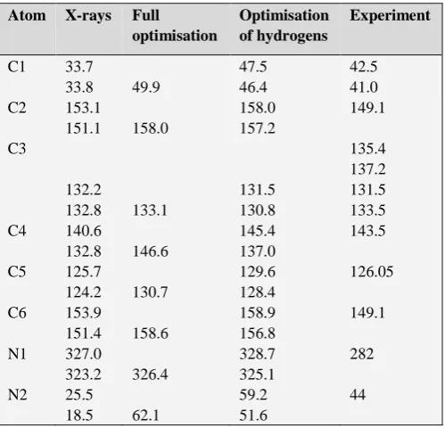
The results are listed in Table 4. Clearly, there is a good agreement between the experimental and theoretical values calculated after optimization of the position of the protons, allowing unambiguously the attribution of the different NMR signals. A key point of the 13C NMR spectrum is the
presence of four resonances for the C3 carbon atom (Fig. 7, Table 4), while only two are expected from the X-ray data. These peaks are grouped in two doublets with an intensity ratio of ca. 4:6. This point is probably related to the fact that there is only 0.305 water molecule per unit cell resulting then in different environments of the aromatic rings. Even if the difference cannot be seen by crystallography it is observed by solid state NMR, which is more sensitive to the local order, The signals of the other carbon atoms are also probably formed of doublets but the linewidths prevent their observation.
The 15N CP-MAS NMR spectrum of the title compound
(Fig.8) two resonance peaks. The first one at 44.0 ppm is related to the two aliphatic nitrogen atoms, while the second one, at 281.3 ppm corresponds to the two aromatic nitrogen atoms (Table 4).
Figure 7. Aromatic region of the experimental and simulated 13C CP-MAS NMR spectrum of
[(C6H9N2)2CdCl4].0.305H2O.
Figure 8. 15N CP-MAS NMR spectrum of
[(C6H9N2)2CdCl4].0.305H2O.
Table 4. Chemical shift values of the carbon atoms in (C6H9N2)2CdCl4×0.305(H2O).
Atom X-rays Full
optimisation
Optimisation of hydrogens
Experiment
C1 33.7
33.8 49.9
47.5 46.4
42.5 41.0 C2 153.1
151.1 158.0
158.0 157.2
149.1
C3
132.2
132.8 133.1
131.5 130.8
135.4 137.2 131.5 133.5 C4 140.6
132.8 146.6
145.4 137.0
143.5
C5 125.7
124.2 130.7
129.6 128.4
126.05
C6 153.9
151.4 158.6
158.9 156.8
149.1
N1 327.0
323.2 326.4
328.7 325.1
282
N2 25.5
18.5 62.1
59.2 51.6
44
IR Spectroscopy at room temperature
The IR spectrum of crystalline [(C6H9N2)2CdCl4].0.305H2O is shown in Figure 9. The most
representative and characteristic vibrational modes of this compound can be compared to those of similar complexes [19-21]. Some aspects of the performed assignments are briefly commented as follows:
In the high-frequency region, the broad bands between 3600 and 2500 cm-1 correspond to the valence vibrations of
C-H, N-H and O-H groups [22].
The bands in the 1630-1100 cm-1 region can be attributed
to the bending vibrations of N-H and O-H groups and to the stretching and bonding modes (C=C), (C-C), (C-N), (C-H) of the aromatic ring [23, 24].
The bands between 1000 and 600 cm-1 are assigned to the
out of plane bending modes γ(Cary-H), γ(Cary-C) and
γ(N-H).25
Conclusion
A new Cd(II) complex, [(C6H9N2)2CdCl4]0.305H2O, was
synthesized in an aqueous medium and characterized by various physico-chemical methods. On the structural level, the metal centers have 6-coordinated octahedral geometry and both Cd atoms exhibit exact crystallographic inversion symmetry. Crystal packing is stabilized by intermolecular N-H...Cl hydrogen bonds that connect individual zwitter-ionic complexes into a three dimensional lattice, which is further stabilized through stacking interactions between aromatic rings of neighboring complexes. Interstitial space is partially filled with water molecules which are connected to the network through O-H...Cl and C-H...O hydrogen bonds. The 13C and 15N CP-MAS NMR spectra are in
agreement with the X-ray structure.
Supplementary data
Crystallographic data for the structural analysis have been deposited with the Cambridge Crystallographic Data Centre, CCDC No 961746. These data can be obtained free of charge via http://www.ccdc.cam.ac.uk/conts/retrieving.html, or from the CCDC, 12Union Road, Cambridge, CB2 1EZ, UK: fax: (+44) 01223-336-033; e-mail: [email protected].
Acknowledgments
We would like to thank the Tunisian Secretariat of State for Scientific Research and Technology for its financial support. The diffractometer was funded by NSF grant 0087210, by Ohio Board of Regents grant CAP-491, and by YSU.
References
1Tao, J., Yin, X., Jiang, L. F., Huang, R. B., Zheng, L. S., Eur.J.
Inorg. Chem., 2003, 2678.
2Wang, X.-Li, Yang, S., Liu, G.-C., Zhang, J.-X, Lin, H.-Y., Tian,
A.-X., Inorg. Chim. Acta, 2011, 375, 70.
3Subashini, A., Muthiah, P. T., Bocelli, G., Cantoni, A. Acta
Cryst., 2008, E64, m250.
4Xiao, J.-M., Acta Cryst., 2010, C66, m348.
5Kaabi, K., El Glaoui, M., Silva, S. P., Silva, M. R., Ben Nasr, C.,
Acta Cryst., 2010, E66, m617.
6Filck, D. F., Kraybill, H. F., Dimitroff, J. M., Environ. Res., 1971,
4, 71
7Caroll, P. E., J. Am. Med. Assoc., 1966, 198, 267.
8Schroeder, H. S ., Balassa, J. J., Am. J. Physiol., 1965, 209, 433.
9Dressing, S. A., Mass, R. P., Weiss, C. M., Bull. Environ.
Contam. Toxicol., 1982, 28, 172.
10Titus, J. A., Pfister, R. M., Bull. Environ. Contam. Toxicol., 1982,
28, 703.
11Hung, Y.W., Bull. Environ. Contam. Toxicol., 1982, 28, 546.
12Apex2 v2011.2-0, Bruker Advanced X-ray Solutions, Bruker
AXS Inc., Madison, Wisconsin, USA.
13Sheldrick, G. M., Acta Cryst., 2008, A64, 112.
14Hübschle, C. B., Sheldrick, G. M., Dittrich, B., J. Appl. Cryst.,
2011, 44, 1281.
15Brandenburg, K., Diamond Version 2.0 Impact GbR, Bonn,
Germany, 1998.
16Macrae, F., Bruno, I. J., Chisholm, J. A., Edgington, P. R.,
McCabe, P., Pidcock, E., Rodriguez-Monge, L., Taylor, R., Van de Streek, J., Wood, P. A., J. Appl. Cryst., 2008, 41, 466.
17Liang, W.-X., Qu, Z.-R., Acta Cryst., 2008, E64, m1254. 18Ha, K., Acta Cryst., 2012, E68, m176.
19Calve, N.L., Romain, F., Limage, M.H., Novak, A., J. Mol.
Struct., 1989,200, 131.
20Ratajczak, H. J., J. Mol. Struct., 1969, 3, 27. 21Navak, A., J. Mol. Struct., 1990, 217, 35.
22Smirani, W., Ben Nasr, C., Rzaigui, M., Mat. Res. Bull., 2004, 39,
1103.
23Kaabi, K., Rayes, A., Ben Nasr, C., Rzaigui, M., Lefebvre, F.,
Mat. Res. Bull., 2003, 38, 741.
24Oueslati, A., Ben Nasr, C., Durif, A., Lefebvre, F., Mat. Res.
Bull., 2005, 40, 970.
25Oueslati, A., Rayes, A., Ben Nasr, C., Lefebvre, F., Mat. Res.
Bull., 2005, 40, 1680.
![Table 1. Experimental details of [(3-Ampy)2CdCl4].0.305H2O.](https://thumb-us.123doks.com/thumbv2/123dok_us/7833618.2089623/2.595.49.283.535.714/table-experimental-details-of-ampy-cdcl-h-o.webp)

![Figure 5. π- π stacking interaction in ([(C6H9N2)2CdCl4].0.305H2O.](https://thumb-us.123doks.com/thumbv2/123dok_us/7833618.2089623/4.595.54.249.287.444/figure-p-p-stacking-interaction-c-h-cdcl.webp)