559
© 2018 by the Serbian Biological Society How to cite this article: Hrnčić D, Grubač Ž, Šutulović N, Rašić-Marković A, Ademović A, Stanojlović O. Modulatory effects of delta sleep-inducing peptide in a lindane model of generalized seizures. Arch Biol Sci. 2018;70(3):559-66.
Modulatory effects of delta sleep-inducing peptide in a lindane model of generalized
seizures
Dragan Hrnčić, Željko Grubač, Nikola Šutulović, Aleksandra Rašić-Marković, Anida Ademović and Olivera Stanojlović*
Laboratory of Neurophysiology, Institute of Medical Physiology “Richard Burian”, Faculty of Medicine, University of Belgrade, 11000 Belgrade, Serbia
*Corresponding author: [email protected]
Received: January 9, 2018; Revised: February 7, 2018 Accepted: March 30, 2018; Published online: April 19, 2018
Abstract: Delta sleep-inducing peptide (DSIP) is an endogenous peptide that is constantly present in several different brain regions. Lindane is used as a pesticide and scabicide, but it also induces seizures refractory to conventional antiepi-leptics. The aim of this paper was to determine whether DSIP modulates lindane-induced seizures in rats in a behavioral and electroencephalographic (EEG) study. DSIP (1 mg/kg, i.p.) or dimethyl-sulfoxide (DMSO, 0.5 ml/kg, intraperitone-ally (i.p.)) were injected 30 min before lindane (8 mg/kg, i.p.) to adult male rats with previously implanted electrodes for EEG registration. During the following 30 min, the EEG was registered, and the following behavioral characteristics of seizures were observed: incidence, latency and intensity. A descriptive scale with grades from 0 to 4 provided an estimate of seizure intensity. In the EEG, the number and duration of ictal periods were analyzed using NeuroSciLaBG (Belgrade, Serbia) software. The lethality rate was also analyzed. DSIP-treated animals showed significantly modified characteristics of lindane-induced seizures when compared to the group without DSIP pretreatment (i.e. a reduced seizure intensity and a prolonged seizure latency period). However, no significant effects of DSIP on seizure incidence and lindane-induced lethality were observed. EEG analyses showed a significantly decreased number of lindane-induced EEG ictal periods in DSIP-treated animals, but with unaltered duration. These results show that DSIP favorably modulates lindane-induced seizures in rats, showing a potential to be an adjuvant component of antiepileptic treatment strategy for refractory seizures. Key words: behavior; delta sleep-inducing peptide (DSIP); EEG; epilepsy; refractory seizures lindane
INTRODUCTION
Epilepsy is one of the most common neurological dis-orders [1,2] with an estimated incidence of 50 cases per 100000 persons annually [3]. The total annual costs of epilepsy treatment is about 15.5 billion euros in Europe [4,5]. The majority of these costs are due to antiepileptic drug treatment, especially in cases with refractory forms requiring polytherapy. Epilepsy is caused by a sudden and excessive hyperactivity of neurons, which is the consequence of excitatory over inhibitory phenomena in the central nervous system (CNS). Usually, it is characterized by distinctive mo-tor manifestations appearing as seizures [1]. Major excitatory phenomena are related to glutamate and its receptors, while major inhibitory phenomena are related to γ-aminobutyric acid (GABA) and its recep-tors. Nonetheless, it has been proven that a variety of
alternative mechanisms are also involved in the patho-physiology of epileptic seizures, such as mutations of ion channel genes, events related to glial cells, other neurotransmitters and neuromodulator disarrange-ments [2].
for sleep and epileptiform activity, increased synchro-nization during slow-wave sleep (SWS), and more complex interactions that depend on the intrinsic physiological and pathophysiological characteristics [11,12]. The observed global decrease in excitability across sleep appears to be differentially regulated by SWS and REM sleep. As reviewed elsewhere [13,14], it has been shown that neocortical and hippocampal neurons decrease firing activity during REM sleep. Corticothalamic projections from the major extrinsic input to the GABAergic reticular thalamic nucleus determine the generation of synchronized activities in the thalamus, which has a pacemaker role in the electrogenesis of thalamic oscillations [15]. When
GABAA-receptor-mediated inhibition is reduced,
spin-dle-like oscillations are replaced by slower spike- and slow-wave types of discharges [11], showing that sleep and epilepsy, especially in terms of sleep spindles and ictal phenomena, are correlated by the dysfunction of excitatory and inhibitory mechanisms.
Delta sleep-inducing peptide (DSIP) is an endoge-nous and somnogenic nonapeptide (Trp-Ala-Gly-Gly-Asp-Ala-Ser-Gly-Glu) [16]. DSIP was discovered by
Monnier et al. [16] who found that it increases delta
activity in an EEG. Shandra et al. [17,18] showed that a neuroprotective mechanism of DSIP probably func-tions through the reduction of excitatory amino acids and the blockage of calcium channels on the post-synaptic membrane. The beneficial effects of DSIP have been presented in different studies dealing with epilepsy, stress and arterial hypertension [19-21].
Lindane (γ-hexachlorocyclohexane) is an organo-chlorine pesticide and scabicide still widely used in ag-riculture, but also in human and veterinary medicine, despite the fact that it was prohibited by the Stockholm Convention because of its neurotoxicity [22]. Lindane is used in underdeveloped and developing countries [23]. Symptoms of toxicity with lindane range from headaches and vertigo to pronounced seizures and death [24]. These symptoms are considered to be the
result of GABA type A (GABAA) receptor blockage,
which reverses the inhibitory effect of GABA on lo-cal discharge and paroxysmal discharge spread [25]. Apart from CNS effects, lindane has a strong toxic impact on numerous other organ systems: the cardio-vascular, gastrointestinal, reproductive systems, as well as on endocrine glands [26]. In order to examine its
mechanisms of action, and the possibility of antiepi-leptic therapy, a model of lindane-induced seizures in rats has been developed [25]. Due to the well-known limitations of clinical studies of epilepsy, the results obtained from animal-based experiments can help in the selection of promising combinations of antiepi-leptic drugs, which beside anticonvulsive activity also have minimal side effects [27]. The lindane model is refractory to numerous classical antiepileptic drugs, like carbamazepine, phenytoin and felbamate [28].
Lindane-induced seizures are predominantly the result of impaired GABAergic neurotransmission, while DSIP is capable of acting neuroprotectively by enhancing GABAergic neurotransmission and reduc-ing excitatory amino acids. Bearreduc-ing this in mind, it is reasonable to examine whether DSIP is capable of beneficially modulating lindane-induced seizures. To this end, we performed the current study with the aim of studying the effects of acute systemic application of DSIP on behavioral signs and on EEG manifestations of lindane-induced seizures in rats, an animal model resembling refractory seizures.
MATERIALS AND METHODS
Animals
All experimental procedures were in full compliance with the European Council Directive (2010/63/EU) and approved by the Ethical Committee of the University of Belgrade (Permission No 298/5-2). Adult male Wi-star albino rats (2 months old, 200-230 g body weight (b.w.)) were used in the study (obtained from the Mili-tary Medical Academy Breeding Laboratory, Belgrade, Serbia). The animals were housed in transparent plastic
cages with ad libitum access to food (Purina rat chow)
and water. They were kept in a sound-attenuated
cham-ber under controlled ambient conditions (22-23oC,
50-60% relative humidity, 12/12 h light/dark cycle with light switched on at 8 a.m.) and habituated to handling. The acclimatization period lasted for 7 days.
Drugs
Experimental groups
The following experimental groups were formed based on our previous results [25] and preliminary experiments: (i) control (group C; DMSO, n=6); (ii) DSIP 1 mg/kg (DSIP, n=8); (iii) lindane 8 mg/kg (L, n=10); (iv) DSIP 1 mg/kg 30 min prior to lindane ad-ministration 8 mg/kg (DSIP+L, n=8). All drugs were freshly dissolved in saline and administered (i.p.) in a volume of 0.1 ml/100 g rat b.w.
Behavioral recordings
Rats that were placed in separate transparent plastic wired-covered cages were observed for 30 min for behavioral manifestations of lindane-induced sei-zures. These were assessed by the incidence of mo-tor seizures and their intensity, as well as duration of latency. Seizure intensity was qualified by a modified descriptive rating scale [29] with grades defined as: grade 1 – head nodding, lower jaw twitching; grade 2 – myoclonic body jerks (hot-plate reaction), bilat-eral forelimb clonus with full rearing (Kangaroo po-sition); grade 3 – progression to generalized clonic convulsions followed by tonic extension of fore and hind limbs and tail; grade 4 – prolonged severe
tonic-clonic convulsions lasting over 10 s (status epilepticus)
or frequent repeated episodes of clonic convulsions for an extended period of time (over 5 min). Latency to seizure was defined as the time from the lindane injection to the first seizure response and was also recorded. For rats without seizures, a 30-min latency time was scored. Lethality was recorded at the end of the observation period.
Surgery
The rats were anesthetized with pentobarbital sodium (50 mg/kg, i.p.). Three gold-plated recording elec-trodes were implanted over the frontal, parietal and occipital cortices in a stereotaxic apparatus. Dental acrylic cement was used to fix this system to the skull. Animals had a recovery period of one week prior to further experiments. A 24-h-long habituation to the recording situation was also applied.
EEG recording
An 8-channel EEG apparatus (RIZ, Zagreb, Croatia) was used. The signals were digitized using a SCB-68 data acquisition card (National Instruments Co, Austin, Texas, USA). A sampling frequency of 512 Hz/chan-nel and 16-bit A/D conversion were used for the EEG signals. The cutoff frequencies for EEG recordings were set at 0.3 Hz and 100 Hz for the high-pass and low-pass filters, respectively. Ambient noise was eliminated us-ing a 50-Hz notch filter. Data acquisition and signal processing were performed with LabVIEW platform software developed in the Laboratory (NeuroSciLaBG [29]). EEGs were recorded in freely moving rats dur-ing a 30-min recorddur-ing session. EEG traces were visu-ally inspected and analyzed during subsequent offline analysis. The power spectra density (obtained by the Fast Fourier transformation method) of the charac-teristic epochs was plotted and the integrated energy
signals expressed as μV2/Hz. Ictal periods in EEG were
defined as follow: (i) spontaneous and generalized spik-ing activity; (ii) lastspik-ing>1 s; (iii) amplitude of at least twice the background EEG activity [29]. The number and duration of ictal periods were calculated during a 30-min period after lindane administration.
Data analysis
Significance of the differences in the seizure inci-dence and lethality were evaluated by Fisher’s exact probability test. Since the normal distribution of the data on seizure latency, intensity of seizure, as well as the number and duration of ictal periods in EEG were not estimated by Kolmogorov-Smirnov test, nonparametric analysis (Mann Whitney U test) was used to determine the statistical significance of the differences between the groups (*p<0.05, **p<0.01).
The results were expressed as medians with 25th and
75th percentiles.
RESULTS
Behavior assessment
any signs of seizures. The incidence of seizures in the group of rats treated with lindane at a convulsive dose (8 mg/kg, i.p., group L) was 80%. In the group of rats which received DSIP (1 mg/kg, i.p.) prior to lindane administration (group DSIP+L), the seizure incidence was lowered to 62.5%. However, according to Fisher’s exact probably test, this difference between DSIP+L and L groups regarding seizure incidence did not at-tain statistical significance (p>0.05, Fig. 1A).
Beside seizure incidence, we analyzed the duration of the latency period to the first sign of seizure and seizure intensity as parameters of convulsive behavior. DSIP administered prior to lindane (group DSIP+L) led to a significant prolongation of the latency period to the first sign of seizure when compared to group L
(DSIP+L vs L, p<0.05 as assessed by the Mann
Whit-ney U test; Fig. 2A). The same holds true regarding seizure intensity. Seizure intensity was significantly lower in the DSIP+L group compared to the L group
(DSIP+L vs L, p<0.05 as assessed by the Mann
Whit-ney U test, Fig. 2B). The maximal seizure intensity in group of rats treated by lindane (group L) was grade 4, while there were no seizures of grade 4 in the group of rats treated with DSIP prior to lindane (group DSIP+L, maximal seizure intensity was grade 3).
A lethal outcome was observed in 30% of animals from group L, and in 25% of animals in group DSIP+L at the end of the observation period. This difference in lethality between groups DSIP+L and L was not statistically significant (p>0.05, Fisher’s exact prob-ability test, Fig. 1B).
EEG analysis
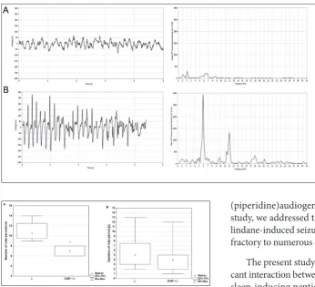
Bioelectrical brain activity registered by EEG in all vehicle-treated rats (group C), as well as in DSIP-treated rats (group DSIP) were without any signs of ictal activity. During the registration procedure the rats were quiet but awake (Fig. 3A). Spontaneous, gen-eralized and sporadic ictal activity (defined as ictal periods, a typical one is presented on Fig. 3B) was recorded in rats treated by lindane alone (group L) and those treated with DSIP prior to lindane (group DSIP+L). Offline analysis of the EEG recordings in-cluded an assessment of the number and duration of these manually identified ictal periods defined accord-ing to criteria described in the Materials and Methods
section. The number of ictal periods in rats treated with lindane alone (group L) ranged from 9 to 14 per rat, while this number ranged from 6 to 8 per rat in the group of rats treated with DSIP prior to lindane (group DSIP+L). Moreover, the median number of ictal periods was significantly lower in the DSIP+L group when compared to the L group according to the Mann Whitney U test (p<0.05, Fig 4A). On the other Fig. 1. The effects of DSIP on the incidence of seizures (A) and lethality (B) induced by lindane in rats. Lindane was administered in a dose of 8 mg/kg (L group). DSIP (1 mg/kg) was administered 30 min prior to lindane (DSIP+L). Seizure incidence: the num-ber of convulsing animals in the group out of the total numnum-ber of animals in the group, expressed in percentages. Lethality was registered at the end of the observation period. Significance of the difference was determined by Fisher’s exact probability test.
hand, the minimal duration of one ictal period was 2 s in the L group and 1 s in the DSIP+L group, while the maximal duration of one ictal period was 13 s in the L group and 12 s in the DSIP+L group. Median duration of ictal periods was not significantly
differ-ent between these two groups (DSIP+L vs L, p>0.05,
Mann Whitney U test, Fig 4B).
DISCUSSION
DSIP is a natural and ubiquitous somnogenic nona-peptide, which has no side effects even at very high doses. DSIP takes part in numerous non-sleep related physiological functions; its antiepileptic role [30,31] was of most interest to us. In our laboratory, we ex-amined DSIP as an anticonvulsive neuropeptide in metaphit (1-(1(3-isothiocyanatophenyl)-cyclohexyl
(piperidine)audiogenic epilepsy [30]. In the current study, we addressed the ability of DSIP to act against lindane-induced seizures, a seizure model which is re-fractory to numerous classical antiepileptic drugs [28]. The present study showed that there was a signifi-cant interaction between the effect of lindane and delta sleep-inducing peptide on the CNS of experimental animals. Actually, DSIP administrated 30 min prior to lindane exhibited a tendency to reduce the incidence of lindane-induced seizures, but the achieved reduction was not statistically significant. Furthermore, DSIP significantly reduced seizure intensity and prolonged the latency period of lindane-induced seizures. More-over, EEG analyses showed significant reduction of the number of ictal periods after DSIP administration, without significant alteration in ictal period duration. Thereby the behavioral and electrical parameters of hyperexcitability caused by lindane were reduced by the protective effects of neuropeptide DSIP.
Epileptic seizures arise from an excessively syn-chronous and sustained discharge of a group of neu-rons, i.e. increased neuronal excitability. Hyperexcit-ability can be caused by alterations in the membrane and metabolic properties of neurons. Therefore, seizure initiation is characterized by high-frequency bursts of action potentials and hypersynchronization of a neuro-nal population with a paroxysmal depolarizing shift in its pathophysiological basis. These synchronized bursts originating simultaneously from numerous neurons, result in ictal activity in the EEG (reviewed in detail Fig. 3. Representative EEG trac-es (left panels) with corrtrac-espond- correspond-ing power spectra diagrams (right panels) in control (A) and lindane-treated rat (B). EEGs were recorded in freely mov-ing rats. Correspondmov-ing power spectra densities were obtained by fast Fourier transformation. Note the burst of spiking activ-ity in the B trace representing a characteristic ictal period in-duced by lindane in rats. Lead: left frontal, right parietal. Am-plitude calibration: 50 µV, time calibration: 1 s. For details see caption to Fig. 1.
elsewhere [2,32]). Herein we quantified and analyzed the number and duration of ictal periods in an EEG induced by lindane and modified by DSIP.
It was shown that lindane induces generalized epileptic seizures in rats in a dose-dependent man-ner, which is distinctive on EEG [25]. The character-istics of lindane-induced seizures are similar to those of kainate-induced seizures [31]. The mechanism by which lindane induces neuronal hyperexcitability and neurotransmitter concentration changes in the neural structures remains unclear, but it is believed that the main effect of lindane is manifested through blockage
of GABAA chloride channels [34, 35]. Furthermore,
the effect on calcium mobilization is certainly one of the contributing factors [36]. We have shown the in-volvement of NO-signaling in lindane proconvulsive effects [29]. However, the latest research has shown that lindane-induced seizures are refractory to numerous antiepileptic drugs (carbamazepine, phenytoin, fel-bamate) [28]. According to the results of the present research, DSIP affected the parameters of convulsive behavior, especially the latency period and the intensity of seizures, as well as the EEG manifestations of lindane proconvulsive effects. The frequency of defined ictal periods was lowered when DSIP preceded lindane, but the duration of these periods remained unaltered. Delta waves in the EEG are those with the smallest frequen-cies and indeed, DSIP-induced delta activity modulated the electrical events in neurons caused by lindane.
Numerous steps have been taken in order to clar-ify DSIP mechanisms of action. DSIP optimizes the excitation/inhibition relationship [36]. Namely, it po-tentiates GABA-activated currents in the hippocampal and cerebellar neurons in rats [37], which could be one of the major mechanisms of action in the model of lindane-induced seizures. On the other hand, Shandra et al. [18] believe that the neuroprotective role of DSIP on NMDA receptors functions through the reduction
of excitatory amino-acids and decrease of Ca2+ influx.
It is important to know that DSIP also affects the ac-tivity of transaminases [38].
Epileptogenesis is also a consequence of oxida-tive stress [39,40]. DSIP optimizes the prooxidant-antioxidant balance by increasing the activities of antioxidant enzymes and the levels of antioxidants [41,42]. These effects of DSIP on antioxidant capaci-ties could be explained by increased expression of
genes for superoxide dismutase 1 and glutathione peroxidase 1, which were evoked by prolonged DSIP treatment [43]. In conjunction with the role of DSIP in amelioration of oxidative stress is its role in the stabi-lization of cell membranes. DSIP increases membrane stability, changes its selective permeability and inhibits the accumulation of lipid peroxidation products by modulating the physicochemical characteristics of the membrane [44]. DSIP has been shown to adopt a compact conformation which permeates the lipid bilayer due to reduced charge, increased lipophilic-ity and intramolecular hydrogen-bond stabilization [45]. Its neuroprotective effects could be related to the role of DSIP in the stabilization of the structure and functioning of neuronal membranes [46]. It is also important to emphasize that DSIP has a tranquil-izing effect on neurons, especially on glutaminergic hippocampal neurons and the neurons of the front hypothalamic nucleus [47].
Careful examination of convulsive behavior param-eters suggests a beneficial modulatory role for DSIP in the lindane model of seizures. However, since the effect of DSIP on lindane seizure incidence was minor, it is not plausible to regard DSIP as a powerful antiepileptic drug for a monotherapy regime in refractory cases. On the other hand, DSIP can be regarded as a valuable add-on drug in the treatment of refractory epilepsies. The following findings additionally support this view: (i) the synergic effect of DSIP as a native neuropeptide together with valproate (a classical antiepileptic drug) in the treatment of the epileptic seizures; (ii) results of numerous studies where the antiepileptic role of DSIP was shown on models of generalized epilepsy induced by picrotoxin, corasol, kainate, NMDA and metaphit [18,30,46,47]; (iii) DSIP, unlike other anticonvulsants drugs, has no side effects, as even an overdose has no negative effect on the CNS [50].
which DSIP is combined with glycine for intranasal administration of an aqueous solution [53]. Subse-quent studies demonstrated geroprotective, anticar-cinogenic and neuroprotective effects of DSIP and Deltaran [53,54].
The results of this research show that DSIP can be considered as an integral constituent of treatments of generalized epilepsy. Among numerous efforts to find an effective add-on drug for alleviating epilepsy with minimal side effects, this research provides a step for-ward.
Acknowledgments: This work was supported by the Ministry of Education, Science and Technological Development of Serbia (grant #175032).
Author contributions: DH, ŽG, OS designed the experiment, performed the experiments and drafted the manuscript. NŠ and AA contributed to the behavioral studies and the draft of the manuscript. ARM contributed to the EEG studies and manuscript draft. All authors reviewed and approved the final manuscript text. Conflict of interest disclosure: The authors do not have conflict-ing interests.
REFERENCES
1. Fisher RS, van Emde Boas W, Blume W, Elger C, Genton P, Lee P, Engel J Jr. Epileptic seizures and epilepsy: defini-tions proposed by the International League Against Epilepsy (ILAE) and the International Bureau for Epilepsy (IBE). Epi-lepsia. 2005;46(4):470-2.
2. Chang BS, Lowenstein DH. Epilepsy. N Engl J Med 2003;349(13):1257-66.
3. Ngugi AK, Kariuki SM, Bottomley C, Kleinschmidt I, Sander JW, Newton CR. Incidence of epilepsy: systematic review and meta-analysis. Neurology. 2011;77(10):1005-12. 4. Pugliatti M, Beghi E, Forsgren L, Ekman M, Sobocki P.
Esti-mating the cost of epilepsy in Europe: a review with eco-nomic modeling. Epilepsia. 2007;48(12):2224-33.
5. Beghi E, Berg A, Carpio A, Forsgren L, Hesdorffer DC, Hauser WA, Malmgren K, Shinnar S, Temkin N, Thurman D, Tomson T. Coment on epileptic seizures and epilepsy: definitions proposed by the International League Against Epilepsy (ILAE) and the International Bureau for Epilepsy (IBE). Epilepsia. 2005;46(10):1698-9.
6. Strine TW, Chapman DP. Associations of frequent sleep insufficiency with health-related quality of life and health behaviors. Sleep Med. 2005;6(1):23-7.
7. Hindmarch I , Dawson J, Stanley N. A double-blind study in healthy volunteers to assess the effects on sleep of pre-gabalin compared with alprazolam and placebo. Sleep. 2005;28(2):187-93.
8. Drake ME Jr, Pakalnis A, Bogner JE, Andrews JM. Outpa-tient sleep recording during antiepileptic drug monotherapy. Clin Electroencephal. 1990;21(3):170-3.
9. Legros B, Bazil CW. Effects of antiepileptic drugs on sleep architecture: a pilot study. Sleep Med 2003;4(1):51-5. 10. Placidi F, Diomedi M, Scalise A, Marciani MG, Romigi A,
Gigli GL. Effect of anticonvulsants on nocturnal sleep in epilepsy. Neurology. 2000;54(5):25-32.
11. Sinha SR. Basic mechanisms of sleep and epilepsy. J Clin Neurophysiol 2011;28(2):103-10.
12. Jiruska P, de Curtis M, Jefferys JG, Schevon CA, Schiff SJ, Schindler K. Synchronization and desynchroniza-tion in epilepsy: controversies and hypotheses. J Physiol. 2013;591(4):787-97.
13. Niethard N, Burgalossi A, Born J. Plasticity during sleep is linked to specific regulation of cortical circuit activity. Front Neural Circuits. 2017;11:65.
14. Halász P. How sleep activates epileptic networks? Epilepsy Res Treat. 2013;2013:425-697.
15. Coenen AM, Van Luijtelaar EL. Genetic animal models for absence epilepsy: a review of the WAG/Rij strain of rats. Behav Genet. 2003;33(6):635-55.
16. Monnier M, Dudler L, Gächter R, Schoenenberger GA. Delta-sleep-inducing peptide (DSIP): EEG and motor activ-ity in rabbits following intravenous administration. Neurosci Lett. 1977;6(1):9-13.
17. Sudakova KV, Umriukhina PE, Rayevskyb KS. Delta-sleep inducing peptide and neuronal activity after glutamate microiontophoresis: the role of NMDA-receptors. Patho-physiol. 2004;11(2):81-6.
18. Shandra AA, Godlevskii LS , Brusentsov AI, Karlyuga VA. Effects of delta-sleep-inducing peptide on NMDA-induced convulsive activity in rats. Neurosci Behav Physiol. 1998;28(6):694-7.
19. Sudakov KV, Coghlan JP, Kotov AV, Salieva RM, Polyntsev YuV, Koplik EV. Delta-sleep inducing peptide sequels in mechanisms of resistance to emotional stress. Ann N Y Acad Sci. 1995;771:240-51.
20. Khvatova EM, Samartzev VN, Zagoskin PP, Prudchenko IA, Mikhaleva II. Delta sleep inducing peptide (DSIP): effect on respiration activity in rat brain mitochondria and stress protective potency under experimental hypoxia. Peptides. 2003;24(2):307-11.
21. Yehuda S, Carasso RL. DSIP--a tool for investigating the sleep onset mechanism: a review. Int J Neurosci 1988;38(3-4):345-53.
22. Secretariat of the Stockholm Convention. Report of the Conference of the Parties of the Stockholm Convention on Persistent Organic Pollutants on the work of its fourth meeting. Geneva (Switzerland): Secretariat of the Stockholm Convention on Persistent Organic Pollutants; 2009 May. 24 p. Report No.:UNEP/POPS/COP.4/6/Rev.1
24. Centers for Disease Control and Prevention (CDC). Unin-tentional topical lindane ingestions-United States. 1998-2003. MMWR Morb Mortal Wkly Rep. 2005;54(21):533-5. 25. Vucević D, Hrncić D, Radosavljević T, Mladenović D,
Rasić-Marković A, Loncar-Stevanović H, Djurić D, Macut D, Susić V, Stanojlović O. Corellation between electrocorticographic and motor phenomena in lindane induced experimental epilepsy in rats. Can J Physiol Pharmacol. 2008;86(4):173-9. 26. Nolan K, Kamrath J, Levitt J. Lindane toxicity: a
compre-hensive review of the medical literature. Pediatr Dermatol. 2012 ;29(2):141-6.
27. Czuszwar SJ, Borowicz KK. Polytherapy in epilepsy: the experimental evidence. Epilepsy Res 2002;52(1):15-23. 28. Tochman AM , Kaminski R , Turski WA , Czuczwar SJ.
Pro-tection by conventional and new antiepileptic drugs against lindane-induced seizures and lethal effects in mice. Neuro-tox Res. 2000;2(1):63-70.
29. Hrnčić D, Rašić-Marković A, Djuric D, Stanojlović O. The role of nitric oxide in convulsions induced by lindane in rats. Food Chem Toxicol. 2011;49(4):947-54.
30. Stanojlović OP, Živanović DP, Mirković SD, Mikhaleva II. Antiepileptic activity of delta sleep inducing peptide and its analogue in metaphit-provoked seizures in rats. Seizures. 2005;14:240-7.
31. Kovalzon VM, Strekalova TV. Delta sleep-inducing pep-tide (DSIP): a still unresolved riddle. J Neurochem. 2006;97(2):303-9.
32. Engelborghs S, D’Hooge R, De Deyn PP. Pathophysiology of epilepsy. Acta Neurol Belg. 2000;100(4):201-13.
33. Suñol C, Tusell JM, Gelpí E, Rodríguez-Farré E. Convulsant effect of lindane and regional brain concentration of GABA and dopamine. Toxicology. 1988;49(2-3):247-52.
34. Anand M., Agrawa AK, Rehmani BNH. Role of GABA receptor complex in low dose lindane (HCH) induced neu-rotoxicity: neurobehavioural, neurochemical and electro-physiological studies. Drug Chem Toxicol. 1998;21(1):35-46. 35. Suñol C, Vale C, Rodríguez-Farré E. Polychlorocycloalkane
insecticide action on GABA- and glycine-dependent chlo-rine flux. Neurotoxicology. 1998;19(4-5):573-80.
36. Rosa R, Sanfeliu C, Suñol C, Pomés A, Rodríguez-Farré E, Schousboe A, Frandsen A. The mechanism for hexachloro-cyclohexane-induced cytotoxicity and changes in intracellu-lar Ca²+ homeostasis in cultured cerebellar granule neurons is different from the gamma- and delta-isomers. Toxicol Appl Pharmacol. 1997;142(1):31-9.
37. Grigor’ev VV, Ivanova TA, Kustova EA, Petrova LN, Serkova TP, Bachurin SO. Effects of delta sleep-inducing peptide on pre- and postsynaptic glutamate and postsynaptic GABA receptors in neurons of the cortex, hippocampus, and cer-ebellum in rats. Bull Exp Biol Med. 2006;142(2):186-8. 38. Mendzheritski AM, Lysenko AV, Uskova NI, Sametkii EA.
Studies of the mechanism of the anticonvulsant effect of delta-sleep-inducing peptide in conditions of increased oxy-gen tension. Neurosci Behav Physiol. 1997;27(6):714-7. 39. Sudha K, Rao AV, Rao A. Oxidative stress and antioxidants
in epilepsy. Clin Chim Acta. 2001;303(1-2):19-24. 40. Puttachary S, Sharma S, Stark S, Thippeswamy T.
Seizure-induced oxidative stress in temporal lobe epilepsy. Biomed Res Int. 2015;2015:745613.
41. Shustanova TA, Bondarenko TI, Milyutina NP, Mikhaleva II. Regulation of free radical processes by delta-sleep inducing peptide in rat tissues under cold stress. Biochemistry (Mosc). 2001;66(6):632-9.
42. Bobyntsev II, Kryukov AA, Belykh AE, Dudka VT. Effect of Delta Sleep-Inducing Peptide on Functional State of Hepa-tocytes in Rats During Restraint Stress. Bull Exp Biol Med. 2016;160(4):421-4.
43. Kutilin DS, Bondarenko TI, Kornienko IV, Mikhaleva II. Effect of delta sleep-inducing peptide on the expression of antioxidant enzyme genes in the brain and blood of rats dur-ing physiological agdur-ing. Bull Exp Biol Med. 2014;157(5):616-9. 44. Bondarenko TI, Miliutina NP, Mikhaleva II, Noskova NV.
Membrane-stabilizing effect of delta sleep-inducing peptide during stress. Bull Exp Biol Med. 1998;126(9):325-7. 45. Polverini E, Casadio R, Neyroz P, Masotti L. Conformational
changes of neuromedin B and delta sleep-inducing pep-tide induced by their interaction with lipid membranes as revealed by spectroscopic techniques and molecular dynam-ics simulation. Arch Biochem Biophys. 1998;349(2):225-35. 46. Mendzeritsky A, Matsionis A, Lysenko A. The protective
effect of delta sleep - inducing peptide under hypokinetic stress. In: Teelken A, Korf J. Neurochemistry. New York: Springer US; 1997. p. 393-43.
47. Umbriukhin PE. Delta sleep-inducing peptide blocks excit-atory effect of glutamate on rat brain neurons. Bull Exp Biol Med. 2002;134(1):5-7.
48. Prudchenko IA, Stashevskaia LV, Mikhaleva II, Ivanov VT, Shandra AA, Godlevskiĭ LS, Mazarati AM. Synthesis and biological properties of a series of new analogues of delta-sleep peptide. Bioorg Khim. 1993;19(1):43-55.
49. Lothman EW, Collins RC, Ferrendelli JA. Kainic acid-induced limbic seizures: electrophysiologic studies. Neurol-ogy. 1881;31(7):806-12.
50. Borowicz KK, Czuczwar SJ. Effects of etomidate, ketamine or propofol and their combinations with conventional anti-epileptic drugs on amygdala-kindled convulsions in rats. Neuropharmacology. 2003;45(3):315-24.
51. Pan W, Kastin AJ. The blood-brain barrier: regula-tory roles in wakefulness and sleep. Neuroscientist. 2016;pii:1073858416639005.
52. Augustijns PF, Borchardt RT. Transport and metabolism of delta sleep-inducing peptide in cultured human intes-tinal epithelial cell monolayers. Drug Metab Dispos. 1995; 23(12):1372-8.
53. Koplik EV, Umryukhin PE, Konorova IL, Terekhina OL, Mikhaleva II, Gannushkina IV, Sudakov KV. Delta sleep-inducing peptide and Deltaran: potential approaches to anti-stress protection. Neurosci Behav Physiol. 2008;38(9):953-7. 54. Popovich IG, Voitenkov BO, Anisimov VN, Ivanov VT,