© 2019 by the Serbian Biological Society How to cite this article: Yavari-Bafghi M, Babavalian H, Amoozegar MA. Isolation, 71 screening and identification of haloarchaea with chitinolytic activity from
hypersaline lakes of Iran. Arch Biol Sci. 2019;71(1):71-81.
Isolation, screening and identification of haloarchaea with chitinolytic activity from
hypersaline lakes of Iran
Maryam Yavari-Bafghi1, Hamid Babavalian2 and Mohammad Ali Amoozegar1,*
1Extremophiles Laboratory, Department of Microbiology, School of Biology, College of Science, University of Tehran,
P. O. Box 14155-6455, Tehran, Iran
2Applied Virology Research Center, Baqiyatallah University of Medical Sciences, Tehran, Iran
*Corresponding author: [email protected]
Received: May 25, 2018; Revised: September 27, 2018; Accepted: October 4, 2018; Published online: October 30, 2018
Abstract: Halophiles produce stable enzymes under extreme conditions. The scant information about chitinolytic haloar-chaea led us to conduct the present study in order to isolate and screen native halophilic arhaloar-chaea with chitinolytic activity and to optimize the enzyme production conditions. Among 500 haloarchaeal strains isolated from water samples from different hypersaline lakes of Iran, five strains showed chitinolytic activity. Based on biochemical, morphological and
molecular analyses, we established that all five potent strains belonged to the genus Natrinema. Besides, observing chitinase
function in culture media, through an additional molecular test the presence of the chitinase gene in chitinase-producing
strains was also confirmed by PCR amplification. Compared with other potent strains, Natrinema sp. strain BS5 showed
significant chitinase production. The production of chitinase in strain BS5 accompanied growth, started at the logarithmic phase and increased to its maximum level at the beginning of the stationary phase. Maximum chitinase production was obtained at 37˚C, pH 7.5, 3 M NaCl and 1% colloidal chitin. The strain BS5 showed 38%, 30%, 24% and 28% decreases in enzyme production at 40˚C, pH 8, 3.5 M NaCl and 0.5% substrate, respectively. This strain was able to produce the enzyme
in NaCl 4 M and in the absence of MgCl2 and MgSO4. This study revealed the strong potentialof the genus Natrinema to
produce chitinase at high salt concentrations without Mg2+ requirement.
Keywords: chitinase, halophile, archaea, Natrinema, chitin
INTRODUCTION
Halophiles are a group of extremophiles that require at least 0.2 M NaCl to grow. Microorganisms living in saline environments are not only compatible with high salinity but also compatible with the other extreme conditions of these habitats such as high or low pH, temperature, and the presence of toxic substances, including heavy metals [1,2]. These peculiarities al-low fermentation processes to run contamination-free under non-sterile conditions in a continuous way. Due to these halophilic properties, interest in using halophiles in industry has recently greatly increased [3]. Halophilic proteins have a unique amino acid composition to adapt to high salt concentrations. The surface of halophilic proteins is highly acidic compared to non-halophilic proteins. This feature is important to prevent aggregation and expose optimal activities at
high salinity, temperature and pH values. Most non-halophilic enzymes do not show the same performance under harsh industrial physicochemical conditions such as high temperature, pH levels and salinity. Thus, enzymes produced by halophiles are good candidates for industrial application, and the sustainability and activity of these enzymes in extreme environments have led to their biotechnological applications [4].
After cellulose, chitin (C8H13O5N)n is the most abundant complex biopolymer that is relatively re-sistant to degradation. However, fungal cell walls and exoskeletons of invertebrates are mostly made of chitin [7] and its resistance has caused many problems, such as protective structures in pest and waste decomposition. Thus, these problems have led to increased interest in chitin degradation [8]. Enzymes with chitinolytic ac-tivity are produced by different organisms, including animals, bacteria, fungi, insects and plants. Animals use chitinase for feeding and defense against chitin-containing pathogens [9]. Most of the information in the field of biochemical properties of chitinase is derived from bacterial and fungal studies. A great variety of bacteria, such as Serratia, Chromobacterium, Klebsi-ella, Pseudomonas, Clostridium, Vibrio, Arthrobacter,
Aeromonas and Streptomyces, are chitinase producers [7]. Chitinase is also found in fungi such as Trichoderma,
Penicillium, Neurospora, Mucor, Lycoperdon, Aspergillus,
Beauveria, Myrothecium, Conidiobolus, Metarhizium,
Stachybotrys and Agaricus [7]. This enzyme could be applied in agriculture, medicine, industry and fishing for the degradation of redundant chitin-containing components, bioremediation, and the production of ethanol, fungal protoplasts and single-cell proteins [10]; however, halophilic archaeal chitinases have not been studied as much as bacterial chitinases [11-13].
Depending on the enzymatic site on the chitin polymer chain, chitinases are categorized into two main groups, endochitinases and exochitinases. Endo-chitinases cause random cleavage in the inner regions of chains that produce low-weight molecules derived from glucosamine residues. Exochitinases break the chain from the ends, producing either chitobiose or N-acetylglucosamine [14]. They are present in 4 fami-lies (18, 19, 23, and 48) of glycosyl hydrolase (GH). The GH families are categorized based on amino acid sequence homology [15]. They can be utilized to pro-duce single-cell proteins (SCP) and fungal protoplasts, to biologically control the plant pathogenic fungi and vector insects and also to produce chitooligosaccharides such as neoglycoproteins, glucosamine, N-acetyl-glucosamine and artificial polysaccharides [7].
An effective way to use chitin-containing waste by chitin bioconversion to SCP was suggested by Re-vah-Moiseev and Carrod in 1981 [16]. Since chitin is a structural compound of the cell wall of fungal
and plant pathogenic insects, it can be considered as a target for chitinases. For instance, the chitinase genes of Trichoderma spp. were transferred to tobacco and potato plants and they show more resistance to phytopathogens [7]. On the other hand, there is an increasing appreciation of the immense pharmaceutical potential of chitooligosaccharides that are potentially useful in human medicines; a chitinase from Vibrio alginolyticus was used to prepare chitopentaose and chitotriose from colloidal chitin [7]. Chitohexaose and chitoheptaose showed antitumor activity; also, chitinases can be used in chitin-containing wastewater and reduce water contamination [7].
Despite the widespread presence of archaea, only a few studies have been conducted on archaeal chitin-ase. Recombinant chitinase activity was investigated in Thermococcus kodakaraensis KOD1, Pyrococcus furiosus and Halobacterium salinarum strain CECT 395 [17-19]. Staufenberger et al. [20] expressed the ORF BAB65950 from Sulfolobus tokodaii in E. coli, encoding functional chitinase. The potential of haloar-chaea to produce novel and stable enzymes in extreme conditions suggest that they may become valuable for biotechnological applications in the future.
In this investigation, the isolation and screening of native halophilic archaea from hypersaline water samples and the assessment of extracellular chitinase production were carried out. Molecular identification of potent isolated strains and the presence of the chitin-ase gene were also performed. A quantitative enzyme assay and the effects of multiple factors on microbial growth and chitinase production were determined to optimize enzyme production conditions.
MATERIALS AND METHODS
Sample collection, growth media and isolation of halophilic archaea
Brine samples were collected from three hypersaline lakes of Iran (Supplementary Fig. S1.) during the dry season in July-October 2013. Lake Urmia is the larg-est hypersaline water body in Iran with a total surface area of about 6100 km2; Lake Aran-Bidgol is the largest
salt lake north of Qom city with a surface area of about 24 km2 with 26% to 28%, 30% to 33% and 25% to 28%
NaCl concentrations, respectively.
Sampling was carried out at the selected sites of the lakes. Samples were collected in sterile plastic bottles, stored at 4˚C and transferred to the laboratory. One hundred µL of each sample were inoculated in modified growth medium (MGM) with 23% (w/v) salt concentration. This medium was used for the isolation and cultivation of organisms. The medium consisted of 1% (w/v) soy peptone, 0.5% yeast extract, 1.5% agar, and salts: 3.1 M NaCl, 0.1 M MgCl2.6H2O, 0.1 M MgSO4.7H2O, 0.07 M KCl and 5 × 10⁻³ M CaCl
2.2H2O
(the CaCl2 solution was first autoclaved separately and then added to the culture medium) (Merck). The pH of cultures was adjusted to 7.2-7.4. Inoculated media were incubated at 40°C for up to one month. Pure strains were isolated during 2013-2016 in the Extremophiles laboratory. Strains were stored in a refrigerator for several months, while liquid nitrogen storage was used for prolonged preservation.
For qualitative screening of chitinase-producing haloarchaea, peptone-free MGM with 1% (w/v) colloi-dal chitin was used. The enzyme production medium for quantitative screening was MGM broth containing 1% (w/v) colloidal chitin without peptone.
In order to select haloarchaeal strains from all isolates, antibiotic susceptibility testing was performed. Haloarchaeal strains were selected among all isolates by their susceptibility to anisomycin antibiotic (Santa Cruz, USA). Antibiotic susceptibility testing was ac-cording to the disk diffusion method with 30 μg of antibiotic per disk [4,21,22].
Chitin extraction from shrimp shells
Shrimp shell is an accessible, inexpensive and rich source of chitin. Due to the high number of strains screened in this investigation, extraction of chitin from shrimp shells was more cost-effective than the one purchased from Sigma-Aldrich. The process of chitin extraction from the shells of saline water shrimps was carried out according to the literature [23]. Shrimp shells were dried and ground. Ten g of this powder was added to 100 mL 3% NaClO– and stirred at 100°C for
10 min. This step was performed twice. The resulting
powder was added to 50 ml HCl 1M and incubated at 75°C for 15 min to remove mineral salts. In order to eliminate proteins, the powder was combined with 50 ml 1 M NaOH and stirred for 20 min at 100°C. The white powder was washed repeatedly with distilled water to reach pH 7. Fourier-transform infrared (FTIR) spectroscopy was performed to determine the purity and the components of the extracted substance.
Colloidal chitin preparation
Colloidal chitin was prepared by a modification of the method of Ahmadian et al. [11]. One g of chitin powder was added slowly to 15 mL of concentrated HCl under vigorous stirring and kept at 4°C for 12 h, after which 300 mL of 96% ice-cold ethanol were added to the mixture. The flask was kept at room temperature with rapid stirring for 12 h. The contents were centrifuged for 20 min at 5000 × g and washed several times with distilled water until the colloidal chitin reached neutral pH.
Screening of isolated strains for extracellular chitinase production
Solid MGM peptone-free medium containing 23% (w/v) salt and 1% (w/v) colloidal-extracted chitin as the sole carbon source was utilized for qualitative screening. Pure archaeal strains were resuspended in 23% NaCl solution [until the turbidity was equal to McFarland standard No. 2 (6 × 10⁸ c.f.u/mL)], and 100
µL of each suspension were inoculated into a colloidal chitin medium incubated at 40°C for 30 days. A clear zone around the archaeal colonies indicated chitinase production. Enzyme activity by potent strains was also verified using Sigma-Aldrich colloidal chitin in solid MGM medium.
Identification of chitinase-producing haloarchaea
determine their phylogenetic relations. The cellular mass was originally prepared from selected isolates and after extracting the chromosomal DNA by the Marmur method [27], the desired gene was ampli-fied. This replication was performed by polymerase chain reaction (PCR) using primers of 16S rRNA, 21F (5’-TTCCGGTTGATCCYGCCGGA-3’) and 1492R (5’-GGTTACCTTGTTACGACTT-3’) [28,29]. Materials and concentrations used in the PCR and the volume of each one for a reaction with a volume of 100 μl were 75 µL distilled water, 10 µL 10X buffer, 3 µL MgCl2 (50 mM), 2.5 µL dNTP (10 mM), 3.5 µL of 21F (10 pmol) primer, 3.5 µL of 1492R (10 pmol) primer, 2 µL template (chromosomal DNA) and 0.5 µL (5 U/µ) Taq DNA polymerase. Samples containing distilled water instead of the template served as a negative control. The PCR was performed according to the following procedures: an initial denaturation step at 94°C for 5 min, followed by 30 cycles of denaturation for 1 min at 94°C, annealing for 1 min at 57°C-60°C, elongation at 72° C for 1 min with a final elongation at 72°C for 7 min. The DNA fragments were separated on agarose gel electrophoresis (1%) in 1X Tris-acetate-EDTA (TAE) buffer. Products were stained with a safe fluorescent loading dye, loaded in each well and electrophoresis was performed at a constant voltage of 100 V and 70 mA; after 60 min, the gel was inspected under UV illumination. The PCR products were sequenced in both directions. The phylogenetic relationship of the chitinase-producing strains was determined by com-paring the sequencing data with the other 16S rRNA genes using EzTaxon-e (http://eztaxon-e.ezbiocloud. net/) [30]. Bioedit and ClastalX software were used to edit and align the nucleotide sequences, respectively. The phylogenetic tree was constructed by the maximum likelihood method using MEGA6 software.
Molecular confirmation of the presence of the chitinase gene in positive strains
PCR was carried out to amplify the chitinase gene [28] and to confirm its presence in positive haloarchaeal strains. PCR primers were designed according to the nucleotide sequence of a chitinase gene in Natrinema
and other close genera (NC_002607.1, NC_018224.1, NC_010364.1 and NC_014731.1) obtained from the NCBI GenBank database. Conserved regions from all strains were utilized to design the primers
us-ing MEGA7 software and the OligoAnalyzer website (https://www.idtdna.com/calc/analyzer). Designed primer sequences were as follows: 611F: (5’ – GGT-GACCGACATGCTGTACG -3’) and 1037R: (5’ – CGTTGGCGACCGACAGG -3’), which contained 20 and 17 oligonucleotides, respectively.
Assays of chitinase activity
Compared to other strains, the strain BS5 displayed a significant clear zone and was selected for further studies. The production medium was MGM broth containing 1% (w/v) colloidal chitin (Sigma-Aldrich) without peptone. A preculture medium was inoculated with the only selected strain and after 4 days, 100 µl of the pre-culture were inoculated into the enzyme produc-tion medium and incubated at 40°C for 14 days with shaking (150 rpm). Chitinase activity and growth of the selected strain were evaluated every 24 h. The degree of turbidity in the liquid inoculated culture at OD600nm is directly related to the number of archaea present and is a convenient method of measuring cell-growth rate.
Microbial growth and enzyme production of strain BS5 under different conditions
The free-peptone MGM broth containing 1% (w/v) colloidal chitin was applied as the base medium to estimate the effect of different conditions on microbial growth and enzyme production of strain BS5. The ef-fects of different temperatures (20-50°C), initial pH values (5.0-11.0), different concentrations of NaCl (0.0-5.0 M), MgCl2 and MgSO4 (0.0-0.5 M), as well as different concentrations of colloidal chitin (0.0, 0.5, 1.0, 1.5, 2.0 and 3%), were determined separately under shaking (150 rpm). After 7 days of incubation at 40°C, chitinase activity and growth of the selected strain were evaluated.
Statistical analysis
The data in this study are presented as the mean±SD. Data were compared using one-way ANOVA with GraphPad Prism 6.0 software. Differences were con-sidered significant at p≤0.05.
RESULTS
Isolation of halophilic archaea
Brine samples were collected from selected sites of lakes Urmia, Aran-Bidgol and Howz-e-Soltan. After successive cultivation and antibiotic susceptibility testing, a total of 500 pure haloarchaeal strains were obtained (Table 1).
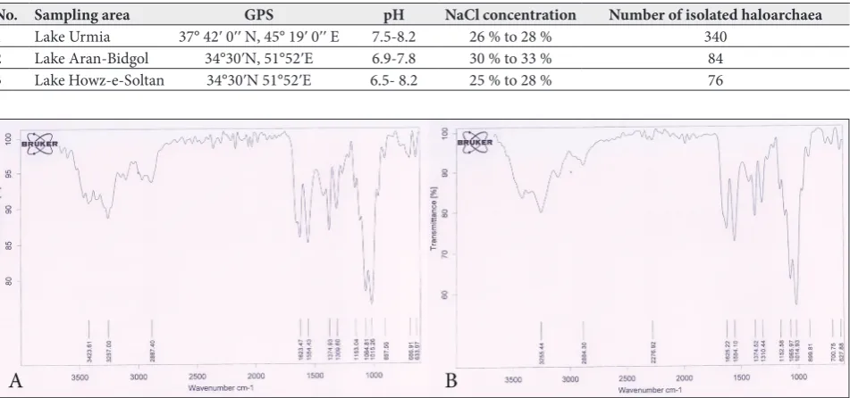
FTIR spectroscopy
To determine the purity level and components of the extracted substance from saline water shrimp shells, FTIR spectroscopy was performed. As can be seen in Fig. 1, spectrum A is related to the extraction compound (chitin) from shrimp shells; spectrum B was obtained after Sigma-Aldrich chitin analysis. In the spectra of both samples, a strong and wide peak at 3000-3500 cm-1
is attributed to the stretching vibration of O-H and N-H. In spectrum A, the absorption peak at 2887 cm-1
is also related to the stretching vibration of C-H in the chitin polysaccharide structure. The stretching vibrations at 1554 cm-1 and 1623 cm-1 belong to the
bending vibrations of O-H and N-H, respectively. The observed peak in the area of 1300-1380 cm-1 is related Table 1. Sampling areas; location and physicochemical characteristics.
No. Sampling area GPS pH NaCl concentration Number of isolated haloarchaea
1 Lake Urmia 37° 42ʹ 0ʹʹ N, 45° 19ʹ 0ʹʹ E 7.5-8.2 26 % to 28 % 340 2 Lake Aran-Bidgol 34°30ʹN, 51°52ʹE 6.9-7.8 30 % to 33 % 84 3 Lake Howz-e-Soltan 34°30ʹN 51°52ʹE 6.5- 8.2 25 % to 28 % 76
to the C-N vibration. The stretching vibrations of C-O and C-O-C were observed in the 1015 and 1064 cm-1 regions, respectively. The observed peak in the
1153 cm-1 area is related to the vibration of the C-C
structure. The rocking vibration of the CH2 group is also seen in the lower range area of 700-800 cm-1. By
comparing the two spectra and verifying the presence of major peaks in the chitin polysaccharide structure (peaks were related to C-C, C-O, C-N, C-H, and O-H vibrations) in spectrum A, the successful extraction of chitin from shrimp shell was confirmed.
Chitinolytic haloarchaea
On the basis of colloidal chitin degradation and zone of clearance on MGM plates, five colonies were selected as chitinase-producing strains. Three potential strains, DC20/BS5/WS8, strain TE14 and strain SM4, were isolated from lakes Urmia, Aran-Bidgol and Howz-e-Soltan, respectively.
Characteristics of chitinase producing strains
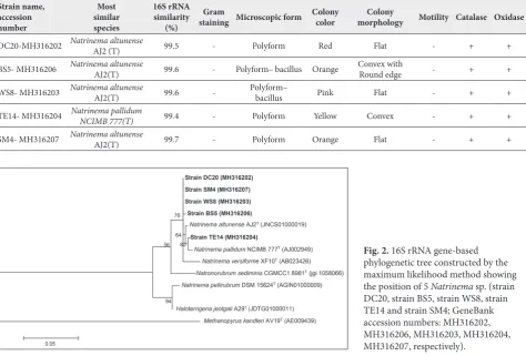
Qualitative screening showed that five strains of the total number of isolates had the ability to produce chitinase. The morphological and physiological char-acteristics of the positive strains demonstrated that all isolated strains were polyform, stained Gram-negative, catalase and oxidase positive and non-motile. How-ever, they differed in morphology and colony color. According to the16S rRNA gene sequence, the strains belonged to the genus Natrinema (Table-2).
A phylogenetic tree was constructed by the maxi-mum likelihood method (Fig. 2). The closest relative of 4 strains DC20, WS8, BS5 and SM4 (Gene Bank ac-cession numbers: MH316202, MH316203, MH316206 and MH316207, respectively) was Natrinema altunense
AJ2 (T), with a Gene Bank access code AY208972 [33], and the closest relative of strain TE14 (Gene Bank accession number: MH316204) was Natrinema pallidum NCIMB 777(T) with a Gene Bank access code AJ002949.
Table 2. Morphological, physiological and molecular characteristics of haloarchaea.
Strain name, accession number
Most similar species
16S rRNA similarity
(%)
Gram
staining Microscopic form Colony color morphologyColony Motility Catalase Oxidase
DC20-MH316202 NatrinemaAJ2 (T)altunense 99.5 - Polyform Red Flat - + +
BS5- MH316206 Natrinema altunense AJ2(T) 99.6 - Polyform– bacillus Orange Convex withRound edge - + +
WS8- MH316203 Natrinema altunense AJ2(T) 99.6 - Polyform– bacillus Pink Flat - + +
TE14- MH316204 Natrinema pallidum NCIMB 777(T) 99.4 - Polyform Yellow Convex - + +
SM4- MH316207 Natrinema altunense AJ2(T) 99.7 - Polyform Orange Flat - + +
Molecular confirmation of chitinase gene
The designed primer pair was used to amplify the chi-tinase gene. Genome extraction of potent strains was performed and amplified bands of 443 bp, detected by agarose gel electrophoresis, confirmed the presence of chitinase gene in chitinase-producing strains (Fig. 3).
Chitinase activity assays
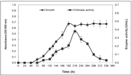
Compared to other strains, Natrinema sp. strain BS5 exhibited a significant clear zone. The growth curve based on the degree of turbidity in the liquid inoculated culture of Natrinema sp. strain BS5 was plotted for 14 days. The production of chitinase corresponded with
Natrinema sp. strain BS5 growth, starting with a loga-rithmic phase, and after 144 h with an 87.2% increase it attained its maximum level (0.36±0.02 U/ml) at the beginning of the stationary phase (Fig. 4). Colloidal chitin was considerably degraded after 14 days.
Microbial growth and enzyme production of the
Natrinema sp. strain under different conditions
The optimal culture conditions (temperature, initial pH, and salt and substrate concentrations) for Natrinema
sp. strain BS5 growth and chitinase production were
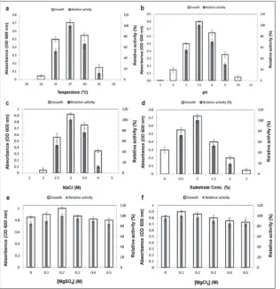
determined. Cell growth occurred over a range of temperatures (30-45˚C), initial pH 6.0-10.0 value, and NaCl (1.5-4 M), colloidal chitin (0-3%), MgSO4 and MgCl2 (0-0.5 M) concentrations. Optimum growth and maximum enzyme production were achieved at 37˚C (Fig. 5a), pH 7.5 (Fig. 5b), 3 M NaCl (Fig. 5c), 1% colloidal chitin (Fig. 5d), 0.2 M MgSO4 (Fig. 5e) and 0.1 M MgCl2 (Fig. 5f) (p≤0.05).
Chitinase production at 35°C and 45°C decreased by 48% and 89%, respectively. The reductions at pH 7 were by 45% and at pH 9 by 70%. At 2.5 M and 4 M NaCl and also at 0.5% and 2% colloidal chitin, 48%, 88%, 28% and 82% reductions in the production of enzyme, respectively, were observed. Although strain BS5 grew at 30°C, pH 6 or 10, 2 M NaCl and 0.0 or 3.0% colloidal chitin, no chitinase production was observed under these conditions. Enzyme production was also obtained in the absence of MgCl2 and MgSO4.
DISCUSSION
Halophilic microorganisms are one of the most impor-tant groups of extremophiles that inhabit hypersaline environments. Their potential applications in several fields of biotechnology have increased because of their survival and adaptation abilities under extreme conditions, such as broad ranges of salinity [34,35]. Compared with other domains of life, archaea are the main group among those with the ability to grow in hypersaline environments. Enzymes produced by halophiles, especially by haloarchaea, can remain active in the presence of high NaCl concentrations,
Fig. 3. Agarose gel electrophoresis of PCR products. M: GeneR-uler™ 1kb DNA ladder (first column represents the number of base pairs (bp) and second column shows the DNA concentration (ng/0.5 µg) in each band and third column indicates the presence percentage of each band in the ladder (%), lane 1: Natrinema sp. strain DC20, lane 2: Natrinema sp. strain BS5, lane 3: Natrinema sp. strain TE14, lane 4: Natrinema sp. strain SM4, lane 5: Natrinema sp. strain WS8, lane 6: negative control.
some organic solvents, low water activity and also in extreme ranges of temperature and pH values [36]. Thus, they have an obvious potential for biotechno-logical applications.
Based on our qualitative screening results using colloidal chitin degradation and zone of clearance, among the 500 isolated haloarchaeal strains from three hypersaline lakes of Iran (Urmia, Aran-Bidgol and Howz-e-Soltan), five strains were chitinase producers.
The use of colloidal chitin in the qualitative screen-ing of microorganisms is a standard procedure [37-40]. The method used to extract chitin from shrimp shells was performed for 1 h compared with other extraction methods which lasted for at least 20 h; it can be said this extraction method is shorter, easier, more economical and faster when compared with other methods [23,41]. The results of the FTIR test indicated that the extrac-tion of chitin was accomplished successfully.
According to the morphological and biochemical tests and 16S rRNA sequence analysis, all 5 strains
(DC20, BS5, WS8, TE14 and SM4) were closely related to the genus Natrinema. This suggests that the genus
Natrinema could potentially be a great producer of chitinase, as Kakhki has also confirmed [4].
The presence of the chitinase gene in the all 5 strains was also confirmed by PCR amplification. Generally, in archaea, sequences of putative chitin-ases were annotated in Halobacteriumsalinarum,
Halomicrobiummukohataei DSM 12286, Haloterrigena turkmenica DSM, Methanoplanuspetrolearius DSM 11571, CandidatusKorarchaeumcryptofilum OPF8 and
CandidatusMethanoregulaboonei 6A8 [20]. Although Kakhki et al. [4] qualitatively showed that 3 isolates of genus Natrinema were able to produce chitinase, our investigation was the first description of chitinase gene activity in this genus.
As the results shows, Natrinema sp. strain BS5 was capable of forming a significant clear zone around its colony compared with other isolated strains; con-sequently, this strain was selected for future studies. The production of chitinase in strain BS5 attained
its maximum level at the beginning of the stationary phase. Maximum enzyme production was at 37°C, pH 7.5, 3M concentration of NaCl, 1% colloidal chitin, 0.2 M and 0.1 M concentration of MgSO4 and MgCl2, respectively. Chitinase production was observed at 35°C up to 45°C, and a broad range of pH (7.0-9.0), indicating the tendency of the strain to produce chi-tinase at alkaline pH. NaCl was a necessary factor for enzyme production because no enzyme production was observed at levels lower than 2.5 M NaCl. The strain had the ability to produce the enzyme even in 4 M NaCl. Since chitinase production was completely dependent on strain growth, reductions in its produc-tion occurred at 35°C and 45°C to 48% and 89%, respec-tively, at pH 7 and pH 9 to 45% and 70%, respecrespec-tively, at 2.5 M and 4M NaCl to 48% and 88%, respectively, and in the presence of 0.5% and 2.0% colloidal chitin to 28% and 82%, respectively. In addition, chitinase production was observed in the absence of MgCl2 and MgSO4: it was therefore concluded that enzyme production is Mg2+-independent. The ability of strain
BS5 to produce chitinase at high salt concentrations with no Mg2+ requirement can be of great use in
in-dustry. Although the optimal temperature for growth and chitinase production in strain BS5 was at 37°C,
Halomicrobium and Salinarchaeum have a maximal growth temperature at 48°C, and Haloterrigena at 50°C in the presence of chitin [42]. In other studies, an optimal temperature between 30°-65°C and pH be-tween 5-9 were reported for the production of bacterial chitinases [8,43]. Also, the study on Halomicrobium
and Salinarchaeum which utilize chitin as growth substrate showed that the concentration of 3.5-4.5 M NaCl is optimal for haloarchaeal growth [42]. In other cases, bioconversion and chitin catabolism in Haloferax mediterranei was accomplished after characterization of the relative genes [44]. Recombinant chitinase activity was also investigatedin Thermococcus kodakaraensis
KOD1, Pyrococcus furiosus, and Halobacterium sa-linarum [17-19]. Staufenberger et al. [20]expressed ORF BAB65950, which encodes functional chitinase, from Sulfolobustokodaii in E. coli. Sorokin et al. [42] examined several halo (natrono) archaeal strains for their ability to grow on insoluble cellulose and chitin as a sole growth substrate in salt-saturated mineral media. In 2011, Kakhki et al. [4] showed that 3 isolates of genus Natrinema were able to produce chitinase. Other studies on Natrinema have also revealed the
ability to produce xylanase, amylase, caseinase and cellulase [45]. A protease gene (sptA), was cloned from the halophilic archaeon Natrinema sp. J7 in Haloferax volcanii WFD11. The SptA protease was optimally active at 50°C in 2.5 M NaCl at pH 8.0 [46]. However, our investigation is not in full agreement with these findings and we did not compare them with our results.
CONCLUSIONS
The genus Natrinema, isolated fromhypersaline lakes in Iran, is a potential source of chitinase production. This investigation, for the first time, describes chi-tinase gene activity in the Natrinema genus using PCR amplifications and quantitative surveying. To our knowledge, this is the first report that this genus possesses the ability to grow and produce chitinase in the absence of Mg2+. Thus, it could possess potential
commercial value.
Funding: This work was supported by the University of Tehran. Acknowledgments: The authors would like to express their gratitude to all individuals in the Extremophiles Laboratory who assisted and supported the project.
Author contributions: M. A. Amoozegar conceived the presented idea. M. Yavari-Bafghi carried out the experiment and wrote the manuscript. M. A. Amoozegar supervised the research. All authors discussed the results and contributed to the final manuscript. Conflict of interest disclosure: The authors declare no conflict of interest regarding the publication of this manuscript.
REFERENCES
1. Ara I, Daram D, Baljinova T, Yamamura H, Bakir M a, Suto M, Ando K. Isolation, classification , phylogenetic analysis and scanning electron microscopy of halophilic , halotolerant and alkaliphilic actinomycetes isolated from hypersaline soil. African J Microbiol Res. 2013;7(4):298-308.
2. Ventosa A. Unusual micro-organisms from unusual habitats: hypersaline environments. In: Logan NA, Lappin-Scott HM, Oyston PCF, editors. Prokaryotic diversity. Cambridge: Cam-bridge University Press; 2006. p. 223-54.
3. Yin J, Chen JC, Wu Q, Chen GQ. Halophiles, coming stars for industrial biotechnology. Vol. 33, Biotechnology Advances. 2015. p. 1433-42.
5. Litchfield CD. Potential for industrial products from the halophilic Archaea. J Ind Microbiol Biotechnol. 2011;38(10):1635–47.
6. Amoozegar MA, Siroosi M, Atashgahi S, Smidt H, Ventosa A. Systematics of haloarchaea and biotechnological potential of their hydrolytic enzymes. Microbiology. 2017;163(5):623-45. 7. Islam R, Datta B. Diversity of chitinases and their industrial
potential. Int J Appl Res. 2015;1:55-60.
8. Stoykov YM, Pavlov AI, Krastanov AI. Chitinase biotechnol-ogy: Production, purification, and application. Vol. 15, Engi-neering in Life Sciences. 2015. p. 30-8.
9. Adrangi S, Faramarzi M, Shahverdi A, Sepehrizadeh Z. Purifi-cation and characterization of two extracellular endochitinases from Massilia timonae. Carbohydr Res. 2010;345(3):402-7. 10. Hashimoto M, Ikegami T, Seino S, Ohuchi N, Fukada H,
Sugiyama J, Shirakawa M, Watanabe T.. Expression and Characterization of the Chitin-Binding Domain of Chi-tinase A1 from Bacillus circulans WL-12. J Bacteriol. 2000;182(11):3045-54.
11. Ahmadian G, Degrassi G, Venturi V, Zeigler DR, Soudi M, Zanguinejad P. Bacillus pumilus SG2 isolated from saline conditions produces and secretes two chitinases. J Appl Microbiol. 2007;103(4):1081-9.
12. Delgado-García M, Aguilar CN, Contreras-Esquivel JC, Rodríguez-Herrera R. Screening for extracellular hydrolytic enzymes production by different halophilic bacteria. Myco-path. 2014;12(1):17-23.
13. Swiontek Brzezinska M, Jankiewicz U, Burkowska A, Wal-czak M. Chitinolytic Microorganisms and Their Possible Application in Environmental Protection. Curr Microbiol. 2014;68(1):71-81.
14. Li H, Greene LH. Sequence and Structural Analysis of the Chitinase Insertion Domain Reveals Two Conserved Motifs Involved in Chitin-Binding. Yang H, editor. PLoS One. 2010 Jan;5(1):e8654.
15. Adrangi S, Faramarzi MA. From bacteria to human: A journey into the world of chitinases. Biotechnol Adv. 2013 Dec;31(8):1786-95.
16. Revah-Moiseev S, Carroad PA. Conversion of the enzymatic hydrolysate of shellfish waste chitin to single-cell protein. Biotechnol Bioeng. 1981;23(5):1067-78.
17. García-Fraga B, da Silva AF, López-Seijas J, Sieiro C. Func-tional expression and characterization of a chitinase from the marine archaeon Halobacterium salinarum CECT 395 in Escherichia coli. Appl Microbiol Biotechnol. 2014 Mar 30;98(5):2133-43.
18. Gao J, Bauer MW, Shockley KR, Pysz MA, Kelly RM. Growth of Hyperthermophilic Archaeon Pyrococcus furiosus on Chi-tin Involves Two Family 18 ChiChi-tinases. Appl Environ Micro-biol. 2003;69(6):3119-28.
19. Tanaka T, Fukui T, Imanaka T. Different Cleavage Specificities of the Dual Catalytic Domains in Chitinase from the Hyper-thermophilic Archaeon Thermococcus kodakaraensis KOD1. J Biol Chem. 2001;276(38):35629-35.
20. Staufenberger T, Imhoff JF, Labes A. First crenarchaeal chitinase found in Sulfolobus tokodaii. Microbiol Res. 2012;167:262-9.
21. Amoozegar MA, Makhdoumi-Kakhki A, Shahzadeh Fazeli SA, Azarbaijani R, Ventosa A. Halopenitus persicus gen. nov.,
sp. nov., an archaeon from an inland salt lake. Int J Syst Evol Microbiol. 2012;62(Pt8):1932-6.
22. Pecher T, Böck A. In vivo susceptibility of halophilic and methanogenic organisms to protein synthesis inhibitors. FEMS Microbiol Lett. 1981;10(3):295-7.
23. Kaya M, Baran T, Karaarslan M. A new method for fast chitin extraction from shells of crab, crayfish and shrimp. Nat Prod Res. 2015;29(15):1477-80.
24. Dassault H. P. An improved technique for staining red halo-philic bacteria. J Bacteriol. 1955;70(4):484-5.
25. Heimbrook ME, Wang WL, Campbell G. Staining bacterial flagella easily. J Clin Microbiol. 1989;27(11):2612-5. 26. Garrity GM, Holt JG. The Road Map to the Manual. In:
Ber-gey’s Manual® of Systematic Bacteriology. 2001. p. 119-66. 27. Marmur J. A procedure for the isolation of deoxyribonucleic
acid from micro-organisms. J Mol Biol. 1961;3(2):208. 28. Spencer JFT, Ragout de Spencer AL, editors. Environmental
Microbiology. Totowa: Humana Press; 2004. 439 p. (Methods in Biotechnology; 16).
29. Klatte T, Evans L, Whitehead RN, Cole JA. Four PCR primers necessary for the detection of periplasmic nitrate reductase genes in all groups of Proteobacteria and in environmental DNA. Biochem Soc Trans. 2011;39(1):321-6.
30. Kim O-S, Cho Y-J, Lee K, Yoon S-H, Kim M, Na H, Park SC, Jeon Y, Lee JH, Yi H, Won S, Chun J. Introducing EzTaxon-e: a prokaryotic 16S rRNA gene sequence database with phylo-types that represent uncultured species. Int J Syst Evol Micro-biol. 2012;62(Pt 3):716-21.
31. Miller GL. Use of Dinitrosalicylic Acid Reagent for Determi-nation of Reducing Sugar. Anal Chem. 1959;31(3):426-8. 32. Nawani NN, Kapadnis BP. Optimization of chitinase
produc-tion using statistics based experimental designs. Process Bio-chem. 2005;40(2):651-60.
33. Xu XW, Ren PG, Liu SJ, Wu M, Zhou PJ. Natrinema altunense sp. nov., an extremely halophilic archaeon isolated from a salt lake in Altun Mountain in Xinjiang, China. Int J Syst Evol Microbiol. 2005;55(3):1311-4.
34. Ali I, Prasongsuk S, Akbar A, Aslam M, Lotrakul P, Pun-napayak H, Rakshit S. Hypersaline habitats and halophilic microorganisms. Maejo Int J Sci Technol. 2016;10(3):330-45. 35. Margesin R, Schinner F. Potential of halotolerant and
halo-philic microorganisms for biotechnology. Extremophiles. 2001;5(2):73-83.
36. Delgado-García M, Valdivia-Urdiales B, Aguilar-González CN, Contreras-Esquivel JC, Rodríguez-Herrera R. Halophilic hydrolases as a new tool for the biotechnological industries. J Sci Food Agric. 2012;92(13):2575-80.
37. Saima, Kuddus M, Roohi, Ahmad IZ. Isolation of novel chi-tinolytic bacteria and production optimization of extracel-lular chitinase. J Genet Eng Biotechnol. 2013;11(1):39-46. 38. Monreal J, Reese ET. The chitinase of Serratia marcescens.
Can J Microbiol. 1969;15(7):689-96.
39. Berger LR RD. The chitinase system of a strain of Streptomy-ces griseus. Biochem Biophys Acta. 1958;29:522-34. 40. Karthik N, Akanksha K PA. Production, purification
and properties of fungal chitinases. Indian J Exp Biol. 2014;11(1025–35.):1025-35.
42. Sorokin DY, Toshchakov S V, Kolganova T V, Kublanov I V. Halo(natrono)archaea isolated from hypersaline lakes utilize cellulose and chitin as growth substrates. Front Microbiol. 2015;6:942.
43. Deeba F, Shakir HA, Irfan M, Qazi JI. Chitinase production in organisms : a review. Punjab Univ J Zoo. 2016;31(1):101-6. 44. Hou J, Han J, Cai L, Zhou J, Lü Y, Jin C, Liu J, Xiang H. Char-acterization of genes for chitin catabolism in Haloferax medi-terranei. Appl Microbiol Biotechnol. 2014;98(3):1185-94. 45. Patil J, Bajekal S. Diversity of hydrolytic enzymes in
Haloal-kaliphilic archaea isolated from Lonar Lake. Microbiology. 2013;2(7).
46. Shi W, Tang X-F, Huang Y, Gan F, Tang B, Shen P. An extra-cellular halophilic protease SptA from a halophilic archaeon Natrinema sp. J7: gene cloning, expression and characteriza-tion. Extremophiles. 2006;10(6):599-606.
Supplementary Data
Supplementary Fig. S1.