The Synthesis of antiCD3-F(ab’)2 and Fab, and the Biodistribution of an antiCD3-F(ab’)2 Radiotracer in C57BL/6J and BALB/c mice
By
Luke B. Soliman
Senior Honors Thesis Department of Chemistry
University of North Carolina at Chapel Hill November 10, 2017
Abstract
Background: The activation of tumor-infiltrating lymphocytes (TILs) has become a prominent immunotherapy technique for the treatment of various cancers. The binding of the antiCD3 immunoglobulin (IgG) to the CD3 T-cell receptor (TCR) produces the cytotoxic T-cell, thereby eliciting an immune response against biological malignancies. In addition to the therapeutic action that these T-cells induce, imaging of these cells serves as a foundation to monitor immunotherapy treatment efficacy. Antigen binding fragments, derived from full-sized therapeutic monoclonal antibodies (mAbs), have been shown to bind identically to their parent mAbs while also exhibiting more rapid clearance from the bloodstream. We developed novel protocols for the production of antiCD3 antigen binding fragments, characterized the fragments, and preliminarily evaluated their capabilities to rapidly and non-invasively analyze the efficacy of TIL therapy.
Result: Protocols for the digestion of antiCD3 to crude Fab and F(ab’)2 were developed and optimized. SE-HPLC purification was successfully performed to isolate the antiCD3-F(ab’)2 fragment. The percent injected dose per gram of tissue (%ID/g) of 64 Cu-NOTA-antiCD3-F(ab’)2 between black and nude mice was statistically significant for the spleen at 6 h, and for the kidney, spleen, cervical lymph node (CLN), and axillary lymph node (ALN) at 24 h.
Conclusion: Novel protocols for the fragmentation and purification of an antiCD3 mAb to Fab and F(ab’)2 were developed. The conjugated, radiolabeled antiCD3-F(ab’)2 fragment showed rapid clearance via the kidney, significant uptake in several lymphoid organs, and further potential for development as an imaging agent for cancer immunotherapy efficacy.
Introduction
Substantial evidence has emerged that many tumors express antigens that can be recognized by the immune system, making immunotherapy a rational approach for the treatment of cancer.1 Of the many cancer immunotherapy techniques under review, one emerging method is the recruitment of cytotoxic T-cells which are signaled by the binding of antibodies to T-cell receptors (TCRs). Additionally, treatment efficacy can be evaluated by monitoring the immune response during therapy.2,3 The CD3 antigen, which is present in TCRs at all stages of T-cell development, makes it a useful target for the signaling and mobilization of T-cells. Thus, the CD3 antigen is also a natural point of interest for the early evaluation of the efficacy of immunotherapy treatments.4
located within an immune cell to thereby stimulate a therapeutic immune response. In addition, this project also makes use of antibody fragments, rather than full
immunoglobulin proteins. The latter, nuanced aim is significant for several reasons. First, the labeling of a suitable radioisotope to an undigested antibody without inflicting
damage to its binding behavior can be demanding.2 Damaged antibodies would fail to serve as effective tumor imaging agents, and would also reduce the potency of
immunotherapy treatment. Second, full antibodies exhibit poorer tissue penetration than antibody fragments.2 Imaging agents that are able to better penetrate tissue and therefore saturate tumor binding sites will optimize the resolution of PET/CT tumor images. Third, whole antibodies ( ~150 kDa) persist in bodily circulation for greater periods than do their fragments (~50 – 110 kDa).2 One challenge of non-invasive tumor imaging is reducing the residual radioactivity that is exposed to the patient – this obstacle is alleviated by the use of antibody fragments which circulate for shorter periods of time.
This project, founded in the two aforementioned nuances, takes advantage of techniques in enzymatic proteolysis and antibody binding behavior to evaluate the biodistribution of antiCD3-F(ab’)2 in vivo – and serves as a basis for subsequent study of rapid and non-invasive evaluation of tumor size, location, and the efficacy of cancer immunotherapy treatments.
We hypothesized that pepsin and ficin would enzymatically proteolyze antiCD3-IgG to F(ab’)2 and Fab respectively, and that an antiCD3-F(ab’)2 fragment radiolabeled with 64Cu would accumulate T-cells with statistical significance between
Materials and Methods Reagents and instruments
AntiCD3 (BE0002) was purchased from BioXCell (West Lebanon, NH).The bifunctional chelator, 2-S-(4-Isothiocyanatobenzyl)-1,4,7-triazacyclononane-1,4,7-triacetic acid (p-SCN-Bn-NOTA) was purchased from Macrocyclics (Dallas, TX, USA). Copper-64 in 1 M oxalic acid was purchased from Washington University (MO, USA). All other
reagents were purchased from Fisher Scientific with the highest purity available. Protein concentration was determined using a Nanodrop 2000/2000c. Radioactivity from non-biological samples was measured in a CRC-55tW dose calibrator/well counter (Capintec, NJ, USA). Radioactivity in biological samples was measured in an automatic γ-counter (2470, Wizard2, Perkin Elmer, Walthem, MA). Size-exclusion high- performance liquid chromatography (SE-HPLC) was performed at room temperature on an Agilent 1200 series chromatographic system equipped with an online flow γ-ray detector (Lablogic). Samples were injected onto Agilent Bio SEC-5 size exclusion column (5 µm, 300 Å; 7.8 x 300 mm) and Agilent Bio SEC-5 size exclusion column (5 µm, 100 Å; 7.8 x 300 mm) connected in series using EDTA 0.01 M in PBS as the isocratic mobile phase. The flow rate was maintained at 1 mL/min and the elution was monitored by UV
spectrophotometer at 254 and 280 nm. Sodium dodecyl sulfate polyacrylamide gel electrophoresis (SDS-PAGE) was carried out in an XCell SureLockTM Mini-Cell electrophoresis system.
Dynamic Light Scattering Analysis of antiCD3-IgG, F(ab’)2, and Fc
Automatic measurement duration and 173° backscatter protein analysis model were used for measurements, which were taken in triplicate.
Fragmentation of antiCD3-IgG to F(ab’)2
To a Vivaspin 2 (30 kDa molecular weight cut off (MWCO)) filter, 1 mg (3.1 mg/mL, 322.5 µL) of antiCD3 IgG in stock solution was added. The solution was concentrated by ultracentrifugation for 10 min at 5000 RCF at 4 oC. The antibody was then buffer
Fragmentation of antiCD3-IgG to Fab
To 10 mL of Fab digestion buffer (ThermoFisher), 43.9 mg L-cysteine•HCl was dissolved to yield 25 mM cysteine solution. An identical fragmentation protocol to the one above was followed, instead substituting 25 mM cysteine buffer rather than F(ab’)2 buffer, using 1 mL of immobilized ficin rather than 500 µL of immobilized pepsin, and allowing the reaction to proceed for 80 min rather than 3 h.
SE-HPLC purification of antiCD3 fragments
Crude F(ab’)2 and Fab were isolated by SE-HPLC purification. Samples of 150 µg were injected onto the column with an isocratic mobile phase of 0.01 M PBS flowing at 1.0 mL/min. Pure F(ab’)2 was collected from 13:55 (mm:ss) to 14:25. Pure Fab was retrieved from 15:01 to 15:31. AntiCD3-Fc was isolated by collecting fractions from 16:07 to 16:25.
Biodistribution of 64Cu-NOTA-antiCD3-F(ab’)
2 in immunocompetent and
immunocompromised mice
Statistical Analysis
Differences between groups were tested for significance using a T-test and Mann-Whitney Test. Outliers were determined by Grubb’s test and removed from future calculation. P values were calculated using a two-tailed test, and values ≤ 0.05 were considered significant. Data are reported as mean ± standard deviation.
Results Fragmentation of antiCD3 to F(ab’)2 and Fab
Although the reaction of antiCD3-IgG with immobilized pepsin is optimized at 3 h, only 0.63% of the full immunoglobulin remains undigested after the first hour. Table 1 indicates the percent conversions between undigested antibody and the fragmented products over time, as calculated by the area under each peak in the respective SE-HPLC chromatogram for that timepoint (λ = 280 nm). After filtration in a 30 kDa MWCO filter, the reaction of antiCD3-IgG with immobilized pepsin, forming F(ab’)2, had a crude yield of 47.46%. SE-HPLC characterization of Crude antiCD3-F(ab’)2 can be found in Figure 1. For comparison, Figure 2 depicts the chromatogram of SE-HPLC analysis of antiCD3-IgG. Of the crude product, 87.97% was antiCD3-F(ab’)2. Therefore, for each 1.0 mg of antiCD3-IgG reacted, 0.4175 mg of unisolated F(ab’)2 is produced.
Table 1. Percent conversions from antiCD3-IgG to F(ab’)2 and Fc fragments during enzymatic proteolysis with pepsin
Time (h) IgG (%) F(ab’)2 (%) Fc (%)
0 100 0 0
1 h 0.63 9.55 89.82
2 h 0.59 8.65 90.76
Crude (post-filtration) 1.19 87.97 10.84
Table 2, shown below, indicates the percent conversions of reactants to products for the digestion of antiCD3-IgG with ficin, as calculated by the area under each peak in the respective SE-HPLC chromatogram for that timepoint (λ = 280 nm). Within the first
40 min of the reaction, 98.87% of the IgG was digested into its constituent fragments. By the end of the digestion at 80 min, 0% of the intact antibody remained. After filtration of the reaction mixture in a 10 kDa MWCO filter, the average crude yield of the reaction was 68.0%. The SE-HPLC analysis of crude antiCD3-Fab is shown in Figure 3. Of the total amount of protein in the crude product, 17.72% was unisolated Fab. Therefore, for a 1:1 reaction of antiCD3-IgG with immobilized ficin slurry, 120 µg of unisolated Fab is produced.
Table 2. Percent conversions of antibody fragments from antiCD3 during fragmentation with ficin
Time (min) IgG (%) Fab (%) Fc (%)
0 95.89 0.12 3.99
20 13.27 22.85 63.88
40 1.13 24.63 74.24
60 0.02 23.71 76.28
80 0 20.73 79.27
Crude (post-filtration) 0 17.72 82.28
SE-HPLC Purification of F(ab’)2 and Fab
Table 3. Yields of total protein and F(ab’)2 during F(ab’)2 fragmentation and SE-HPLC purification
Percent (%) Mass (µg)
Total Crude Protein Yield 47.46 474.60
Unisolated F(ab’)2 within Total Crude Protein
87.97 417.50
HPLC Purified F(ab’)2 Yield 3.38 16.03
Overall antiCD3-IgG to Pure F(ab’)2 Yield
1.60 16.03
Table 4. Yields of total protein and Fab during Fab fragmentation and SE-HPLC purification
Percent (%) Mass (µg)
Total Crude Protein Yield 68.00 680.00
Unisolated Fab within Total
Crude Protein 17.72 120.50
HPLC Purified Fab Yield 0.33 2.27
Overall antiCD3-IgG to Pure Fab Yield
0.227 2.27
Dynamic Light Scattering of antiCD3 IgG, F(ab’)2, and Fc
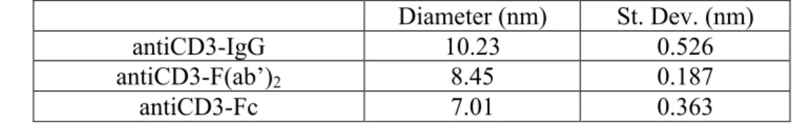
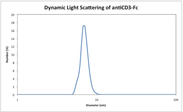
Isolated antiCD3-IgG, F(ab’)2, and Fc were evaluated by dynamic light scattering to estimate the spherical diameter of each molecule. Table 5 below indicates the measured diameters and their respective standard deviations.
Table 5. antiCD3 immunoglobulin and fragment diameters, as characterized by dynamic light scattering
Diameter (nm) St. Dev. (nm)
antiCD3-IgG 10.23 0.526
antiCD3-F(ab’)2 8.45 0.187
antiCD3-Fc 7.01 0.363
Figures 5-7 graphically depict the size measurements for each of the proteins.
Biodistribution of 64Cu-NOTA-antiCD3-F(ab’)2 in immunocompetent and
immunocompromised mice
measurement of radioactivity in target tissues serves as a rational foundation for the determination of antiCD3-F(ab’)2 biodistribution.
Figure 8 graphically illustrates the percent injected dose per gram of tissue (%ID/g) for both immunocompetent and immunocompromised mice at 6 h post tail vein injection. AntiCD3-F(ab’)2 exhibited rapid clearing via the kidney for both types of mice, averaging 69.16 and 73.23 %ID/g for black and nude mice respectively. The lymphoid organs, which serve as storehouses for immune cells, are the only tissues at both 6 h and 24 h to show a statistically significant difference in uptake of antiCD3-F(ab’)2 between black and nude mice. At 6 h, the cervical lymph node (CLN) and axillary lymph node (ALN) showed significantly greater uptake in black mice than in nude (p < 0.01). The spleen showed even greater significance between black and nude mice in F(ab’)2 uptake (p < 0.001). Other ex vivo samples, such as the liver, bone, blood, and carcass failed to show significance in fragment uptake between black and nude mice.
Ex vivo analysis of target tissues at 24 h post tail vein injection (Figure 9)
Table 6. Average percent injected dose per gram of tissue (%ID/g) at 6 h and 24 h for both immunocompetent and immunocompromised mice, with corresponding P value and significance level (* p<0.05, ** p<0.01, *** p<0.001, ns = not significant).
*Note: one data point (inguinal LN from a black mouse at 6 h) was removed as it was a significant outlier (p < 0.05) by Grubbs’ test
Discussion
as PET/CT imaging agents for the evaluation of immunotherapy treatment efficacy, as well as potential use in immunotherapy.
One approach of cancer immunotherapy hinges on the ability of the treatment to successfully produce cytotoxic T-cells and to traffic tumor infiltrating lymphocytes (TILs) in the fight against bodily malignancies. The lymphoid organs, which serve as storehouses for immune cells, are integral players in the mobilization of T-cells. The biodistribution data discussed in this report demonstrate statistically significant
differential uptake of 64Cu-NOTA-antiCD3-F(ab’)2. between black and nude mice in the spleen, CLN, and ALN at 6 h. Nude mice showed a significantly higher uptake of radioactivity in the kidney than did black mice at 24 h. This could possibly indicate that immunocompetent mice, which bear T-cells, uptake and retain antiCD3-F(ab’)2 in other tissues. This conclusion is supported by the data, which indicates that at 24 h, lymphoid organs such as the spleen, CLN, and ALN showed statistically significant higher uptakes of radioactivity in black mice than in nude mice. AntiCD3-F(ab’)2 therefore shows incredible promise in its ability to as a useful imaging marker to monitor lymphocytes and analyze TIL recruitment efforts in immunotherapy treatment.
Conclusions
An antiCD3 immunoglobulin was successfully fragmented and purified to F(ab’)2 and Fab. AntiCD3-IgG, F(ab’)2, and Fc were characterized by dynamic light scattering for their diameters. A radiolabeled antiCD3-F(ab’)2 fragment was injected into
immunocompromised mice. AntiCD3-F(ab’)2 demonstrates incredible future promise as a PET/CT imaging agent and analytical tool for evaluating the efficacy for cancer immunotherapy treatment.
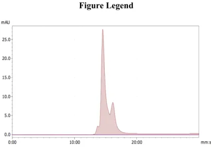
Figure Legend
Figure 1. Crude antiCD3-F(ab’)2, post-30 kDa MWCO filtration. In order of earliest elution time to latest: antiCD3-IgG (13:45), F(ab’)2 (14:32), Fc (16:09)
Figure 3. Crude antiCD3-Fab post-10 kDa MWCO filtration. In order of earliest elution time to latest: antiCD3-IgG (13:45), Fab (14:58), Fc (16:09)
Figure 5. Dynamic light scattering analysis of antiCD3-IgG (Diameter = 10.23 nm)
Figure 7. Dynamic light scattering analysis of antiCD3-Fc (Diameter = 7.01 nm)
Figure 9. Biodistribution of antiCD3-F(ab’)2 in black and nude mice at 24 h.
Supplemental Information
Supplemental Figures:
Figure S2. Purified NOTA-antiCD3-F(ab’)2 (14:12)
Figure S3. 64Cu-NOTA-antiCD3-F(ab’)2 (14:11)
Supplemental Materials and Methods:
One area of consideration for future antiCD3-F(ab’)2 fragmentations lies in the accuracy of the amount of pepsin used. While the novel reaction protocol discussed here calls for 500 µL of immobilized pepsin for each 1.0 mg of antiCD3-IgG, the density of the pepsin slurry can vary from 2 mg/mL to 3 mg/mL.5 Although gently stirring the resin does help to evenly distribute the enzyme throughout the mixture, there still exists a margin of uncertainty for the exact stoichiometric ratio of digestive enzyme to antibody.
References
1. Parish, C. R. Cancer immunotherapy: The past, the present and the future. Immunology and Cell Biology 81, 106–113 (2003).
2. Rashidian, M. et al. Noninvasive imaging of immune responses. Proc. Natl. Acad. Sci. U. S. A. 112, 6146–51 (2015).
3. McCracken MN, Tavaré R, Witte ON, Wu AM. Advances in PET Detection of the Antitumor T Cell Response. 2016;187–231. Available from:
http://linkinghub.elsevier.com/retrieve/pii/S0065277616300190
4. Beckford Vera, DR. Immuno-PET imaging of tumor-infiltrating lymphocytes using Zirconium-89 radiolabeled antiCD3 antibody in immune-competent mice bearing syngenic tumors.
5. ThermoFisher. Pierce TM