Therapeutic Hypothermia to Reduce Cardiac Arrest-Related Neurological
Deficits and Mortality
By
Angel Legare, PA-S
A Capstone Paper submitted to the faculty of the University of North Carolina at Chapel Hill
in partial fulfillment of the requirements for the degree of Master of Health Sciences
in the Physician Assistant Program
Chapel Hill
December 2017
________________________________
Name and title of First Reader
______________________
Date
________________________________
Name and title of Second Reader
ii
TABLE OF CONTENTS
ABSTRACT ... iii
1. INTRODUCTION ...1
2. BACKGROUND ... 1-9
2A. HISTORY OF THERAPEUTIC HYPOTHERMIA ... 1-4
2B. PHYSIOLOGY OF THERAPEUTIC HYPOTHERMIA ... 4-9
3. METHODOLOGY ... 9-10
4. REVIEW OF LITERATURE RESULTS ... 10-18
4A. HOLZER AT AL. ... 10-13
4B. SUNDE AT AL ... 13-15
4C. BERNARD AT AL ... 15-18
5. DISCUSSION ... 18-21
6. CONCLUSION ... 21-22
REFERENCES ... 23-25
APPENDIX
A. MESH TERMS ...26
B. TABLE 1 ... 27-31
C. RISK OF BIAS EVALUATION ... 32-33
iii Abstract
Purpose
The purpose of this clinical review is to determine if induced therapeutic hypothermia is
effective in reducing neurological deficits and mortality in cardiac arrest patients.
Methods
PubMed, Cochrane Review Database, Embase, and Trip database were utilized. A
common set of search terms including "induced hypothermia", "therapeutic hypothermia”,
"cardiovascular resuscitation", “cardiac arrest”, "neurological deficits", “neurological outcome”
and “mortality” guided the search. A total of three individual studies that were included for final
results review.
Results
Clinically based trials revealed that induced therapeutic hypothermia can improve both
survival rates and neurological outcomes in cardiac arrest patients compared to cardiac arrest
patients that do not receive therapeutic hypothermia.
Conclusion
Although more research needs to be done to make therapeutic hypothermia implemented
across all cardiac arrest patients, current research has suggested that therapeutic hypothermia
implantation in cardiac arrest protocols can prove to be extremely beneficial in improving patient
1 Introduction
Death rates among patients that suffer sudden cardiac arrest out of hospital are high. In
the United States alone, more than 350,000 individuals had in incidence of out of hospital
cardiac arrest in 2016.10 Out of these 350,000 patients that suffered cardiac arrest, ~200,000 of
them resulted in death. This clinical condition affects many individuals without warning as it is
difficult to predict. One adverse effect of cardiac arrest is persistent neurological deficits as a
consequence of spontaneous reperfusion. Surviving with good neurological outcome is difficult
to achieve and experimental evidence proposes that therapeutic hypothermia is beneficial11.
Research has suggested that therapeutic hypothermia implantation in cardiac arrest protocols can
prove to be extremely supportive in giving the patient the very best neurological outcome.
The concept of therapeutic hypothermia has been around for over 2000 years and while
the principle has advanced since then, there is still research that needs to be done to establish
guidelines for its widespread implementation. With the continuous advances in medicine there is
no set limit to how it can drastically change our patients outcomes for the better. In the
meantime, while we can continue to work to keep ourselves as healthy as possible, cardiac arrest
can still happen. It is what happens after cardiac arrest that can be improved, and by improving
that we can improve survival rates as well as quality of life.
Background
History of Therapeutic Hypothermia
While therapeutic hypothermia may seem futuristic, it has been around far longer than
2
chamber suspending one’s animation in time while medical staff races to find a cure like a sci-fi
movie would suggest, medicine still has made great advances since it was first introduced. The
concept of therapeutic hypothermia has been used dating all the way back to the father of
medicine himself, Hippocrates. He saw the value of it and mentioned the use of snow and ice to
reduce hemorrhage in his wounded and critically ill patients.1
This treatment has emerged continually throughout history. In 1954 a study by Rosomoff
and Holaday2 found a surprising fall in cerebral oxygen consumption in dogs that underwent
therapeutic hypothermia. They documented this decrease in oxygen consumption occurred when
the dogs temperatures were lowered from 35°C to 26°C. Furthermore, it was found that there
was an astonishing three-fold reduction in oxygen consumption when the dogs were cooled
down to 26°C2. Through their experiments and others that followed, this helped catalyze what
would then lead to predictions in how this concept could be used in human medicine. With these
studies, it could predict how it could be used for cardiac arrest patients.
In 1958 the very first study on humans using therapeutic hypothermia was conducted
with cardiac arrest patients3. The study suggested decreased mortality in the patients that
received that therapeutic hypothermia. Although this study did not look at neurological outcome
in these patients. This groundbreaking study included 19 patients that were resuscitated after
suffering perioperative cardiac arrest. All patients had their thoracic cavity opened and their heart
was documented to be in fibrillation or asystole. Then the subjects that were chosen for
therapeutic hypothermia were cooled using a circulating coolant blanket. The subjects were
cooled and maintained at approximately 31–32°C.3 There was no set time that the subjects were
cooled, and clinical judgment was used to determine the duration of hypothermia. Although,
3
slowly and gradually returned to normal temperature. It was discovered that out of the 19
patients included in the study, 7 patients in the control group did not receive therapeutic
hypothermia, while 12 patients in the treatment group received therapeutic hypothermia.3 The
conclusion showed that the survival rate was 50 percent in the treatment group compared to 14
percent in the control group.
Although the outcomes of utilizing therapeutic hypothermia to improve survival of
cardiac arrest were very promising, until twenty years ago few studies on the treatment existed.
The studies that were published documented favorable results and in 2002 the advisory panel
from the International Liaison Committee on Resuscitation (ILCOR) implemented the
recommendation of its use in common practice. In 2002, two studies were published out of
Europe4 and Australia5 that demonstrated improved survival rates and improved neurological
outcomes when using mild therapeutic hypothermia. The studies studied that effect of
therapeutic hypothermia on comatose survivors of Out of Hospital Cardiac Arrest (OHCA) that
were due to Ventricular Fibrillation (VF). The Hypothermia after Cardiac Arrest Study Group
(HACA), including nine hospitals in five European countries, showed that mild hypothermia
(cooling to 32–34ºC for 24 h) in 274 OHCA patients with return of spontaneous circulation
(ROSC) provided significant improvement in functional recovery and good neurological
outcome after hospital discharge (55% vs. 39%).5 It also led to a lower 6-month mortality rate
when compared with patients who were not cooled (41% vs. 55%).5 In Australia, Bernard et al.
examined the endpoint of survival to hospital discharge in 77 patients.4 This showed a 49%
survival rate in the therapeutic hypothermia group which were cooled to 33°C for 12 hours,
4
Even though there were published guidelines in by the International Liaison Committee
on Resuscitation (ILCOR) supporting the use of therapeutic hypothermia6 in 2005, the use of
hypothermia as treatment still lagged in utilization. Adoption of hypothermia in North America
was even slower with far higher utilization rates in North European countries.7 There were many
different reasons for its delay in implementation. These reasons included insufficient knowledge
of effective hypothermia techniques, lack of belief that therapeutic hypothermia would improve
the outcome for individual patients, and controversies regarding the best method to reach the
target temperatures6.
The ILCOR guidelines in 2005 did not clarify a set cooling protocol. In 2010, newer
guidelines were published by the International Liaison Committee on Resuscitation (ILCOR)8.
This progressive advancement by the ILCOR helped provide the material for the American Heart
Association (AHA)9 to write their new resuscitation guidelines in 2010. Now with the AHA
advocating for the implementation of the use of therapeutic hypothermia, there was a higher
likelihood that therapeutic hypothermia would be utilized in North America to truly see how it
could impact cardiac arrest patient outcomes. While it may have started with the father of
medicine more than 2000 years ago, with some modern improvisation, therapeutic hypothermia
will continue to strive to benefit patient outcomes.
Physiology of Therapeutic Hypothermia
The act of simply “cooling” a patient seems almost elementary in comparison to other
lifesaving procedures that occur during cardiac arrest resuscitation. While it may seem that way
at first, after considering what the active cooling does to the patient’s individual systems it
uncovers itself to be quite complex. That is the exquisite nature of the physiology of therapeutic
5
arrest, there is secondary damage to the brain and other vital organs due to hypoperfusion that
can ultimately lead to neurological deficits and death. Therapeutic hypothermia is being used to
improve this secondary impact that causes profound damage to the brain and other organ tissue.
Unfortunately, what is not yet fully understood is the complete mechanism of action of
hypothermia and how it effects every organ system differently. This is due to how incredibly
complex it is. Although, research has been conducted to further understand how something like
hypothermia works to help individuals from this secondary damage.
Therapeutic hypothermia and its protective effects on the body, especially from
secondary damage can be broken down into several pathways. A decreased metabolic state
resulting in lower oxygen and energy consumptionalong with carbon dioxide productionmay
prevent secondary injury when oxygen supply is interrupted or, at least, impaired.12 This is
especially important with patients suffering from cardiac arrest. Regardless if a patient suffers
cardiac arrest out of hospital or in hospital there will almost always be a period when their
oxygen supply to the brain is interrupted. When there is no oxygenation through ventilation this
causes delayed perfusion to the brain and organ tissues. After the oxygen supply has been
interrupted and then reintroduced this causes a rush of oxygenated blood. This rush of re
oxygenated blood while necessary, can cause reperfusion injury. Therapeutic hypothermia has
been speculated and studied to demonstrate protection against such reperfusion injuries through
various mechanisms. 13 In 2003 a study was published that went to explain some of these cellular
pathways.
Among the many cellular pathways that may be responsible for the beneficial effects of
cooling, a central hypothesis is that hypothermia reduces cellular metabolism and oxygen
6
Ischemia-induced cellular abnormalities
(ROS, Ca+, pH)
a
Cell death and inflammatory
signaling
Normothermia
Normoxia Ischemia Reperfusion
Normoxia Ischemia Reperfusion
Normoxia Ischemia Reperfusion
b Post-reperfusion cooling
c Intra-ischemia cooling
abnormal free radical production, improves cellular ion handling, and improves cellular pH
balance.15 Hypothermia also reduces cell death and inflammatory signaling.16 Reperfusion
following ischemia resultsin a short period of excessive free radicalproduction.
Reperfusion-induced free radical productionsuggests that reperfusion injury is mediated by oxidativestress.18
Studies suggest that the mitochondrialelectron transport chain is an important siteof
post-reperfusion free radical generation.13 Mitochondrial free radical productionis an important target
mechanism during thefirst window of opportunity for hypothermiatreatment. This window
likely requires the initiation of therapeutic hypothermia during the active cardiac arrest so that
when spontaneous reperfusion is finally obtained the blood that is circulating is already cooled.
A second window of opportunity for hypothermiaafter reperfusion injury targets the
inflammatorycascade and cell death pathways known as apoptosis and necrosis. After a patient’s
oxygen supply is interrupted, apoptotic processes may be initiated in brain tissue and neuronal
cells may start to die and become necrotic. In these earliest stages these pathways may be
blocked by hypothermia.12 This second windowlasts several hours and is targeted by our current
clinical practice of initiating therapeutic hypothermia after spontaneous reperfusion. Lampe, J.
W., et al. illustrated this concept of the effects of hypothermia on reperfusion injury. As shown in
Figure 1, initiation of therapeutic hypothermia after spontaneous reperfusion occurs offers
protection but is considered to only help with
later cell death and inflammatory
signaling. The illustration also shows
that initiation of therapeutic hyperthermia
during active cardiac arrest and before
spontaneous reperfusion, is considered
7
to target cellular abnormalities that are induced by ischemia as well as cell signaling after
spontaneous reperfusion is obtained.
Ischemia reperfusion injury can also lead to significant disruptions in the blood-brain
barrier. This disruption can initiate the development of brain edema in patients which can be
problematic. The use of therapeutic hypothermia can significantly reduce the blood-brain barrier
disruption, and also can decrease the vascular permeability following ischemia reperfusion. This
in hand, can decrease the brain edema from forming.17 Other studies such as the Eurotherm3235
Trial ISRCTN34555414 have branched off from this evidence to use therapeutic hypothermia to
reduce secondary brain injury and thus the development of cerebral edema or prophylactic
hypothermia.19 Also, the use of rescue hypothermia as an exact treatment of raised intracranial
pressure. This showed that therapies such as therapeutic hypothermia that reduce intracranial
pressure also improve neurological outcome. Nevertheless, the association between controlled
intracranial pressure and exactly how it can improve neurological outcome is still uncertain.20
Appendix B further summarizes the changes to the blood-brain barrier and intracranial pressure
related to therapeutic hypothermia.
Until the 1990s, it was presumed that the neurological protective effects of hypothermia
were exclusively due to a reduction in cerebral metabolism.21 Even though there is a distinct
decrease in metabolism, it is now understood that this decrease is only one of numerous
mechanisms behind the protective effects of hypothermia. In therapeutic hypothermia when the
patients core temperature drops and the metabolic rate decreases, glucose and oxygen
consumption, as well as carbon dioxide production also decrease. This helps prevent or decrease
injury when oxygen supply is interrupted or limited. It is important to note that this reduction in
8
in ventilator settings to ensure a patient maintains normal arterial carbon dioxide pressures.17
Since glucose consumption decreases as well as insulin sensitivity, this may also require needed
adjustments in insulin infusion rates. The rate of these changes depends on how quickly a patient
was cooled due to therapeutic hypothermia induction.22
A patient’s electrolytes are also affected by therapeutic hypothermia due to intracellular
shift and tubular dysfunction. When considering that low magnesium and potassium levels can
elevate the risk of arrhythmias, and low levels of phosphate can increase the risk of infection it is
important to maintain these electrolytes on the higher end of normal range during and after
initiation of therapeutic hypothermia.19 When potassium supplementation is administered during
the cooling it is important to be aware of the possibility of rebound hyperkalaemia that is a risk
during the rewarming phase. Cooling can also have effects on intracellular/extracellular acidosis
and cellular metabolism. The reduction in cell membrane integrity, ion-pump failure,
mitochondrial dysfunction, and cellular hyperactivity all contribute to the development of
intracellular acidosis which stimulates many of these harmful processes.19 The risk of these
potentially harmful processes can be lessened by the use of therapeutic hypothermia. Appendix B
further summarizes the changes to metabolism and electrolytes related to therapeutic
hypothermia.
Cardiovascular changes and hemodynamic effects occur as well during therapeutic
hypothermia. The effects of cooling on the myocardium and its contractility moderately depend
on the patient’s volume status and capability of sedation. This decrease in metabolic rate usually
matches or exceeds the reduction in cardiac output which then maintains or improves the
equilibrium between supply and demand.22 After therapeutic hypothermia has been initiated there
9
it is generally advisable to allow this occur and to not attempt to increase heart rate
pharmacologically. Since myocardial contractility improves when heart rate is decreased during
therapeutic hypothermia, if heart rate is artificially increased it can decrease myocardial
contractility.23 In regards to coronary perfusion, cooling can provide protection for ischemic
myocardium, although patients with preexisting coronary artery disease may not receive this
protection. There is preliminary evidence available that suggests that early initiation of
therapeutic hypothermia after myocardial infarction may help lessen subsequent myocardial
injury.21 Appendix B further summarizes the changes to heart function as well as other
mechanisms of action, risks and changes to body regulations related to therapeutic hypothermia.
Methodology
PubMed, Cochrane Review Database, Embase, and Trip database, were utilized. The
search terms "induced hypothermia", "therapeutic hypothermia”, "cardiovascular resuscitation",
“cardiac arrest”, "neurological deficits", “neurological outcome” and “mortality” guided the
literature search. Please see appendix A for Mesh terms. Supplemental resources included JAMA
evidence, Society of Emergency Medicine Physician Assistants (SEMPA), and UpToDate,
utilized to provide background information and provide structure for further searches. Excluded
studies in my review included those that were: not randomized clinical trials, studies that did not
focus on neurological outcomes or survival rates, studies that only focused on optimal target
temperature management, studies on animals, studies focusing on timing of neurologic
prognostication, studies focusing solely on adverse events, studies focusing on other initial
arrhythmias than ventricular in nature, and studies focusing on therapeutic hypothermia’s effects
10
trials, written in English, and published within the last 15 years. There were 3 final individual
studies that were included for a final results review.
Review of Literature Results
Holzer at al.
Holzer at al. in association with the HACA (The Hypothermia After Cardiac Arrest Study
Group) conducted a RCT study with 275 patients. They sought to understand in patients with
cardiac arrest due to VF (ventricular fibrillation) or pulseless VT (ventricular tachycardia),
whether mild hypothermia improve neurologic outcomes compared with standard care
normothermia. Their study design was a randomized, controlled trial with blinded assessment of
the outcome. The patients were then randomly assigned to either the hypothermia group or the
normothermia group (received standard care after resuscitation).5 137 of the patients were
randomly assigned to the hypothermia group and 138 to the normothermia group. Unfortunately,
14 patients were discontinued early in the hypothermia group due to a variety of reasons
including: death, arrhythmias, hemodynamic instability, cooling device technical problems, liver
rupture, and assignment/cooling errors. All of the randomized patients were included in the
analysis of mortality.5
To be eligible in the study the patient had to meet the following criteria: witnessed
cardiac arrest, ventricular fibrillation or pulseless ventricular tachycardia as the initial cardiac
rhythm, presumed cardiac origin of the arrest, age of 18 to 75 years, estimated interval of 5 to 15
minutes from the witness of patient’s collapse to the first attempt at resuscitation by emergency
medical personnel, and an interval of no more than 60 minutes from collapse to restoration of
11
they met any of the following criteria: tympanic-membrane temperature below 30°C on
admission, a comatose state before the cardiac arrest due to the administration of anesthetic
medications, pregnancy, response to verbal commands after the return of spontaneous circulation
and before randomization, evidence of hypotension (mean arterial pressure, less than 60 mm Hg)
for more than 30 minutes after the return of spontaneous circulation and before randomization,
evidence of hypoxemia (arterial oxygen saturation, less than 85 percent) for more than 15
minutes after the return of spontaneous circulation and before randomization, a terminal illness
that preceded the arrest, factors that made participation in follow-up unlikely, enrollment in
another study, the occurrence of cardiac arrest after the arrival of emergency medical personnel,
or a known preexisting coagulopathy.5
The patients in the study that were randomly assigned to the group receiving therapeutic
hypothermia, had a median interval of ~105 minutes between the return of spontaneous
circulation and the initiation of cooling. The median interval between the return of spontaneous
circulation and when the target temperature of between 32°C and 34°C was reached was ~8
hours. Unfortunately, the target temperature could not be reached in 19 patients.5 Some of the
complications that arose from the study included bleeding, pneumonia, sepsis, pancreatitis, renal
failure, hemodialysis, pulmonary edema, seizures, lethal or long-lasting arrhythmias, and
pressure ulcers. Despite a variety of complications that arose, the proportion of patients with
complications did not greatly differ from the hypothermic group and the normothermic group.
Seventy-three percent of patients in the hypothermic group suffered from complications, while
70 percent of patients in the normothermic group reported complications. The greatest difference
12
significant, with incidences reported in: 7 percent normothermic group and, 13 percent in the
hypothermic group.5 The risk of bias assessment is discussed in Appendix C.
Neurological outcome
Good neurological outcome within 6 months was found in 55 percent of the patients that
received therapeutic hypothermia. In the normothermic group that did not receive therapeutic
hypothermia 39 percent of the patients had a favorable neurologic outcome within 6 months. To
be considered to have a favorable neurological outcome, this meant that the patient demonstrated
a cerebral performance category of 1 (good recovery) or a cerebral performance category 2
(moderate disability). They calculated a risk ratio of 1.40 (1.08-1.81), with a 95 percent
confidence interval. This is shown in Table 1. It was shown that 6 patients would have to be
treated with therapeutic hypothermia to prevent 1 unfavorable neurological outcome.5
Morbidity and Mortality
The therapeutic hypothermia group had a mortality rate of 56 percent within 6 months,
while the normothermic group had a mortality rate of 76 percent. This showed that the patients
that were cooled had a mortality rate that was 14 percent lower than the patients that were not in
the hypothermic group. They calculated a risk ratio for the hypothermic group of 0.74
(0.58-0.95), with a 95 percent confidence interval. This is shown in Table 1. It was shown that 7
patients would have to be treated with therapeutic hypothermia to prevent 1 death. The study also
reported that most of the patients that had unfavorable neurological outcome died within 6
13
Table 1: Neurologic Outcome and Mortality at Six Months
Outcome Nonrmothermic
No./total no.
(%)
Hypothermic
No./total no. (%)
Risk Ratio
95% CI
P Value
Good neuro outcome 54/137 (39%) 75/136 (55%) 1.40 (1.08-1.81) 0.009
Death 76/138 (55%) 56/137 (41%) 0.74 (0.58-0.95) 0.02
Table 1: Hypothermia after Cardiac Arrest Study Group. (2002). Mild therapeutic hypothermia to improve the neurologic outcome after cardiac arrest. N Engl J
Med, 2002(346), 549-556.
Sunde et al.
Sunde et al. conducted a study where they designed a standardized treatment protocol for
post resuscitation care following an out of hospital cardiac arrest. The treatment protocol that
they designed included therapeutic hypothermia, percutaneous coronary intervention (PCI) only
if indicated, and standardized goals for factors such as blood glucose, hemodynamics,
ventilation, and handling of seizures.32 Since their study was a multivariate system approach, the
patients could not be randomized to either select patients to receive therapeutic hypothermia or
not in the emergency department and intensive care unit. Instead they compared their results to
patients that were admitted to Ulleval University Hospital in a recently published study from a
preceding period that Sunde was a part of.33
In this study, all patients included sustained return of spontaneous circulation (ROSC) in
the emergency department after suffering out of hospital cardiac arrest (OHCA) of cardiac
etiology. To be included they also had to be admitted to the ICU in the two-year interval for
14
to the patients in Sunde’s previous study which also looked at patients in a two-year interval.
This included 58 patients in total.
This article’s major endpoints were neurological outcome at time of discharge and
survival after 1 year. With their overall goal of improving patient survival and good quality of
life due to a favorable neurological outcome they created a standardized post resuscitation
treatment protocol (see Appendix D) that was implemented and distributed to all involved
hospital personnel. Some of the complications that arose in both the earlier control study and this
study included general complications, pneumonia, sepsis, severe arrhythmias, and seizures.32
They did not include bleeding complications since it was not looked at in the control study.
There were no significant complication differences found between the groups. The risk of bias
assessment is discussed in Appendix C.
Neurological outcome
Good neurological outcome= patient demonstrated a cerebral performance category of 1
(good recovery) or a cerebral performance category 2 (moderate disability). Good neurological
outcome at time of discharge was found in all patients that survived and were discharged from
the hospital of the intervention group that received therapeutic hypothermia. This included 34
patients out of the initial 34 patients initially admitted to the ICU that survived. In the control
group for which patients did not receive therapeutic hypothermia there was good neurological
outcome at the time of discharge for 15 patients out of the 18 patients that survived and were
admitted to the ICU. The intervention group initially had 69 patients that were admitted to the
ER but 8 patients were excluded due to death before ICU admission. 61 patients were admitted
15
patients that were admitte3d to the ER but 10 patients were excluded due to death before ICU
admission. 58 patients were admitted to the ICU and 18 patients survived.
Morbidity and Mortality
In the therapeutic hypothermia group, there were 34 patients out of the initial 69 patients
with a one-year survival after discharge. Although 34 patients were alive at discharge so this
means that 100 percent of the patients that survived discharge were alive a year later. The
intervention group had 56 percent of their patients with one-year survival. In the control group,
there were 15 out of the initial 68 patients with a one-year survival after discharge. This
normothermic group had 31 percent of their patients with one-year survival.32 In conclusion all
survivors with a favorable neurological outcome in both groups were still alive one year after
discharge. The data shows that significantly more patients survived with a favorable outcome in
the therapeutic hypothermia group than the control group or normothermic group.
Bernard et al.
In a highly reviewed and cited study by Bernard et al, they enrolled patients over a 3-year
period to look at patient outcomes. They examined patients suffering out of hospital cardiac
arrest with VF (ventricular fibrillation) as their first cardiac rhythm who were comatose after
they achieved ROSC (return of spontaneous circulation). This was a randomized controlled trial
that was set up with the help of Melbourne EMS and 4 adjacent emergency departments and
intensive care units. To be eligible to be included in the study the patient had to meet the
following criteria: VF as an initial cardiac rhythm at the arrival of EMS, successful return of
spontaneous circulation, persistent coma after the return of spontaneous circulation, and transfer
16
from the study even if they met all of the initial criteria if there was no ICU bed available at the
chosen hospital. A patient would initially be excluded if they met any of the following criteria:
men with age less than 18 years old, females with age less than 50 years old (due to the
possibility of pregnancy), cardiogenic shock (a systolic blood pressure of less than 90 mm Hg
despite epinephrine infusion), or possible causes of coma other than cardiac arrest (such as drug
overdose, head trauma, or cerebrovascular accident).4
Patients were randomly assigned to either the normothermic group (control), or the
therapeutic hypothermia group (treatment). After meeting inclusion criteria, there were initially
84 patients selected for this study. Seven patients were excluded from the study due to either
being transferred to another hospital that was not participating in the study or due to the next of
kin refusing consent for data collection. Of the remaining 77 patients that met inclusion criteria,
34 patients were assigned to the control/normothermic group, and 43 patients were assigned to
the treatment/therapeutic hypothermic group. If patients were assigned to the treatment group
EMS would initiate the cooling by placing ice packs around the head, neck, torso, and limbs. If
patients were assigned to the control group EMS would provide standard therapy without
therapeutic hypothermia. After initial stabilization of the patient in the emergency department,
the patient was admitted to the ICU and a pulmonary artery catheter was inserted. The target core
temperature for the treatment group was 33◦C which needed to be maintained over a 24-hour
period.4 After the 24-hour cooling period the patients underwent a 8 hours of passive rewarming.
After rewarming the treatment group patients received standard intensive care unit protocols
which mirrored those patients in the control group. The risk of bias assessment is discussed in
17
Neurological outcome
Examining patients in the treatment group, 21 of the 43 patients that underwent
therapeutic hypothermia had good neurological outcome at the time of discharge. The control
group had 9 patients out of the 34 that were discharged with good neurological outcomes. In the
treatment group out of the 43 patients, 15 patients had normal of minimal disability (good
ADLs/able to take care of themselves) and were discharged directly to home. 6 patients had
moderate disability and were discharged to a rehab facility. In the control group, out of the 34
patients, 7 patients had normal of minimal disability (good ADLs/able to take care of
themselves) and were discharged directly to home. Two patients had moderate disability and
were discharged to a rehab facility, 1 patient had severe disability (awake but completely
dependent) and were discharged to a long-term care facility, and 1 patient was unconscious with
a severe disability and discharged to a long-term care facility. Forty-nine percent of the
therapeutic hypothermic patients had good neurological outcomes in comparison to 26 percent of
the normothermic patients.4
Morbidity and Mortality
Examining patients in the treatment group, 21 patients survived to be discharged from the
hospital that underwent therapeutic hypothermia. In the control group, 11 patients survived.
Some factors that were found to have affected outcome were age, time from collapse to return of
spontaneous circulation, and CPR administrated by a bystander. It was found that for each 2-year
increase in age there was a 9 percent decrease in likelihood of a good outcome or survival. An
odds ratio was calculated: 0.91; 95 percent confidence interval, 0.84 to 0.98; P=0.014.4
Consequently, for each additional 1.5 minutes from the time of collapse to the time of return of
18
survival. An odds ratio was calculated: 0.86; 95 percent confidence interval, 0.78 to 0.94;
P=0.001. CPR administered by a bystander was found to be associated with a nonsignificant
improvement in outcome as well. An adjusted analysis for base-line differences in age, time of
collapse to time of return of spontaneous circulation, the odds ratio for good outcome in the
treatment/ therapeutic hypothermia group compared to the control/ normothermia group was
5.25.4 In the data for mortality the patients that underwent therapeutic hypothermia totaled at 51
percent and the patients that were not cooled totaled at 68% for death. This showed that the
difference in mortality rates between the treatment group and the normothermia group did not
reach statistical significance (P=0.145).4
Discussion
Limited randomized clinical trials have been conducted to study the use of therapeutic
hypothermia in the setting of cardiac arrest patients. The studies that have been performed look
to determine effectiveness in reducing neurological deficits and mortality. The use of therapeutic
hypothermia is not yet the standard of care among health care providers in the United States.
Although, through this clinical review it has shown great promise in displaying the increased
chance of survival and good neurological outcome of cardiac arrest patients that have underwent
therapeutic hypothermia. Granted there are limitations of these studies along with their strengths.
The results of the Holzer at al. study showed that for the patients in their treatment group
which received therapeutic hypothermia for the 24 hours did show to have an increased chance
of survival as well as better neurological outcomes. These results were compared to the patients
that did not receive any cooling and were treated with standard life support protocols. There also
wasn’t a great significant difference in rate of complications between the treatment and control
19
receiving the therapy. While there are many strengths of this study that is not to say that it was
without limitations.
A limitation of this study was that the health care providers taking care of the patients in
the study could not be blinded to whether the patient was chosen to be in the control or the
treatment group. This is to be expected considering the nature of this study. Although a patient
may be randomly assigned to the treatment group, the health care team could not be blinded to
their treatment due to the fact that after cooling was initiated it would need to be continued in the
ICU as well as the rewarming phase. Another limitation of this study was the small sample size.
Due to the importance of eligibility criteria it was found that only 8 percent of all patients that
were initially assessed for inclusion were actually included in the study.5
The results of the Sunde et al. study showed when using their standardized post
resuscitation care treatment protocol that included therapeutic hypothermia patient outcomes
improved. The authors were looking at survival to hospital discharge with favorable neurological
outcomes and well as one-year survival rates. Their survival rates were high in comparison to the
survival rates of the previous results reported. The reported a 56 percent32 survival rate at
hospital discharge with good neurological outcomes and a one-year survival among the patients
that had out of hospital cardiac arrest that were admitted to the ICU. While their findings are
very encouraging for the benefits of implementing therapeutic hypothermia in the standard of
care for these patients, limitations were not absent.
One limitation that was found was patients that were included in the treatment group
phase of the study to receive therapeutic hypothermia were found to be younger than the patients
in the control phase. It was reported that the younger age could be a huge factor associated with
20
the mean age for both groups was below 70 years of age.32 Another limitation was the patients in
the study could not be randomized for this study. If eligible patients were admitted to the
participating hospitals during the treatment phases when the new protocol had been initiated they
would receive therapeutic hypothermia. Since these participating hospitals had been trained on
the new protocol and may have had increased enthusiasm for this new treatment plan it was
thought that this could influence good patient outcomes. While staff enthusiasm cannot be
controlled this has to be included as a possible limitation of the study.
The results of the Bernard et al. study showed that not only did the patients in the
treatment group that received therapeutic hypothermia have better outcomes than the patients in
the control group but their results also suggested that the use of therapeutic hypothermia did not
seem to show any association to significant adverse effects.4 This evidence of no increased
clinically significant adverse reactions is very important information to gain from the study.
Since the use of therapeutic hypothermia in cardiac arrest is not entirely implemented in the
United States yet, it is important to look at the evidence that shows it does not have any
increased harm to patients. It is important to note that their study looked at cooling for 12 hours
but nothing beyond that time frame. While there are many strengths of this study and it was a
major strength to also show no increase in adverse effects, like other studies reviewed this study
was not without limitations.
Much like other studies in the treatment of cardiac arrest patients, a limitation of this
study was being unable to blind the health care providers from whether a patient was in the
treatment or control group. Another limitation of the study was the out of hospital randomization
of patients in the designated emergency medical systems.4 The found this to be very challenging
21
patients into either the treatment group that received therapeutic hypothermia or the control
group which received standard protocol management. Despite their studies limitations, they
concluded that induced hypothermia does improve patient outcomes in those patients that are
comatose after resuscitation from out of hospital cardiac arrest.4
Conclusion
While patients resuscitated from cardiac arrest remain at high risk of neurologic deficits
or death, huge strides are starting to be implemented to try to improve these outcomes.
Therapeutic hypothermia has been demonstrated to help improve survival rates and improve
better neurological outcomes. Not only has it been shown to improve patient outcomes, it also
has not been shown to cause increased adverse effects. With the use of high quality critical care
during cardiac arrest and after the return of spontaneous circulation, this care based on the most
recent evidence based results will improve patient outcomes. Although more studies are needed
to make the use of therapeutic hypothermia evidence stronger. The optimal core temperature of
cooling to result in the best possible patient outcome needs further studies and evaluations.
Along with looking into the optimal temperature, the duration of the cooling needs to also
be studied more. There is not enough evidence on different durations of therapeutic hypothermia
to make an evidence based decision on duration of cooling prior to the rewarming phase.
Another area of evidence that needs further studies is patients that suffer in hospital arrest and
patients that suffer cardiac arrest that is not due to reasons of cardiac etiology. After more studies
show good evidence for the use of therapeutic hypothermia in adults the next population that
needs to be studied is the effect on children. Although more research needs to be done to make
22
suggested that therapeutic hypothermia implantation in cardiac arrest protocols can prove to be
23 References
1. Bonaventura, J., Alan, D., Vejvoda, J., Honek, J., & Veselka, J. (2016). History and current use of mild therapeutic hypothermia after cardiac arrest. Archives of medical science: AMS, 12(5), 1135.
2. Rosomoff, H. L., & Holaday, D. A. (1954). Cerebral blood flow and cerebral oxygen consumption during hypothermia. American Journal of Physiology--Legacy
Content, 179(1), 85-88.
3. Benson, D. W., Williams JR, G. R., Spencer, F. C., & Yates, A. J. (1959). The use of hypothermia after cardiac arrest. Anesthesia & Analgesia, 38(6), 423-428.
4. Bernard, S. A., Gray, T. W., Buist, M. D., Jones, B. M., Silvester, W., Gutteridge, G., & Smith, K. (2002). Treatment of comatose survivors of out-of-hospital cardiac arrest with induced hypothermia. New England Journal of Medicine, 346(8), 557-563.
5. Hypothermia after Cardiac Arrest Study Group. (2002). Mild therapeutic hypothermia to improve the neurologic outcome after cardiac arrest. N Engl J Med, 2002(346), 549-556.
6. Biarent, D. (2005). International Liaison Committee on Resuscitation.: 2005 International Consensus on Cardiopulmonary Resuscitation and Emergency Cardiovascular Care Science with Treatment Recommendations. Circulation, 112(22), 1-136.
7. Arrich, J., & European Resuscitation Council Hypothermia After Cardiac Arrest Registry Study Group. (2007). Clinical application of mild therapeutic hypothermia after cardiac arrest. Critical care medicine, 35(4), 1041-1047.
8. Sayre, M. R., Koster, R. W., Botha, M., Cave, D. M., Cudnik, M. T., Handley, A. J., ... & Morley, P. T. (2010). 2010 International consensus on cardiopulmonary resuscitation and emergency cardiovascular care science with treatment recommendations, part 5: adult basic life support. Circulation, 122(16 (Suppl. 2)), 8298-8324.
9. Peberdy, M. A., Callaway, C. W., Neumar, R. W., Geocadin, R. G., Zimmerman, J. L., Donnino, M., ... & Hoek, T. L. V. (2010). Part 9: Post–cardiac arrest
care. Circulation, 122(18 suppl 3), S768-S786.
10. Bernard, S. A., Smith, K., Finn, J., Hein, C., Grantham, H., Bray, J. E., ... & Brink, D.
(2016). induction of therapeutic hypothermia During Out-of-hospital cardiac arrest Using a rapid infusion of cold saline. Circulation, 134(11), 797-805.
11. Arrich J, Holzer M, Herkner H, Müllner M. Hypothermia for neuroprotection in adults after cardiopulmonary resuscitation. Cochrane Database Syst Rev 2009; 4: CD004128
24
13. Lampe, J. W., & Becker, L. B. (2011). State of the art in therapeutic hypothermia. Annual
review of medicine, 62, 79-93.
14. Erecinska M, Thoresen M, Silver IA. 2003. Effects of hypothermia on energy metabolism in mammalian central nervous system. J. Cereb. Blood Flow Metab. 23(5):513–30
15. ShaoZ, SharpWW,Wojcik KR, et al. 2010. Therapeutic hypothermia cardioprotection viaAkt- and nitric oxide-mediated attenuation ofmitochondrial oxidants. Am. J. Physiol.Heart Circ. Physiol. 298(6):H2164–73
16. Yang D, Guo S, Zhang T, et al. 2009. Hypothermia attenuates ischemia/reperfusion-induced endothelial cell apoptosis via alterations in apoptotic pathways and JNK signaling. FEBS Lett. 583(15):2500–6
17. Polderman KH. 2009. Mechanisms of action, physiological effects, and complications of hypothermia. Crit. Care Med. 37(Suppl.):S186–S202
18. Becker LB. 2004. New concepts in reactive oxygen species and cardiovascular reperfusion physiology. Cardiovasc. Res. 61(3):461–70
19. Moore, E. M., Nichol, A. D., Bernard, S. A., & Bellomo, R. (2011). Therapeutic
hypothermia: benefits, mechanisms and potential clinical applications in neurological, cardiac and kidney injury. Injury, 42(9), 843-854.
20. Polderman KH, Ely EW, Badr AE, Girbes ARJ. Induced hypothermia in traumatic brain injury: considering the conflicting results of meta-analyses and moving forward. Intensive Care Med 2004;30:1860–4.
21. Polderman KH. Induced hypothermia and fever control for prevention and treatment of neurological injuries. Lancet 2008;371:1955–69.
22. Polderman KH, Herold I. Therapeutic hypothermia and controlled normothermia in the intensive care unit: practical considerations, side effects, and cooling methods. Crit Care Med 2009;37:1101–20.
23. Lewis ME, Al-Khalidi AH, Townend JN, et al. The effects of hypothermia on human left ventricular contractile function during cardiac surgery. J Am Coll Cardiol 2002;39:102– 8.
24. Wong, K. C. (1983). Physiology and pharmacology of hypothermia. Western Journal of
Medicine, 138(2), 227.
25
26. Badjatia, N., Strongilis, E., Gordon, E., Prescutti, M., Fernandez, L., Fernandez, A., ... & Mayer, S. A. (2008). Metabolic impact of shivering during therapeutic temperature modulation. Stroke, 39(12), 3242-3247.
27. Crepeau, A. Z., Fugate, J. E., Mandrekar, J., White, R. D., Wijdicks, E. F., Rabinstein, A. A., & Britton, J. W. (2014). Value analysis of continuous EEG in patients during therapeutic hypothermia after cardiac arrest. Resuscitation, 85(6), 785-789.
28. Raper, J. D., & Wang, H. E. (2013). Urine output changes during postcardiac arrest
therapeutic hypothermia. Therapeutic hypothermia and temperature management, 3(4), 173-177.
29. Nielsen, N., Sunde, K., Hovdenes, J., Riker, R. R., Rubertsson, S., Stammet, P., ... & Friberg, H. (2011). Adverse events and their relation to mortality in out-of-hospital cardiac arrest patients treated with therapeutic hypothermia. Critical care medicine, 39(1), 57-6
30. Perman, S. M., Kirkpatrick, J. N., Reitsma, A. M., Gaieski, D. F., Lau, B., Smith, T. M., ... & Becker, L. B. (2012). Timing of neuroprognostication in postcardiac arrest therapeutic hypothermia. Critical care medicine, 40(3), 719.
31. Liu, Y., Li, S., Li, Z., Zhang, J., Han, J. S., Zhang, Y., ... & Wang, H. S. (2017). A safety evaluation of profound hypothermia-induced suspended animation for delayed resuscitation at 90 or 120 min. Military Medical Research, 4(1), 16.
32. Sunde, K., Pytte, M., Jacobsen, D., Mangschau, A., Jensen, L. P., Smedsrud, C., ... & Steen, P. A. (2007). Implementation of a standardised treatment protocol for post resuscitation care after out-of-hospital cardiac arrest. Resuscitation, 73(1), 29-39.
26 Appendix A
PubMed
“hypothermia, induced; patients; heart arrest; mortality, heart arrest; hypothermia, induced; patients, heart arrest; resuscitation; cardiovascular system”
Cochrane Review Database
“Induced hypothermia, Hypothermia, Induced, Therapeutic Hypothermia; Hypothermia, Therapeutic; Induced Hypothermia; Moderate Hypothermia, Induced; Induced Moderate Hypothermia; Induced Moderate Hypothermias; Moderate Hypothermias, Induced; Mild Hypothermia, Induced; Induced Mild Hypothermia; Induced Mild Hypothermias; Mild
Hypothermias, Induced, Heart Arrest, Induced, Induced Heart Arrest; Cardiac Arrest, Induced; Induced Cardiac Arrest, Hyperthermia, Induced, Induced Hyperthermia; Therapeutic
Hyperthermia; Hyperthermia, Therapeutic, Heart Arrest, Arrest, Heart; Cardiac Arrest; Arrest, Cardiac; Cardiopulmonary Arrest; Arrest, Cardiopulmonary, Neurologic Manifestations, Neurological Manifestations; Manifestations, Neurological; Manifestation, Neurological; Neurological Manifestation; Neurologic Deficits; Deficit, Neurologic; Deficits, Neurologic; Neurologic Deficit; Focal Neurologic Deficits; Deficit, Focal Neurologic; Deficits, Focal Neurologic; Focal Neurologic Deficit; Neurologic Deficit, Focal; Neurologic Deficits, Focal Death, Sudden, Cardiac, Sudden Cardiac Death; Cardiac Death, Sudden; Death, Sudden Cardiac; Cardiac Sudden Death; Death, Cardiac Sudden; Sudden Death, Cardiac; Sudden Cardiac Arrest; Arrest, Sudden Cardiac; Cardiac Arrests, Sudden; Cardiac Arrest, Sudden. Cardiopulmonary Resuscitation, Resuscitation, Cardiopulmonary; Cardio-Pulmonary Resuscitation; Cardio Pulmonary Resuscitation; Resuscitation, Cardio-Pulmonary; Mouth-to-Mouth Resuscitation; Mouth to Mouth Resuscitation; Mouth Resuscitations; Resuscitation, Mouth-to-Mouth; Resuscitations, Mouth-to-Mouth-to-Mouth; Basic Cardiac Life Support; Life Support, Basic Cardiac.”
Embase
('induced hypothermia'/exp OR 'induced hypothermia') AND therapeutic AND
('hypothermia'/exp OR hypothermia) AND ('cardiovascular'/exp OR cardiovascular) AND ('resuscitation'/exp OR resuscitation) AND cardiac AND ('arrest'/exp OR arrest) AND deficits AND neurological AND ('outcome'/exp OR outcome) AND ('mortality'/exp OR mortality)
Trip database
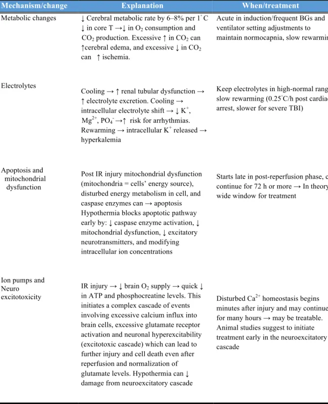
27 Appendix B
Table 1: Potential mechanisms of action, risks and changes with hypothermia
Mechanism/change Explanation When/treatment
Metabolic changes
Electrolytes
Apoptosis and mitochondrial dysfunction
Ion pumps and Neuro
excitotoxicity
↓ Cerebral metabolic rate by 6–8% per 1◦C
↓ in core T →↓ in O2 consumption and
CO2 production. Excessive ↑ in CO2 can ↑cerebral edema, and excessive ↓ in CO2
can ↑ ischemia.
Cooling →↑ renal tubular dysfunction → ↑ electrolyte excretion. Cooling →
intracellular electrolyte shift →↓ K+, Mg2+, PO4- →↑ risk for arrhythmias.
Rewarming → intracellular K+ released →
hyperkalemia
Post IR injury mitochondrial dysfunction (mitochondria = cells’ energy source), disturbed energy metabolism in cell, and caspase enzymes can → apoptosis Hypothermia blocks apoptotic pathway early by: ↓ caspase enzyme activation, ↓
mitochondrial dysfunction, ↓ excitatory neurotransmitters, and modifying intracellular ion concentrations
IR injury →↓ brain O2 supply → quick ↓
in ATP and phosphocreatine levels. This initiates a complex cascade of events involving excessive calcium influx into brain cells, excessive glutamate receptor activation and neuronal hyperexcitability (excitotoxic cascade) which can lead to further injury and cell death even after reperfusion and normalization of glutamate levels. Hypothermia can ↓
damage from neuroexcitatory cascade
Acute in induction/frequent BGs and ventilator setting adjustments to maintain normocapnia, slow rewarming
Keep electrolytes in high-normal range, slow rewarming (0.25◦C/h post cardiac
arrest, slower for severe TBI)
Starts late in post-reperfusion phase, can continue for 72 h or more → In theory wide window for treatment
Disturbed Ca2+ homeostasis begins
28 Inflammation Free radials Blood-brain Barrier/vascular permeability
Acidosis and cellular Metabolism
Brain injury → Proinflammatory mediators released ++ → leukocytes
drawn across BBB →↑ inflammatory cells
in brain → passage of neutrophils, phagocytic monocytes and macrophages into brain → phagocytic action and toxin production → further injury by stimulating further immune reactions. Some of this is neuroprotective, but if continual and excessive →↑ injury. Hypothermia →↑
ischemia-induced inflammatory and immune reactions, ↓ NO production (key agent in developing brain injury post-ischemia), ↓ neutrophil/macrophage function and ↓ WCC
IR injury →↑ free radicals that oxidize and damage cell components → brain’s defense mechanisms likely overwhelmed. Hypothermia →↑ release of free radicals
→ endogenous antioxidants more able to
meet demand
Traumatic/IR injury can disrupt BBB →
brain edema. Mild hypothermia ↓ BBB
disruptions and vascular permeability after IR injury →↓ brain edema. Brain edema and ICH play key role in neurological injury in severe TBI and ischemic stroke, and ICH is a marker for neurological injury → plausible that therapies to ↓ ICP may also improve neurological outcome Hypothermia has been used to ↓ ICP in neurological injury including TBI, ischemic stroke, meningitis and SAH
Ion-pump failure, mitochondrial
dysfunction, cellular hyperactivity and ↓ in cell membrane integrity → intracellular acidosis →↑ harmful processes. Hypothermia can alleviate this, may improve brain glucose metabolism and when induced early enhances speed of metabolic recovery →↓ toxic metabolite accumulation →↓ acidosis
Begins ~ 1 h after ischemia and persists for up to 5 days, suggesting a
therapeutic window for these mechanisms
Brain edema peaks after 24–72 h → this mechanism could offer a wide
29
Brain temperature
Coagulation
Vasoactive mediators
Brain temperature slightly higher than core temperature and can ↑ 0.1–2.0 ◦C post-injury (more with fever). Injured areas are hotter than uninjured areas due to cellular hyperactivity. Dissipation of heat by lymph/venous drainage is hampered by local brain edema (cerebral thermo-pooling) →↑ hyperthermia related injury Hypothermia in brain-injured patients may
↓ potential hyperthermia-related adverse effects
Activation of coagulation seems to be involved in developing IR injury. Its reversal, whilst targeting other mechanisms, could improve outcomes Hypothermia induces anticoagulatory effects: mild platelet dysfunction at 33–35◦
C; can affect clotting factors at ≤ 33◦C, and a potential reduction in platelet count, may influence synthesis and kinetics of clotting enzymes and plasminogen activator inhibitors. This anticoagulation effect could provide protection, but not investigated. Cooling to 35◦ C – no effect
on coagulation
Secretion of vasoactive substances endothelin and TxA2 (vasoconstrictors)
and prostaglandin I2 (vasodilator) is
affected by hypothermia. TxA2 and
prostaglandin I2 regulate cerebral blood
flow. Their balanced production is required to maintain homeostasis. If
disrupted by ischemia/trauma TxA2
production increases which can →
vasoconstriction and hypoperfusion in injured brain. Hypothermia →↓imbalance, but regulation of cerebral perfusion is complex and influenced by cerebral autoregulation and patient management. Influence of hypothermia on secretion of
30
Improved tolerance of ischemia (pre-conditioning)
Reduction of epileptic activity
Early gene activation
Shivering Insulin sensitivity and secretion Cardiovascular/ hemodynamic effects
vasoactive mediator in brain-injured patients requires further investigation
In animal models ‘preconditioning’ with hypothermia improves tolerance for ischemia. As brain injury is frequently complicated by ischemic events after the initial insult, this could be a valuable neuroprotective mechanism
Epileptic activity without signs and symptoms (non-convulsive) occurs frequently in brain-injured patients and if it occurs in the acute phase of brain injury the combined effect is destructive. Evidence indicates that hypothermia # epileptic activity; another mechanism through which it could provide neuroprotection
Hypothermia →↑ early gene activation which is part of the protective cellular stress response to injury and →↑
production of cold shock proteins that can be cryoprotective in the presence of ischemic and traumatic injury
↑ metabolic rate, O2 consumption, work of
breathing, heart rate and myocardial O2
consumption
↓ with cooling → hyperglycemia or ↑
insulin required
Mild hypothermia: In euvolemic, adequately sedated pts ↓ HR, ↑
myocardial contractility, → or slightly ↑
BP, ↓ CO. ↓ metabolic rate matches or exceeds ↓ CO → balance maintained Initial transient ↑ HR due to ↑ venous return (↑ if sedation inadequate, shivering untreated) Stabilizes cell membranes →↓
risk of arrhythmias, ↑ successful
defibrillation. Deep hypothermia: (≤30◦C)
↓ contractility, ↑ risk for arrhythmias, ↓
successful defibrillation, ↓ response to antiarrhythmics Cold diuresis: the result of
↑ venous return (due to peripheral vessel constriction), atrial natriuretic peptide activation, ↓ ADH and renal ADH receptor
34–35◦ C/opiates, sedation, paralysis if
required, other agents
Induction and rewarming/frequent BGL checks and insulin adjustments, slow rewarming
Sedate adequately
Allow ↓ HR 45–55 at 33◦C (artificial ↑
HR →↓ contractility)
31
Coronary perfusion
Drug clearance
Infection
Gut
levels, and tubular dysfunction →
hypovolemia.
↓ metabolic rate and HR protects ischemic myocardium, ↑ coronary vasodilation and perfusion. But in severely atherosclerosed coronaries, vasoconstriction can occur →
may affect result of hypothermia
Shivering can ↑ myocardial O2
consumption
Most enzyme-based reactions slowed →↓
drug clearance by liver. Tubular
dysfunction may also affect clearance, and response to some drugs alters e.g. ↓ effect of adrenaline and noradrenaline. BUT most drug levels ↑→↑strength and duration of effect
↓ leukocyte migration and phagocytosis, ↓
proinflammatory cytokine synthesis →↓
proinflammatory response → may protect
against damaging neuroinflammation, but
↑ risk for infection (↑ risk with ↑ duration)
↑ risk for wound infection due to cutaneous vasoconstriction
Signs of infection: e.g. fever and possibly
CRP and WCC ↓.
↓ gut function and gastric emptying, ↓
metabolic rate
Sedate adequately-prevent shivering
Modify doses of certain drugs
Low threshold for antibiotic treatment may be advisable
↑ in ‘cooling power’ required may indicate fever and infection
Reduce feeding target in maintenance phase
↓: decrease (d), →: leads to, ↑: increase (d), T: temperature, O2: oxygen, CO2: carbon dioxide, BGs: blood gases, IR: ischemia reperfusion, ATP:
adenosine triphosphate, Ca2+: calcium, BBB: blood–brain barrier, NO: nitric-oxide, WCC: white cell count, ICH: intracranial hypertension, ICP:
intracranial pressure, TxA2: thromboxane A2, ◦C: degrees Celsius, BGL: blood glucose level, K+ : potassium, Mg2+: magnesium, PO4- : phosphate,
TBI: traumatic brain injury, HR: heart rate, BP: blood pressure, CO: cardiac output, ADH: antidiuretic hormone, CRP: C-reactive protein.
32 Appendix C
The Cochrane Risk of Bias Tool was my quality evaluation choice.
Holzer at al.
Sunde
at al.
Bernard at al.
Selection bias
Treatmentassignments were randomly generated by computer in blocks of 10
The patients in this study could not be randomized. They were either in the control group (1st phase), or the treatment group (2nd phase)
The patients were randomized using the method of odd and even days. Despite the potential for bias in randomization, it appears that the two patient groups were comparable.
Performance bias
Patient care providers involved in the care of patients during the first 48 hours after cardiac arrest could not be blinded with respect to treatment assignments. Physicians responsible for assessing the neurologic outcome within the first six months after the arrest were however blinded of the patients treatment assignmentTreatment
assignment could not be blinded. Medical providers were not blinded to patient treatment plans. Possible bias due to increased awareness and enthusiasm for the new treatment approach
Treatment
assignment was not blinded. It was not feasible to blind medical providers to the patients' treatment group assignments. There is a possibility that bias could have affected patient care and outcome
Detection bias
Physicians responsible for assessing the neurologic outcome within the first six months after the arrest were blinded of the patients treatment assignmentPhysicians
responsible of patient care were not blinded of their patients outcome, however they were blinded to the patient outcomes in the control phase
Patient was
discharged by a rehab center physicians who was unaware of initial treatment protocols and were blinded to the
33
Attrition bias
Minimal incompleteoutcome data reported Minimal incomplete outcome data reported Minimal incomplete outcome data reported
Reporting bias
No evidence of selective outcome reporting foundNo evidence of selective outcome reporting found
No evidence of selective outcome reporting found
Other
The requirement ofinformed consent was waived in accordance to ethical standards and local guidelines. The patient’s family was informed about the trial, and the studies protocol stated that if there were any protests, the patient would be withdrawn from the study.
Written consent to be included in this study could not be obtained from patient. Written informed consent for participation in this study was sought from the next of kin as soon as possible after the arrival of the patient at the hospital. Also there could be possible bias due to conflict of interest since Dr. Sunde received research grants from Laerdal Foundation for Acute Medicine and
Professor Steen is a member of the Board of Laerdal Medical.
34 Appendix D
Standardized post resuscitation treatment protocol
In hospital standardized treatment plan after ROSC at Ulleval University Hospital Goal: to reduce the vital organ injuries (brain, heart), through:
1. Initial optimizing hemodynamics and oxygenation
2. (a) Treat the cause of arrest; reperfusion (PCI) after STEMI and
(b) Therapeutic hypothermia (33 ◦C in comatose patients for 24 h) Start as early as possible after decision making in the ED Initially 1-3 l of ice-cold 0.9% NaCl i.v. together with ice bags
Endovascular cooling/external cooling for maintenance after arrival at the ICU
3. A standardized treatment protocol for the following days
3.1. Factor Goal Strategy
Reperfusion Reperfusion PCI in STEMI
Blood pressure MAP > 65—70 mmHg Volume, vasopressors, inotropic
agents, IABP
Central venous pressure 8—12 mmHg Volume, glyceryl trinitrate,
diuretics
ECG, rate/ischemia 60—100/min Volume, sedation, glyceryl
trinitrate, beta-blocker (normally not indicated when using therapeutic hypothermia because of relative bradycardia)
Temperature 33 ◦C for 24 h Initially ice cold (4 ◦C) NaCl 0.9%
i.v.
and icepacks, then
internal/external cooling device
Ventilator SpO2 95—98 Respiratory control, FiO2, PEEP
pCO2 5—6 kPa (NB! Avoid hyperventilation)
Blood glucose 5—8 mmol/l Actrapid-infusion (NB! Avoid
hypoglycemia/hypokalemia)
Electrolytes Normal values Replacement/specific treatment
Hemoglobin >9—10 g/dl Transfusion if necessary
Diuresis >1 ml/kg/h Volume, diuretics or pressors
Buffers pH > 7.1, BE >−10 When indicated, trometamol
125—250 ml i.v.
Seizures Prevent/treat seizures Increase sedation, or specific
anticonvulsive medication
EEG when indicated (early contact with a neurologist)
3.2. Sedation
35
3.3. Monitoring Arterial catheter O2-saturation Continuous ECG
Central venous line with central venous pressure Temperature (bladder)
Arterial blood gases (pH, BE, pCO2, pO2) Blood glucose and electrolytes Echocardiography, chest X-ray EEG and SEP
3.4. Vasopressors/inotropic agents
First choice: dopamine If tachycardia, check volume status, (2—10 µg/kg/min) or change to noradrenaline
(norepinephrine) (0.02—.3 µg/kg/min)
If pump failure/cardiogenic shock IABP
Dobutamine (2—10 µg/kg/min) and if necessary adrenaline (epinephrine)(0.02—0.3 µg/kg/min)
(levosimedane as last resort)
3.5. Awakening protocol/respirator weaning
After 24 h of cooling, patients should be slowly rewarmed (0.5 ◦C/h). Sedation may be stopped after the body temperature has reached 35.5 ◦C. Extubation using normal indications. Avoid long term ventilator treatment (if no complications are present)