Vaccination against
atherosclerosis
Vaccination against
atherosclerosis
A novel therapeutic approach
PROEFSCHRIFT
ter verkrijging van
de graad van Doctor aan de Universiteit Leiden,
op gezag van de Rector Magnificus Prof. mr. P.F. van der Heijden, volgens besluit van het College voor Promoties
te verdedigen op donderdag 29 januari 2009 klokke 16.15 uur
door
Thomas van Es
Promotores: Prof. dr. Th.J.C. van Berkel Prof. dr. J. Kuiper
Referent: Prof. dr. P. H. A. Quax
Overige leden: Prof. dr. J.D. Laman (Erasmus MC) Prof. dr. M. Danhof
Prof. dr. W. Jiskoot
The studies described in this thesis were supported by a grant of the Netherlands Heart Foundation (NHF-2000T040) and were performed at the Division of Biopharmaceutics, Leiden/Amsterdam Center for Drug Research, Leiden University, Leiden, the Netherlands. Financial support by the Netherlands Heart Foundation for publication of this thesis is greatefully acknowledged.
The realization of this thesis was also financially supported by: -Leiden University
-LACDR -Pfizer
Welke fakkel wij ook ontsteken en hoever zij haar licht ook verspreiden moge: Onze horizon blijft steeds de diepere duisternis
-Schoppenhauer-
Cover designed by C.J.N. Verkleij and T. van Es Printing: Wöhrmann Print Service
ISBN: 978-90-9023853-1 van Es, Thomas
Vaccination against atherosclerosis A novel therapeutic approach Proefschrift Leiden
Met literatuur opgave – Met samenvatting in het Nederlands © 2009 T. van Es
TABLE OF CONTENT
Page
CHAPTER 1: General introduction and outline of the thesis 9
CHAPTER 2: IL-15 aggravates atherosclerotic lesion development 47 in LDL receptor deficient mice
CHAPTER 3: Vaccination against interleukin-17 attenuates 63 atherosclerosis in LDL receptor deficient mice
CHAPTER 4: Attenuated atherosclerosis upon interleukin-17 81 receptor signaling disruption in LDL receptor
deficient mice
CHAPTER 5: Bone marrow transplantation with p19 deficient 97 bone marrow does not alter the atherosclerotic
burden in LDL receptor deficient mice
CHAPTER 6: Vaccination against the p28 subunit of 109 interleukin-27 aggravates atherosclerosis in
LDL receptor deficient mice
CHAPTER 7: Induction of oral tolerance to HSP60 or an 129 HSP60-peptide activates T cell regulation and
reduces atherosclerosis
CHAPTER 8: Vaccination against Foxp3 regulatory T cells 151 aggravates atherosclerosis
CHAPTER 9: A vaccine against atherosclerosis: myth or reality? 169
CHAPTER 10: Summary and perspectives 185
Nederlandse samenvatting 199
List of abbreviations 209
List of publications 215
Curriculum Vitae 219
Chapter 1
General introduction and
outline of the thesis
1
1 ATHEROSCLEROSIS
1.1 GENERAL
Cardiovascular disease (CVD) is still the number one cause of death in the Western world. There are many different clinical manifestations of cardiovascular disease such as angina pectoris, cardiomyopathy, endo-myocarditis and aneurysm.
Atherosclerosis affects the medium and large sized arteries of the heart and blood vessels and is the underlying cause of many clinical symptoms of CVD. Major risk factors for CVD, such as smoking, obesity and a high fat diet are well recognized. In 2004, 45445 people died of the consequences of CVD, accounting for 33%1 of all reported deaths in the Netherlands, whereas 28% (38824 people) of all
reported deaths death were related to cancer.2 This states the importance of
cardiovascular disease in the Dutch society.
The current treatment of atherosclerosis is mostly aimed at the reduction of risk factors by life style advice (stop smoking, more exercise, and lower cholesterol intake) and by subscribing drugs, such as statins, that lower plasma cholesterol levels. Additionaly drugs that lower blood coagulation and blood pressure are prescribed. However, these interventions cannot prevent that CVD is still the leading cause of death in the Western world. Therefore, there is an urge to develop new therapies targeting the different molecular pathways and stages of atherosclerosis.
1.2 PATHOGENESIS OF ATHEROSCLEROSIS
1.2.1 Initiation of the lesion
The first stage of atherosclerosis is fatty-streak formation. Fatty streak formation is asymptomatic and is already found during the first decades of life in medium and large sized arteries, at predisposed sites. The typical atherosclerotic prone sites are characterized by low shear stress and high oscillatory shear stress, which increase adhesion of leukocytes and the expression of inflammatory genes.3
Under normal conditions, healthy endothelium is able to respond to physical and chemical signals by the production of a wide range of factors that regulate vascular tone, cellular adhesion, thrombus resistance, smooth muscle cell proliferation, and vessel wall inflammation.4 It is generally accepted that the
Circulating lipoproteins, in particularly very low-density lipoproteins (VLDL) and low-density lipoproteins (LDL) infiltrate into the arterial intima and become modified through processes, such as oxidation, glycation, aggregation, association with proteoglycans or incorporation into immune complexes.7-10 The
modified LDL particles are highly immunogenic and activate the endothelial cells. Stimulated endothelial cells undergo a switch from a quiescent phenotype towards a phenotype that initiates a defense response. Most cardiovascular risk factors, such as smoking and high blood pressure, activate the molecular machinery in the endothelium, resulting in the expression of chemokines, cytokines and adhesion molecules designed to interact with leukocytes and platelets and designed to target inflammatory cells to specific tissues to clear invading microorganisms or to respond to vascular injury.11 Activated endothelial
cells express adhesion molecules, like vascular cell adhesion molecule-1 (VCAM-1), intercellular adhesion molecule-1 (ICAM-1), P-selectin and E-selectin.3, 12
Leukocytes (i.e. monocytes and lymphocytes) express counter receptors for these adhesion molecules and decelerate via interaction with P- and E-selectin. Once slowed down, a more firm adhesion is facilitated via interaction with VCAM-1 and ICAM-1 with very late antigen-4 (VLA-4) and lymphocyte function associated antigen-1 (LFA-1), respectively, which are expressed on leukocytes.13-15
Once a firm adhesion is established, leukocytes migrate through the interendothelial junction into the subendothelial space (diapedesis) (Figure 1.2A). This process is facilitated by additional adhesion molecules, such as platelet/endothelial cell adhesion molecule-1 (PECAM-1) and junctional adhesion molecule-1 (JAM-1).16, 17 Furthermore, activated endothelial cells produce several
chemokines and interleukins (IL), which enhance diapedesis 18 and the recruitment
of leukocytes into the lesion. CCL2 (MCP-1), produced by vascular endothelial cells, is an important chemoattractant for monocytes and T cells. These cells play an important role in lesion development.19-22 Activated vascular cells but also
macrophages within the lesion continue to produce chemokines such as, CCL5 (RANTES), CXCL10 (IP-10) and CCL11 (eotaxin) to further enhance the immune response (Figure 1.2B).23-25 Within the lesion, monocytes differentiate into
macrophages by stimulation of macrophage-colony-stimulating factor (M-CSF), which is produced by endothelial and smooth muscle cells.26 The macrophages
become activated by the uptake of modified LDL, thereby transforming into lipid loaded “foam cells”. This process will be discussed in more detail later on.
1
FIGURE 1.1: DEVELOPMENT OF ATHEROSCLEROSIS.
A, fatty streak formation: Endothelial cells in lesion prone areas become activated leading to permeabilety for lipoproteins, such as LDL. Within the intima, LDL is modified by processes, like oxidation and becomes immunogenic. Monocyte derived macrophages migrate into the intima and start to phagocytose the modified LDL particles and become activated. Macrophages, trapped in the intima and loaded with cholesterol are now called “foam cells”. B, progression of the plaque: vascular smooth muscle cells become activated. They migrate and produce extracellular matrix proteins, in particular collagen, to form a cap structure to protect the lesion from the blood flow. More leukocytes are recruited to the intima, such as T cells and monocytes and these cells enhance the inflammation. To compensate for the narrowed lumen, outward remodeling takes place. C, necrotic core and thrombus formation: “foam cells” become apoptotic and eventually form a necrotic core, consisting of cellular debris and free cholesterol. The necrotic core is highly immunogenic, which results in the recruitment of more inflammatory cells to the intima. The fibrous cap formed by the smooth muscle cells protects the lesion from the bloodflow, however this may not prevent rupture. When the plaque ruptures, a thrombus will form and can cause clinical symptoms, such as acute coronary syndrome. D, obstructive lesion: when the plaque does not rupture, the lesion can grow by the ongoing inflammation. When outward remodeling is not sufficient, the lesion becomes obstructive and causes clinical symptoms, such as angina pectoris. More details are described in the text. (Adapted from Rader and Daugherty).236
1.2.2 Progression of the lesion
Atherosclerotic lesion progression is associated with the continuous influx of inflammatory cells due to the local production of chemokines in the plaque. T cells interact with activated macrophages (“foam cells”), which express class II and class I histocompatiblity complexes (MHC II and MHC I) and present antigens to T cells. CD4+ and CD8+ T cells are associated with all stages of atherosclerotic lesion
development and activation of T cells results in a broad range of immune responses and the acquisition of many features of a chronic inflammatory state.27
Activated T cells produce several cytokines such as IFN-γ and TNF-α.28 These
cytokines activate other cells within the lesion, such as endothelial cells, macrophages and smooth muscle cells.
The necrotic core becomes covered by a fibrous cap, which consists of smooth muscle cells and extracellular matrix proteins like collagen. The formation of the cap structure is facilitated by cytokines and growth factors, which are produced by activated macrophages and T cells. The cytokines and growth factors stimulate smooth muscle cells to proliferate and migrate to the cap.5, 6Activated smooth
muscle cells can migrate from the media to the intima, where they are able to internalize lipids and transform into smooth muscle cell derived foam cells and can produce matrix proteins.30
In this stage of lesion development, the outward remodeling of the vessel takes place to compensate for the increase in lesion size. The process of outward remodeling is necessary to prevent severe narrowing of the vessel and to preserve blood flow.31 The above-described process is schematically depicted in figure 1.1B
and C.
FIGURE 1.2: THE ROLE OF MACROPHAGES AND T CELLS IN ATHEROSCLEROSIS.
1
1.2.3 Lesion stability and rupture
When outward remodeling can not compensate for the reduced volume, the advanced atherosclerotic lesion leads to insufficient blood flow to distal tissues, causing clinical symptoms, such as angina pectoris (Figure 1.1D). However, acute cardiovascular events are associated with myocardial infarction and stroke as a result of a ruptured plaque and a subsequent thrombotic event (Figure 1.1C).32, 33
Plaque rupture is the final outcome of plaque destabilization. During lesion development activated macrophages, T cells and other immune cells accumulate around the necrotic core and in the shoulder regions of the plaque.6, 34
Furthermore, activated macrophages, T cells, and mast cells have been observed at
sites of plaque rupture, indicating their potential relation to the rupture process.
35-37 T cells, predominantly of the T helper 1 phenotype, produce high amounts of
IFN-γ, which inhibit the production of collagen by vascular smooth muscle cells
and their cell proliferation, thereby negatively influencing plaque stability.28, 38
Activated macrophages produce several proteases, which destabilize the plaque, such as matrix metalloproteinases (MMPs), cysteine proteases, and
chymases.39-41 Members of these families of enzymes are found in the
atherosclerotic plaque and can degrade the matrix. Especially MMP-1, MMP-8,
MMP-9 and MMP-13 may be important.42 In addition, macrophages induce
apoptosis of vascular smooth muscle cells, thereby negatively influencing the
collagen production and subsequent plaque stability.43, 44
Since the weakened cap structure cannot withstand the hemodynamic forces, the unstable atherosclerotic lesion may rupture and consequently expose thrombogenic plaque material (lipids/necrotic core) to the blood. The subsequent aggregation of platelets and coagulation forms a thrombus, which obstructs blood
flow and results in clinical symptoms such as myocardial infarction and stroke.34
Plaques that are prone to rupture contain high numbers of activated immune cells. Furthermore, patients with acute coronary syndromes (ACS) demonstrate signs of inflammation, with elevated levels of circulating cytokines,
acute phase reactants, and not only activated T cells.45, 46 Therefore the immune
system and inflammation play an important role throughout atherosclerotic plaque development, but are also crucial in the final stage of atherosclerosis.
2 ATHEROSCLEROSIS: AN INFLAMMATORY DISEASE
immune response, both with their specific role in host defense.47,48 This is
illustrated in different mouse models for atherosclerosis with a specific depletion of components of the innate as well as the adaptive immune system.49, 50 For
example, when LDL receptor deficient mice were cross-bred with lymphocyte-deficient (RAG1 lymphocyte-deficient) mice, a reduction in lesion size was observed.51
Further evidence supporting the relation between atherosclerosis and inflammation is found in gene polymorphisms involved in inflammation, such as TLR4 polymorphisms.52-54 There are associations found between an increased risk
of cardiovascular events and autoimmune diseases, such as rheumatoid arthritis and systemic lupus erythematosus.55 However, besides inflammation,
atherosclerosis is also associated with metabolic and hemodynamic factors, thereby placing atherosclerosis as an unique disease.
Metabolic and hemodynamic factors are likely to play a role in the initiation of autoimmunity by activation of endothelial cells and subsequent recruitment of immune cells. This process leads to initial atherosclerosis via the innate immune system and gradually evolves into a chronic, autoimmune like, inflammatory disease via the adaptive immune system. Therefore, the regulation and crosstalk between innate and adaptive immune cells is very important in the initiation and development of atherosclerosis.
Although several exogenous stimuli, such as cytomegalovirus (CMV) and
Chlamydia pneumoniae have been identified in atherosclerosis,56 there is also
evidence that endogenous stimuli are involved in the process of atherosclerosis.57, 58
2.1 INNATE IMMUNE SYSTEM
The innate immune response is the fist line of defense against pathogenic stimuli and is characterized by a natural selection of germline-encoded receptors, which focuses on highly conserved motifs in pathogens. It provides the first line of defense for the host and is characterized by fast (minutes to hours) and blunt (lacking exquisite structural specificity) responses. It is a very conserved system, which is already present in many lower organisms.
Important cells involved in the innate immune response are macrophages, neutrophils, mast cells and natural killer (NK) cells. The exact role of neutrophils in atherosclerotic lesion initiation is not known yet, but neutrophils are found in ruptured or eroded plaques, indicating that they are recruited in a later phase in response to injury.59 However, upon endothelial activation, P-selectin and
1
Another kind of innate immune cells are mast cells. These cells are present in atherosclerotic lesions and are activated at sites of plaque rupture, indicating
that mast cells are involved in the rupture process of advanced lesions.64 Mast cells
store granules, which contain growth factors, chymases and pro-inflammatory cytokines.65,66 Once stimulated, the mast cells degranulate and exocytose the
granule-associated effector substances into their microenvironment, thereby negatively influencing plaque stability.67, 68
NK cells are found to play a role in early atherosclerosis.69, 70 Their role
however, is not yet completely understood. Reduced atherosclerosis was observed in LDLr-/- mice after bone marrow transplantation from transgenic mice
overexpressing the Ly49A receptor, which results in dysfunctional NK cells.70
Noteworthy, in these transgenic mice, not only NK cells are affected, but also NK T cells, CD8+ cytotoxic cells and other lymphocytes expressing granzyme A.
Therefore, it is difficult to determine the exact role of NK cells in the initiation of atherosclerosis.
The key inflammatory cell during atherosclerotic plaque formation is the macrophage. Macrophages are part of the innate immune system and have an important “bridge” function between the innate and adaptive immune system by presenting innate immune signals to the adaptive immune system. In atherosclerosis, macrophages play an important role in the various phases of lesion formation and progression.12 Infiltrated monocytes differentiate into macrophages
and start to express cytokines and receptors71 by stimulation via M-CSF, which is
produced by endothelial and smooth muscle cells.26 Monocyte-derived
macrophages express pattern recognition receptors (PRRs), which are involved in the innate immune response. PRRs recognize a restricted pattern of ligands called pathogen-associated molecular patterns (PAMPs). PAMPS consist of many different ligands such as lipopolysaccharides, aldehyde-derivatized proteins, bacterial DNAs and denatured DNAs, resulting in endocytoses and lysosomal degradation of the ligand72,73 and activation of nuclear factor-κB, resulting in an
inflammatory response.74
Two important groups of PRRs are the scavenger receptors (ScRs) and the toll-like receptors (TLRs), which are both expressed by macrophages.47, 75 The ScR
family, which includes CD36, CD68, SR-A and SR-B, mediates the internalization of modified lipoproteins (e.g. oxLDL) via endocytosis and contributes to foam cell formation, a hallmark of the atherosclerotic lesion.9,76,77 The uptake of modified
lipoproteins and their constituents via ScRs is important in triggering the production of the mediators of innate immunity such as, IL-1β and TNF-α.78
The other important pattern recognition receptor family, the TLRs, are involved in the activation of macrophages.80 Studies with TLR4 and ApoE double
knock out mice, demonstrate a reduction in atherosclerotic plaque development, thereby further illustrating the importance of the innate immune system in atherogenesis.81 Activation of ScRs and TLRs on macrophages within the plaque
results in a proinflammatory environment capable of activation of other vascular and immune cells, thereby enhancing lesion progression.
Within the lesion, foam cells contribute to the production of reactive oxygen species, cytokines and other molecules, thereby amplifying and sustaining the inflammatory response in the plaque.80 Additional cells in the early plaque,
such as smooth muscle cells and endothelial cells become activated by these molecules and start producing interleukins and chemokines, i.e. IL-1β, IL-6, IL-18, TNF-α and CCL2.82-84 Besides production of these cytokines, macrophages and
other vascular cells start to produce T cells attractants, like CCL5, CXCR3, CXCL10 and CXCL11.24 The pro-atherosclerotic effect of T cells in atherosclerosis is
demonstrated by blocking for example CCL5 or CXCR3 and thereby attenuating atherosclerosis.85,25
Besides attracting T cells, macrophages also express MHC class II in association with a specific epitope. This is a pivotal step in bridging the innate immune response to the adaptive immune response. Within the lesion MHC class
II expressing macrophages (and also dendritic cells, discussed in more detail later
on) can be detected close to T cells, which suggests that there is an ongoing
immune activation of the adaptive immune response in the plaque.86-88
2.2 ADAPTIVE IMMUNE SYSTEM
1
The role of the adaptive immune system, especially the T and B cells, is nicely demonstrated in studies in which these cells were not functional by for example using severe combined immunodeficient (SCID) mice, resulting in decreased lesion formation.89
2.3 AUTOANTIGENES
Many exogenous and endogenous antigens have been suggested to be presented in atherosclerosis. As mentioned before, T cells recognize specific antigens presented by APCs with their TCR. Upon activation, T cells that recognize a specific antigen will start proliferating, resulting in clonal T cell expansion. Clonal T cell expansion has been detected in atherosclerotic lesions of mice and humans, suggesting specific TCR activation by an (auto) antigen.90-92
Since inflammation has a prominent role in atherosclerosis, it has been suggested that exogenous stimuli may initiate atherosclerotic lesion development. Many virus and bacteria related antigens have been identified in atherosclerosis, but most extensively studied are the Cytomegalovirus (CMV)93 and Chlamydia
pneumoniae.94, 95 Both pathogens are associated with aggravating atherosclerosis in
mice and humans, since antibodies against these pathogens have been correlated with the severity of cardiovascular disease in patients.93, 96-98 Furthermore,
experimental data have shown that infection with Chlamydia pneumoniae enhances atherosclerosis.97 However, large clinical trials on the treatment of cardiovascular
patients with antibiotics directed against Chlamydia pneumoniae did not result in a reduction of cardiovascular events in antibiotic treated patients.99, 100
Another group of antigens is of endogenous origin, but is related to exogenous antigens via a process called molecular mimicry.101 An example are the
heat shock proteins (HSPs), a group of highly conserved proteins. These proteins are highly conserved and immune responses induced against bacterial HSP60 may cross-react with responses against endogenous hsp60. Endogenous HSP60 is induced when cells are exposed to different stress stimuli.102 HSP60 is expressed on
endothelial cells, vascular smooth muscle cells and mononuclear cells in human atherosclerotic plaques.103 Furthermore, circulating antibodies against HSP60 were
detected in patients with atherosclerosis and HSP60 specific T cells were detected within the atherosclerotic plaque.104,105 Therefore an immune response against
HSP60 may contribute to endothelial damage and subsequent enhancement of atherosclerosis.106,107 Interestingly, antibodies against HSP60 and its prokaryote
homologue HSP65 are also detected in other autoimmune diseases such as rheumatoid arthritis.108 In relation to this, patients with rheumatoid arthritis have a
A third group of antigens is derived from endogenous sources. This group mainly contains altered self-proteins and the autoimmune response against these proteins is directed against the neo-epitopes of the altered proteins. T cells do not react with native LDL, as there is immunological tolerance against self-antigens. However, modifications of LDL lead to non-self epitopes (neo-epitopes) and increased autoreactivity of T cells in mice and human.110, 111 LDL in the
atherosclerotic lesion can be modified by various processes as described before, and are accountable for the development of neo-epitopes.112 An example of a
neo-epitope related to LDL is oxLDL. OxLDL specific T cells are identified in human plaques and circulating antibodies against epitopes of oxLDL are detected in serum samples of patients with cardiovascular disease.110, 113 Furthermore, lymph
nodes and spleens of ApoE-deficient mice gave rise to an oxLDL specific T and B cell line displaying a strong humoral and cellular immune response against these modified lipoproteins, indicating the role of oxLDL in immune activation.114, 115
2.4 DENDRITIC CELLS
Although activated macrophages effectively present antigens to T cells, dendritic cells (DCs) are the most potent APCs of the immune system and are the key players in the regulation of the adaptive immune response. 116
Immature DCs efficiently take “samples” of their antigenic microenvironment through macropinocytoses and receptor mediated endocytosis. Depending on the triggered PRRs, DCs present the antigen in context of either MHC class I or MHC class II and produce cytokines to evoke an appropriate immune response.117 Therefore, DCs are crucial for an adequate clearance of the
infection, but DCs are also responsible for pathogenic immunological responses. Dendritic cells are a component of the vasculature associated lymphoid tissue and low numbers are found in the intima of healthy, but susceptible arteries before atherosclerotic lesion development is initiated. 118 Furthermore, DCs
increase in number in the intima during the progression of atherosclerosis.119 This
indicates a role for DCs in the initiation and regulation of arterial inflammation. Immature DCs capture antigens at the site of inflammation and migrate to secondary lymphoid organs, such as the spleen and lymph nodes. The migration is orchestrated by various chemokines, such as CCR-2, -5, -6, -7 and CXCR1 and CXCR2.120, 121 Within the secondary organs the maturated DCs are able to stimulate
antigen specific T cells, which is further enhanced by the fact that mature DCs starts to express co-stimulatory molecules, such as CD40, thereby enabling them to interact with CD40L expressing T cells.
1
However, in hypercholesterolemic conditions these monocyte derived DCs are possibly not able to migrate out of the atherosclerotic lesion to the secondary lymph organs, thereby directly activating residential cells in the lesion such as T cells, which leads to aggravated atherosclerosis.122-124
2.5 T CELLS
T cells are activated in the lymphoid organs by APCs by recognizing a specific antigen and by costimulatory signals such as CD40L-CD40 and CD80/CD86-CD28 interactions.125 The microenvironment determines the type of T cell response. For
example IL-12 production by APCs lead to the development of T helper 1 (Th1) cells, which have been shown to aggravate atherosclerosis.126 Activated T cells
migrate from the lymphoid organs to the site of inflammation e.g. the atherosclerotic lesion via chemokine signaling and are reactivated upon recognition of the antigen presented by APCs. Interference in T cell migration via the inhibition of the CXCR3 and CCL5 pathway, results in reduced Th1 cell influx into the lesion and subsequently in reduced atherosclerosis. 85, 127
Most T cells within the atherosclerotic lesions are CD3+ CD4+ TCR αβ+
cells.128, 129 Although CD8+ T cells are also found in atherosclerotic plaques, their
role in atherosclerosis is not yet clear. Furthermore, CD4- CD8- TCR γδ T cells are
found in plaques and may play a role in relation to IL-17 production.129 IL-17 will
be discussed later in more detail.
In relation to the topic of this thesis, the discussed T cell populations will be limited to Th1, T helper 2 (Th2), T helper 17 (Th17) and regulatory T (Treg) cells (Figure 1.3).
2.5.1 T helper 1 cells
Data obtained from patients with CVD illustrated a predominant Th1 pattern within atherosclerotic plaques. This is also observed in mouse models for atherosclerosis. T cells differentiate into Th1 cells through stimulation of naive T
cells with Th1 polarizing cytokines, such as IL-12 and IL-18.130, 131 The production
of interferon (IFN)-γ hallmarks Th1 cells, which has pleiotropic pro-atherosclerotic effects. IFN-γ promotes activation of macrophages and endothelial cells to produce more adhesion molecules, proinflammtory cytokines and chemokines, which results in more T cell recruitment. Furthermore, IFN-γ promotes the production of proteases and inhibits collagen production, thereby interfering in plaque
stability.132 Another Th1 associated cytokine is TNF-α, which also exerts pleiotropic
The role of Th1 cells in the aggravation of atherosclerosis has already been
demonstrated. For example, vaccination against IL-12 in LDLr-/- mice results in a
reduction in IFN-γ expression within the lesion and reduced atherosclerotic lesion
development.131 Buono et al. demonstrated in LDLr and IFN-γ deficient mice a 75%
reduction in lesion size, indicating the pro-atherosclerotic nature of Th1 cells.134
Furthermore, IL-18 deficient ApoE-/- mice also showed reduced atherosclerosis.130
FIGURE 1.3: DIFFERENTIATION OF T HELPER CELLS.
Naive T cells differentiate into either Th1, Th2, Th17 or Treg cells upon stimulation with specific interleukins. Every Th cell subset has its own specific function in the immune response. However, a disproportional proliferation of a Th cell subset may lead to autoimmune diseases or chronic inflammation. The role of T helper cells in atherosclerosis is also depicted in this figure. See text for a more detailed description of the different Th subsets. (Adapted and modified from Tato and O´Shea)237
2.5.2 T helper 2 cells
Traditionally, Th2 cells are considered anti-atherosclerotic due to the production of IL-4, IL-5, IL-9, IL-13, IL-10 and IL-3.135 An athero-protective role for these cells is
suggested since the Th2 interleukins inhibit Th1 cells. Overexpressing IL-10 in LDLr-/- mice for example, exerts anti-atherosclerotic effects.136, 137 Furthermore, IL-5
1
thereby attenuate atherosclerosis.138 This effect is nicely illustrated by Binder et al.,
where IL-5 deficient bone marrow is transplanted into a LDLr-/- recipient mice
leading to aggravated atherosclerosis and a decrease in natural autoantibodies.139
Conflicting data exist on the role of IL-4 in atherosclerotic plaque development. An athero-protective effect on initial plaque development was found after injection of IL-4 into mild hypercholesterolemic mice.140 On the other hand
IL-4 deficient ApoE-/- mice demonstrate reduced atherosclerosis, illustrating a
pro-atherosclerotic effect of IL-4.141 Moreover, IL-4 is associated with increasing MMP-1
production by macrophages and may therefore have negative effects on plaque stability.142
For long, it was thought that a disturbed balance between Th1 and Th2 cells was causative for many autoimmune diseases, including atherosclerosis. However, the aforementioned findings on Th2 cells show that the classical Th1/Th2 balance cannot adequately explain the inflammatory process in atherosclerosis.
Recently, two new T cell subsets were identified: the Th17 cell and the Treg cell. These cells provide more complexity and may provide an opportunity to explain the observed conflicting experimental effects.
2.5.3 Regulatory T cells
The regulatory T cells (Tregs) are important in maintaining immune homeostasis and preventing autoimmunity.143 Tregs develop in the thymus and display a
diverse TCR repertoire specific for autoantigens. Tregs migrate from the thymus into the peripheral tissues and exert their anti-inflammatory response by recognition of specific autoantigens.144
There are various mechanisms of immune suppression by Tregs.182 Firstly,
the suppression of immune cells by cytolysis, via granzymes and perforins. Secondly, suppression by metabolic disruption, via “consuming” IL-2. Thirdly, suppression by inhibitory cytokines, such as TGF-β and IL-10. Finally, suppression by targeting DCs, via the interference in maturation and function of DCs, for example by cell-cell inhibition via CTLA4 (on Tregs) and CD80/CD86 (on APCs).146
Tregs can generally be divided into two groups, the natural occurring Treg (nTreg) cells and the inducible T regulatory (iTreg) cells.147, 148 nTreg cells express
CD4, CD25 (IL-2Rα), cytotoxic T lymphocyte antigen (CTLA)-4 and forkhead box P3 (Foxp3).145 nTreg cells exert their immunosupressive action predominantly by
expressing membrane bound TGF-β, which suppress cells via cell-cell contact in paracrine fashion.146, 149 Furthermore, nTreg cells are able to bind to CD80 and/or
There are also Foxp3 negative Treg cells leaving the thymus, which can be induced in the periphery to become immunosuppressive Treg cells, called inducible Tregs (iTregs). Depending on the suppressive action, these cells can be divided in Tr1 cells, which predominantly produce IL-10151 and Th3 cells, which
exert their immunosuppressive function predominantly via TGF-β. Interestingly, Th3 cells do transiently express FoxP3.152, 153
Mallat et al. hypothesized that adaptive or natural regulatory T cells may play an important role in the regulation of pathogenic T cells in atherosclerosis. There are several studies underlining this hypothesis. Depletion of Treg cells by
treatment with anti-CD25 antibodies results in aggravated atherosclerosis in ApoE
-/- mice.125 Furthermore, van Puijvelde et al. demonstrated that the induction of Treg
cells via tolerance induction against oxLDL or HSP60 leads to attenuated atherosclerosis.154 Moreover, Foxp3 expression has been detected in human
atherosclerotic plaques, indicating that Treg cells are present.155
2.5.4 T helper 17 cells
Th17 cells are a novel T cell subset with a separate lineage. Langrish et al. showed that IL-23 selectively induces the proliferation of in vivo-primed IL-17-expressing Th cells and that these cells do not produce IL-4 or IFN-γ, indicating a separate Th subset.156 Harrington et al. and Park et al. further established the idea of a separated
lineage of Th cells distinct from the T helper type 1 and 2 lineages. A naive precursor T cell is potently inhibited by IFN-γ and IL-4 in differentiation towards Th17 cells, whereas committed Th17 cells are resistant to suppression by Th1 or Th2 cytokines.157 Together these data provide evidence for a new Th subset, which
is regulated by cytokines of Th1/Th2 cells and is involved in autoimmunity. Hence the name, IL-17 is the most prominent cytokine produced by Th17 cells. The 17 family consists of six members, 17A, 17B, 17C, 17D, IL-17E and IL-17F. IL-17A and F are most related to each other and share a 50% homology in protein sequence.158 As IL-17A was the first member of the IL-17
family which was identified, it is mostly designated as IL-17.
The 17R family consists of five members, designated as 17RA, IL-17RB, IL-17RC, IL-17RD and IL-17RE.158 The best-studied IL-17R is IL-17RA, also
designated as IL-17R, and is expressed ubiquitously through the body, which explains the pleiotropic effects of IL-17. The primary source of IL-17A and F are Th17 cells.159 However, there are other cells of the innate and adaptive immune
system which produce IL-17A and F. It has been shown that CD8+ T cells produce
IL17A and F as well as the γδ T cells.160, 161 In many different cell types, binding of
1
chemotactic effect in recruiting and activating neutrophils, providing a mechanism by which Th17 cells can mediate the crosstalk between innate and adaptive immune responses.158,165,166 IL-17 exhibits pleiotropic biological effects on various
atherosclerotic lesion-associated cell types, such as endothelial cells, vascular smooth muscle cells and macrophages.167-169 Upon activation by IL-17 these cells
produce pro-inflammatory cytokines, chemokines and matrix metalloproteinases (MMPs), includingIL-6, CXCL8, CCL2 and MMP-9.169, 170 However, the role of
IL-17 in atherosclerosis is not investigated yet. In this thesis, the role of IL-IL-17 and its receptor in atherosclerosis will be discussed in chapter 3 and 4.
Besides IL-17, Th17 cells also produce IL-21, IL-22 and CCL20. IL-21 is expressed by Th17 cells171, but also by other cell types, such as IL-6 stimulated T
cells.172,173 The receptor for IL-21 is expressed only on lymphoid cells and
predominantly on B cells.174 IL-21 has pleiotropic effects, such as stimulating
proliferation and differentiation of CD8+ T cells175 and it promotes differentiation
and isotype switching in B cells.176 IL-21 induces CXCL8 expression in
macrophages177, which is involved in the recruitment of monocytes to the early
lesion, thereby aggravating atherosclerosis.178 Furthermore, IL-21 regulates the
differentiation of CD4+ T cells to Th17 cells in an autocrine manner.171, 179
IL-22 is a member of the IL-10 family 180and the IL-22 receptor subunits are
primarily expressed on epithelial and parenchymal tissues. 181 IL-22 protects
against liver damage in an acute inflammation model.182
Finally, Th17 cells produce CCL20.183, 184 Interestingly, its receptor CCR6, is
also expressed by Th17 cells.183, 185 This may imply an autocrine mechanism to
regulate its own recruitment in inflamed tissues.The development pathway of the Th17 cell lineage is actively investigated. Initially is was thought that IL-23 was the driving force behind Th17 development.156
IL-23 is a heterodimeric interleukin consisting of a p40 and a p19 subunit. IL-23 is closely related to IL-12 as they both share the p40 subunit. 186 The receptor
of IL-23 consists of a heterodimeric complex consisting of IL-12Rβ1 shared with the 12 receptor and a unique 23R. The 23R shares many features with IL-12Rβ2, which is the other part of the IL-12 receptor. The IL-23R is mainly expressed on effector T cells and not on naive T cells, suggesting an important role for IL-23 in ongoing inflammation.186, 187
IL-23 is mainly produced by macrophages and dendritic cells. Like the subunits of IL-27, EBI3 and p28, p19 must be expressed together with p40 in the same cell in order to be functionally excreted as IL-23.186 Recently, it is
that IL-23p19-deficient animals do not develop EAE and do not develop IL-17-producing T cells.188 In patients with Crohn’s disease, a single nucleotide
polymorphism (SNP) in the coding sequence of IL-23R results in a strong protection against this disease, indicating a pathogenic role of IL-23 in chronic inflammation. Since IL-23 can expand a population of IL-17-producing pathogenic cells, an important role of IL-23 in the development of autoimmune diseases was suggested.156 These data imply that IL-23 may be responsible for the differentiation
of Th17 cells. However, in vivo experiments demonstrated that IL-23 functioned more like a maintenance interleukin for the Th17 cell population.189 Therefore, it
has been proposed that IL-23 may play a role in maintaining or stabilizing the Th17 cell phenotype, or in the survival of Th17 cells (Figure 1.4).190
Interestingly, as research continued, two “old” cytokines with opposing effects, IL-6 and TGF-β, were associated with Th17 cell differentiation.171, 191 IL-6 is
a pro-inflammatory cytokine and was shown to aggravate atherosclerosis.192 On
the other hand, TGF-β is an anti-inflammatory cytokine, which is associated with the differentiation of natural Treg cells and attenuation of atherosclerosis.193194
Most research on Th17 cells is done in mice. However, there are differences observed in Th17 development between mice and humans. Recently, some studies showed that also TGF-β, in combination with IL-1β, IL-6 or IL-21 is able to induce the differentiation of human Th17 cells,195, 196 indicating some overlap. More
research has to be performed to further clarify the different aspects of Th17 cell development in different organisms.
FIGURE 1.4: DETAILED OVERVIEW OF TH17 CELL DIFFERENTIATION.
Since the discovery of Th17 cells many research has been done to elaborate the developmental pathway of Th17 cells. The insight in the role of IL-23 in Th17 cell development changed over time and is now assigned as a stabilizing factor for Th17 cell differentiation. TFG-β and IL-6 are associated with the differentiation of naive T cells to Th17 cells. Furthermore, the recently identified IL-21 is assigned to amplify Th17 cell development via an autocrine positive feedback. (Adapted and modified from Bettelli and Kuchroo)190
1
et al. recently identified increased Th17 cells in patients with acute coronary syndrome.197
Nevertheless, there are indications that Th17 cells may play an important role in atherosclerosis. The prominent effector cytokine, IL-17 is discussed in chapter 3 and 4 of this thesis. Furthermore, the fact that IL-23 is involved in several autoimmune diseases, together with the finding that IL-23 is involved in the Th17 pathway, which is likely to be pro-atherosclerotic, indicates a role for IL-23 in atherosclerosis. Again, there are hardly any reports addressing IL-23 in atherosclerosis. In chapter 5 the role of the IL-23R in atherosclerosis will be discussed. Finally, we also studied the role of IL-27, which can suppress the development of Th17 cells,198 as is described in chapter 6.
2.5.5 Th17 and Treg cells, two different subsets with a close relationship
Both, the Th17 lineage as well as the Treg lineage requires TGF-β for their development. Therefore, a reciprocal relationship between these two cell populations is likely.191 TGF-β is needed for both cell types and IL-6 has a pivotal
role in shifting the balance toward Th17 cells.171, 191 Furthermore Laurence et al.
demonstrated that IL-2, which is necessary for Treg cells, inhibits Th17 cells in a STAT5 dependent way.189Interestingly, IL-21 synergizes with TGF-β to promote
the differentiation of Th17 cells in mice and, as mentioned before, IL-21 has an autocrine loop to enhance its own production by inducing proliferation and recruitment of Th17 cells. 171, 179
There is also an interesting relation between Treg cells and Th17 cells on the level of transcription factors (Figure 1.5). The Th17 cell specific transcription factors, RORγ and RORα, are able to bind and antagonize Foxp3, a Treg transcription factor, and vice versa.199, 200 To underline this fact, conditional
deletion of Foxp3 protein in Treg cells in vivo results in an increase in RORγ and subsequent upregulated IL-17 and IL-21 expression.201, 202 These data may be
FIGURE 1.5: REGULATION OF TH17 AND TREG SPECIFIC TRANSCRIPTION FACTORS.
The transcription factor STAT3 is crucial for Th17 development. IL-6 and IL-21 activate this pathway, however the exact role for TGF-β on transcriptional level remains to be elucidated (black arrow). Recently, IL-27 has been shown to inhibit Th17 cells and this process is probably mediated by STAT1 dependent STAT3 inhibition. Furthermore, IL-2 is able to suppress Th17 development in a STAT1 dependent way. These data suggest a reciprocal relationship between Th17 and Treg cells. (Adapted and modified from Dong)238
2.6 B CELLS
B cells are also an important cell type of the adaptive immune system and are pivotal in the production of antibodies against specific antigens. Depletion of B cells results in aggravated atherosclerosis.203, 204 A specific subpopulation of B cells,
B-1 cells, produce IgM class antibodies (T15/EO6) directed against antigens that exert an atheroprotective function.139, 205 Therefore, it is speculated that B cells are
atheroprotective.206
However, B cells are also able to present antigens and produce cytokines. Positive correlations have been found between atherosclerosis burden in the carotid artery and activated B cells in the circulation.207, 208 Furthermore, these B
1
3 COMMUNICATION BETWEEN IMMUNE CELLS, A CRUCIAL
STEP
The immune system is a complex system capable of defending its host against many different pathogenic organisms. However, although tightly regulated, the immune system can become pathogenic when targeting autoantigens or by overexerting the immune response against a certain pathogenic antigen. The adaptive immune system depends on signals of the innate immune response to adjust and adapt the response against the antigen. However, when this line of communication is disturbed or deregulated, it may result in chronic or autoimmune diseases.
Within atherosclerosis, the adaptive and innate immune response have an important role in all stages of the disease. The innate immune system initiates atherosclerosis and the adaptive immune system is involved in development of the lesion. An important way of communication between immune cells and non-immune cells is done by interleukins. A number of interleukins of special interest for this thesis, as they are involved in autoimmune diseases or in the polarization of autoimmune associated immune cell population, are discussed below.
3.1 INTERLEUKIN 15
Interleukin 15 (IL-15) is a pro-inflammatory cytokine which is first described in 1994 by Grabstein et al. as a T cell activating factor with structural resemblance to IL-2.210 Although, little primary homology on protein level is found, high
homology on secondary level is observed between 15 and 2. In addition, IL-15 is designated as a member of the α-helix bundle cytokine family, which also includes IL-2.210
The IL-15 receptor shares two subunits, the ß and γc subunit, with the IL-2
receptor. The third subunit is formed by a unique α-chain, IL-15Rα.211 The third
subunit of the IL-2R is the IL-2Rα (CD25).
IL-15 is expressed by several immune cells, in particular by monocytes and macrophages, but also by some non-lymphoid cells, such as fibroblasts.212 IL-15 is
also involved in expansion and survival of Natural Killer T (NKT) cells, which form an important link between the innate and adaptive immune response and enhance atherosclerosis.213 IL-15 is also expressed in a biologically active form in
association with IL-15Rα on the surface of monocytes and activated macrophages. This surface expressed IL-15 is approximately five times more effective than soluble IL-15 in the induction of T cell proliferation and is able to signal in a cis and
trans fashion to neighboring cells.214-216 Since soluble IL-15 is difficult to detect in
IL-15 is expressed in human and murine atherosclerotic lesions218, 219 and
may therefore affect T cells within the plaque. Besides activating T cells, IL-15 is a strong chemo-attractant for T cells and Natural killer (NK) cells220, 221 and it
enhances CD44 mediated T cell adhesion to endothelial cells.220, 222 Sanchi et al.
demonstrated that in the presence of IL-15, activated endothelium at sites of chronic inflammation is able to recruit and activate peripheral blood T cells to the site of inflammation.223 Furthermore, IL-15 can indirectly aggravate atherosclerosis
by autocrine regulation of the production of pro-inflammatory cytokines by macrophages, such as TNF-α, IL-6 and IL-1β224 and fibroblasts produce matrix
metalloproteinases upon stimulation by IL-15.225
These data suggest a direct and indirect role for IL-15 in atherosclerosis. The role of IL-15 in atherosclerosis will be addressed in chapter 2 of this thesis.
3.2 INTERLEUKIN 27
Recently, a new interleukin was identified with structural resemblance with IL-12 and IL-23, called IL-27 and is composed of Epstein-Barr virus induced gene 3 (EBI3) and p28.226 It is produced by activated antigen presenting cells and by
resident macrophages.227 The IL-27 p28 is poorly secreted unless it is co-expressed
with its partner EBI3 and thus creating a situation where expression of IL-27 can be tightly controlled during an immune response.187 IL-27 is therefore an important
regulator of the adaptive immune response by interpreting signals of the innate immune system.
The receptor for IL-27 is a heterodimeric complex of gp130 (part of the IL-6 receptor) and the novel IL-27R (also designated as WSX-1 or TCCR).228 IL-27
receptor is expressed on lymphocytes, such as B cells and Tregs, but also on natural killer (NK) cells, NK T cells, activated endothelial cells, activated epithelial cells, activated DCs, monocytes and mast cells.187
Interestingly, IL-27 is also associated with several autoimmune diseases. Initially IL-27 was assigned to have proinflammatory properties, based on early reports of the group of Goldberg et al. Theyillustrated that vaccination against p28 resulted in the suppression of EAE and adjuvant induced arthritis.229, 230 Recent
studies showed a more complex dualistic role for IL-27 with anti- and pro-inflammatory properties.
IL-27 is able to induce differentiation of naive CD4+ T cells to Th1 cells, but
is also able to suppress the development of Th17 cells in EAE models thereby attenuating the disease.198, 227, 231 Furthermore, IL-27 inhibits the development of
Tregs and Th2 cells.232, 233 IL-27 has an unexpected activity in the immune system,
1
et al. proposed a mechanism where IL-27 stimulates STAT1 and STAT3 in naive Th cells, whereas only STAT3 is activated in activated Th cells.234 Thus, IL-27 is
capable to inhibit or stimulate T cells, depending on the IL-27R expression of the target cells and subsequent signal transduction. However, more research needs to be done to study the divergent effects of IL-27 on different cell types under different conditions.
The role of IL-27 in atherosclerosis is not yet investigated. The complex role of IL-27 in atherosclerosis is studied in this thesis and will be discussed in chapter 6.
4 VACCINATION AS RESEARCH AND THERAPEUTIC TOOL
The current treatment of atherosclerosis is focused on reducing risk factors, such as hypercholesterolemia by the administration of statins and a change in life style. Since the inflammatory aspects of atherosclerosis are getting more and more elucidated, novel strategies may arise as potential therapy, such as vaccination. Vaccination is an ideal tool to generate a desired immune response against an antigen. In atherosclerosis, vaccinations may be directed against one or several autoantigens involved in this disease. This approach is already successfully demonstrated by vaccinating mice against oxLDL154 and HSP60235 (chapter 7),
which resulted in attenuated atherosclerosis. Another possibility is the targeting of certain cell types to restore or shift a balance towards a favorable outcome. For example, vaccination against IL-12 resulted in a decrease of Th1 cells and subsequently in the reduction of atherosclerosis.131
A relative new development is DNA vaccination and this may prove to be a promising strategy in the future. DNA vaccination is based on, hence the name, DNA and can be produced at relatively low costs. Furthermore, the manufacturing and storage conditions are less stringent compared to protein-based vaccines and thereby facilitating a broader distribution and availability of anti-atherosclerotic medicine. As atherosclerosis and its related symptoms are getting pandemic proportions, these issues may be considered.
Furthermore, DNA vaccination may provide a good and again relatively cheap research tool to investigate the function of certain cell types, by inducing a cytotoxic response against these cells. Signaling molecules, such as interleukins, can be neutralized by raising a humoral response against them. Additionally, the effect of depletion or neutralization of the targets can be studied in different phases of the disease. This contributes to a better understanding of the disease and ultimately to its cure.
5 OUTLINE OF THE THESIS
In this thesis, the role of several key interleukins and inflammatory cells is studied in relation to atherosclerosis.
In chapter 2 a DNA vaccination strategy is used, which makes use of a living carrier, the Salmonella typhimurium to induce a cytotoxic T cell response. In this study, we investigated the effect of IL-15 neutralization in atherosclerosis. We observed a strong reduction in atherosclerosis, which suggest an important role for this cytokine in the initiation of atherosclerosis.
Another proinflammatory interleukin, IL-17, is studied in chapter 3. In this study we made use of a novel vaccination strategy, where we inject a DNA vaccine in the muscle. By neutralizing IL-17 in LDLr-/- mice with a HEL-IL-17 DNA
vaccine, a dramatic decrease in atherosclerotic lesion development was observed. In chapter 4 the role of IL-17 signaling is studied by performing a bone marrow transplantation of IL-17 receptor deficient bone marrow into LDLr-/-
recipient mice. This study illustrates an important role for IL-17 signaling in atherosclerosis.
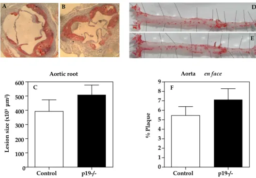
In chapter 5, another bone marrow transplantation is described with p19 (a subunit of IL-23) deficient bone marrow to study the contribution of IL-23 signaling in atherosclerosis. However, there is no change in plaque size observed in this experiment.
In chapter 6, again a DNA vaccination strategy was used, although with another immunodominant T helper cell epitope, PADRE, to break the tolerance against the p28 subunit of IL-27. In this study, the effect of IL-27 depletion in atherosclerosis results in aggravated atherosclerosis, indicating an atherosclerotic protective role for IL-27.
To study whether regulatory T cells can be induced against an atherosclerotic related autoantigen, we induced tolerance against HSP60 as described in chapter 7. The tolerance induction results in an increased number of Treg cells and attenuated atherosclerosis. These results further establish the protective role of regulatory T cells in atherosclerosis.
Dendritic cell based vaccination is described in chapter 8. Here we used a novel mRNA based vaccine against the specific regulatory T cell transcription factor, Foxp3. Via electroporation, the dendritic cells are “loaded” with the mRNA and subsequently injected in LDLr-/- mice. We demonstrate again that regulatory T
cells have a protective role in atherosclerose, as we observed aggravated atherosclerosis in Foxp3 vaccinated mice.
1
Furthermore, we discuss, to our opinion, the best approach to vaccinate against atherosclerosis.
References
1. NHS. Hoofdstuk 1 HVZ 2006: Hart- en vaatziekten in Nederland; 2006. 2. KWF. sterftecijfers door kanker in Nederland: KWF Kanker bestrijding; 2004.
3. Dai G, Kaazempur-Mofrad MR, Natarajan S, Zhang Y, Vaughn S, Blackman BR, Kamm RD, Garcia-Cardena G, Gimbrone MA, Jr. Distinct endothelial phenotypes evoked by arterial waveforms derived from atherosclerosis-susceptible and -resistant regions of human vasculature. Proc Natl Acad Sci U S A. 2004;101:14871-14876.
4. Deanfield JE, Halcox JP, Rabelink TJ. Endothelial function and dysfunction: testing and clinical relevance. Circulation. 2007;115:1285-1295.
5. Ross R. Atherosclerosis--an inflammatory disease. N Engl J Med. 1999;340:115-126. 6. Lusis AJ. Atherosclerosis. Nature. 2000;407:233-241.
7. Khoo JC, Miller E, McLoughlin P, Steinberg D. Enhanced macrophage uptake of low density lipoprotein after self-aggregation. Arteriosclerosis. 1988;8:348-358.
8. Khoo JC, Miller E, Pio F, Steinberg D, Witztum JL. Monoclonal antibodies against LDL further enhance macrophage uptake of LDL aggregates. Arterioscler Thromb. 1992;12:1258-1266. 9. Steinberg D. Low density lipoprotein oxidation and its pathobiological significance. J Biol
Chem. 1997;272:20963-20966.
10. Skalen K, Gustafsson M, Rydberg EK, Hulten LM, Wiklund O, Innerarity TL, Boren J. Subendothelial retention of atherogenic lipoproteins in early atherosclerosis. Nature. 2002;417:750-754.
11. Hansson GK. Inflammation, atherosclerosis, and coronary artery disease. N Engl J Med. 2005;352:1685-1695.
12. Cybulsky MI, Gimbrone MA, Jr. Endothelial expression of a mononuclear leukocyte adhesion molecule during atherogenesis. Science. 1991;251:788-791.
13. Collins RG, Velji R, Guevara NV, Hicks MJ, Chan L, Beaudet AL. P-Selectin or intercellular adhesion molecule (ICAM)-1 deficiency substantially protects against atherosclerosis in apolipoprotein E-deficient mice. J Exp Med. 2000;191:189-194.
14. Nageh MF, Sandberg ET, Marotti KR, Lin AH, Melchior EP, Bullard DC, Beaudet AL. Deficiency of inflammatory cell adhesion molecules protects against atherosclerosis in mice. Arterioscler Thromb Vasc Biol. 1997;17:1517-1520.
15. Shih PT, Brennan ML, Vora DK, Territo MC, Strahl D, Elices MJ, Lusis AJ, Berliner JA. Blocking very late antigen-4 integrin decreases leukocyte entry and fatty streak formation in mice fed an atherogenic diet. Circ Res. 1999;84:345-351.
16. Lutters BC, Leeuwenburgh MA, Appeldoorn CC, Molenaar TJ, Van Berkel TJ, Biessen EA. Blocking endothelial adhesion molecules: a potential therapeutic strategy to combat atherogenesis. Curr Opin Lipidol. 2004;15:545-552.
17. Ostermann G, Weber KS, Zernecke A, Schroder A, Weber C. JAM-1 is a ligand of the beta(2) integrin LFA-1 involved in transendothelial migration of leukocytes. Nat Immunol. 2002;3:151-158.
18. Rao RM, Yang L, Garcia-Cardena G, Luscinskas FW. Endothelial-dependent mechanisms of leukocyte recruitment to the vascular wall. Circ Res. 2007;101:234-247.
19. Boring L, Gosling J, Cleary M, Charo IF. Decreased lesion formation in CCR2-/- mice reveals a role for chemokines in the initiation of atherosclerosis. Nature. 1998;394:894-897.
20. Guo J, Van Eck M, de Waard V, Maeda N, Benson GM, Groot PH, Van Berkel TJ. The presence of leukocyte CC-chemokine receptor 2 in CCR2 knockout mice promotes atherogenesis. Biochim Biophys Acta. 2005;1740:453-459.
21. Nelken NA, Coughlin SR, Gordon D, Wilcox JN. Monocyte chemoattractant protein-1 in human atheromatous plaques. J Clin Invest. 1991;88:1121-1127.
22. Peters W, Charo IF. Involvement of chemokine receptor 2 and its ligand, monocyte chemoattractant protein-1, in the development of atherosclerosis: lessons from knockout mice. Curr Opin Lipidol. 2001;12:175-180.
1
24. Mach F, Sauty A, Iarossi AS, Sukhova GK, Neote K, Libby P, Luster AD. Differential expression of three T lymphocyte-activating CXC chemokines by human atheroma-associated cells. J Clin Invest. 1999;104:1041-1050.
25. Veillard NR, Kwak B, Pelli G, Mulhaupt F, James RW, Proudfoot AE, Mach F. Antagonism of RANTES receptors reduces atherosclerotic plaque formation in mice. Circ Res. 2004;94:253-261.
26. Rosenfeld ME, Yla-Herttuala S, Lipton BA, Ord VA, Witztum JL, Steinberg D. Macrophage colony-stimulating factor mRNA and protein in atherosclerotic lesions of rabbits and humans. Am J Pathol. 1992;140:291-300.
27. Hansson GK, Holm J, Jonasson L. Detection of activated T lymphocytes in the human atherosclerotic plaque. Am J Pathol. 1989;135:169-175.
28. Hansson GK, Hellstrand M, Rymo L, Rubbia L, Gabbiani G. Interferon gamma inhibits both proliferation and expression of differentiation-specific alpha-smooth muscle actin in arterial smooth muscle cells. J Exp Med. 1989;170:1595-1608.
29. Colles SM, Irwin KC, Chisolm GM. Roles of multiple oxidized LDL lipids in cellular injury: dominance of 7 beta-hydroperoxycholesterol. J Lipid Res. 1996;37:2018-2028.
30. Campbell JH, Campbell GR. The role of smooth muscle cells in atherosclerosis. Curr Opin Lipidol. 1994;5:323-330.
31. Herity NA, Ward MR, Lo S, Yeung AC. Review: Clinical aspects of vascular remodeling. J Cardiovasc Electrophysiol. 1999;10:1016-1024.
32. Davies MJ, Richardson PD, Woolf N, Katz DR, Mann J. Risk of thrombosis in human atherosclerotic plaques: role of extracellular lipid, macrophage, and smooth muscle cell content. Br Heart J. 1993;69:377-381.
33. Lee RT, Libby P. The unstable atheroma. Arterioscler Thromb Vasc Biol. 1997;17:1859-1867. 34. Glass CK, Witztum JL. Atherosclerosis. the road ahead. Cell. 2001;104:503-516.
35. Kaartinen M, van der Wal AC, van der Loos CM, Piek JJ, Koch KT, Becker AE, Kovanen PT. Mast cell infiltration in acute coronary syndromes: implications for plaque rupture. J Am Coll Cardiol. 1998;32:606-612.
36. Moreno PR, Falk E, Palacios IF, Newell JB, Fuster V, Fallon JT. Macrophage infiltration in acute coronary syndromes. Implications for plaque rupture. Circulation. 1994;90:775-778. 37. van der Wal AC, Becker AE, van der Loos CM, Das PK. Site of intimal rupture or erosion of
thrombosed coronary atherosclerotic plaques is characterized by an inflammatory process irrespective of the dominant plaque morphology. Circulation. 1994;89:36-44.
38. Amento EP, Ehsani N, Palmer H, Libby P. Cytokines and growth factors positively and negatively regulate interstitial collagen gene expression in human vascular smooth muscle cells. Arterioscler Thromb. 1991;11:1223-1230.
39. Jones CB, Sane DC, Herrington DM. Matrix metalloproteinases: a review of their structure and role in acute coronary syndrome. Cardiovasc Res. 2003;59:812-823.
40. Lindstedt KA, Kovanen PT. Mast cells in vulnerable coronary plaques: potential mechanisms linking mast cell activation to plaque erosion and rupture. Curr Opin Lipidol. 2004;15:567-573. 41. Liu J, Sukhova GK, Sun JS, Xu WH, Libby P, Shi GP. Lysosomal cysteine proteases in
atherosclerosis. Arterioscler Thromb Vasc Biol. 2004;24:1359-1366.
42. Dollery CM, Libby P. Atherosclerosis and proteinase activation. Cardiovasc Res. 2006;69:625-635.
43. Mallat Z, Tedgui A. Apoptosis in the vasculature: mechanisms and functional importance. Br J Pharmacol. 2000;130:947-962.
44. Shah PK, Falk E, Badimon JJ, Fernandez-Ortiz A, Mailhac A, Villareal-Levy G, Fallon JT, Regnstrom J, Fuster V. Human monocyte-derived macrophages induce collagen breakdown in fibrous caps of atherosclerotic plaques. Potential role of matrix-degrading metalloproteinases and implications for plaque rupture. Circulation. 1995;92:1565-1569. 45. Caligiuri G, Paulsson G, Nicoletti A, Maseri A, Hansson GK. Evidence for antigen-driven
T-cell response in unstable angina. Circulation. 2000;102:1114-1119.
46. Liuzzo G, Biasucci LM, Gallimore JR, Grillo RL, Rebuzzi AG, Pepys MB, Maseri A. The prognostic value of C-reactive protein and serum amyloid a protein in severe unstable angina. N Engl J Med. 1994;331:417-424.