Rugosibacter aromaticivorans
gen. nov., sp. nov., a bacterium
within the family
Rhodocyclaceae
, isolated from contaminated
soil, capable of degrading aromatic compounds
Elizabeth M. Corteselli, Michael D. Aitken and David R. Singleton*
Abstract
A bacterial strain designated Ca6Twas isolated from polycyclic aromatic hydrocarbon (PAH)-contaminated soil from the site of a former manufactured gas plant in Charlotte, NC, USA, and linked phylogenetically to the familyRhodocyclaceae of the classBetaproteobacteria. Its 16S rRNA gene sequence was highly similar to globally distributed environmental sequences, including those previously designated ‘Pyrene Group 1’demonstrated to grow on the PAHs phenanthrene and pyrene by stable-isotope probing. The most closely related described relative was Sulfuritalea hydrogenivoransstrain sk43HT(93.6 % 16S rRNA gene sequence identity). In addition to a limited number of organic acids, Ca6T was capable of growth on the monoaromatic compounds benzene and toluene, and the azaarene carbazole, as sole sources of carbon and energy. Growth on the PAHs phenanthrene and pyrene was also confirmed. Optimal growth was observed aerobically under mesophilic temperature, neutral pH and low salinity conditions. Major fatty acids present included summed feature 3 (C16 : 1!7cor C16 : 1 !6c) and C16 : 0. The DNA G+C content of the single chromosome was 55.14 mol% as determined by complete genome sequencing. Due to its distinct genetic and physiological properties, strain Ca6Tis proposed as a member of a novel genus
and species within the family Rhodocyclaceae, for which the name Rugosibacter aromaticivorans gen. nov., sp. nov. is proposed. The type strain of the species is Ca6T(=ATCC TSD-59T=DSM 103039T).
In a prior experiment that used stable-isotope probing (SIP) to reveal bacteria involved in the removal of the polycyclic aromatic hydrocarbon (PAH) pyrene in an aerobic, slurry-phase bioreactor treating contaminated soil from a former manufactured gas plant (MGP) in Charlotte, NC, USA, we identified three distinct groups of 16S rRNA genes derived from uncharacterized Proteobacteria. One of these groups was a monophyletic cluster associated with the family Rho-docyclaceae of the classBetaproteobacteriadesignated‘ Pyr-ene Group 1’ (PG1) [1]. Subsequent culture-independent experiments suggested that members of PG1 could also degrade the three-ring PAH phenanthrene [2]. A metage-nome constructed from a mixed culture dominated by members of PG1 contained multiple genes for PAH degra-dation, some of which were heterologously expressed in
Escherichia coliand their products shown to transform mul-tiple PAHs [3].
16S rRNA gene sequences highly similar to those designated as PG1 have since been recovered from a variety of environ-ments, suggesting widespread distribution of organisms
associated with the clade in both water and soil. DNA sequences highly similar to both the 16S rRNA gene and functional genes associated with PG1 have also been detected in geographically distant soils contaminated by PAHs [4, 5]. Other PG1 16S rRNA gene sequences have appeared in samples contaminated with trichloroethylene [6], copper [7] and asphalts [8], and from such diverse loca-tions as oilfields [9] to pristine water environments [10, 11]. In our own laboratory, PG1 sequences were among the most abundant in the bacterial community of a contami-nated soil recovering from chemical oxidation as part of a remediation scheme [12]. In each of these instances, how-ever, no isolates were obtained to represent the group. The most closely related characterized isolate to organisms represented by SIP- and environmentally-derived PG1 sequences isSulfuritalea hydrogenivorans strain sk43HT, a facultatively autotrophic freshwater bacterium [13]. Cultiva-tion efforts in our lab using a modified medium derived from the aerobic growth medium of strain sk43HTresulted in the successful isolation of a strain from the
bioreactor-Author affiliation:Department of Environmental Sciences and Engineering, Gillings School of Global Public Health, University of North Carolina at Chapel Hill, NC 27599-7431, USA.
*Correspondence:David R. Singleton, [email protected]
Keywords:polycyclic aromatic hydrocarbons; biodegradation; aromatics;Rhodocyclaceae.
Abbreviations:MGP, manufactured gas plant; PAH, polycyclic aromatic hydrocarbon; PG1,’Pyrene Group 1’; SIP, stable-isotope probing. The GenBank/EMBL/DDBJ accession number for the whole-genome sequence of strain Ca6Tis CP010554.
treated soil from the MGP site in Charlotte, NC, represent-ing PG1 and designated Ca6T. The modified sk43HT isola-tion medium used to isolate strain Ca6T originally contained 5 mM sodium-potassium phosphate buffer (pH 7.0) and 5 mM NH4NO3(collectively referred to as‘reactor
buffer’), 1 mM MgSO4. 7H2O, 1 mM CaCl2. 2 H2O, 1 ml
trace element solution l 1[containing per litre; 12.5 ml HCl (25 %), 2.1g FeSO4. 7 H2O, 30 mg H3BO3, 100 mg MnCl2. 4
H2O, 190 mg CoCl2. 6 H2O, 24 mg NaCl2. 6 H2O, 2 mg
CuCl2. 2H2O, 144 mg ZnSO4. 7H2O, 36 mg Na2MoO4. 2
H2O] and 1 ml selenium-tungsten solution l 1(containing
per litre; 0.4 g NaOH, 6 mg Na2SeO3. 5H2O, 8 mg Na2WO4
. 2H2O). After autoclaving for 15 min at 250
C, the follow-ing filter-sterilized solutions (0.2 µm pore size) were added aseptically to the cooled media (per l): 1 ml vitamin solution (containing 4 mg p-aminobenzoic acid, 1 mg biotin, 10 mg nicotinic acid, 5 mg calcium pantothenate, 15 mg pyridoxine hydrochloride, in 100 ml of a 10 mM phosphate buffer, pH 7.1), 1 ml thiamine solution (containing 10 mg thiamine in 100 ml of 25 mM sodium phosphate buffer, pH 3.4), 1 ml vitamin B12 solution (containing 5 mg cyanocobalamin in 100 ml distilled water) and 1.5 ml thiosulfate solution (24.8 g Na2S2O3. 5H2O in 100 ml distilled water, stored
under N2 gas). Carbon was added before autoclaving as
either 0.02 % pyrene dissolved in acetone to the flask, allow-ing the solvent to evaporate prior to addallow-ing other compo-nents, or as 0.2 % sodium pyruvate. Subsequent tests of strain Ca6Tgrown on the isolation medium excluding indi-vidual components indicated that only the reactor buffer, MgSO4, trace element solution and a carbon source were
required for growth at a rate equivalent to that of the com-plete medium. We observed that growth in the liquid medium was slower and to a lower cell density in the absence of amended MgSO4, and that no growth at all
occurred in the absence of the trace element solution. All subsequent tests used a medium containing only the required components: reactor buffer, MgSO4and trace
ele-ment solution (referred to as sRB1 medium,‘supplemented reactor buffer’) with sodium pyruvate as a carbon source unless otherwise noted. For solid media (‘sRB1-agar’), 1.5 % agar (Acros Organics) was added to liquid medium prior to autoclaving.
The optimal growth conditions of strain Ca6T in sRB1 medium with pyruvate as a carbon source were determined for a range of temperatures, pH and salinities. Optimal growth was defined as the maximum growth rate during the exponential growth phase as measured by turbidity at OD600with a HACH DR3000 spectrophotometer. Optimal
temperature for growth was determined in triplicate 5 ml cultures in liquid sRB1 medium at pH 7.0 and 225 r.p.m. for temperatures of 23, 26, 28, 30, 32, 34, 35, 36 and 37
C. Additional temperatures were evaluated on solid sRB1 plates at 20 and 4
C. The effect of pH was tested in tripli-cate 5 ml tubes of sRB1 medium buffered to pH values of pH 5.0, 5.5, 6.0, 6.5 [buffered with 2-(N -morpholino)etha-nesulfonic acid, 50 mM], 7.0, 7.5, 8.0 [4-(2-hydroxyethyl) 1-piperazineethanesulfonic acid, 50 mM] and 8.5, 9.0 [Tris/
HCl, 50 mM] and incubated at 225 r.p.m. and 30
C. Salinity effects were determined using triplicate 5 ml cultures of sRB1, pH 7.0, incubated at 30
C and 225 r.p.m., amended with either 0, 0.25, 0.5, 1, 2, 3, 4 or 5 % NaCl (w/v). The optimal temperature for growth was between 30 and 34
C, with obvious growth between 20 and 35
C. Strain Ca6T grew between pH 6.5 and 7.5 with an optimum of pH 6.5. No growth was observed at salt concentrations >0.5 %, but strain Ca6Tgrew equally well with either 0.25 % or no NaCl. Under optimal conditions in sRB1-pyruvate liquid media with constant shaking at 225 r.p.m., strain Ca6T grew on pyruvate to maximum turbidity in approximately 3 days. Colonies on sRB1-agar plates with pyruvate as a carbon source developed over a period of 2 weeks when incubated at 30
C. Strain Ca6Tcolonies on sRB1 plates with pyruvate were generally small (0.5–0.8 mm in diameter), circular, with a convex elevation and were pale yellow–white in colour.
The ability of Ca6T to grow under low oxygen conditions was assessed using the GasPak 100 system containing a Gas-Pak EZ Anaerobe Container System sachet (BD Bioscien-ces). Plates comprising sRB1-agar and pyruvate were inoculated and sealed in the system according to the manu-facturer’s directions. According to the manufacturer, the system achieves an atmosphere of <1 % oxygen and13 % carbon dioxide within 2.5 h. Low-oxygen conditions were confirmed after 48 h by visual observation of an indicator strip soaked in 1 mM resazurin solution. Plates were incu-bated at 30
C and compared with concurrent aerobic con-trols. Ca6T did not display growth under low oxygen conditions after 4 weeks. Unless otherwise noted, all subse-quent tests of Ca6Toccurred at 30
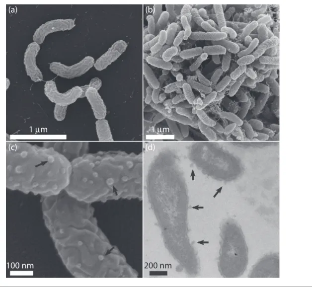
apparent (Fig. 1a, b). Electron-transparent inclusions were evident in some pyrene-grown cells examined by TEM, which were absent in cells grown on pyruvate (not shown). Gram staining performed using the standard reaction and visualized by light microscopy indicated strain Ca6T was Gram-type-negative.
Cellular motility was tested using sRB1-agar stab-tubes with agar added at 0.3 % (w/v) and pyruvate as the carbon source. No outward motility from the stab was observed. No flagella were observed during microscopy, and few genes for flagellar synthesis were indicated in the annotated genome [14]. Metabolism of a variety of carbon substrates was tested using the Biolog GN2 Microplate. For each plate, cells were grown in liquid sRB1 medium amended with pyruvate and washed three times in PBS (pH 7.5) before being resuspended in Biolog GN/GP Inoculating Fluid as per the manufacturer’s instructions. The microplates were incubated overnight at
30
C and scored through visual examination in comparison with the no-carbon control. In triplicate samples, the only substrates that strain Ca6Tactively metabolized were formic acid, methyl pyruvate, monomethyl succinate and b -hydroxybutyric acid. The other 91 substrates, including a variety of sugars, other organic acids, nucleosides and amino acids were not metabolized.
Utilization of nitrogen sources was tested by substituting either 5 mM KNO3 or NH4Cl for NH4NO3 in sRB1 liquid
media. Strain Ca6Tdisplayed growth on pyruvate with both nitrogen sources tested. Nitrate reduction was evaluated using a culture grown in sRB1 media with NH4NO3
substi-tuted with 5 mM KNO3. No nitrate reduction to nitrite
beyond what was required for assimilation and growth was observed. Starch hydrolysis and cellulase activity were eval-uated by supplementing sRB1 plates with either starch (10 g l 1) or cellulose (1 g l 1) (adapted from Kasanaet al.[15]). Inoculated plates were incubated for 2 weeks at 30
C, after
(a)
1
μ
m
1
μ
m
100 nm
200 nm
(b)
(c)
(d)
Fig. 1.Scanning electron micrographs (a, b, c) and transmission electron micrograph (d) of strain Ca6Tgrown on pyruvate (a, c) or
which Gram’s iodine was used to identify clear zones around colonies. Protease activity was tested by adding a skimmed milk solution aseptically (10 % skimmed milk powder dissolved in distilled water) to sRB1-agar after autoclaving. Plates were incubated at 30
C for 1 month and monitored for zones of clearing. Gelatin hydrolysis was assessed using a nutrient gelatin stab method wherein pow-dered gelatin (120 g l 1) was added to sRB1, gently heated to dissolve and aliquoted into 5 ml tubes. Gelatin tubes con-taining Ca6Twere incubated at 30
C and monitored for liq-uefaction of the media. Ca6T was negative for starch hydrolysis, cellulase, skimmed milk protease and gelatinase activities.
Lipase activity was assessed by adding tributyrin (98 %; Acros Organics) to sRB1-agar (1 %, v/v). Urease activity was determined using Stuart’s urea broth, which was prepared by amending phosphate buffer with urea (20 g l 1) and phe-nol red (10 mg l 1) in addition to pyruvate, MgSO4 and trace element solution. Urea tubes were monitored for 24 h for a colour change indicating urease production. Strain Ca6Twas positive for lipase activity and negative for urease activity.
Strain Ca6Twas tested for catalase activity by adding a 3 % hydrogen peroxide solution (v/v) to cells freshly scraped from the surface of an sRB1 plate. Oxidase activity was determined by adding a few drops of freshly-prepared 1 %N¢,N¢,N¢,N¢-tetramethyl-p-phenylenediamine dihydro-chloride (Acros Organics) to cells scraped from the same plate onto filter paper. Strain Ca6T was catalase-negative and oxidase-positive.
Indole production was tested by supplementing sRB1 media with tryptone (10 g l 1). After growth to turbidity, a few drops of Kovac’s reagent were added to each tube. The lack of a colour change indicated no production of indole by strain Ca6T. As indigo production from indole is indicative of activity by some ring-hydroxylating dioxygenases (RHD), enzymes involved in the initial step in the aerobic degrada-tion of PAHs by bacteria [16, 17], the lack of indole produc-tion was anticipated. To further investigate this phenotype, indole was added as a small crystal to the lid of inverted Petri plates of sRB1-pyruvate medium 24 h after inoculation and incubated. A blue/purple colour developed by colonies on the plate due to indigo production confirmed the likely presence of at least one active RHD.
Growth of strain Ca6Ton select PAHs as sole sources of car-bon and energy was tested in liquid sRB1 media amended with individual PAHs (final concentration 0.2 g l 1). PAHs were added to tubes in a solvent (either acetone or dichloro-methane), and the solvent was allowed to evaporate prior to adding the remaining media components. Tubes were addi-tionally sonicated after autoclaving to help break up PAH crystals. Ca6T cells were grown in sRB1-pyruvate and washed three times with reactor buffer prior to inoculation, and triplicate PAH-containing tubes were incubated at 30
C at 225 r.p.m. for up to 19 days. As spectrophotometric
measurements of turbidity were not possible due to the presence of PAH crystals in the media, growth of Ca6Ton PAHs was defined by both protein accumulation and disap-pearance of the parent compound. Protein concentrations were calculated from aliquots of culture approximately every 48 h using a Pierce BCA Protein Assay according to manufacturer’s instructions (ThermoFisher Scientific). Dis-appearance of each PAH was determined using a liquid-liq-uid extraction with an equal volume of n-hexanes and quantification using a HPLC system with fluorescence detection as previously described [12]. Extraction efficiency was determined from uninoculated tubes with PAH added. For all substrates except naphthalene, this method was able to quantify PAH concentration; naphthalene presumably volatilized from the tubes during either the incubation or extraction. Removal of pyrene (47 % of the initial mass) and phenanthrene (48 %) by Ca6T was correlated with protein accumulation, indicating growth on those substrates. Incu-bation with benz[a]anthracene resulted in modest protein accumulation and removal of the PAH from the medium compared with phenanthrene and pyrene, and the protein concentration did not display a steady increase typical of exponential growth. This suggests that Ca6Tmay be capable of weakly positive growth on this compound. Incubation with fluorene and anthracene resulted in PAH removal without protein accumulation, suggesting transformation but not growth on these substrates. Ca6Twas negative for growth on or transformation of chrysene, benzo[a]pyrene, fluoranthene, naphthalene and acenaphthene. Growth on the azaarene carbazole was determined by visual turbidity and protein accumulation over the course of the incubation, as the HPLC assay employed for quantifying PAHs was not suitable for this compound. Strain Ca6T was positive for growth on carbazole.
Mineralization of the partially14C-labelled PAHs phenan-threne, fluoranthene, chrysene, benz[a]anthracene and benzo[a]pyrene was performed as previously described [18]. Ca6Tcells grown on sRB1-pyrene and washed three times with reactor buffer were used to inoculate the flasks. The extent of mineralization was measured after 24 and 48 h. Mineralization was assessed as a percentage of added PAH mineralized, and biotic samples were compared with acidi-fied killed control replicates. Statistically significant miner-alization occurred for phenanthrene (29 % minerminer-alization,
mixture of xylenes (o-xylene 98.5 %, m- 99 %, p- 99 %, in equal volumes) by adding 0.5 ml of the chemical(s) to a piece of filter paper in a glass beaker. Inoculated sRB1-agar plates without additional carbon were incubated in these containers at room temperature for 1 month. Strain Ca6T grew well on benzene, weakly on toluene, but did not grow on either ethylbenzene or mixed xylenes. Inoculated plates of sRB1-agar without amended carbon in normal atmo-sphere showed no growth over the same time period. Cellular fatty acid profiling was performed using the MIDI Sherlock Microbial Identification System by Microbial ID on cells grown on sRB1 plates with pyruvate. The dominant fatty acids in Ca6T were summed feature 3 (C16 : 1!7c or
C16 : 1!6c) (42.5 %) and C16 : 0(36.7 %). Ca6Talso contained
C17 : 0 cyclo (6.5 %), C18 : 1!7c (4.9 %), C10 : 0 3-OH (3.9 %)
and C12 : 0 (3.0 %). Trace amounts (<1 % each) of C10 : 0,
C14 : 0, C16 : 1!5c, C17 : 1!7c, C18 : 2!6,9c, C18 : 0, C19 : 0 cyclo !8c and C19 : 0 were also detected. Analysis of respiratory
quinones and polar lipids was carried out by the Identifica-tion Service, DSMZ, Braunschweig, Germany. Major polar
lipids in Ca6Twere phosphatidylethanolamine, phosphati-dylglycerol, diphosphatidylglycerol and phospholipid, with minor amounts of aminolipid, glycolipid and lipid present (Fig. S1, available in the online Supplementary Material). Respiratory quinones present were Q7 (9 %), Q8 (80 %) and Q9 (11 %).
The genome of strain Ca6T was determined as described previously [14] and was composed of a singular, circular chromosome of 2 934 611 bp with a DNA G+C content of 55.14 mol%. No plasmids were detected. The two copies of the 16S rRNA gene detected were identical, and the closest described relatives of Ca6TwereSulfuritalea hydro-genivorans strain sk43HT(93.6 % 16S rRNA gene similar-ity) [13], Denitratisoma sp. TSA61 (93.2 %) [19],
Georgfuchsia toluolica G5G6T (92.0 %) [20], Sulfurisoma sediminicolaBSN1T(91.8 %) [21],Denitratisoma oestradio-licumAcBE2-1T(91.6 %) [22] andSterolibacterium denitri-ficans ChoI-1ST (91.5 %) [23]; these organisms are all members of genera within the betaproteobacterial family
Rhodocyclaceae(Fig. 2).
Ferribacterium limneticum CdA-1T (Y17060) 88
87 98 81 98 82 69 0.01
57
54 62
76 69 64 71
63 66
63 64
92
55 92 86
98 99
92 82
Quatrionicoccus australiensis Ben 117T (NR_029035)
Dechloromonas agitata CKBT (AF047462)
Azonexus fungiphilus BS5-8T (AF011350)
‘Dechlorobacter hydrogenophilus’ LT-1 (AY124797)
Propionivibrio dicarboxylicus CreMal1T (Y17601)
Rhodocyclus purpureus 6770T (M34132)
Azospira oryzae 6a3T (AF011347)
Azovibrio restrictus S5b2T (AF011346)
Thauera selenatis AX39T (Y17591)
Azoarcus indigens VB32T (L15531)
Uliginosibacterium gangwonense 5YN10-9T (DQ665916)
Zoogloea ramigera NBRC 15342T (AB680846)
Sulfurisoma sediminicola BSN1T (AB842427)
Georgfuchsia toluolica G5G6T (EF219370)
Sulfuritalea hydrogenivorans sk43HT (NR_113147)
Rugosibacter aromaticivoransCa6T (CP010554)
Denitratisoma oestradiolicum AcBE2-1T (AY879297)
Methyloversatilis universalis FAM5T (DQ442273)
Sterolibacterium denitrificans Chol-1ST (AJ306683)
Fig. 2.16S rRNA gene neighbour-joining phylogenetic tree of Ca6Twith type strains of genera from the family
Rhodocyclaceae. Per-centage bootstrap values of >50 % based on 1000 iterations are shown for the neighbour-joining [26] and maximum-parsimony algo-rithms [27] above and below nodes, respectively. The consensus neighbour-joining tree was reconstructed including all sequence information available and without considering positions with gaps.Nitrosomonas europaeastrain ATCC 25978T(GenBank accession
In addition to high 16S rRNA gene sequence divergence from described species of phylogenetically related genera, Ca6Twas also differentiated by its physiological characteris-tics (Table 1). Most species from closely related genera within the familyRhodocyclaceaewere isolated from aquatic or anoxic environments, while Ca6Twas isolated from con-taminated soil. Representatives from all closely related gen-era were also gengen-erally capable of facultative or anaerobic growth and nitrate reduction, while Ca6Trequired an aero-bic environment for growth under the growth conditions tested and was not able to reduce nitrate. None of the phylo-genetically closest described genera to Ca6Tcontain species reported to degrade PAHs. Among the genera examined, strain Ca6Twas also the only isolate reported to contain the respiratory quinones Q7 and Q9; all isolates for which such information is available, including Ca6T, indicated Q8 as the major or sole respiratory quinone. Similarly, while spe-cies from all closely related genera (including strain Ca6T) contained C16 : 1!7c(generally as part of a summed feature)
and C16 : 0as major fatty acids, strain Ca6Tcould be
distin-guished by the presence or absence of other, less abundant fatty acids. A significantly lower genomic DNA G+C con-tent (mol%) further distinguished Ca6T from strains of related genera. A complete genome comparison between Ca6Tand Sulfuritalea hydrogenivorans sk43HT (the closest relative by 16S rRNA gene sequence identity) approximated a DNA–DNA hybridization of 19.00 % (16.8–21.4 %) based
on a linear regression model [24]. The average nucleotide identity (ANI) between the two genomes was 76.69 % [25]. On the basis of genomic and physiological differences, we propose Ca6Tas the type strain of a novel species and genus, for which the nameRugosibacter aromaticivoransgen. nov., sp. nov. is proposed.
DESCRIPTION OF
RUGOSIBACTER
GEN. NOV.
Rugosibacter(Ru.go.si.bac¢ter. L. adj.rugosuswrinkled; N. L. masc. n.bactera rod; N. L. masc. n.Rugosibactera wrinkled rod).Cells are Gram-type-negative, non-motile and grow aerobi-cally. Catalase-negative and oxidase-positive. Heterotrophic growth occurs on a limited number of organic acids. Pre-dominant fatty acids are summed feature 3 (C16 : 1!7c or
C16 : 1!6c) and C16 : 0. The major polar lipids are
phosphati-dylethanolamine, phosphatidylglycerol, diphosphatidylgly-cerol and phospholipid. The major respiratory quinone is Q8. Phylogenetically, the genus is a member of the family
Rhodocyclaceae in the class Betaproteobacteria. The type species isRugosibacter aromaticivorans.
DESCRIPTION OF
RUGOSIBACTER
AROMATICIVORANS
SP. NOV.
Rugosibacter aromaticivorans [a.ro.ma.ti.ci.vo¢rans. L. adj.
aromaticus aromatic, fragrant; L. pres. part. vorans
Table 1.Differential characteristics of strain Ca6Tand closely related taxa within the familyRhodocyclaceae
Strains: 1, Ca6T; 2,
Denitratisoma oestradiolicumAcBE2-1T[22]; 3,
Georgfuchsia toluolicaG5G6T[20]; 4, species of the genus
Methyloversatilis including
Methyloversatilis universalisstrains FAM5Tand Ehg5 [28],
Methyloversatilis discipulorumstrains FAM1T, RZ18-153 and RZ94 [29], and
Methyloversatilis thermotolerans3tT[30]; 5,
Sterolibacterium denitrificansChoI-1ST[23]; 6,
Sulfurisoma sediminicolaBSN1T[21]; 7,
Sulfuritalea hydrogenivoranssk34HT
[13]; 8, species of the genusRhodocyclusincludingRhodocyclus purpureusstrain‘Ames’6770T[31, 32] and
Rhodocyclus tenuisstrain ATCC 25 093T
[31, 33, 34]. +, Positive; , negative; var., variable results among strains;NR, not reported. DNA G+C content (mol%) values for the genus Methylover-satilisbased on genomic data presented by Smalleyet al.[29].
Characteristic 1 2 3 4 5 6 7 8
Isolation source(s) Contaminated soil, USA
Activated sludge, Germany
Iron-reducing
aquifer, Netherlands
Lake sediment, USA; hot springs,
Russia
Anoxic reactor treating landfill
leachate, Uruguay
Freshwater lake sediment,
Japan
Freshwater lake, Japan
Swine waste lagoon, USA; freshwater pond,
Germany
DNA G+C content (mol%)
55.1 61.4 NR 65.6–67 65.3 67 67 64.1–65.1
Diagnostic fatty acids
C8 : 03-OH + +
C10 : 03-OH + var. + + +
C17 : 0cyclo + + var.
C17 : 1!7c +
C19 : 0cyclo !8c
+
Respiratory quinones Q8, Q9, Q7 Q8 NR Q8 Q8 NR NR Q8, MK8
Catalase NR + + + +
Motility + + + + NR var.
Oxygen requirement Aerobic Facultative Anaerobic Facultative Facultative Facultative Facultative Facultative
devouring; N.L. part. adj. aromaticivorans devouring aro-matic (compounds)].
Cells are curved rods (0.77–1.150.23–0.27 µm). On solid media, colonies are small, convex and circular with a yel-low–white colour. Growth occurs aerobically between 20 and 35
C (optimum 30–34
C), at pH 6.5–7.5 (optimum pH 6.5) and with salinity 0.5 %. Cells can grow on select monocyclic and polycyclic aromatic compounds as a sole source of carbon, and use both nitrate and ammo-nia as nitrogen sources. Cells are negative for gelatin hydrolysis, cellulase, skimmed milk protease, starch hydrolysis, nitrate reduction and urease activity, and posi-tive for lipase activity. In addition to those listed in the genus description, fatty acids present in minor amounts are C17 : 0 cyclo, C18 : 0!7c, C10 : 0 3-OH and C12 : 0. Polar
lipids and respiratory quinones are consistent with the genus description.
The type strain, Ca6T (=ATCC TSD-59T=DSM 103039T), was isolated from PAH-contaminated soil from Charlotte, NC, USA. The genomic DNA G+C content of the type strain is 55.14 mol%.
Funding information
This work was supported by the US National Institute of Environmental Health Sciences (NIEHS) as part of the Superfund Research Program (5 P42ES005948).
Conflicts of interest
The authors declare that there are no conflicts of interest.
References
1. Singleton DR, Sangaiah R, Gold A, Ball LM, Aitken MD. Identifica-tion and quantificaIdentifica-tion of uncultivated Proteobacteria associated with pyrene degradation in a bioreactor treating PAH-contami-nated soil.Environ Microbiol2006;8:1736–1745.
2. Singleton DR, Hunt M, Powell SN, Frontera-Suau R, Aitken MD.
Stable-isotope probing with multiple growth substrates to deter-mine substrate specificity of uncultivated bacteria. J Microbiol Methods2007;69:180–187.
3. Singleton DR, Hu J, Aitken MD.Heterologous expression of poly-cyclic aromatic hydrocarbon ring-hydroxylating dioxygenase genes from a novel pyrene-degrading betaproteobacterium.Appl Environ Microbiol2012;78:3552–3559.
4. Martin F, Torelli S, Le Paslier D, Barbance A, Martin-Laurent F et al. Betaproteobacteria dominance and diversity shifts in the bacterial community of a PAH-contaminated soil exposed to phen-anthrene.Environ Pollut2012;162:345–353.
5. Regonne RK, Martin F, Mbawala A, Ngassoum MB, Jouanneau Y.
Identification of soil bacteria able to degrade phenanthrene bound to a hydrophobic sorbent insitu.Environ Pollut2013;180: 145–151.
6. Humphries JA, Ashe AM, Smiley JA, Johnston CG.Microbial com-munity structure and trichloroethylene degradation in groundwa-ter.Can J Microbiol2005;51:433–439.
7. He Z, Xie X, Xiao S, Liu J, Qiu G.Microbial diversity of mine water at Zhong Tiaoshan copper mine, China.J Basic Microbiol2007;47: 485–495.
8. Kim JS, Crowley DE.Microbial diversity in natural asphalts of the Rancho La Brea Tar Pits. Appl Environ Microbiol 2007; 73:4579–4591.
9. Tang YQ, Li Y, Zhao JY, Chi CQ, Huang LXet al.Microbial commu-nities in long-term, water-flooded petroleum reservoirs with
different insitutemperatures in the Huabei Oilfield, China.PLoS One2012;7:e33535.
10. Gihring TM, Moser DP, Lin LH, Davidson M, Onstott TCet al.The distribution of microbial taxa in the subsurface water of the Kala-hari shield, South Africa.Geomicrobiol J2006;23:415–430. 11. Zhang L, Gao G, Tang X, Shao K.Impacts of different salinities on
bacterial biofilm communities in fresh water. Can J Microbiol
2014;60:319–326.
12. Richardson SD, Lebron BL, Miller CT, Aitken MD. Recovery of phenanthrene-degrading bacteria after simulated insitu persul-fate oxidation in contaminated soil.Environ Sci Technol2011;45: 719–725.
13. Kojima H, Fukui M.Sulfuritalea hydrogenivoransgen. nov., sp. nov., a facultative autotroph isolated from a freshwater lake.Int J Syst Evol Microbiol2011;61:1651–1655.
14. Singleton DR, Dickey AN, Scholl EH, Wright FA, Aitken MD. Com-plete genome sequence of a novel bacterium within the family
Rhodocyclaceaethat degrades polycyclic aromatic hydrocarbons.
Genome Announc2015;3:e00251-15.
15. Kasana RC, Salwan R, Dhar H, Dutt S, Gulati A.A rapid and easy method for the detection of microbial cellulases on agar plates using gram’s iodine.Curr Microbiol2008;57:503–507.
16. Ensley BD, Ratzkin BJ, Osslund TD, Simon MJ, Wackett LP et al.
Expression of naphthalene oxidation genes inEscherichia coliresults in the biosynthesis of indigo.Science1983;222:167–169.
17. Schell MA.Cloning and expression inEscherichia coliof the naph-thalene degradation genes from plasmid NAH7.J Bacteriol1983; 153:822–829.
18. Singleton DR, Ramirez LG, Aitken MD.Characterization of a poly-cyclic aromatic hydrocarbon degradation gene cluster in a phen-anthrene-degradingAcidovoraxstrain.Appl Environ Microbiol2009; 75:2613–2620.
19. Ishii S, Ashida N, Otsuka S, Senoo K.Isolation of oligotrophic deni-trifiers carrying previously uncharacterized functional gene sequences.Appl Environ Microbiol2011;77:338–342.
20. Weelink SA, van Doesburg W, Saia FT, Rijpstra WI, Röling WFet al.A strictly anaerobic betaproteobacterium Georgfuchsia toluolica gen. nov., sp. nov. degrades aromatic compounds with Fe(III), Mn(IV) or nitrate as an electron acceptor.FEMS Microbiol Ecol2009;70:575– 585.
21. Kojima H, Fukui M.Sulfurisoma sediminicolagen. nov., sp. nov., a facultative autotroph isolated from a freshwater lake.Int J Syst Evol Microbiol2014;64:1587–1592.
22. Fahrbach M, Kuever J, Meinke R, K€ampfer P, Hollender J. Deni-tratisoma oestradiolicum gen. nov., sp. nov., a 17b -oestradiol-degrading, denitrifying betaproteobacterium. Int J Syst Evol Microbiol2006;56:1547–1552.
23. Tarlera S, Denner EB.Sterolibacterium denitrificansgen. nov., sp. nov., a novel cholesterol-oxidizing, denitrifying member of theb
-Proteobacteria.Int J Syst Evol Microbiol2003;53:1085–1091. 24. Meier-Kolthoff JP, Auch AF, Klenk HP, Göker M. Genome
sequence-based species delimitation with confidence intervals and improved distance functions.BMC Bioinformatics2013;14:60. 25. Goris J, Konstantinidis KT, Klappenbach JA, Coenye T,
Vandamme Pet al.DNA–DNA hybridization values and their rela-tionship to whole-genome sequence similarities.Int J Syst Evol Microbiol2007;57:81–91.
26. Thompson JD, Higgins DG, Gibson TJ.CLUSTAL W: improving the sensitivity of progressive multiple sequence alignment through sequence weighting, position-specific gap penalties and weight matrix choice.Nucleic Acids Res1994;22:4673–4680.
27. Cummings MP, Hancock JM, Zvelebil MJ.PAUP* (phylogenetic anal-ysis using parsimony (and other methods). In: Dictionary of Bioinformatics and Computational Biology. John Wiley & Sons, Ltd; 2004.
within theBetaproteobacteriarepresented by three methylotrophic isolates.Int J Syst Evol Microbiol2006;56:2517–2522.
29. Smalley NE, Taipale S, De Marco P, Doronina NV, Kyrpides N et al.Functional and genomic diversity of methylotrophic Rhodocy-claceae: description ofMethyloversatilis discipulorumsp. nov.Int J Syst Evol Microbiol2015;65:2227–2233.
30. Doronina NV, Kaparullina EN, Trotsenko YA.Methyloversatilis ther-motoleranssp. nov., a novel thermotolerant facultative methylo-troph isolated from a hot spring.Int J Syst Evol Microbiol2014;64: 158–164.
31. Hiraishi A, Hoshino Y, Satoh T.Rhodoferax fermentansgen. nov., sp. nov., a phototrophic purple nonsulfur bacterium previously
referred to as the ’Rhodocyclus gelatinosus-like’ group. Arch Microbiol1991;155:330–336.
32. Pfennig N.Rhodocyclus purpureusgen. nov. and sp. nov., a ring-shaped, vitamin B12-requiring member of the family
Rhodospirilla-ceae.Int J Syst Evol Microbiol1978;28:283–288.
33. Imhoff JF.Genus I.RhodocyclusPfennig 1978, 285AL. In: Brenner
DJ and Staley JT (editors). Bergey’s Manual of Systematic Bacteriology, The Proteobacteria Part B, the Betaproteobacteria, vol. 2. New York: Springer; 2005.
34. Pfennig N.Rhodospirillum tenuesp. n., a new species of the purple nonsulfur bacteria.J Bacteriol1969;99:619–620.
Five reasons to publish your next article with a Microbiology Society journal 1. The Microbiology Society is a not-for-profit organization.
2. We offer fast and rigorous peer review–average time to first decision is 4–6 weeks. 3. Our journals have a global readership with subscriptions held in research institutions around
the world.
4. 80% of our authors rate our submission process as‘excellent’or‘very good’.
5. Your article will be published on an interactive journal platform with advanced metrics.