DEVELOPMENT OF PROTEASE-RESISTANT β-HAIRPIN PEPTIDES FOR THE DETECTION OF ENZYMATIC ACTIVITY IN CANCER CELLS
Kaiulani Michelle Houston
A dissertation submitted to the faculty at the University of North Carolina at Chapel Hill in partial fulfillment of the requirements for the degree of Doctor of Philosophy in the
Department of Chemistry (Biological Division).
Chapel Hill 2014
©2014
ABSTRACT
Kaiulani Michelle Houston: Development of Protease-Resistant β-Hairpin Peptides for the Detection of Enzymatic Activity in Cancer Cells
(Under the direction of Marcey L. Waters)
Multiple myeloma (MM) is an incurable malignancy of the antibody-producing plasma cells in the bone marrow and is characterized by the targeted degradation of anti-tumor regulatory proteins by the ubiquitin proteasome system (UPS). The development of proteasomal inhibitors, such as bortezomib (Velcade®), has greatly improved the median survival time of MM patients. Inappropriate activation of protein kinase B (PKB or Akt) by bortezomib has been observed leading to refractory disease. Inhibitors of PKB activity are now in clinical trials for the treatment of MM. Chronic myelogenous leukemia (CML), the malignancy of myeloid cells in the bone marrow, is treated predominantly with Abl kinase inhibitors. We have designed protease-resistant fluorescent peptide probes that, once optimized, can be used to determine the effectiveness of these inhibitors by direct measurement of proteasomal and Abl kinase hyperactivity and PKB activation in patient cells.
degradation in the cell by cytosolic proteases. Unstructured peptides in the cytosol of immune cells are threaded into protease active sites at the N-terminus.
We designed and synthesized short, well-folded protease resistant β-hairpin peptides through the incorporation of favorable cross-strand side chain interactions and a
stabilizing turn nucleating sequence. Designing the β-hairpins to include ornithine, a noncanonical amino acid analog of lysine, and D-amino acids, produced long-lived peptides with half-lives of 2-6 hours and non-degradable peptides, in the presence of specific and nonspecific proteases in vitro. These β-hairpin peptides were covalently attached to the N-terminus of unstructured kinase substrate peptides resulting in increased stability to peptidases in vitro while maintaining efficacy as kinase substrates.
To the Teacher, S. D. G.
ACKNOWLEDGEMENTS
I would like to thank Professor Marcey L. Waters for giving me the greatest opportunity of my lifetime thus far. Her enthusiasm for interdisciplinary science is contagious and her dedication to her family, students, and the advancement of women in science is inspirational. I’m so grateful for her advocacy, her belief in my abilities, and her encouragement in pursuing new research ideas. She is truly an awesome lady!
I would like to thank my committee members, Professor Gary Pielak, Professor, David Lawrence, Professor Eric Brustad, and particularly Professor Nancy Allbritton for their open-door policy and willingness to share ideas about my science. I am particularly grateful to Professor Allbritton for her constant guidance and input and the sharing of her instruments, reagents and lab family. I would also like to thank Professor Kevin Weeks for his infectious enthusiasm for chemical biology, which caused be to join the
Department of Chemistry. I want to thank Dr. Ashutosh Tripathy, the Director of the Macromolecular Interactions Facility, for his instruction and for being a great neighbor. Thanks to Dr. Sohrab Habibi, Director of the Mass Spectrometry Facility, for his
patience, sense of humor, great advice, and instruction. Thanks to Professor David Jones for his encouragement and support. Aloha to Lars Sahl who always greets me with a smile. Thanks to all faculty and staff of the Department of Chemistry for providing a pleasant environment in which to work.
Thanks to BBSP, the IMSD family, and the Translation Medicine Program,
me to UNC-Chapel Hill and for being strong reliable support systems throughout my graduate school career.
I must thank the most incredible group of people on the planet, the members of the Waters Group. I could not have been blessed with a better group of people with which to spend the past four years in an exciting but trying environment. I found some of my very best friends among the group and could no longer imagine life without them. Thanks to all of the group members for their encouragement, scientific input, enthusiastic
discussions about research and life, and for showing me adventures beyond the
laboratory. You’ve been a wonderful family and it has been a privilege to know you all and work with you.
Thanks to the Allbritton Group for welcoming me with open arms and adopting me into their family. I would like to thank them for their graciousness with their time and instruments and the sharing of their scientific knowledge. I have truly enjoyed the work of the culinary artists and the great conversations that took places while indulging in great food. Thanks particularly to Dr. Adam Melvin, Greg Woss, Dr. Angela Proctor, Dr. Imola Zigoneanu, Abby Turner, Nick Dobes, and Dr. Shan Yang for their instruction and great humor.
Thanks to all of my friends and church family members within Chapel Hill and beyond it borders. They kept my soul uplifted throughout this journey. Thanks to the ladies of the House of Prayer for blessing me with a wonderful home. I would especially like to thank my Centering Prayer family for their spirits and love.
TABLE OF CONTENTS
LIST OF TABLES ………xii
LIST OF FIGURES………..xiii
LIST OF SCHEMES……….xv
LIST OF ABBREVIATIONS………...…xvi
CHAPTER I: PEPTIDES USED TO MONITOR ENZYMATIC ACTIVITY……...……1
A. Introduction………....1
B. Multiple myeloma diagnosis and treatment today………...2
C. Peptides as substrates for the detection of enzymatic activity in vitro....3
i. Chelation-enhanced fluorescence (CHEF)………..…8
ii. Quencher Dyes……….9
iii. Fluorescence detection by capillary electrophoresis……… 10
iv. Detection of UPS activity in vitro using short peptides………10
D. Peptides for the detection of intracellular enzymatic activity……….11
i. Caged compounds……….12
ii. Single cell CE………13
iii. Detection of proteasome activity in live cells………14
E. Purpose of this work………15
CHAPTER II: DESIGN, SYNTHESIS, AND CHARACTERIZATION OF PROTEASE-RESISTANT β-HAIRLIN PEPTIDES CALLED PROTECTIDES……….……16
B. Design and synthesis………23
C. Results and discussion……….29
i. Structural characterization by circular dichroism………..29
ii. Determination proteolytic stability in vitro………35
D. Conclusions………..39
E. Ongoing Work……….…40
F. Experimental………...42
CHAPTER III: COVALENT LINKAGE OF PROTECTIDES TO UNSTRUCTURED KINASE AND PROTEASOME SUBSTRATE FOR ENHANCED PROTEASE STABILITY……….…..48
A. Background………..…48
i. Emergence of molecularly targeted therapeutics………...48
ii. The fate of peptides in the immune cell……….51
iii. N-terminal protection………...….52
B. Design and Synthesis………..….54
i. The β-Hairpin protectide………..….54
ii. Kinase and proteasome substrate peptides………54
iii. Protectide-substrate synthesis………55
C. Results and Discussion………61
i. Secondary structure characterization of PKB reporter peptides….……...61
ii. Investigation of protectide-PKB substrate reporters in vitro by Pronase E……….64
iii. Investigation of SMP4B and MP4B in OPM-2 cell lysates………..67
iv. Investigation of protectide-Abl kinase reporter peptide………69
E. Ongoing Work……….74
F. Experimental………77
CHAPTER IV: β-HAIRPIN PEPTIDES AS SUBSTRATES AND REPORTERS OF UBIQUITIN PROTEASOME SYSTEM ACTIVITY……….…..83
A. Background………..83
i. Degradation of proteins in the cell……….83
ii. Clinical Relevance of UPS Activity………..87
iii. Detection of UPS Activity using peptides synthesized by solid phase peptide synthesis……….89
iv. Degrons recognized by the UPS………90
B. Design and synthesis………93
C. Results and Discussion………97
i. Ornithine ubiquitination in vitro………97
ii. Determination of the primary site of ornithine ubiquitination………...100
iii. Secondary Structure Determination……….103
iv. Role of Secondary Structure………106
v. Resistance to proteolytic degradation in OPM-2 cell lysates…………..108
vi. Investigation of possible acting E3 ligase enzymes……….…112
vii. Cell permeability of ornithine degrons………116
viii. Ex vivo ubiquitination OWOWO degron……….117
D. Conclusions………121
E. Ongoing work………122
F. Experimental………..123
LIST OF TABLES
Table 2.1 Name, amino acid sequence and secondary structure of peptides
investigated for protease resistance……….…..26 .
Table 2.2 Peptide Dimensions and Resistance to proteolysis by pronase E………….….26 Table 3.1 Sequences of Protectides and Kinase substrates………57 Table 3.2 Names and Sequences of Protected and Unprotected
LIST OF FIGURES
Figure 2.1 Anti-Parallel β-hairpin peptide design features………....…20 Figure 2.2 Structure of the WKWK pGturnβ-hairpin peptide………...….…22 Figure 2.3 Structures of protectides and unstructured control peptide………..27 Figure 2.4 Structures of β-hairpin protectides containing the Andersen capping motif…28 Figure 2.5 Circular Dichroism spectra of the ScramOWOWO, OWOWO,
and Trp Pocket dPro peptides………30 .
Figure 2.6 Circular Dichroism Spectra of the Andersen Capped peptides,
AC1F, AC2F, AC4F, and AC5F………..….32
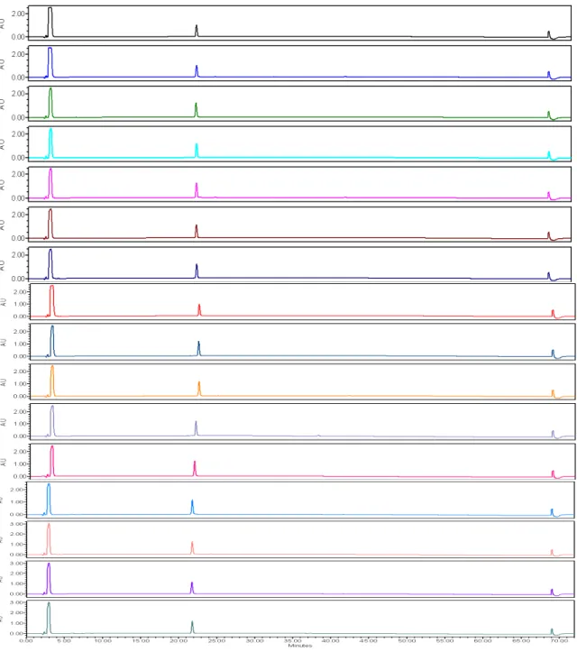
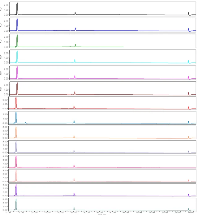
Figure 2.7 Circular Dichroism Spectra of the Mirror and ScramMirror peptides…….34 Figure 2.8 Analytical RP-HPLC analysis of peptide degradation by pronase E……..….36 Figure 2.9 Structures of the OWOC and the OWOCyc peptides………...……..41 Figure 2.10 Analytic RP-HPLC chromatograms of the Mirror peptide
incubated with pronase E for different time periods……….….46 Figure 2.11 Analytic RP-HPLC chromatograms of the ScramMirror peptide
incubated with pronase E for different time periods……….….47 Figure 3.1 Structures of protectides Mirror, ScramOWOWO, ScamMirror…………58 Figure 3.2 Structures of kinase substrates, FPKB, Abl……….59 Figure 3.3 Structures of protectide-kinase substrate full-length
reporter peptides………..….60 Figure 3.4 Circular dichroism spectra of FPKB, MP4B,
and SMP4B reporter peptides………..…63 Figure 3.5 Degradation of the ScramOWOWO peptide in the
Figure 3.8 Assessment of the stability of Abl and MED-PEG4-Abl
in presence of Ba/F3 BCR-Abl cell lysates……...………..…72 Figure 4.1 The Ubiquitin Proteasome System………..84 Figure 4.2 Structures of the ornithine-containing degrons………95 Figure 4.3 Structures of OWOWO Arg-substituted degrons investigated for
ubiquitination………..…….96 Figure 4.4 Structure of OWOWO-RRRGproteasome reporter……….…..97 Figure 4.5 The ubiquitination of Orn residues by endogenous UPS enzymes
from HeLa S100 lysates………...99 Figure 4.6 Determination of the primary site of Orn ubiquitination
on the OWOWO peptide………..102 Figure 4.7 Circular dichroism spectra of the ScramOWOWO, OWOWO,
OWRWR degron peptides………105 Figure 4.8 Determination of the significance of secondary structure
in the ubiquitination of Orn residues……….107 Figure 4.9 Electropherogram of III-67B peptide after degradation by proteolytic
enzymes present in OPM-2 lysates as determined by CE-LIF………..111 Figure 4.10 Structures of E3 ligase inhibitors and of E1 conjugating enzyme
inhibitor used in the ubiquitination pull-down assay……….….…114 Figure 4.11 E3 Ligase Inhibitor Assay……….……...115 Figure 4.12 Electroporation of OPM-2 cells for the membrane permeation of the
LIST OF SCHEMES
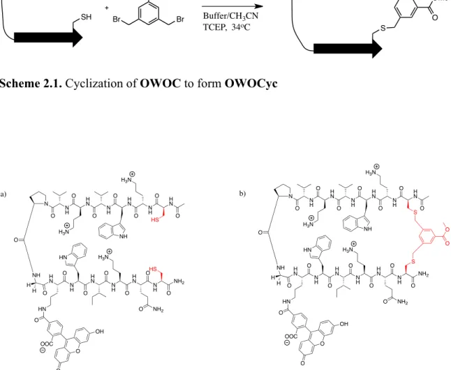
SCHEME 2.1 Cyclization of OWOC to form OWOCyc……….41 SCHEME 3.1 Using click chemistry to conjugate kinase
LIST OF ABBREVIATIONS
Ac Acetyl
Akt Protein kinase B Ala, A Alanine
Alloc Allyloxycarbonyl Arg, R Arginine
Asn, N Asparagine Asp, D Aspartic Acid Azk Azidolysine
BME 2-mercaptoethanol, β-mercaptoethanol Boc tert-Butyloxycarbonyl
CD Circular dichroism
CE-LIF Capillary electrophoresis with laser induced fluorescence Cys, C Cysteine
DCM Dichloromethane
DEDTC Sodium diethyldithiocarbamate trihydrate DIPEA N,N’-Diisopropylethylamine
DMF Dimethylforamide DMSO Dimethylsulfoxide DTT Dithiothreitol EDT 1,2-Ethanedithiol ESI Electrospray ionization
FlAsH Fluorescein Arsenical Hairpin FMOC 9-Fluorenylmethoxycarbonyl Gln, Q Glutamine
Glu, E Glutamic Acid Gly, G Glycine
H2O Water
HBTU O-(Benzotriazol-1-yl)-N,N,N’N’-tetramethyluronium hexafluorophosphate
HOBt 1-Hydroxybenzotriazole Ile, I Isoleucine
ivDde 1-(4,4-dimethyl-2,6-dioxocyclohex-1-ylidene)-3-methylbutyl Leu, L Leucine
Lys, K Lysine
MALDI Matrix-assisted laser desorption ionization MeOH Methanol
MeUb Methylated ubiquitin MM Multiple myeloma
NMR Nuclear magnetic spectroscopy No Lys Ub No lysine ubiquitin
Orn, O Ornithine
Pbf 2,2,4,6,7-Pentamethyldihydrobenzofuran-5-sulfonyl PBS Phosphate buffered saline
PEG Polyethylene glycol Phe, F Phenylalanine PKB Protein kinase B Prg Propargyl glycine
D-Pro, p D-Proline
Pro, P Proline
RFU Relative fluorescence units SDC Sodium deoxycholate SDS Sodium dodecyl sulfate SPPS Solid phase peptide synthesis
tBu Tert-butyl
TFA Trifluoroacetic acid Thr, T Threonine
TIPS Triisopropylsilane
Tris Tris(hydroxymethyl)aminoethane Trp, W Tryptophan
Trt Trityl
TUBEs Tandem ubiquitin binding entities Tyr, Y Tyrosine
Ub Ubiquitin
Chapter I
PEPTIDES USED TO MONITOR ENZYMATIC ACTIVITY
A. Introduction
During the 20th and early 21st centuries, clinical diagnosis and treatment went from being predominantly macroscopic to being mostly microscopic. Nearly every medical diagnosis in well-developed countries is validated by one or more molecular tests that screen for a microscopic indication of disease.1 The development and advancement of microscopy, spectroscopy, antibody-based assays, and the mapping of the human genome and proteome, has allowed for an increased understanding of disease causation and the determination of the mechanisms of action of drugs on their molecular targets.
Knowledge of pharmaceutical targets and drug modes of action has led to the development of ways to monitor disease progression and remission at the molecular level.2 Clinicians and bench scientists are designing ways to directly and molecularly measure the effects of therapeutic agents on their molecular targets during the course of the treatment.
________________________
1Yager, P.; Edwards, T.; Fu, E.; Helton, K.; Nelson, K.; Tam, M.R.; Weigl, B.H.
Nature2006, 442, 412-18.
The treatment of multiple myeloma (MM), an incurable hematologic malignancy of the plasma cells in the bone marrow, has greatly advanced with the FDA approval of molecularly targeted therapeutics.3 However, none of the current diagnostic tests directly measure the activity of enzymes and protein-protein interactions targeted by these novel treatments.3 In this chapter, we will briefly discuss the current state of MM diagnostics and the ways in which the effectiveness of the molecularly-targeted therapeutics used to treat the disease can be measured during the course of treatment. Specifically, we will highlight examples of how peptides can be used in the future to monitor disease progression and eventual remission by directly evaluating cytosolic enzymatic activity relevant to disease states. We will conclude with our proposal to design and use fluorescent peptide-based probes for the monitoring of therapeutic agent effectiveness during MM treatment with enzyme inhibitors.
B. Multiple myeloma diagnosis and treatment today
Multiple myeloma (MM) is the monoclonal proliferation of plasma cells, the
antibody-producing white blood cells present in the bone marrow, and accounts for 10% of all hematologic malignancies in the Unites States.4 MM arises from the accumulation of genetic errors in plasma cells that lead to constitutively active, hypoactive or
dysregulated protein activity.5 ________________________
3Palumbo, A.; Anderson, K. N Engl J Med2011, 364, 1046-60. 4Kyle, R.A.; Rajkuma, S.V. Blood2008, 111, 2962-72.
5Ocio, E.M.; Mateos, M.V.; Maiso, P.; Pandiella, A.; San-Miguel, J.F. Lancet
Currently, a MM diagnosis depends on the combined outcome of several tests including, serum immunoglobulin levels (the heavy chain of antibodies), light chain protein
concentration in the urine, bone marrow plasma cell population density, and the karyotyping of the plasma cells in the bone marrow.3 High concentrations of bone marrow plasma cells and monoclonal antibodies, cytogenetic abnormalities within the nuclei of myeloma cells, and physical symptoms such as anemia and bone lesions constitutes a diagnosis. Further investigation of chromosome abnormalities using fluorescence in situ hybridization (FISH), is done to determine the prognosis of the patient, with certain karyotypes associated a better outcome than others. The high concentrations of proteins including beta2-microglobulin and M-protein in the serum and
urine are associated with more advanced stage disease.3 Patients with a good immediate prognosis are first treated with an autologous stem cell transplant and then with a
combination of chemotherapeutic agents targeting the catalytic activity of enzymes such as the 26S proteasome and histone deacetylases (HDACs), E3 ligase protein-protein interactions, and DNA replication.5 Though all patients with treatable MM are
homeostasis, namely through directing intracellular trafficking and protein degradation by the 26S proteasome.6 Since the FDA approval of the 26S proteasome inhibitor bortezomib (Velcade) in 2003, the overall median survival of patients with MM has lengthened from 2-3 years to 5-10+ years depending on age and cytogenetic factors.3,7 Several next generation proteasome inhibitors have been developed and targeting the entire (UPS) for the treatment of several diseases in addition to MM, is projected in the next decade to become as popular as targeting kinase/phosphatase signaling, which dominates 25% percent of the drug market today.8
Some clinical trials associated with inhibitors of kinases and UPS enzymes have failed in the late stages due to underwhelming effects on patient overall survival.9 MM patients enrolled the Stage II/III clinical trial for perifosine, an allosterictic inhibitor of protein kinase B (PKB or Akt) activity, were screened for cytogenetic abnormalities associated with improper immunoglobulin production and excretion, and for their status as relapsed/refractory in response to combination therapies including
bortezomib/thalidomide (immunomodulatory agent)/lenolidomide. The patient tumor cell-associated PKB activity was not determined prior to enrollment. Though aberrant PKB activity has been implicated in tumor growth presumably due to its roles in cellular metabolism, motility, proliferation, and angiogenesis, PKB may not be activated or hyperactive in all cancers.10
_______________________
6Nandi, D.; Tahiliani, P.; Kumar, A.; Chandu, D. J Biosci2006, 31, 137-55. 7Samson, D. Postgrad Med J 1994, 70, 404-10.
8(a) Cohen, P.; Tcherpakov, M.; Cell2010, 143, 686-93 (b) Gonzalez-Vera, J.A. Chem
Perifosine investigation was halted in Stage III though it showed synergistic activity with bortezomib.9-10 Though bortezomib has been shown to activate PKB in some MM tumors, it is not known whether or not the activity of PKB was dysregulated in all MM patient tumors investigated and therefore the inhibitor may be useful for only a subset of patients.11 Despite the short-term and long-term effectiveness of enzyme inhibitors in the treatment of MM, no diagnostic testing is associated with determining enzyme activity in biopsied myeloma cells. This gap in diagnostic testing is due to the absence of high throughput assays that can detect enzymatic activity in a small number of cells. As low as 10% of the bone marrow cells collected from patient biopsies must be plasma cells in order to constitute a possible malignant diagnosis.3 Many techniques that measure enzymatic expression and activity such as Western Blotting, immunocoprecipitation, and ELISA require one million cells for an accurate readout while MM biopsies can
sometimes yield less than 1000 malignant cells. Chemical cytometry assessing enzymatic activity relevant to disease progression in patient tumor cells at diagnosis would give clinicians the information needed to personalize treatments for their patients. Assessing targeted enzymatic activity prior to clinical trial enrollment would allow investigators the ability to enroll patients based on their aberrant enzyme activity status and thus more accurately determine who would most benefit from treatment.
________________________
9Richardson, P.G.; Nagler, A.; Yehuda, D.B.; Badros, Hari, P.; Hajek, R.; Spicka, I.;
Kaya, H.; Le Blanc, R.; Yoon, S.S.; Kim, K.; Martinez-Lopez, J.; Mittelman, M.; Shpilberg, O.; Tothova, E.; Laubach, J.P.; Ghobrial, I.M.; Leiba, M.; Gatt, M.E.; Sportelli, P.; Chen, M.; Anderson, K.C. Blood2013, 122, 3189.
10 Richardson, P.G.; Eng, C.; Kolesar, J.; Hideshima, T.; Anderson, K.C. Expert Opin
Another advantage of being able to directly measure enzymatic activity during treatment and/or clinical trial investigation of an enzyme inhibitor would be the ability to directly monitor the drug inhibitor’s effect on its targeted activity. As in the case with MM where traditional treatment regiments include a combination of multiple enzyme inhibitors, a rapid high-throughput screening of several enzymes simultaneously would be ideal. Another factor to take into consideration is tumor heterogeneity. The
enzymatic activity in tumor cells is not homogenous even when the tumor cells are all taken out of the same patient.12 Early detection of treatment-resistant tumor cell populations would allow clinicians to implement an alternative treatment plan long before indirect measurements of relapse become detectable.
Below we will highlight a few types fluorescent peptide-based probes developed by others that are capable of detecting enzymatic activity in vitro, in vivo, and ex vivo in just a single cell. Our primary focus is on the detection of kinase activity and UPS enzymatic activity as these are relevant drug targets for the treatment of MM. We will briefly discuss the major challenges of using peptide-based probes to assess enzymatic activity in the cytosol of cells and introduce our proposed method to overcome these challenges. The use of the methods discussed is restricted mostly to bench research analysis rather than for a clinical testing. We hope the work described in the remaining chapters will be a foundation for the translation of direct enzymatic activity assays from bench top experimentation only to clinical diagnostics and disease-monitoring
technology.
________________________
11Mahindra, A.; Laubach, J.; Raje, N.; Munshi, N.; Richardson, P.G., Anderson, K. Nat
C. Peptides as substrates for the detection of enzymatic activity in vitro
Commonly, enzyme expression, activation and regulation are determined by Western Blot, enzyme-linked immunosorbant assay (ELISA), small molecule activity-based probes, and peptide-modification activity-based probes. All of the above methods rely on the mimic of protein-protein interactions essential to the maintenance of homeostasis in the body. While the above assays can give information about the activation, mutation and indirectly the catalytic activity of an enzyme, all require a large number of cells if used to investigate cell lysates and none are capable of giving real-time kinetic data.8b Assay sensitivity with fewer cells required for detection has been achieved through the use of fluorescent peptide-based probes. Peptides-derived from natural protein substrates have been successfully used as catalytic substrates of enzymes present in the cytosol of the cell.13 Peptides can be designed to be recognized and modified by the enzyme of interest with high specificity and activity.14 The addition of a fluorophore to the substrate peptide allows for quick sensitive detection of enzymatic activity that can be visualized by several types of instrumentation including fluorescence microscopy, fluorescence imaging of agarose gels, liquid chromatography coupled with mass spectrometry (LC/MS), and capillary electrophoresis with laser-induced fluorescence (CE-LIF). Below are four examples of how kinase and UPS activity have been detected in vitro
using fluorescent peptides. _______________________
12Marusyk, A.; Almendro, V.; Polyak, K. Nat Rev Canc2012, 12, 323-34. 13 Kennelly P.J.; Krebs, E.G. J Biol Chem1991, 266,15555-8.
14Wu, D.; Sylvester, J.E.; Parker, L.L.;, Zhou, G.; Kron, S.J. Biopolymers2010, 94,
i. Chelation enhanced fluorescence (CHEF)
One way to detect kinase activity is to measure a change in the fluorescence of a kinase substrate peptide due to phosphorylation of the substrate. This can be achieved by chelation enhanced fluorescence (CHEF) of the kinase substrate peptide through the phosphorylation-dependent coordination of a metal by a chromophore covalently attached to the kinase substrate peptide.15 Notable examples of such probes were reported by the Imperiali Group whose probes included a cysteine conjugated
sulfonamide-oxime (C-Sox) chromophore within the region C-terminal or N-terminal to the kinase substrate region.16 Upon phosphorylation of the substrate peptide by kinases in in mammalian cell lysates, Mg2+ was simultaneously chelated by both the C-Sox chromophore and the phosphoryl group on the modified Ser/Thr residue which led to a 2-5 fold increase in fluorescence. Lukovic et al. were able to design CHEF-based probes with a higher degree of substrate specificity for several kinases including protein kinase C (PKC), Abelson kinase (Abl) and Akt1 (PKB) through a recognition-domain focused design strategy. In addition, they tailored the kinase activity detection to a 96-well format that allowed for kinetic data to be determined based on fluorescence enhancement
in vitro, demonstrating the potential for this assay to evolve into a high-throughput technique.
___________________________
15Shults, M.D.; Janes, K.A.; Lauffenburger, D.A.; Imperiali, B. Nature Methods 2005, 2,
277-84.
While the majority of the activity measured by these CSox probes was due to phosphorylation by the desired kinase as shown by the use of kinase inhibitors and immunodepletion of the kinase of interest, there is still some probe modification by other kinases. Since there are over 500 known kinases in the cell, cross-reactivity in not
unexpected but not desired. Therefore continued improvement of substrate specificity is necessary before this technology can be applied to cell lysate assays in the clinic.
ii. Quencher dyes
Another strategy for specific kinase activity detection by a change in fluorescence is “Deep Quench” developed by the Lawrence Group. Deep quench detects the restoration of fluorescence of the kinase-specific peptide substrate upon modification by the desired kinase.17 Kinase substrates designed for deep quench possessed a coumarin moiety conjugated to the positively charged peptide N-terminal to the modifiable Ser/Thr modification site. Prior to phosphorylation, coumarin fluorescence was quenched in the presence of the negatively charged Acid Green dye. Phosphorylation of the kinase substrate peptide and subsequent binding of the phosphorylated substrate to the 14-3-3 protein domain displaced the Acid Green dye and led to the fluorescence of modified substrates. This method allowed for the detection of protein kinase A (PKA) activity in mitochondrial membranes, with phosphorylation associated fluorescence observed over the autofluorescence associated with the organelles.
_________________________
The dramatic fluorescence enhancement upon modification of these probes highlights the potential for similar peptide-based probes to be used ex vivo to monitor organelle specific-kinase activity in small sample sizes.
iii. Fluorescence detection by capillary electrophoresis
It is possible to detect kinase activity through the use of fluorescent peptide-based probes without monitoring a change in fluorescence. The Allbritton Group, in
collaboration with the Lawrence Group, has reported the detection of Abl kinase activity in cell lysates using capillary electrophoresis coupled with laser-induced fluorescence (CE-LIF).18 This technique allows for the separation and fluorescence detection of both the modified and unmodified reporters within a capillary based on the difference in electrophoretic mobility due to the differing net charge of the phosphorylated and nonphosphorylated substrates.
This method of detection is highly sensitive requiring only subnanomolar concentrations of modified peptides for fluorescence detection and was used to determine the catalytic efficiency of substrate phosphorylation.18 This technique, is also amenable to the simultaneous screening of several substrates at once, allowing for the concurrent detection of the activity of multiple enzymes.
iv. Detection of UPS activity in vitro using short peptides
In addition to kinase activity, the activity of UPS enzymes, including the proteasome, can be detected using peptide substrates as well.
_________________________
Recently, the Allbritton Group has examined the collective activity of E1, E2, and E3 UPS enzymes through the detection of ubiquitinated peptides bearing short modifiable recognition motifs known as degrons.19 Short fluorescent peptide sequences, 9-26 amino acids long, derived from proteins regulated by UPS-directed ubiquitination in the cell, were modified by E1, E2 and E3 enzymes in vitro. Both monoubiquitination and polyubiquitination of the peptides substrates was observed by fluorescence imaging of the ubiquitinated substrates separated by gel electrophoresis. The kinetics of peptide substrate ubiquitination and the minimal sequence required for modification were also assessed. The Allbritton Group is currently adapting the assay for analysis by CE-LIF.
D. Peptides for the detection of intracellular enzymatic activity
The methods for kinase and UPS activity detection mentioned above as well as many other noteworthy approaches not mentioned here, mark great strides in the advancement toward enzymatic activity assessment in patient samples in the clinic. However, all of the above efforts were done in the context of purified enzymes or cell lysates and not within the cytosolic environment of intact cells. Detection of intracellular enzymatic activity requires the use of stable substrates and sensitive detection methods. Peptide-based fluorescent probes that meet the demands of ex vivo kinase activity
detection have been developed by several groups using a variety of methodologies. Below we will briefly discuss three approaches used to successfully detect kinase or UPS enzymatic activity within intact cells.
________________________
19Melvin, A.T.; Woss, G.S.; Park, J.H.; Dumberger, L.D.; Waters, M.L.; Allbritton, N.L.
i. Caged compounds
A major challenge when measuring kinase activity inside of cells is the inability to accurately control and measure the start point of enzymatic activity. One way to gain control of the intracellular kinase activity initiation is to use “caged” substrates.
Molecular caging involves masking the substrate functional group required for enzymatic activity with an optically silent moiety via a photocleavable bond. Photolysis of the caged compound at the desired zero time point, liberates the substrate to an optically active form.20 The caging strategy was first applied to the masking of end phosphate groups on cyclic AMP and ATP with a photolabile nitromethoxy phenyl group. Upon photolysis biologically active forms of cyclic AMP and ATP were released and available for hydrolysis by cellular enzymes.21Recently, the caging strategy has been applied to the detection of PKA activity in erythrocytes by using a peptide-based substrate. The
Lawrence Group reported controllable PKA activity detection through the combined strategies of fluorescence quenching and phosphorylation site caging.22 The PKA substrate peptide contained a N-terminal fluorophore that was quenched in the presence of a negatively charged dye that noncovalently bound positively charged residues in the peptide chain via electrostatic interactions.
________________________
20Lee, H.M.; Larson, D.R.; Lawrence, D.S. ACS Chem Biol2009, 4, 409-27. 21(a) Forbus III, B.; Kaplan, J.H.; Hoffman, J.F. Biochemistry1978, 17, 3667-76.
(b)Kaplan, J.H.; Ellis-Davies, G.C.; Proc Natl Acad Sci USA1988, 85, 6571-75. (c)Nargeot, J.; Nerbonne, J.M.; Engels, J.; Lester H.A. Proc Natl Acad Sci USA 1983, 80, 2395-99.
22Oien, N.P.; Nguyen, L.T.; Jernigan, F.E.; Priestman, M.A.; Lawrence, D.S. Angew
The phosphorylatable Ser residue was masked with the photolabile 4,5-dimethoxy-2-nitrobenzyl group prior to loading into intact erythrocytes (red blood cells). The reporter peptide was loaded into the red blood cells and fluorescence was detected only upon photolysis which liberated the Ser hydroxyl group for phosphorylation by PKA and the subsequent displacement of the negatively charged quencher dye, restoring the
fluorescence of the N-terminal fluorophore. This strategy demonstrated the power of using temporal detection for the measurement of kinase activity ex vivo.
ii. Single cell CE
Collaborative efforts of both the Allbritton and Lawrence Groups has allowed for the detection of kinase activity in single cells using CE-LIF.23 PKB activity was detected using peptide substrates with unnatural amino acids incorporated intermittently
throughout the substrate peptide chain to increase resistance to degradation by proteases without greatly sacrificing substrate efficacy. These fluorescent probes were used to measure both the rates of phosphorylation and the rates of protease degradation based on the differences of the electrophoretic mobility of the phosphorylated substrates,
nonphosphorylated substrates, intact reporter peptides and digestion fragments. These probes also gave information about differing sites of substrate hydrolysis by proteases in cell lysates versus intact cells. This report showed the potential of detecting kinase activity when the number of cells is the limiting factor such as the case with plasma cell analysis of MM patient tumors. Their results also showed the potential of redesigning substrates for increased stability to proteolysis without sacrificing the capability of being modified by the desired enzyme.
_________________________
iii. Detection of proteasome activity in live cells
The success of bortezomib in the clinic has increased the need for probes that can be used to detect proteasome activity inside intact patient cells. The proteasome utilizes three different types of catalytic activity to degrade proteins, trypsin-like activity, chymotrypsin-like activity, and caspase-like activity.24 When screening potential proteasome inhibitors knowledge of which enzymatic activity is being blocked is highly valued. Several fluorescent peptide-based probes with terminal electrophiles have been used to label proteasome substrates. The general structure of several of these probes is a dye conjugated to the N-terminus of a tri-aminohexanoic acid (Ahx), tri-leucine (Leu), vinyl-sulfone (Dye-(Ahx)3-L3-VS) probe. One example reported by Verdoes et al.
utilized a membrane-permeable Bodipy-TMR-Ahx3L3VS which they used to detect
proteasome activity in cell lysates, intact cells and in mice.24 When incubated with cell lysates in the presence of known proteasome inhibitors, the degree of proteasome
labeling correlated to the degree of inhibitor. These probes were even used to determine which subunit of the proteasome was being inhibited. Though detection of proteasome labeling required cell lysis or fixation, the combination of membrane permeability and fluorescence detection of subunit specific labeling, lays a solid foundation for the use of these probes in a clinical setting.
_________________________
24Verdoes, M.; Florea, B.I.; Menendez-Benito, V.; Maynar C.J.; Witte, M.D.; van der
E. Purpose of this work
Though the use of peptides for the detection of enzymatic activity in the cell is advantageous due to their being excellent mimics of natural enzyme substrates, peptides comprised of canonical amino acids are also substrates for cytosolic peptidases. Peptide hydrolysis by aminopeptidases in the cytosol of immune cells such as plasma cells reduces peptides to their amino acid building blocks rapidly.25 Peptide-based enzymatic activity probes without features to make them resistant to hydrolysis by cytosolic proteases, are subject to destruction in the cellular environment before an activity-based readout can be detected.
In the remaining three chapters we will describe our work to improve the protease resistance of fluorescent peptide probes used to detect kinase and UPS activity in
malignant mammalian cell lines. Specifically we will describe the design, synthesis, and performance of orninthine-rich β-hairpin peptide probes of enzymatic activity and the advantages and of utilizing secondary structure and noncanonical amino acids to impart protease resistance.
_______________________
Chapter II
DESIGN, SYNTHESIS, AND CHARACTERIZATION OF
PROTEASE-RESISTANT β-HAIRPIN PEPTIDES CALLED PROTECTIDES
A. Background
Synthetic peptides have emerged as popular tools used to study cellular events, including molecular recognition, enzymatic activity, and membrane trafficking.1 The natural existence of peptides in biological systems and their vast potential for
diversification allows them to be used as pharmaceuticals and diagnostics as well. There is a need for diagnostic testing that can measure enzymatic activity associated with the effectiveness of drug inhibitors in patient cells ex vivo.2 This need can be met through the use of fluorescently tagged peptide substrates that can be easily detected by spectroscopy, spectrometry, and/or electrophoresis, all methods already used in a clinical setting.3 _____________________
1Ruttekolk, I.R.; Witsenburg, J.J.; Glauner, H.; Bovee-Geurts, P.H.M.; Ferro, E.S.;
Verdurmen, W.P.R.; Brock, R. Mol Pharmaceutics2012, 9, 1077-86.
2Mahindra, A.; Laubach, J.; Raje, N.; Munshi, N.; Richardson, P.G., Anderson, K. Nat
Rev Clin Oncol2012, 9, 135-43.
The detection of kinase activity, the target of many pharmaceutical inhibitors, in live cells and in cell lysates has been reported using short unstructured peptides (6-30 residues) mimicking the native protein substrates.3-4 Incorporating such reporters as part of regular diagnostic screening would be particularly helpful in cancers of the immune cells like multiple myeloma (MM) and chronic myelogenous leukemia (CML) where kinase and other enzyme inhibitors are part of the first line of treatment.5 Currently, there are no diagnostic tests for either of these diseases that screen for the targeted enzyme activity, prior to treatment with drug inhbitors.6
While proteins, due to their tertiary structure, are not susceptible to degradation unless specifically targeted in live cells by degradation pathway proteins, unstructured peptides are vulnerable because of their shorter length and lack of secondary and tertiary structure.7 In the cytosol of immune cells, aminopeptidases are predominantly
responsible for peptidic digestion though the presence of endopeptidase activity has been reported.8 The crystal structures of two aminopeptidases, tripeptidyl peptidase II (TPP II) and leucine aminopeptidase (LAP) reveal catalytic cleft entrance dimensions of 30 Å × 15 Å and 30 Å × 10 Å respectively, restricting access to short unstructured peptides.9 _________________________
4Kunkel, M.T.; Ni, Q.; Tsien, R.Y.; Zhang, J.; Newton, A.C. J Biol Chem 2005, 280,
5581-7.
5(a) Placzek, E.A., Plebanek, M.P.; Lipchik; A.M.; Kidd, S.R.; Parker, L.L. Anal
Biochem2010, 397, 73-8. (b) El-Amm, J.; Tabbara, I.A. Am J Clin Oncol 2013 Aug 7, [Epub ahead of print].
6Palumbo, A.; Anderson, K. N Engl J Med2011, 364, 1046-60.
7(a)Reits, E.; Griekspoor. A.; Neijssen, J.; Groothuis, T.; Jalink, K.; van Veelen, P.;
Well-folded β-hairpin peptides at the N-terminus of kinase activity reporter peptides can be used to increase their resistance to such proteases.We recently reported that the attachment of β-turn peptides, also known as β-bends, to the N-terminus of an unstructured protein kinase reporter peptide, successfully slowed its degradation by cellular proteases.10
In this work, β-hairpin peptides were used to protect unstructured peptide-based probes from proteolytic degradation. A β-hairpin peptide consists of two parallel or antiparallel peptide strands connected by a rigid turn or an unstructured loop, forming the most basic unit of a β-sheet, one of the most prevalent types of secondary structure found in proteins.11 In the past two decades, several studies have been dedicated to
understanding the essential noncovalent and covalent interactions necessary to form stable well-folded β-sheets and β-hairpins outside of the context of full protein structure and tertiary contacts.12 There are five basic features that allow for short peptides to adopt stable β-hairpin conformations; 1) a turn-nucleating sequence, 2) cross-strand backbone H-bonding, 3) sidechain noncovalent interactions, 4) backbone rigidity, and 5) terminal covalent and noncovalent interactions to prevent fraying (Figure 2.1).
__________________________
8Akkad, N.; Schatz, M.; Dengjel, J.; Tenzer, S.; Schild, H. Med Microbiol Immunol2012,
201, 463-73.
9Rockel, B.; Kopec, K.O.; Lupas, A. N.; Baumeister, W. Biochim Biophys Acta 2012,
1824(1), 237-45. (b) Burley, S.K.; David, P.D.;, Taylor, A.; Lipscomb, W.N. Proc Natl Acad Sci1990, 87, 6878-82.
10(a) Eker, F.; Griebenow, K.; Schweitzer-Stenner, R. J Am Chem Sci 2003, 125,
8178-85. (b) Yang, S.; Proctor, A.; Cline, L.L.; Houston, K.M.; Waters, M.L.; Allbritton, N.L.
Investigation of these key features has allowed for the design of well-folded β-hairpin peptides with multiple applications, including binding nucleic acids, mimicking protein-protein interactions, and imparting protease resistance.13 Additionally, folded β-bend peptides which contain only the amino acid residues necessary to nucleate a β-turn,have been reported .10
β-hairpins were chosen as ideal protectides over other secondary structures such as alpha helices, due to their ability to tolerate diverse functionalization (including
appending a peptide at the C-terminus) while maintaining their conformational folding in aqueous solutions. While the use of peptidomimetics, including the incorporation of β and ϒ amino acids, has allowed for the development of short, stable peptide mimics with remarkable resistance to proteolytic degradation, the building blocks are generally moreexpensive.14 Also, we are designing protease resistant moieties to be covalently attached to L-amino acid peptide kinase substrates, and the use of peptidomimetics may necessitate additional synthetic steps.
___________________________________
11Voet, D.; Voet, J. Biochemistry; 3rd ed.; John Wiley & Sons, Inc: Hoboken, NJ, 2004,
pg 227-9.
12Hughes, R. M.; Waters, M.L. Curr Opin Struct Biol2006, 16, 514-24.
13(a) Stewart, A.L.; Waters, M.L. ChemBioChem2009,10, 539-44. (b) Wilger, D.J.; Park,
Short, thermodynamically stable, β-hairpins have been designed and synthesized through the optimization of turn sequences, the utilization of favorable
sidechain-sidechain interactions, the use of amino acids with high β-sheet propensity, and the use of salt bridges and capping motifs to prevent terminal fraying. 12,15 It has been reported by our group and others that optimization of beta-hairpin secondary structure conveys substantial resistance to proteolytic degradation.13c,e-f
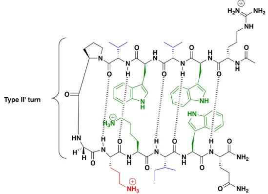
Figure 2.1 Anti-parallel β-hairpin peptide design features. Highlighted in green are
residues participating in noncovalent sidechain interactions, in blue are residues with high β-sheet propensity, and the residue in red is the non-canonical amino acid ornithine. Backbone H-bonding is indicated by the hashed lines.
H N N O O H N N O O O HN H H N
O NH
N N H O O O N O N NH2 O O H H H H H Type II' turn
NH3 NH HN N H O NH NH2
H2N
NH2
O N
H
Previously, our group confirmed a direct correlation between secondary structure and the resistance of β-hairpin peptides to degradation by proteolytic enzymes.13c,f The
WKWK and the Trp Pocketβ-hairpin peptides (Table 2.1) were determined to be 95% and 99% folded in aqueous solvent, respectively, despite being only 12 amino acids in length.16 Each contained 11 canonical amino acids and 1 non-canonical amino acid (ornithine, Orn) and were found to be substantially more resistant to degradation by specific and nonspecific proteases than the unstructured control.13cThe sequence of
WKWK was modified by replacing the turn sequence Asn-Gly (type I’ turn) with
D-Pro-Gly (type II’ turn). The incorporation of the D-Pro-D-Pro-Gly turn sequence has been shown to enhance the stability of β-hairpins by nucleating a tighter turn.17 The resulting
WKWK-pGturn peptide (Figure 2.2 and Table 2.1), rich in basic amino acids (cleavage site of
trypsin) and aromatic amino acids (cleavage site of α-chymotrypsin) showed resistance to degradation by trypsin, α-chymotrypsin and pronase E (a mixture of nonspecific
proteases).13cThe WKWK-pGturn peptide showed a 10-fold greater stability to
proteolytic degradation by pronase E in vitro over the unstructured control with a similar primary amino acid sequence.13cThis increased stability was attributed to its higher degree of fraction folded afforded by the type II’ turn sequence.
________________________
14Frackenpohl, J.; Arvidsson, P.I.; Schreiber, J.V.; Seebach, D. ChemBioChem 2001,
2,445-55.
15Kier, B.; Shu, I.; Eidenschink, L.A.; Andersen, N.H. Proc Natl Acad Sci2010, 107,
10466-71.
16 (a) Butterfield, S. M.; Waters, M.L.; J Am Chem Soc2003, 125, 9580-1. (b) Butterfield,
S.M.; Sweeney, M.M.; Waters, M.L. J Org Chem2005, 70 1105-14. (c) Rieman, A.J.; Waters, M.L. Biochemistry2009, 48, 1525-31.
The following report describes ours efforts to further increase the lifetime of
WKWK-pGturn by modifying its amino acid structure to increase folding and to slow
or prevent hydrolysis by cytosolic peptidases. The modified β-hairpin peptides were designed to last for hours in vitro under harsh proteolytic conditions. The resulting highly resistant β-hairpin peptides, which we call “protectides”, were then covalently attached to the N-termini of unstructured kinase and proteasome substrates to slow their degradation in cell lysates. The synthesis and performance of these full-length reporters is discussed in Chapter III of this work.
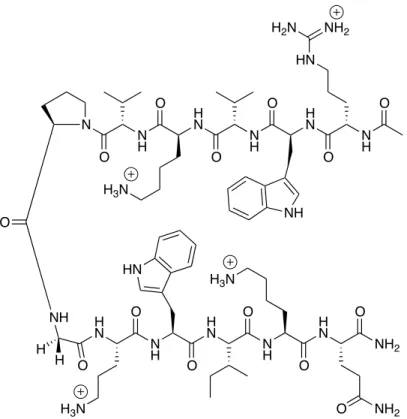
Figure 2.2 Structure of the WKWK pGturnβ-hairpin peptide.13c
N H H N N H H N O O O O N H O N O HN H2N NH2
NH O NH H N N H H N N H O O HN O O H N NH2 O O
H3N O NH2
H H
H3N
B. Design and Synthesis
Building on the work published previously by our group (Cline and Waters, 2009), we made key changes to the amino acid sequence of WKWK-pGturn, based on the five design features mentioned above as well as the addition of non-canonical amino acids.
The OWOWO peptide has the same primary sequence as WKWK pGturn except all of
the basic natural amino acids were replaced with the non-canonical amino acid analog of lysine, ornithine (Figure 2.3, Table 2.1). Although trypsin-like peptidases cleave at the carboxyl group of basic amino acids, the shortening of the Lys sidechain by one
methylene group as is the case with Orn, slows the rate of hydrolysis by such enzymes.18 Our group has demonstrated that Orn residues are capable of participating in diagonal cation-π interactions with aromatic sidechains and was therefore expected to participate in favorable interactions in the OWOWOpeptide.19 Since Orn carries a positive charge under physiological conditions, Orn residues were also added to increase the water solubility of otherwise hydrophobic peptides. The ScramOWOWO (Scrambled-OWOWO) peptide has the same primary amino acid sequence as the OWOWO peptide except the turn residues were positioned at the C-terminus of the peptide rather than in the middle (Figure 2.3). This peptide served as an unstructured control. The Trp
Pocket peptide, which demonstrated proteolytic stability when tested previously, was
also modified and investigated.13cThe Asn-Gly turn sequence was replaced with the stronger turn-nucleating sequence D-Pro-Gly, the N-terminal Arg was replaced with Orn,
and 5(6)-carboxyfluorescein was attached to the sidechain of the Lys8 (Figure 2.3, Table 2.1).
_______________________
A long-standing challenge of designing and synthesizing short tightly-folded β-hairpin peptides is terminal fraying. To alleviate this problem, β-β-hairpins are commonly cyclized forming a covalent bond between the termini.4 However, one solution to fraying using noncovalent interactions was reported by Kier et al. and is referred to here as the Andersen capping motif.15The Andersen capping motif, “acyl-W-loop-WTG”, added +6 kJ/mol of stability to the β-hairpins reported.15This capping motif confers additive stability through a face-to-edge Trp-Trp interaction, bifurcated hydrogen
bonding of the Thr residue with the N-terminal acyl group and the HN of Gly, and a CH-π
interaction between the N-terminal Trp and the C-terminal Gly (Figure 2.4).15 The Andersen capping motif was incorporated into the primary amino acid sequences of the
AC1F, AC2F, AC4F, and AC5Fβ-hairpin peptides (Figure 2.4, Table 2.1) The cross-strand cation-π sidechain-sidechain interaction that was present in the WKWK-pGturn
peptide and the Trp Pocket peptide was incorporated into the AC4F peptide by
positioning a Lys residue opposite a Trp residue on the non-hydrogen bonding face of the peptide. The “tryptophan zipper” (trpzip) motif reported by Cochran and coworkers was incorporated into the AC5F peptide through the placement of four Trp residues cross-strand from each other on the non-hydrogen bonded face of the β-hairpin.20 The edge-to-face π-π interaction between the indole sidechains of the trpzip motif have allowed for short β-hairpin peptides to be over 99% folded under physiological conditions.16c
________________________
19Hughes, R.M.; Benshoff, M.L.; Waters, M.L.Chem-Eur J2007 13, 5753-64.
20 (a) Cochran, A.G.; Skelton, N.J.; Starovasnik, M.A. Proc Natl Acad Sci2001, 98,
Finally, the D-isomer of the WKWK-pGturn peptide was made by replacing all of
the L-amino acids with their D-isomer and the D-Pro residue was replaced with L-Pro. As
Gly is achiral it remained the same. The D-isomer of WKWK-pGturn, referred to as
theMirrorβ-hairpin peptide, was expected to have the greatest resistance to degradation
since proteases are highly enantiospecific. The scrambled version of the Mirror peptide,
the ScramMirror peptide, was synthesized as an unstructured control (Figure 2.3).
The fluorophore 5(6)-carboxyfluorescein (FAM) was appended to the sidechain of the Lys or Orn at the i+4 position just after the turn sequence of the β-hairpin peptides and to Orn7 (Lys7) of the scrambled peptides. The fluorophore was attached next to a turn residue of all β-hairpin peptides while all of the aromatic residues were placed on the opposite face to prevent the quenching of the FAM.21We expected that the incorporation
of the fluorophore FAM to the sidechain of Lys or Orn, would also increase protectide resistance to trypsin-like peptidases, as the FAM would likely prevent proteolytic degradation due to steric hindrance. The attachment of FAM to each protectide allowed for their detection in cell-based assays described in Chapters III and IV of this work.
________________________
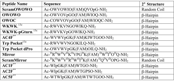
Table 2.1 Name, amino acid sequence and secondary structure of peptides investigated for protease resistancea
Peptide Name Sequence 2° Structure
ScramOWOWO Ac-OWVOWIO(FAM)QVOpG-NH2 Random Coil
OWOWO Ac-OWVOVpGO(FAM)WIOQ-NH2 β-Hairpin
OWOC Ac-COWVOVpGO(FAM)WIOQC-NH2 β-Hairpin
WKWK.13c Ac-RWVKVNGOWIKQ-NH2 β-Hairpin
WKWK-pGturn.13c Ac-RWVKVpGOWIKQ-NH2 β-Hairpin
AC4F** Ac-WVWVpGK(FAM)KIWTGOO-NH2 β-Hairpin
Trp Pocket13c Ac-RWVWVNGOKILQ-NH2 β-Hairpin
Trp Pocket dPro Ac-OWVWVpGK(FAM)OILQ-NH2 β-Hairpin
Mirror Ac-DRDWDVDKDVPGDK(FAM) DWDIDODQ-NH2 β-Hairpin
ScramMirror Ac-DKDWDVDRDWDIDK(FAM) DQDVDOPG-NH2 Random Coil
AC1F** Ac-WIpGK(FAM)WTGO-NH2 β-Hairpin
AC2F** Ac-WIpGK(FAM)WTGPSO-NH2 β-Hairpin
AC5F** Ac-WTWIpGK(FAM)WTWTGOO-NH2 β-Hairpin
________________________
aO represents the non-canonical amino acid ornithine. “Ac” represents acetylation of the
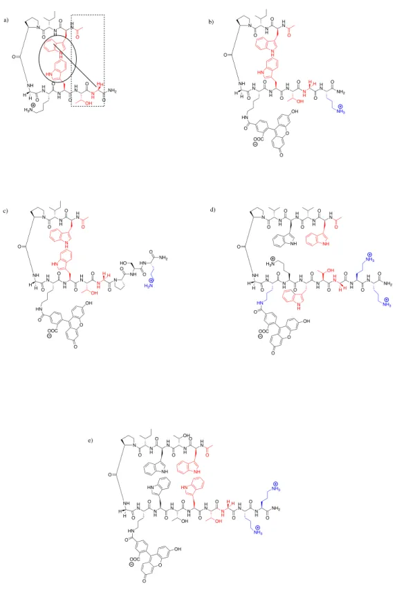
Figure 2.3 Structured protectides and the unstructured control peptide evaluated in this work. (a) OWOWO (b) ScramOWOWO (c) Trp Pocket dPro (d) Mirror (e)
ScramMirror. Ornithine residues are highlighted in blue.
N H H N N H H N N H H N N H H N O O O O O O O O N H H N O O N O NH O NH2 O NH NH3 NH NH2 O H3N HN H H N H H N N H H N O O O O N H O N O H3N NH O NH H N N H H N N H O O HN H3N O O H N NH2 O O HN H3N NH2 O O O O OH OOC H H N H H N N H H N O O O O N H O N O H3N NH O NH H N N H H N N H O O O O H N NH2 O O NH2 O H H NH HN O O O OH OOC H3N N H H N N H H N O O O O N H O N O HN H2N NH2
NH O NH H N N H H N N H O O HN O O H N NH2 O O HN H3N NH2 O O O OH OOC O H H N H H N N H H N N H H N N H H N O O O O O O O O N H H N O O N O NH O NH2 O NH3 NH NH NH2
H2N
HN NH2 O H3N HN O O O OH H H H3N O O O HO COO COO
H3N
a) b)
c) d)
Figure 2.4Structures of β-hairpin protectides containing the Andersen capping motif. (a) Example peptide highlighting the noncovalent interactions of the Andersen capping motif. (b) AC1F (c) AC2F (d) AC4F (e) AC5F. Residues part of the capping motif are highlighted in red. Ornithine residues are highlighted in blue.
H N N H H N O O O N H O N O NH O NH H N N H H N N H O O O O H N N H O O HN O O O OH OOC H H NH H N O NH2 O N H OH NH3 NH3 H N O N H O N O NH H N N H H N N H O O O O O OH N H H N HN H H O HN O O OH O NH2 O NH3 H N O N H O N O NH H N N H H N N H O O O O O OH N H N HN H H O HN O O OH O H N N H H N O O O N H O N O NH OH O NH H N N H H N N H O O O O H N N H O O HN O O O OH OOC H H NH H N O N H O HN OH HN OH O NH2 NH3 NH3 H N O N H O N O O NH H N N H H N N H O O O O O OH N H NH2 HN HH H H OOC OOC H3N
H H
H H
H3N
H H H H NH
O HN O NH2 O HO
H2N
a) b)
c) d)
C. Results and discussion
i. Structural characterization by circular dichroism
The secondary structure of the WKWK-pGturn peptide was characterized previously using circular dichroism (CD) and NMR. This 12-mer was folded greater than 98% at 25°C in aqueous solvent.13c The secondary structure of the peptides listed in Table
2.1 was determined CD spectroscopy (Figures 2.5, 2.6, 2.7). The OWOWOpeptide had a local minimum near 218 nm indicative of a β-hairpin fold but it also had a minimum near 205 nm which has been reported in the spectra of other β-hairpin peptides with type II’ turns and aromatic residues (Figure 2.5).22 The exciton coupling between the indole rings of the Trp residues was observed by the presence of a local maximum at 225 nm and may be responsible for the shift in the minimum at 205 nm. The minimum at 205 nm may also represent a distortion of the fold to accommodate the cross-strand cation-π interaction between Orn and Trp residues on the non-hydrogen bonded face. The scrambled peptide, ScramOWOWO, had a minimum near 195 nm which is
characteristic of a random coil (Figure 2.5).13c This observation confirmed that moving the turn sequence to the C-terminus of the peptide prevented the formation of any secondary structure though the identification of the amino acids and the chain length of the primary sequence was identical to that of the OWOWOβ-hairpin peptide. The
Trp-Pocket dPro peptide had a minimum near 218 nm and maximum near 230 nm indicative
Figure 2.5 Circular dichroism spectra of the ScramOWOWO, OWOWO, and Trp
Pocket dPro peptides. Experiments were done at 40 µM peptide concentration in 10 mM
sodium phosphate buffer, pH 8 at 25°C. ScramOWOWO (blue) was found to be a
random coil while OWOWO and Trp Pocket dPro (green and purple respectively) both fold into a β-sheet conformation.
-‐60000 -‐50000 -‐40000 -‐30000 -‐20000 -‐10000 0 10000 20000 30000 40000
185 190 195 200 205 210 215 220 225 230 235 240 245 250
Θ (deg
cm
2dmol -‐1)
Wavelength (nm)
ScramOWOWO
OWOWO
The CD spectra of the Andersen capped peptides was similar to that of the trizip peptides reported by Cochran and coworkers (Figure 2.6).20a Since a major component of the noncovalent capping motif is a cross-strand face-to-edge Trp-Trp interaction, the similarity between the spectra was expected. Exciton coupling again was observed by the presence of a minimum near 218 nm and a local maximum near 230 nm. The Trp-Trp interactions masked the β-sheet peak at 215 nm in the spectra of all the Andersen capped β-hairpins (Figure 2.6).
___________________
22Mahalashmi, R.; Shanmugam, G.;Polavarapu, P.L.; Balaram, P. Chembiochem 2005, 6,
Figure 2.6 Circular Dichroism Spectra of the Andersen Capped peptides, AC1F (blue),
AC2F (red), AC4F (black), and AC5F (green). Experiments were done at 40 µM
peptide concentration in 10 mM sodium phosphate buffer at 25°C. All capped peptides
fold into a β-sheet conformation and display strong Trp-Trp interactions with a minimum mean residue ellipticity near 218 nm and a maximum near 230 nm.
-‐100000 -‐80000 -‐60000 -‐40000 -‐20000 0 20000 40000 60000 80000 100000
185 190 195 200 205 210 215 220 225 230 235 240 245 250
Θ (deg
cm
2dmol -‐1)
Wavelength (nm)
AC1F
AC2F
AC4F
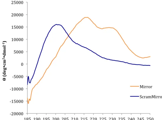
Figure 2.7 Circular dichroism Spectra of the Mirror and ScramMirror peptides. Experiments were done at 40 µM peptide concentration in 10 mM sodium phosphate
buffer, pH 8 at 25°C. The Mirror (orange) peptide folds into a β-sheet conformation indicated by a maximum near 215 nm. The local maximum near 230 nm is due to Trp-Trp interactions. The ScramMirror (orange) peptide has a random coil conformation shown by the maximum near 195 nm.
-‐20000 -‐15000 -‐10000 -‐5000 0 5000 10000 15000 20000 25000
185 190 195 200 205 210 215 220 225 230 235 240 245 250
Θ (deg
cm
2dmol -‐1)
Wavelength (nm)
Mirror
ii. Determination of proteolytic stability in vitro
Based on the work published by Cline and Waters as well as ease of synthesis, we selected six peptides to be tested in vitro for resistance to proteolytic degradation by a mixture of peptidases. Pronase E, a mixture of endopeptidases and exopeptidases isolated from Streptomyces griseus, was chosen because of the wide range of targeted cleavage sites. Most peptidases in the cell are contained within organelles and not free in the cytosol, thus pronase E in vitro conditions were expected to be harsher than that of the cytosolic environment. The WKWK-pGturnβ-hairpin peptide showed substantially greater resistance to pronase E than its unstructured counterpart even though its
secondary structure was not locked by covalent cyclization.13cThe peptides chosen for the pronase E degradation assay are listed in Table 2.2. The AC4F peptide was tested to determine the feasibility of using noncovalent capping motifs to increase resistance of β-hairpin peptides to degradation by proteases. The OWOWO peptide was tested to determine if any resistance to proteolytic degradation could be gained by the
incorporation of a noncanonical amino acid with the same stereochemistry found in natural proteins. The ScramOWOWO peptide was included to determine the necessity of secondary structure when nearly half of the primary amino acid sequence contained noncanonical amino acids. While the four peptides mentioned above were tested in triplicate, the ScramMirror and Mirror peptides were tested in duplicate and served as negative degradation controls since natural enzymes are highly stereospecific and thus these peptides were expected to be resistant to degradation. Since there was one natural amide bond in both peptides, (L-Pro-Gly), these peptides would also serve to give insight
Each peptide was incubated with pronase E at 37°C for 24 hours, at the enzyme
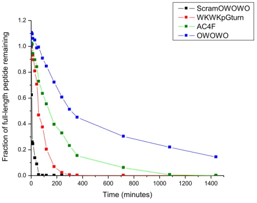
concentration necessary for the ScramOWOWO peptide to have a half-life of less than 10 minutes. At 16 different time points, aliquots of the reaction mixtures were quenched with acetic acid and the amount of peptide degradation at each time point was determined by analytical RP-HPLC. The acetic acid used to quench the reaction also served as an internal standard. To determine the fraction of intact peptide remaining, the parent peak was integrated in the Empower Pro 2 analytical RP-HPLC software and was graphed as a function of time (Figure 2.8).
Figure 2.8Analytical RP-HPLC analysis of peptide degradation by pronase E.
Experiments performed with 100 µM initial peptide concentration, in 10 mM phosphate
buffer, 100 mM NaCl, pH 7.6 at 37°C. The ScramOWOWO peptide degraded fully
after 15 minutes. The WKWK pGturn, AC4F, and OWOWOβ-hairpin peptides show greater resistance to proteolysis. Graph represents the average peptide degradation of experiments done in triplicate.
0 200 400 600 800 1000 1200 1400
0.0 0.2 0.4 0.6 0.8 1.0 1.2
F
rac
tion
of
full
-lengt
h
pept
ide
rem
aini
ng
Time (minutes)
ScramOWOWO WKWKpGturn AC4F
The unstructured ScramOWOWO degraded quickly relative to the β-hairpin peptides including relative to the OWOWO peptide, as expected with half-lives of 7 minutes and 315 minutes, respectively(Figure 2.8). Unexpectedly, the ScramOWOWO peptide with four Orn residues and a bulky fluorophore attached which were absent from the
WKWK-pGturn peptide, still degraded much faster with a half-life of 7 minutes in comparison to
40 minutes for WKWK pGturn. This suggests that the inclusion of sidechain
unnatural functional groups alone, in the absence of increased secondary structure, is not enough to impart substantial resistance to proteolytic degradation.
The AC4F and the OWOWOβ-hairpin peptides both contain residues capable of engaging in cross-strand cation-π interactions to impart stability just like those found in
WKWK pGturn, however these two peptides have additional features to increase
stability to proteolytic degradation. The AC4F peptide contained the Andersen capping motif, which includes a number of energetically favorable noncovalent terminal
interactions.15 These additional terminal contacts, which presumably reduced terminal fraying,are likely responsible for the increased half-life of AC4F over WKWK pGturn. Since the addition of Orn residues and a fluorophore alone did not make much difference in the rate of degradation of the ScramOWOWO peptide, this greater half-life of AC4F
over WKWK pGturn can be attributed to its improved folding. The most stable peptide however was the OWOWO peptide, with 4 internal and terminal Orn residues. Though it lacks the capping motif, the combination of non-canonical amino acids and secondary structure proved to be ideal, resulting in half-life of 315 minutes.
The Mirror and ScramMirror peptides appeared to show no degradation over 24
without mass spectrometry confirmation, it cannot be said with certainty that these two peptides were not cleaved. The only scissile bond of the Mirrorβ-hairpin available to the proteases is in the middle of the peptide. If the proteases had cleaved the Mirror peptide, the resulting fragments would have differed significantly in polarity and size and therefore would have likely had an elution time different from the intact parent peptide. Since only one peak was seen throughout the 24-hour study, it is highly likely that no degradation took place. The native amide bond of the ScramMirror peptide however was at the C-terminus and could have been readily accessed by the pronase E proteases. Cleavage at this bond would have resulted in a peptide fragment that would be similar in size and polarity to the parent peptide and could therefore co-migrate with the parent peptide. Any future studies done with these peptides would require mass spectrometry confirmation of their resistance to proteolytic degradation.
Table 2.2. Peptide Dimensions and Resistance to proteolysis by pronase Ea
Peptide Name β-Hairpin Dimensions (Å) t1/2 (min)
ScramOWOWO ---- 7
WKWK-pGturn 21 x 13 40
AC4F ---- 143
OWOWO 21 x 12 315
Mirror 21 x 13 ND
ScramMirror ---- ND
___________________
at
1/2 time necessary for 50% of peptide to be degraded by pronase E; ND denotes No
Degradation observed; Studies with ScramOWOWO, WKWK pGturn, AC4F and
OWOWO were done in triplicate; ScramMirror and Mirror were tested in