A NOVEL, MATRIX-SPECIFIC GEF/GAP INTERACTION REGULATES RHO GTPASE CROSSTALK CRITICAL FOR 3D COLLAGEN MIGRATION
Matthew L. Kutys
A dissertation submitted to the faculty at the University of North Carolina at Chapel Hill in partial fulfillment of requirements for the degree of Doctor of Philosophy in the
Department of Cell Biology and Physiology in the School of Medicine.
Chapel Hill 2014
© 2014 Matthew L. Kutys ALL RIGHTS RESERVED
ABSTRACT
Matthew L. Kutys: A novel, matrix-specific GEF/GAP interaction regulates Rho GTPase crosstalk critical for 3D collagen migration
(Under the direction of Kenneth M. Yamada)
Differential activation of the Rho family GTPases, Cdc42, Rac1, and RhoA, helps to govern the distinct morphological and migratory phenotypes downstream of adhesion to different extracellular matrix (ECM) proteins. However, it is not known how specific GTPase-dependent signaling pathways are activated in response to different ECM ligands. We hypothesized that adhesion to different ECM molecules, such as collagen and fibronectin, will trigger selective regulation of guanine nucleotide exchange factors (GEFs) to regulate the appropriate matrix-specific cell migratory response. We utilized an affinity precipitation-based mass spectrometry screen to isolate active GEFs from primary human fibroblasts migrating in collagen, fibronectin, or ECM-free environments. Among the GEFs identified, we found that βPix, a Rac1/Cdc42 GEF, was robustly activated only during migration in collagen matrices. Knockdown of βPix led to a collagen-specific migration defect characterized by rapid, spatially-deregulated protrusions, rounded morphology, the absence of stable leading and trailing edges, and robust contraction of the adjacent collagen matrix. In contrast to fibroblasts migrating on fibronectin, βPix in cells migrating in collagen did not localize to focal adhesions, but instead transiently accumulated on the membrane adjacent to areas of cellular protrusion as determined by live cell imaging, immunofluorescence staining, and biochemical
fractionation. Mechanistically, we found that βPix is critical for efficient migration in fibrillar collagen environments by restraining RhoA signaling. Live FRET imaging and RNAi knockdown established this suppression occurs through a mechanism of Rho GTPase crosstalk between Cdc42 and RhoA that is regulated by a collagen-specific functional interaction between βPix and the GTPase activating protein (GAP) srGAP1. Additionally, we identified that binding of α2β1 integrin to fibrillar collagen
leads, through PP2A, to loss of phosphorylation at T526 on βPix and promotes association with srGAP1. We conclude that ECM-dependent regulation of a specific GEF is a fundamental mechanism of migration in different microenvironments. Our results reveal a conserved, matrix-specific pathway controlling migration involving a GEF/GAP interaction of βPix with srGAP1 that is critical for maintaining suppressive crosstalk between Cdc42 and RhoA during 3D collagen migration.
ACKNOWLEDGEMENTS
To my advisor Ken, thank you for providing the ideal environment and mentoring style to foster and develop my ability to conduct independent research. I will look to your example as a guide for not only exceptional research vision, but as a benchmark for integrity and managerial excellence throughout the course of my scientific career.
To my PhD advisory committee, Drs. Keith Burridge, Ken Jacobson, Stephanie Gupton, and Steve Rogers, thank you for providing advice, criticism, and direction throughout the course of my PhD. Your input was critical to surpassing many of the hurdles encountered in this project. I would also like to thank Dr. Rafael Garcia-Mata for previously serving on this committee and remaining a source of counsel thereafter.
Thank you to the members of the Yamada lab, particularly Andrew Doyle, Kaz Matsumoto, Jill Harunaga, Emily Joo, Ryan Petrie, and Will Daley, who were always willing to lend a critical eye, provide technical advice, or listen to vented frustrations. You made the lab an easy place to spend each day.
To my parents, Larry and Susan, words cannot express all that you have done for me, but they can convey my gratitude. Thank you to my mom, to whose memory this dissertation is dedicated, for teaching me to never be satisfied with achieving anything less than my best. Thank you to my dad, I would have undoubtedly never reached this point without your support. Thank you for being the first person to teach me how to
analytically approach a problem, to value both working with my hands and my mind, and for being a model of perseverance in the face of adversity. To the rest of my family, Anna, Greg, and Ginny, thank you for your unwavering support and for being an outlet for my sanity.
TABLE OF CONTENTS
LIST OF TABLES ...x LIST OF FIGURES ... xi LIST OF ABBREVIATIONS ... xiii INTRODUCTION: EXTRACELLULAR MATRIX AND THE CELL: A
SYMBIOTIC RELATIONSHIP
Composition and organization of the extracellular matrix ...2 Cell migration and the extracellular matrix in
homeostasis and disease ...5 Dimensionality, cell migration, and in vitro extracellular
matrix models ...8 Mechanisms of cell migration in response to the
extracellular matrix ...12 Figures and legends ...18 CHAPTER 1: IDENTIFICATION OF EXTRACELLULAR MATRIX-
SPECIFIC GEFS
1.1 Introduction: GEFs and cell migration ...21 1.2 Fibronectin and collagen are sufficient to trigger
Rac1 activation and cell migration ...24 1.3 Development of an ECM-based GEF activity screen ...25 Figures and legends ...28 CHAPTER 2: βPIX: A COLLAGEN-SPECIFIC GEF CRITICAL FOR
CELL MIGRATION
2.1 Introduction: The versatile roles of βPix ...31 2.2 Differential localization of βPix on fibronectin and collagen ...34
2.3 Collagen-specific cell morphological defects of
βPix knockdown ...35 2.4 Collagen-specific cell migratory defects with
knockdown of βPix ...37 2.5 Loss of βPix leads to collagen matrix remodeling and
cell-cell adhesion specifically in collagen environments ...38 2.6 Conservation of collagen-specific βPix function in multiple
cell types with implications during cancer cell motility ...39 Figures and legends ...41 CHAPTER 3: βPIX REGULATES CDC42/RHOA CROSSTALK THROUGH
A COLLAGEN-SPECIFIC INTERACTION WITH SRGAP1 3.1 Introduction: Cdc42, Rac1, and RhoA: 3D migration
and crosstalk ...51 3.2 βPix acts through Cdc42, but not Rac1, during
collagen migration ...55 3.3 βPix/Cdc42 suppress and localize RhoA activity during
collagen migration ...58 3.4 Modulation of RhoA activity is sufficient to mimic or suppress
the βPix knockdown phenotype in collagen microenvironments ...59 3.5 Identifying novel collagen-specific βPix interacting proteins ...60 3.6 βPix has a collagen-specific association with the RhoA GAP
srGAP1 that is essential for its collagen-specific function ...61 Figures and legends ...63 CHAPTER 4: PHOSPHO-REGULATION OF βPIX BY FIBRILLAR COLLAGEN
THROUGH α2β1 INTEGRIN AND PROTEIN PHOSPHATASE-2A
4.1 Introduction: The regulation of βPix by fibrillar collagen ...77 4.2 α2β1integrin controls βPix function downstream
of fibrillar collagen ...80 4.3 Collagen-specific phospho-regulation at threonine 526
4.4 βPix has a collagen-specific association with protein
phosphatase-2A that regulates phosphorylation at T526 ...83 4.5 Protein phosphatase-2A activity is necessary for
collagen-specific βPix function ...85 Figures and legends ...87 CONCLUSION: THE ROLE OF βPIX DURING MIGRATION IN
FIBRILLAR COLLAGEN MICROENVIRONMENTS ...98 MATERIALS AND METHODS ...106 REFERENCES ...119
LIST OF TABLES CHAPTER 4
Table 1 ECM-specific βPix phospho-peptides ...89 METHODS
Table 2 RNAi sequence information ...118
LIST OF FIGURES INTRODUCTION
Figure 1 Dimensional regulation of cell migration ...18
Figure 2 Examples 3D in vitro ECM models and their generation ...19
Figure 3 Extracellular matrix-driven cell migration ...20
CHAPTER 1 Figure 4 Screen for ECM-specific regulation of Rac1 GEFs ...28
Figure 5 FN and COL are sufficient to activate Rac1 and trigger cell motility ...29
Figure 6 Results of the ECM-GEF activity screen ...30
CHAPTER 2 Figure 7 ECM-dependent localization of βPix ...41
Figure 8 Collagen-specific morphological defects of βPix knockdown ...43
Figure 9 Collagen-specific migratory defects of βPix knockdown ...45
Figure 10 Loss of βPix leads to robust collagen remodeling ...47
Figure 11 Conservation of the βPix pathway in controlling cell migration in fibrillar collagen environments ...49
CHAPTER 3 Figure 12 βPix regulates cell morphology through Cdc42, but not Rac1...63
Figure 13 βPix regulates cell migration through Cdc42, but not Rac1...65
Figure 14 FRET analysis reveals collagen-specific loss of polarized Cdc42 activity during migration ...67
Figure 15 βPix/Cdc42 suppress and localize RhoA activity during collagen migration ...69
Figure 16 Modulation of intracellular RhoA activity is sufficient to mimic or suppress the βPix knockdown phenotype ...71
Figure 17 βPix binds srGAP1 specifically during migration in
fibrillar collagen ...73 Figure 18 Knockdown of srGAP1 mimics βPix/Cdc42 knockdown
in fibrillar collagen ...75 CHAPTER 4
Figure 19 α2β1integrin controls βPix function downstream of
fibrillar collagen ...87 Figure 20 Collagen-specific phospho-regulation at threonine 526 is
critical for βPix function and association with srGAP1 ...91 Figure 21 Association between βPix and PPP2R1A regulates T526
dephosphorylation in response to fibrillar collagen ...93 Figure 22 Knockdown (PPP2R1A) or inhibition (OKA) of PP2A
phenocopies βPix knockdown in 3D collagen matrices ...95 Figure 23 T526A mutation in βPix knockdown/rescue cells is
sufficient to rescue knockdown of PPP2R1A ...97 CONCLUSION
Figure 24 Summary model of the collagen-specific role of βPix
during migration in fibrillar collagen environments ...105
LIST OF ABREVIATIONS
3D Three dimensional
BME Basement membrane extract CDM Cell-derived matrix
COL Collagen
ECM Extracellular matrix
FN Fibronectin
GAG Glycosaminogylcan
GAP GTPase-activating protein
GEF Guanine nucleotide exchange factor GDP Guanosine diphosphate
GTP Guanosine triphosphate HFF Human foreskin fibroblast
OKA Okadaic acid
NS Non-specific
p-Thr Phosphothreonine
PBD p21 binding domain of Pak1
PG Proteoglycan
PKA Protein kinase A
PP2A Protein phosphatase 2A
PPP2R1A Protein phosphatase 2, regulatory subunit A, alpha isoform RBD Rho binding domain of Rhotekin
TIRF Total internal reflection fluorescence
INTRODUCTION
EXTRACELLULAR MATRIX AND THE CELL: A SYMBIOTIC RELATIONSHIP
The extracellular matrix (ECM) is the ubiquitous, non-cellular component found in all tissues and organs. Once merely considered a passive scaffold providing support for cells and tissues, we now appreciate that the ECM defines the chemical and physical interactions of the cell, directly influencing cellular physiology and behavior. Core ECM proteins such as collagens and laminins are highly conserved in metazoans, serving as adhesive substrates necessary for proper tissue development, differentiation, survival, and structural homeostasis (Engler et al., 2006; Frantz et al., 2010; Hynes, 2012). However, the distinct chemical composition and physical arrangement of the ECM are the dynamic product of cellular synthesis and remodeling, which are often unique to specific tissues. Thus a synergy exists between ECM matrix assembly/remodeling by the cell and the influence of the ECM on cellular and tissue function. This synergy plays a key role in determining how cells interact and respond to their environment, imbalance of which is the direct cause of many pathological conditions (Byron et al., 2013). To begin to elucidate the diverse cellular functions of the multitude of distinct ECM molecules found in vivo, this dissertation focuses on understanding how specific fibrous ECM proteins signal to stimulate distinct cellular migratory pathways.
Composition and organization of the extracellular matrix
The ECM is composed of several distinct families of complex, multifunctional molecules. While conserved proteins domains do exist across these families, arising over the course metazoan evolution through exon shuffling, ECM molecules are generally disparate in both origin and function (Hynes, 2009; Rozario and DeSimone, 2010). Over one hundred ECM proteins have been identified, with ~10-30% of that total being tissue-specific (Naba et al., 2012). This introduction will give a general overview of ECM composition and structure, and will focus on those ECM proteins relevant to this dissertation.
ECM proteins can be broadly grouped into two classes of macromolecules: proteoglycans (PGs) and fibrous proteins. PGs are ubiquitous molecules classified by their diverse protein cores and linked glycosaminoglycan (GAG) polysaccharide chains. PGs are secreted by the cell and either functionally integrate with other ECM constituents (perlecan and decorin) or exist on the cell surface/span the plasma membrane (syndecans and glypicans). PGs have a wide variety of functions, including contributing to tissue mechanical resistance, proliferation, hydration, and solute buffering (Aszodi et al., 2006; Friedl, 2010). In addition, through binding interactions, PGs can modulate the activity of secreted growth factors and cell-surface receptors to stimulate cell migration. In particular, the syndecan family of transmembrane PGs has recently been discovered to have significant influence on cell migration in response to ECM adhesion. Syndecan-4 regulates the directionality of cell migration in concert with fibronectin by modulating Rac1 activity through integrin recycling (Bass et al., 2007; Morgan et al., 2013), while
syndecan-1 controls cell migration by triggering focal adhesion turnover in response to type I collagen (Altemeier et al., 2012).
The second class of ECM macromolecules is fibrous matrix proteins, consisting mainly of laminins, fibronectins, collagens, and elastins. Collagens are composed of a triple helical organization of α chain subunits, where the differential combination of α chains defines the collagen type. While there are 28 known collagens, type I collagen is the most abundant protein in the human body and found in generally all interstitial ECMs, where the bulk of collagen is synthesized, secreted, and organized by fibroblasts, osteoblasts, and macrophages. In the case of type I collagen, two pro-α(1) chains combine with a pro-α(2) chain to form a triple-stranded procollagen precursor molecule. These collagen precursor molecules are secreted and cleaved in the extracellular environment by peptidases, which results in a collagen molecule capable of forming and crosslinking with collagen fibrils (Shoulders and Raines, 2009). The role of collagen was originally thought to be entirely structural, providing tensile strength and maintaining tissue integrity through crosslinking, fibril bundling, and cooperative interaction with the ECM protein elastin (Frantz et al., 2010; Wise and Weiss, 2009). However, collagens also regulate cell adhesion, haptotaxis and migration, cell polarity, and tissue development (Rozario and DeSimone, 2010). Mice deficient for the α(1) chain of type I collagen, COL1A1, are embryonic lethal due to impaired tissue development, morphogenesis, and severe integrity defects (Liu et al., 1995).
Fibronectin is another fibrous matrix protein that exists as a dimer of two polypeptide chains composed of a series of repeating modules (type I, II, III repeats), each with distinct functions and ability to bind other ECM components such as tenascins
and collagens (Singh et al., 2010). Fibroblasts secrete soluble fibronectin dimers into the surrounding microenvironment, which through concerted remodeling and deposition by additional cells form larger, insoluble fibronectin fibers (Wierzbicka-Patynowski and Schwarzbauer, 2003). Fibronectin is essential for cell migration during development; deletion of the fibronectin gene FN1 results in embryonic lethality due to a multitude of morphogenic, developmental, and migratory defects in the mesoderm, neural tube, and vasculature (Rozario and DeSimone, 2010; Tsang et al., 2010). An important property of fibronectin required for both fiber assembly and signaling is that cellular traction on fibronectin fibrils causes it to stretch several times over its resting length, exposing cryptic binding sites. These sites promote self-association between fibronectin fibers, and cell adhesion to these cryptic sites has drastic effects on cellular behavior, implicating fibronectin as an extracellular mechanosensor (Klotzsch et al., 2009).
On a macroscale, the ECM is arranged primarily into basement membranes and interstitial matrix structures. Basement membranes surround epithelial and endothelial tissues, where cell adhesion to the basement membrane defines epithelial apical-basal polarity, and are essential for proper development and tissue homeostasis. The basement membrane confines the epithelium and separates it from the surrounding tissue stroma. All basement membranes are composed of a common set of interacting ECM proteins (type IV collagen, laminin, nidogen, and perlecan) (Daley and Yamada, 2013; Yurchenco, 2011). Surrounding the basement membrane layer, fibroblasts in the stroma assemble the interstitial matrix, consisting primarily of type I and III collagen, elastin, fibronectin, and a multitude of different PGs. The interstitial matrix maintains the structural integrity of the tissue and participates in dynamic regulatory crosstalk with the
epithelium that is necessary for the chemical and mechanical homeostatic maintenance of the entire tissue (Frantz et al., 2010).
Cell migration and the extracellular matrix in homeostasis and disease
The aim of this dissertation is to understand how cellular interaction with fibrous ECM proteins such as collagen and fibronectin stimulates cell migration. Physiologically this is most relevant to the interstitial matrix or stroma, an ECM structure that is essential for tissue homeostasis and often deregulated during disease. The distinct groups of ECM molecules previously discussed provide the capacity for a high degree of functional complexity and tissue-specific ECM composition. When assembled under normal conditions, these components are able to generate an interstitial matrix exhibiting diverse biochemical and biophysical properties that are necessary for regulation of cell behavior (Lu et al., 2012).
The compliance, or elasticity, of the stroma is attributed to a relaxed network of type I and III collagen, elastin, and fibronectin, which are surrounded by a hydrogel of GAG-containing PGs. This ECM network imparts resistance to the entire tissue against tensile and compressive stresses (Scott, 2003) and defines the physical properties of the interstitial matrix, which include its porosity, rigidity, elastic behavior, and topography. In addition to maintaining tissue integrity, these physical properties provide major environmental cues that determine cellular behaviors that include differentiation and gene expression, morphology, and migration (Engler et al., 2006; Petrie et al., 2012; Wolf et al., 2013). In regard to cell migration, differential matrix rigidity stimulates directed cell migration toward a substrate of greater stiffness in a process known as durotaxis (Lo et al., 2000), and ECM porosity can inhibit or direct the mode by which a cell migrates
(Wolf and Friedl, 2011). From a cellular perspective, physical ECM homeostasis is mediated by the coordinated deposition and arrangement of ECM molecules, secretion of ECM-degrading metalloproteinases (MMPs) and their agonist tissue inhibitors of metalloproteinases (TIMPs), controlled activity of ECM crosslinking enzymes such as lysyl oxidase and transglutaminase, and regulating the transmission of actomyosin-generated cellular forces to the ECM (Lucero and Kagan, 2006; Mott and Werb, 2004; Provenzano et al., 2008b).
The highly dynamic molecular composition of the stroma also imparts direct and indirect biochemical signals that influence cellular behavior. Charged polysaccharide GAG chains, such as heparan sulfate, bind a host of growth factors including BMPs, FGFs, and WNTs. Through this process, the interstitial matrix is able to limit the accessibility of growth factors to their receptors, establish gradients for chemotactic signaling, and create reservoirs of enzymatically accessible growth factors (Hynes, 2009). Additionally, fibrous proteins in the ECM can also initiate signaling by directly engaging cell-surface receptors. Fibroblast adhesion to fibronectin triggers migratory haptotaxis (Hynes and Yamada, 1982), and fibronectin deposition is essential for driving epithelial morphogenesis and cleft formation in the developing salivary gland (Sakai et al., 2003). Adhesion to type I collagen is also sufficient to stimulate migration and morphogenetic changes in human fibroblasts (da Rocha-Azevedo and Grinnell, 2013), and increased type I collagen deposition is a common marker of tissues undergoing epithelial-to-mesenchymal transitions (Kalluri and Weinberg, 2009).
The significance of maintaining the proper chemical and physical characteristics of the ECM for tissue and cell homeostasis is evident by the dysregulation of the ECM in
multiple diseases. Tissue fibrosis is the result of an abnormal collective wound healing response, characterized by hyperproliferation of fibroblasts, their differentiation into myofibroblasts, and excessive ECM synthesis, deposition, and remodeling (Cox and Erler, 2011). This excessive ECM deposition of collagen type I and III, fibronectin, and hyaluronic acid leads to elevated mechanical stress, which disrupts normal tissue function. During pulmonary fibrosis, increases in collagen concentration and crosslinking lead to drastic changes in tissue elastic properties and result in severe respiratory deficiencies (Suki and Bates, 2008). The increased deposition and remodeling of the ECM also promotes the directional migration of cells within the tissue toward the wound site. Fibronectin directs the migration of activated macrophages, which secrete and release growth factors and cytokines to promote angiogenesis, differentiation, and epithelial-to-mesenchymal transition (Schultz and Wysocki, 2009). Continual injury or failure to suppress the normal wound healing response compounds upon existing ECM changes, leading to further ECM synthesis, remodeling, and enhanced crosslinking, which results in chronic fibrosis.
Tumor progression involves the loss of tissue organization through aberrant behavior of transformed cellular components. Similar to fibrosis, the tumor microenvironment is comparable to wounds that have failed to heal, such that tumor cells and cancer-associated fibroblasts manipulate the surrounding microenvironment to enhance their survival. Tumors are characteristically more rigid than normal tissue with stiffening induced by ECM deposition, remodeling, and crosslinking by activated fibroblasts and the subsequent increased contractility of the transformed epithelium (Levental et al., 2009). Type I collagen and fibronectin are the most common and
abundant ECM components deposited during tumor progression, resulting in a dense fibrous tissue that typically surrounds the tumor (Provenzano et al., 2008a). In particular, the remodeling of fibrillar type I collagen surrounding the tumor is associated with metastatic progression. Linearization and perpendicular reorganization of collagen fibers to the tumor front is a classic marker of malignant transformation and metastatic potential (Levental et al., 2009; Provenzano et al., 2006). These changes in composition of the local ECM microenvironment are significant, as malignant breast cancer cells will not invade in a ECM consisting of basement membrane extract (primarily laminin), but do invade when the local ECM is changed to fibrillar type I collagen (Nguyen-Ngoc et al., 2012). Consequently, intravital imaging has revealed that tumor cells travel along aligned collagen fibers to facilitate invasion (Condeelis and Pollard, 2006), highlighting the importance of understanding the migratory signaling pathways downstream of adhesion to ECM fibers as potential targets of therapeutic intervention.
Dimensionality, migration, and in vitro extracellular matrix models
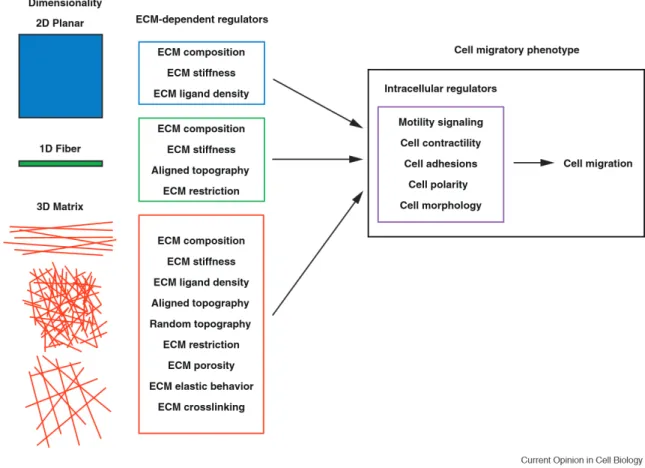
Characterization of cell migration signaling pathways in tissue culture in response to ECM proteins like fibronectin and collagen have helped establish the identity of the receptors and core cytoskeletal machinery involved in the migratory response. While these observations have guided our current understanding, recent investigations into cell migration in three-dimensional (3D) ECM models have revealed substantial differences between 2D and 3D migration (Baker and Chen, 2012; Doyle et al., 2013). However, as evident in Figure 1, these fundamental differences in cell migration are not simply a product of a change in “dimensionality,” but the many chemical and physical features that are inherent to each specific 3D ECM environment. Therefore, rather than simply
concluding that dimensionality directly affects cell migration, it is necessary to identify and understand the cell-regulatory features of each 3D ECM and to elucidate exactly what 3D migratory stimuli dominate that differ from more traditional 2D settings.
Cells migrating on 2D demonstrate a biphasic velocity dependence on ECM ligand concentration. In contrast, cells migrating on a 1D fibrillar ECM (which mimics migration along a 3D fiber) display increasing migration rates with increased ECM concentrations until reaching a plateau, with no inhibition of migration at high ligand concentration (Doyle et al., 2009). This conundrum is compounded further when extended to 3D systems, as changing the ECM protein concentration also alters stiffness, matrix pore size, degree of crosslinking, and topography. Recent investigations into ECM-dependent regulators of 3D migration concluded that deformation of the nucleus through the 3D porous matrix structure is generally the rate limiting factor during migration (Wolf et al., 2013). Inhibition of cell-generated actomyosin contractility decreases migration rates in 1D and 3D environments, but increases rates on 2D ECM (Doyle et al., 2012). Additionally, fibroblasts can respond to the elastic behavior of the ECM and switch their mode of migration from lobopodia-driven in linearly elastic environments (3D CDM) to lamellipodia-driven in nonlinear elastic environments (3D collagen or fibrin) (Petrie et al., 2012). However in addition to these physical parameters, it is clear that the chemical composition of 3D ECM significantly affects both the morphology and migratory behavior of cells. Direct comparisons of fibroblast morphology, migration rates, and focal adhesion structure across four different 3D ECM models revealed quantitative differences that were dependent on the specific ECM chemical composition (Hakkinen et al., 2011). Therefore, it is necessary to carefully
dissect both the physical and chemical cell-ECM interactions that stimulate migration, which is achieved through the combined analysis of 3D in vivo and 3D in vitro reductionist models.
Undoubtedly, the optimal way to study cell-ECM interactions is in vivo. Mouse models provide accurate, physiologically representative insight into cellular interactions with complex ECM microenvironments. However, mouse models often focus on manipulation of cellular aspects rather than directly on the ECM, have limited ability directly to induce chemical and mechanical changes in the local ECM environment, require sensitive and sophisticated imaging/quantification techniques, and are both time- and cost-intensive (Cox and Erler, 2011; Yamada and Cukierman, 2007). In vitro 3D ECM models, while sacrificing the physiological accuracy of mouse models, provide environments to precisely manipulate and quantify chemical and physical changes in the ECM and their effect on cell migration in a controlled setting.
In vitro models for 3D ECM study fall into two categories: synthetic, functionalized hydrogels and hydrogels based on natural ECM proteins. Polyethylene glycol (PEG) hydrogels have been developed with tunable mechanics and incorporated functionalities (such as tethered ECM adhesive domains, growth factors, and cleavable sites) mimicking natural ECM properties (Hern and Hubbell, 1998). Other common synthetic substrates are based on sugars (hyaluronan and dextran), which due to the abundance of functional sites along the polymer backbone, offer more flexibility than PEG in terms of chemical modifications (Trappmann and Chen, 2013). Recently, “designer ECMs” have combined the complex physical features of natural matrices with the versatility of synthetic matrices. For example, a recent PEG hydrogel system
incorporated a collagen peptide mimetic that allowed for in vivo-like triple helical assembly, allowing cells to naturally crosslink the synthetic matrix (Stahl et al., 2010; Trappmann and Chen, 2013). While synthetic matrices allow for heightened manipulation of precise physical/structural parameters, they lack the many physiological subtleties found in natural protein gels.
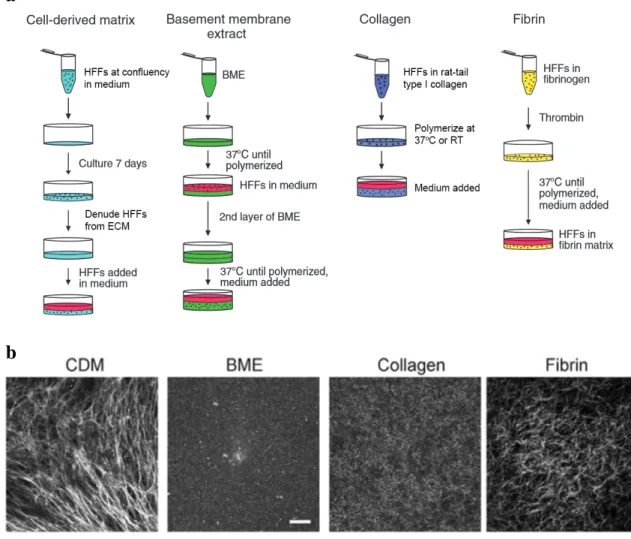
Natural ECM protein gels such as type I collagen, fibrin, and basement membrane extract are composed of proteins that self-assemble in vitro under proper experimental conditions into 3D fibrous networks mimicking in vivo environments. While these ECMs provide a clear physiological advantage in replicating in vivo chemical interactions between the cell and fibrous ECMs, it is difficult in these systems to isolate and manipulate individual properties, both physical and chemical, without inducing additional changes to the matrix. Figure 2a illustrates the methodology behind the preparation of cell-derived matrix (which is a product of cellular synthesis and discussed below), fibrin, basement membrane extract, and collagen gels. In cell migration studies using 3D collagen gels, globular, acid-solubilized rat tail type I collagen is equilibrated with fibroblasts at proper pH and ionic concentration and polymerized into a hydrated collagen lattice when incubated at the proper temperature (Grinnell and Petroll, 2010). 3D collagen gels generally consist of a dense network of collagen fibrils that lack any specific orientation and can range to depths of 100 - 200 microns. It is important to note that collagen gel polymerization can vary greatly between research groups and even individual experiments if there are differences in the preparation conditions. For example, polymerizing collagen at lower temperature (4oC) yields a matrix with thick collagen bundles and large pore sizes, while polymerization at higher temperature (37oC) yields a
reticular network of short fibers and smaller pore size (Raub et al., 2007), both of which will significantly affect cell migratory behavior.
Fibroblast-generated cell-derived matrix (CDM) is a heterogeneous fibrous matrix consisting primarily of a meshwork of linear fibronectin fibrils, the predominant adhesive ligand, which can be oriented in parallel or more random in organization (Figure 2a). Additional matrix proteins such as collagen I and IV, perlecan, tenascin-C, hyaluronic acid, and heparan sulfate proteoglycans are present in lower abundance, as well as sequestered growth factors (Beacham et al., 2007). This diversity and spatial heterogeneity of CDM components mimic more closely what is found in in vivo matrix, providing physiological properties not commonly found in traditional native protein gels. Because CDM is generated by the secretion and assembly of ECM fibers from layers of confluent cells in vitro, its topography consists of arrays of fibronectin fibers that are stacked to an approximate depth of 5-20 microns (Kutys et al., 2013). As evident in Figure 2b, each of the above described 3D in vitro ECM models provides a unique complement of matrix composition and physical architecture for studying cell migration. In this dissertation, direct matrix-specific comparisons of primary fibroblast migratory behavior are made between 3D cell-derived and type I collagen matrices.
Mechanisms of cell migration in response to the extracellular matrix
Interactions between cells and the ECM can profoundly affect migration rate and the particular migratory phenotype. As highlighted previously in Figure 1, a multitude of physical and chemical properties of the ECM are each sufficient to alter migratory signaling, requiring the cell to integrate each of these inputs for directed, persistent migration. Much of what is known about how the cell senses the ECM, translates and
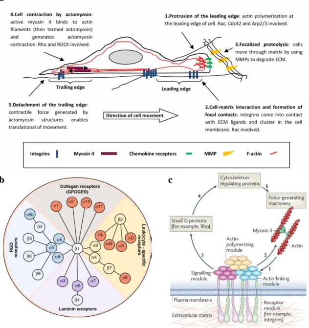
integrates these signals, and transduces them to the migratory machinery has been elucidated from studies in 2D culture. For efficient mesenchymal migration, cells require an asymmetric morphology with defined leading and trailing edges. Polarized intracellular signaling orients protrusion of the cell leading edge, followed by integrin-mediated adhesion to the ECM, coordinated contraction of the actomyosin machinery at adhesion sites, and disassembly of adhesions at rear regions of the cell, leading to cell translocation (Figure 3a) (Lauffenburger and Horwitz, 1996; Ridley et al., 2003).
Cells interact with the ECM through cell-surface receptors, which include proteoglycans such as syndecans, the DDR-family of collagen receptors, growth factor receptors, and the 24 human integrins which are the dominant class of ECM receptor (van Dijk et al., 2013). All integrins are transmembrane, non-covalently linked heterodimers consisting of an α and β subunit. As seen in Figure 3b, mammalian genomes contain eighteen α and eight β subunit genes, whose different combination determines the ECM ligand specificity of the integrin dimer (Humphries et al., 2006). While multiple pairs of α and β integrin subunits can mediate adhesion to the same type of ECM protein, the strength of their association can vary greatly. For example, both αvβ3and α5β1 can bind to
fibronectin, the primary component of 3D CDMs, yet the adhesion strength of α5β1
-containing adhesions can be six-fold higher (Roca-Cusachs et al., 2009). This family of integrins recognize the ECM tripeptide RGD domain (Arg-Gly-Asp) motif, which binds the integrin pair at the active site between the α and β subunits. RGD sequences are found not only in fibronectin, but other matrix ligands such as vitronectin, tenascin, fibrinogen, and laminin, where the specificity and affinity of each integrin is dependent on the fit between the protein’s RGD conformation and the specific α-β active site (Humphries et
al., 2006). Far less is known about the structural recognition of the ECM by laminin/collagen binding integrins. A crystal structure of the binding between α2 and a
type I collagen peptide identified a key motif (GFOGER) on collagen that is critical for its interaction (Emsley et al., 2000). Additionally, mutational analysis has revealed that α2
integrin preferentially recognizes fibrillar type I collagen, while α1 binds globular type I
collagen with higher affinity (Kapyla et al., 2000). Integrin adhesion to specific ECM proteins is transduced to the to the intracellular face of the plasma membrane (termed outside-in signaling), where the integrin cytoplasmic tails assemble variable, multiprotein signaling/structural complexes called focal adhesions, whose individual protein components are known collectively as the adhesome.
Molecular analyses of integrin-mediated adhesions have identified ~160 distinct protein components in the adhesome (Geiger et al., 2009). When assembled into focal adhesions, these molecules serve a variety of functions that include linking ECM-bound integrins to the actomyosin machinery, nucleating cytoskeletal polymerization, and serving as a nexus for a variety of signaling events to the rest of the cell (Figure 3c). The unique molecular composition of a focal adhesion is dictated by both the specific engaged ECM ligand-integrin complex (Byron et al., 2011) and tensional forces imposed on it by actomyosin-dependent intracellular contractility in response to rigidity of the surrounding matrix (Kuo et al., 2011). Acting in concert, these ECM-specific and mechanosensitive proteins combine to form a focal adhesion signature that is unique to the surrounding matrix environment. These proteins then translate signals to the cell that are required for proliferation, polarity, differentiation, and migration. In regards to cell migration, signaling from ECM-integrin adhesions leads to the activation of the Rho
family of GTPases, which act through cytoskeleton-regulating proteins to direct cell motility.
The Rho family of GTPases consists of twenty mammalian proteins characterized by the similarity of their amino acid sequence to the first family member to be characterized, RhoA (Heasman and Ridley, 2008). These proteins are relatively small and nearly all possess the intrinsic ability to convert guanosine triphosphate (GTP) to guanosine diphosphate (GDP). This bound nucleotide regulates the activity of the GTPase through conformational changes, rendering it inactive or active in the case of GDP or GTP, respectively. The Rho GTPases mediate a variety of intracellular signaling pathways involved in the regulation of cell cycle progression (Olson et al., 1995), gene transcription (Hill et al., 1995), differentiation (Keung et al., 2011), and cell transformation (Khosravi-Far et al., 1995). However during cell migration, the major function of the Rho GTPases is regulating the polarization and assembly of the actin cytoskeleton and contractile myosin II machinery. For these roles, the best characterized Rho GTPases are Cdc42, Rac1, and RhoA.
A role for the Rho family GTPases in regulating cytoskeletal dynamics was first identified when RhoA was found to be a substrate of C3 transferase, an exoenzyme that induces cell rounding and filamentous actin disassembly in eukaryotic cells (Aktories et al., 1989). Additionally, the constitutively active form of RhoA (RhoAV14) was found to trigger the formation of bundles of parallel F-actin or stress fibers when injected into fibroblasts (Paterson et al., 1990). The ability of the Rho family of GTPases to regulate cytoskeletal remodeling was reinforced when fibroblast were injected with active RhoA, Rac1, or Cdc42 which generated stress fibers, lamellipodia and membrane ruffles, or
filopodia, respectively (Hall, 1998). Further, each of these GTPases was able to trigger the contextual formation, maturation, and disassembly of focal adhesions (Nobes and Hall, 1995). These initial demonstrations of the RhoA, Rac1, and Cdc42 function in fibroblasts have become synonymous with their general function during cell migration. The property of the Rho GTPase family to impinge on each other’s activity, or crosstalk, was also observed where active Cdc42 activated Rac1 and active Rac1, in turn, could activate or inactivate RhoA (Arthur and Burridge, 2001; Nobes and Hall, 1995). More recently, there has been increasing evidence for their diverse and non-canonical functions in many different cell types.
During traditional 2D migration, Rac1, Cdc42, and RhoA are spatiotemporally polarized and activated at the leading edge of cells (Machacek et al., 2009). However during migration in 3D, polarization of Rac1 and Cdc42 can be absent during migration (Petrie et al., 2012) or the cell can exhibit variable dependence on the activity of a particular GTPase depending on matrix rigidity and composition (Deakin and Turner, 2011). Requirements for the Cdc42 and Rac1 effectors N-WASP and Scar/WAVE, regulators of actin assembly and lamellipodial protrusions, also differ during 2D and 3D migration (Tang et al., 2013). In the case of RhoA signaling, modulating RhoA-ROCK signaling switches 3D modes of motility in primary fibroblasts between lamellipodial and lobopodial-driven migration in 3D cell-derived matrix. Additionally, the mode of cancer cell migration in 3D collagen matrices depends on both traditional and non-canonical RhoA signaling pathways: amoeboid (RhoA-ROCK) and mesenchymal migration (Cdc42-MRCK) (Sahai and Marshall, 2003), as well as migratory efficiency (RhoA-ROCK1/ROCK2, RhoC) (Vega et al., 2011). Furthermore, activation of
regulated contractility is necessary for remodeling and alignment of matrix fibers to provide contact guidance during 3D malignant epithelial cell migration (Provenzano et al., 2008b). It is clear that the understanding of signaling mechanisms driving contextual migration in 3D, especially in the case of the Rho family GTPases, is in its infancy, with most observed differences attributed to simply changes in dimensionality rather than to a specific aspect of the ECM.
To address this outstanding and complicated question, it is important to begin by isolating experimentally and conceptually the specific roles of each of the many ECM regulators of 3D cell migration. In this dissertation, we focus on the migratory response of primary human fibroblasts upon adhesion to fibronectin or type I collagen ligands to gain a better understanding of the mechanisms behind the differential 3D migratory behaviors observed between these ECM conditions. We investigate how interaction with these ECM ligands modulates activators of the Rho family of GTPases, first specifically Rac1 for its well-documented role in regulating leading edge protrusion (Figure 3a). We then analyze how these ECM-specific signaling pathways translate to migration in 3D environments, with the goal of identifying a conserved migratory pathway that is unique to fibronectin or collagen microenvironments.
Figure 1: Dimensional regulation of cell migration. Illustration of the numerous unique ECM-dependent regulators (center column) associated with migration 2D, 1D, and 3D environments. These microenvironment regulators in turn influence intracellular regulatory pathways that govern the migratory phenotype (right panel) and determine how cell migration proceeds. Adapted from (Doyle et al., 2013).
Figure 2: Examples 3D in vitro ECM models and their generation. a Simplified methodology for preparation of 3D in vitro cell-derived matrix, basement membrane extract, type I collagen, and fibrin ECM models. See Materials and Methods for expanded procedures for preparation of cell-derived matrix and collagen matrix. b Protein fiber structure of each 3D matrix. Cell-derived matrix (CDM) was visualized by fibronectin immunofluorescence stain, fibrin by fluorescently labeled fibrinogen, and basement membrane extract (BME) and collagen were visualized using reflection microscopy. Adapted from: (Hakkinen et al., 2011).
a
b
Figure 3: Extracellular matrix-driven cell migration a Sequential diagram of the major events during mesenchymal cell migration. b Diagram of the 24 human integrins and their ECM ligands. c Schematic of integrin-mediated focal adhesion signaling. Adapted from: (Barczyk et al., 2010; Geiger et al., 2009; Parri and Chiarugi, 2010)
a
b c
CHAPTER 1
IDENTIFICATION OF EXTRACELLULAR MATRIX-SPECIFIC GEFS
1.1 Introduction: GEFs and cell migration
Distinct migratory responses of cells to interactions with different ECM proteins is necessary for efficient tissue development and wound repair, and is often deregulated in cancer (Daley and Yamada, 2013; Frantz et al., 2010; Petrie et al., 2012; Provenzano et al., 2006). Integrin binding to ECM proteins triggers selective activation of the Rho GTPases, which induce cell polarization, cytoskeletal rearrangements, and contractile responses required for efficient migration in different microenvironments (Huttenlocher and Horwitz, 2011; Petrie et al., 2009; Raftopoulou and Hall, 2004). However, a fundamental unanswered question is how specific Rho GTPase signaling pathways governing migration are regulated differentially by specific ECM proteins.
Rho GTPases function as molecular switches that cycle between an inactive GDP-bound and an active GTP-GDP-bound conformation. The type of nucleotide (GDP or GTP) that is bound modulates conformational changes within the Rho GTPase switch domain region and directs effector interactions. Nucleotides are additionally stabilized by a Mg2+ cation binding pocket, which is required for high-affinity binding to the GTPase (Goicoechea et al., 2014). The activation of Rho GTPases is mediated by guanine nucleotide exchange factors (GEFs), which catalyze the exchange of GDP for GTP. GEFs facilitate the exchange of GDP for GTP by promoting GTPase conformational
intermediates that lack both nucleotide and Mg2+. In cells, GTP is preferentially loaded onto Rho GTPases during nucleotide exchange because GTP is present at substantially higher intracellular concentrations than GDP (Rossman et al., 2005).
To date there have been approximately 80 GEFs toward Rho GTPases identified in the human genome, which are classified into two distinct GEF families by protein structure: the Dbl family, which comprises 69 members in humans, and the DOCK family with 11 members (Meller et al., 2005; Rossman et al., 2005). The Dbl family of GEFs is named after the first mammalian GEF isolated, which was a Cdc42 GEF identified as a transforming gene from human diffuse B-lymphoma cells and subsequently designated Dbl (Schmidt and Hall, 2002). GEFs in this family are characterized by the presence of a Dbl homology (DH) catalytic domain followed by an adjacent pleckstrin homology (PH) domain. Under most conditions, the PH domain binds to phosphoinositides and localizes the GEF to plasma membranes where exchange activity commonly occurs. This conserved DH-PH motif defines the minimal structural unit required to trigger the GDP-GTP exchange reaction (Rossman et al., 2005). Outside the DH–PH domain, Dbl-family GEFs are significantly divergent and contain other protein domains that regulate the intrinsic catalytic activity of the GEF, their GTPase specificity, intracellular localization, and direct GTPase-effector targeting through additional protein-protein interactions (Goicoechea et al., 2014).
Currently, the number of GEFs in the human genome is four times higher than the number of their target Rho GTPases and continues to grow as non-canonical GEFs are continually being discovered. This complexity is compounded further by the fact that many GEFs have the ability to activate more than one GTPase, and the specificity of the
majority of GEFs has yet to be fully characterized. While tissue specificity could explain this apparent redundancy, most GEFs are ubiquitously expressed (Garcia-Mata and Burridge, 2007). A paradigm that has recently evolved is that GEFs not only serve to activate a particular GTPase, but also to facilitate and localize the connection between an upstream stimulus and downstream specific GTPase effector. This is achieved through both diversity in GEF protein domain structures and tightly regulated post-translational modifications.
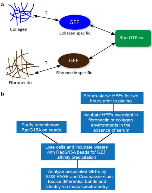
There is evidence for the extracellular regulation of GEF activity by the ECM microenvironment. The Rac1 GEF P-Rex1 mediates the ErbB receptor response in breast tumorigenesis, regulating Rac1-directed cell proliferation and motility (Sosa et al., 2010). Additionally, matrix mechanical stresses translated through fibronectin adhesions activate the RhoA GEFs LARG and GEF-H1 to regulate the contractile response of the cell (Guilluy et al., 2011b). Further, certain GEFs have to been shown to associate directly, or in complex, with specific integrin subtypes suggesting their restricted, specific activation (Humphries et al., 2009; Samson et al., 2007). As emerging evidence continues to support the contextual activation of particular Rho GEFs, a model is developing in which the specific activity of Rho GTPases is controlled by the localization and activation of particular GEFs and associated with a specific cellular stimulus. Considering the reports of complex regulation of the Rho GTPases in different ECM microenvironments, it is plausible that particular GEFs are associated with specific matrix-integrin complexes governing the migratory response. We therefore hypothesized that adhesion to specific ECM molecules, such as collagen and fibronectin, would trigger differential GEF activation to regulate cell migratory responses (Figure 4a). We initially focused on
differential regulation of GEFs toward Rac1 in response to fibronectin and collagen for its well-documented roles in governing protrusion and leading edge dynamics during cell migration.
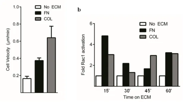
1.2 Fibronectin and collagen are sufficient to trigger Rac1 activation and cell migration
Previous work from our laboratory has demonstrated the role of Rac1 activity in determining the directionality, persistence, and rate of 2D cell migration. Additionally, investigations of cell motility in 3D ECM have also reported a contextual requirement for Rac1 activity for the efficient coordination of lamellipodial dynamics and mesenchymal-type migration (Pankov et al., 2005; Petrie and Yamada, 2012; Sanz-Moreno et al., 2008). Before screening for ECM-specific Rac1 GEFs, we first ensured that fibroblast adhesion to fibronectin or collagen alone was sufficient to trigger Rac1 activity and subsequent migration.
Primary human foreskin fibroblasts (HFFs) were plated onto MatTek dishes coated with human fibronectin or type I rat tail collagen in the absence of serum and incubated overnight to allow cells to reach steady-state migration. The following day, the cells were assayed for migratory behavior by timelapse microscopy for 24 hours. We observed that adhesion to either fibronectin or collagen alone was sufficient to induce cell migration in comparison to a no ECM control (Figure 5a). Characteristically, cells on fibronectin migrated in a persistent fashion, with broad, stable lamellipodia. On collagen, migration velocity was increased, yet less persistent, with a higher frequency of protrusion and less stable lamellipodia. We then assayed whether adhesion to solely fibronectin or collagen was sufficient to induce intracellular Rac1 activity. Fibroblasts were allowed to spread on fibronectin or collagen in the absence of serum over a time
course in which Rac1 activity was measured (Figure 5b). Adhesion to both fibronectin and collagen led to increases in Rac1 activity relative to a no-ECM control; however we observed different kinetics of activation over the time course, suggesting different molecular mechanisms for each ECM condition.
1.3 Development of an ECM-based GEF activity screen
Having established that adhesion to both collagen and fibronectin alone was sufficient to activate Rac1 and induce cell migration in HFFs, we next sought to develop a screen to identify and isolate novel Rac1 GEFs uniquely active under the two ECM conditions. It has been demonstrated previously that recombinant dominant-negative Rho GTPase mutants can be used for affinity-isolation of activated GEFs (Dubash et al., 2007; Garcia-Mata et al., 2006). Particularly, mutants that mimic the conformation of a nucleotide-free GTPase, which is an intermediate in the GDP-GTP exchange reaction, are able to form high-affinity complexes with active GEFs (Cherfils and Chardin, 1999). Taking advantage of this principle, mutant Rac1 constructs were generated containing the nucleotide-free, dominant-negative mutation RacG15A to isolate GEFs toward Rac1. Using this purified recombinant mutant, we developed an unbiased screening approach for isolating and identifying activate Rac1 GEFs from lysates of fibroblasts migrating in fibronectin- or collagen-based microenvironments.
A schematic diagram illustrating the ECM-GEF screen can be found in Figure 4b. Briefly, HFFs were serum-starved for two hours prior to plating on fibronectin or collagen-coated dishes. To avoid studying an artifact of cell spreading in response to matrix, primary fibroblasts were cultured overnight in the absence of serum to ensure that cells were undergoing steady-state migration at the time of analysis. The following day,
HFFs were lysed, and the cell lysates were incubated with GST-RacG15A conjugated to agarose beads in order to extract active GEFs. GEFs that bound to RacG15A were analyzed by SDS-PAGE, visualized by Coomassie staining, and mass spectrometry was performed on ECM-specific, excised protein bands for GEF identification. All GEFs identified using mass spectrometry were confirmed through western blot quantification.
A critical aspect during development of the ECM-GEF screen was ensuring that all relevant active GEFs were being solubilized during cell lysis. To evaluate the effects that different lysis approaches had on effectively isolating GEFs, we initially compared the profiles of GEFs associated with the recombinant RacG15A probe from HFFs under standard culture conditions using SDS-PAGE and Coomassie staining. By systematically varying lysis buffer detergents and solubilization methods, we determined the optimal strategy for extracting active GEFs by observing which method yielded the most unique protein bands on Coomassie stained gels while maintaining constant culture conditions. Surprisingly, addition of deoxycholate or NP-40 did not significantly affect the number of extracted GEFs in comparison to a 1% Triton X-100 base buffer. However comparing lysates that had been briefly sonicated to those incubated on ice yielded significantly more unique protein bands associated with the RacG15A probe. Therefore brief sonication was determined to be a crucial step for efficiently extracting the total active GEF population.
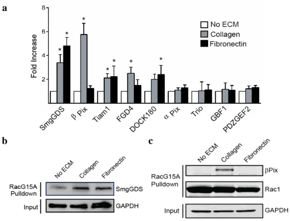
Quantitative results of select GEFs isolated from the ECM-based screen are shown in Figure 6a. Results depict the relative Western blot band intensity of mass spectrometry-identified GEFs in comparison to a no-ECM control. The majority of GEFs that were isolated showed increased activity on both fibronectin and collagen. In
particular, the promiscuous GEF SmgGDS, which has been reported to activate a wide variety of GTPases, was observed to increase its association with RacG15A strongly during cell migration on both fibronectin and collagen (Figure 6b). This result contradicts previous reports that SmgGDS does not have exchange activity toward Rac1 (Hamel et al., 2011); however, preliminary analysis of SmgGDS knockdown in HFFs suggested an inhibition of proliferation in response to fibronectin or collagen (data not shown) and warrants future investigation. Although isolating Rac1 GEFs that showed increased activity on both matrix ligands was interesting and provided insight into their function, the goal of this screen was to isolate a Rac1 GEF whose activity was specific to either fibronectin or collagen. Therefore, the primary novel finding of this screen and focus of this dissertation is that the activity of the Rac1/Cdc42 GEF βPix is specifically and robustly increased during cell migration in response to collagen versus fibronectin and null ECM controls (Figure 6c).
Figure 4: Screen for ECM-specific regulation of Rac1 GEFs. a Central hypothesis: adhesion to ECM ligands such as fibronectin or collagen specifically activates GEFs to modulate Rho GTPase activity and subsequent cell migration in different ECM environments. b Schematic diagram of the screen for ECM-specific GEFs. Briefly, HFFs were plated on ECM-coated dishes, allowed to reach steady-state migration overnight in the absence of serum, lysed, and incubated with GST-RacG15A conjugated to beads to extract active GEFs. Beads were analyzed by SDS-PAGE, Coomassie staining, and mass spectrometry of excised protein bands for identification.
a
b
Figure 5: FN and COL are sufficient to activate Rac1 and trigger cell motility. a HFFs were allowed to reach steady-state migration overnight in the absence of serum on dishes coated with 10 µg/ml fibronectin (FN) or 50 µg/ml type I collagen (COL). The following day migration was observed by timelapse microscopy over a 24 hour period. Adhesion to both fibronectin and collagen was found to trigger motility in comparison to no-ECM control (n = 10, 13, 15 cells). b Time course of intracellular Rac1 activity was measured using ELISA-based activity assays (G-LISA, Cytoskeleton, Inc.), in fibroblasts spreading on fibronectin or collagen in the absence of serum. Values are reported as relative Rac1 activity increase in comparison to a no-ECM control. We observed that both fibronectin and collagen stimulated Rac1 activity in the absence of serum, but had different kinetics of activation, eventually stabilizing at similar levels of Rac1 activity (n = 2 dishes per time point).
Figure 6: Results of the ECM-GEF activity screen. a Quantification of western blot band intensities of select GEFs isolated from the RacG15A ECM-GEF screen. Values are fold intensity increase above a no-ECM condition (n = 3 blots, error bars represent s.e.m, one-way ANOVA with Bonferroni multiple comparisons correction). b Western blot confirms up-regulation of the GEF SmgGDS binding to RacG15A when migrating in the presence of a collagen or fibronectin ECM ligand. c Western blot validation of βPix binding to RacG15A during migration on collagen. We observed a specific association between βPix and RacG15A only during collagen-based migration. * P < 0.05.
a
b
c
CHAPTER 2
βPIX: A COLLAGEN-SPECIFIC GEF CRITICAL FOR CELL MIGRATION
1.1 Introduction: the versatile roles of βPix
βPix was originally discovered in 1997 when it was isolated from a mouse thymus cDNA expression library screened with a monoclonal antibody recognizing a common SH3 epitope and was designated Cool-1 (Cloned out of library-1). It was observed to be widely expressed in mouse tissues and localize to the nucleus, cytoplasm, and focal complexes on the cellular level (Oh et al., 1997). Two additional Cool family members were also identified based on homology to Cool-1, and the Cool family of proteins soon encompassed p50Cool-1, p85Cool-1, and Cool-2 (Koh et al., 2001). The function of these proteins remained largely unknown, although GEF activity was suspected due to the presence of a tandem DH-PH motif in each of the Cool proteins. One year later, both a yeast two-hybrid screen searching for Pak3 binding proteins (Bagrodia et al., 1998) and immunoprecipitation of Pak1 (Manser et al., 1998) indicated direct binding to p50Cool-1 and p85Cool-1. Surprisingly, p50Cool-1 was found to be incapable of stimulating Pak1 activity, as would have been expected if it were acting as a GEF for Cdc42 or Rac1, and it also inhibited Pak1 activation by Dbl or by activated forms of Cdc42. The p85Cool-1 protein did not inhibit Dbl- or activated Cdc42-stimulated Pak1 activity, but itself was incapable of directly stimulating Pak1 activity (Feng et al., 2002). After this, the Cool family of proteins was re-designated Pix (Pak-interactive exchange factor) proteins, with
p85Cool-1as βPix, p50Cool-1 as a smaller, inhibitory splice isoform of βPix, and the separate gene product Cool-2 as αPix.
The gene containing βPix, ARHGEF7, yields five alternatively spliced mRNA transcripts, yet only four distinct isoforms of βPix have been characterized. While less abundant isoforms are exclusively expressed in the brain and central nervous system, the primary 85 kDa isoform, designated βPix, is expressed ubiquitously in humans (Koh et al., 2001). The domain structure of βPix is outlined in Figure 7a. βPix is a member of the Dbl family of GEFs, containing a classical DH-PH motif flanked by a T1 domain that is also critical for GEF activity (Feng et al., 2006). Outside of this conserved region, there are numerous protein-protein interacting domains: a SH3 domain, a proline-rich domain (PRD), a Cat (Cool-associated tyrosine phosphosubstrate)/GIT (G protein-coupled receptor kinase interactor)-binding (CBD) domain, and a leucine zipper (LZ). This diverse array of domains provides the capacity for the many specialized cellular functions that have been ascribed to βPix.
For each specific intracellular function, βPix exhibits exchange activity exclusively on Cdc42 or Rac1 GTPases (Feng et al., 2002; Manser et al., 1998). This specificity is determined by whether βPix is a dimer or monomer, protein-protein interactions, and post-translational modifications (Baird et al., 2005). Outside of traditional roles in cell migration, βPix acts through Rac1 to regulate cell apoptosis by controlling adhesion-dependent epithelial survival through EBP50, as well as cytokinesis by initiating centralspindlin complex formation through a cooperative balance with the Rac1 GAP CYK4 (Bastos et al., 2012; Chen et al., 2012). Additionally, βPix also acts through Cdc42 in specialized cellular functions. The importance of βPix/Cdc42 signaling
has been reported to include regulation of proper insulin secretion in beta cells, directing EGF receptor degradation in cooperation with the E3 ligase Cbl, and modulation of β-catenin transcriptional activity in colon cancer cells (Chahdi and Raufman, 2013; Feng et al., 2006; Kepner et al., 2011).
However, what makes βPix a particularly attractive candidate GEF for an ECM-specific role in governing cell migration are previous reports of its involvement in cell polarity, protrusion, and focal adhesion turnover. In the canonical βPix pathway, the SH3 domain of βPix binds to a unique proline-rich sequence on Pak1, which is essential for Rac1 activation, and this complex localizes to focal adhesions (Manser et al., 1998). This focal adhesion localization is achieved by the interaction of βPix with proteins such as GIT1/2 (G-protein-coupled receptor kinase interactor) and PKL (paxillin kinase linker), which bind βPix at the CBD domain and also functionally link to the focal adhesion protein paxillin (Turner et al., 1999). During migration on flat two-dimensional fibronectin substrates, localization of βPix-Pak1 to focal adhesions was demonstrated to trigger focal adhesion disassembly, which was indirectly attributed to the activity of Rac1 (Nayal et al., 2006; Zhao et al., 2000). More recently, the localization of βPix to focal adhesions has been described to be negatively regulated by cellular contractility, with inhibition of myosin II leading to βPix enrichment in adhesion complexes (Kuo et al., 2011). Further investigation into the regulation of the βPix-Pak1 pathway has shown that Cdc42 acts upstream to direct βPix-Pak1 assembly. In this context, Cdc42-Pak1-βPix acts to control the polarization of cell protrusions during migration in a scratch wound assay, where Pak1 acts through βPix to spatially restrict Rac1-dependent actin polymerization to the leading edge (Cau and Hall, 2005). In addition, recent studies have begun to uncover
non-traditional roles for βPix in cell migration. βPix association with the membrane scaffolding protein Scrib leads to localized Cdc42 activity during astrocyte migration (Osmani et al., 2006), and phosphorylation of βPix by ERK/Pak2 in response to bFGF localizes βPix to lamellipodia in neuronal growth cones, controlling neurite outgrowth (Shin et al., 2002).
With the multitude of different roles reported for βPix in regulating cell physiology, it is conceivable that many are highly contextual and thus require tight regulation of βPix function. One hypothesis is that the regulation of βPix function is due to multiple phosphorylation sites on the protein (Mayhew et al., 2007). Additionally, efforts have been made to define a βPix-Pak1 “interactome” and characterize unique scaffolding and adaptor proteins that may be directing this signaling complex (Mayhew et al., 2006). However, these efforts fall short in effectively recapitulating all the stimuli, particularly extracellular, that direct βPix interactions. Therefore, it is feasible that βPix is serving a specialized role during migration in collagen environments. In this chapter, we build upon the previous observation of the collagen-specific association between βPix and RacG15A and investigate whether βPix is important for regulating cell morphology and migration in collagen.
2.2 Differential localization of βPix on fibronectin and collagen
βPix exists at multiple subcellular sites, including focal adhesions and plasma membrane, which is consistent with differential functions (Cau and Hall, 2005; Kuo et al., 2011; Liu et al., 2010). As an initial test for whether βPix has ECM-specific functions, we examined for altered localization of βPix during fibroblast migration on fibronectin versus fibrillar collagen. βPix has been previously shown to localize to focal
adhesions in cells migrating on fibronectin. As expected, both immunofluorescence staining for endogenous βPix and live-cell imaging of GFP-βPix showed strong localization to focal adhesions during migration on fibronectin (Figure 7b) and 3D cell-derived matrix (Figure 7c), where the primary ECM ligand is fibronectin (Kutys et al., 2013). Surprisingly, we observed a dramatic decrease in both endogenous and GFP-βPix focal adhesion localization in fibroblasts migrating on both fibrillar collagen and 3D collagen (Figure 7b, c). Instead, on fibrillar collagen βPix transitioned to non-paxillin containing structures that were localized to lamellipodia and appeared to be plasma membrane-associated. Live-cell GFP-βPix imaging provided further insight into this unique localization: βPix displayed patchwork localization on ventral cell membranes in amorphous, persistent aggregates of variable size that, while polarized to leading-edge protrusions, did not co-localize with paxillin. To further confirm this unique localization, fibroblasts migrating on fibronectin or fibrillar collagen were subjected to Triton X-100 fractionation. Subcellular fractionation revealed that on fibrillar collagen, endogenous βPix transitioned from detergent-soluble to -insoluble fractions (Figure 7d). These data demonstrate that the intracellular location of βPix changes dramatically when cells migrate on collagen compared to fibronectin, supporting the existence of ECM-specific functions observed in the initial GEF screen.
2.3 Collagen-specific cell morphological defects of βPix knockdown
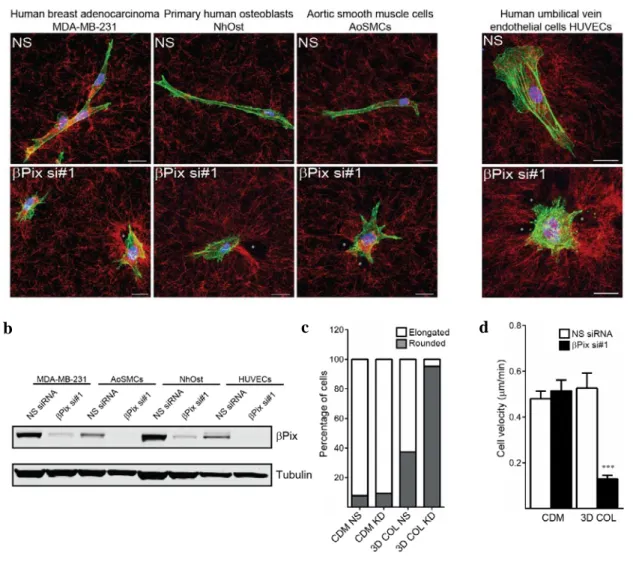
The differential βPix focal adhesion/plasma membrane localization observed during migration on fibronectin versus fibrillar collagen strongly suggested distinct molecular functions between the two ECM conditions. To parse out these functions, we generated stable βPix knockdown lines in primary human fibroblasts using
based shRNA delivery. The pLentiLox 3.7 lentiviral packaging system has proven efficient in delivering shRNA hairpins and cDNA at the single-cell and organ level (Cai et al., 2007), with the viral vector (pLL 3.7) consisting of two distinct promoters governing a multiple cloning-GFP site and a shRNA hairpin site. Using this system, two distinct hairpins, one previously reported (shRNA #4, Table 2 in Materials and Methods) (Kuo et al., 2011), and one unique to this study (shRNA #2, Table 2 in Materials and Methods), were transduced into fibroblasts. FACS sorting was performed on infected populations of HFFs by using the GFP reporter to isolate high expressers and ensure adequate βPix knockdown. Both shRNA hairpins and a single, independent siRNA efficiently depleted βPix protein in HFFs (Figure 8b).
Using stable βPix knockdown fibroblasts, we next tested whether βPix had any collagen-specific functions regulating cell morphology. Nonspecific (NS) shRNA control and βPix knockdown fibroblasts were plated onto CDM or 3D collagen gels and incubated overnight in complete media. The following morning, cells were fixed and visualized by phalloidin staining, while the surrounding ECM was imaged using either fibronectin immunostaining or reflection microscopy. We observed that loss of βPix resulted in cells with a severe, rounded morphology and inability to spread in 3D collagen matrices, with a ~75% decrease in cell elongation (Figure 8c). In contrast, there were no effects on cell elongation in CDM (Figure 8a, c). Surprisingly, phalloidin staining revealed that these rounded cells were also hyper-protrusive, with a nearly three-fold increase in the number of protrusions per cell in comparison to NS fibroblasts (Figure 8d). We next investigated whether this collagen-specific morphological phenotype translated into a migratory defect.
2.4 Collagen-specific cell migratory defects after knockdown of βPix
To assay for collagen-specific migratory defects in the absence of βPix, stable NS and βPix shRNA fibroblasts were incubated overnight in CDM or 3D collagen matrices in complete media and migratory phase timelapse movies were obtained over 24 hours the following morning. Migration assays uncovered severe defects in motility after βPix knockdown that were specific to 3D collagen, as evident in the representative phase timelapse image (Figure 9a) and velocity quantification across ECMs (Figure 9c). This phenotype was characterized by rapid, transient formation of spatially deregulated cell protrusions that exhibited apparent deformation of adjacent collagen fibers and resulted in minimal cell motility compared to nonspecific shRNA control cells in 3D collagen. Representative migratory tracks of βPix knockdown fibroblasts in 3D collagen show that any residual motility of these cells lacks any persistence and appear to be due to stochastic oscillations of the cells within the collagen matrix (Figure 9b).
We assayed the effects of βPix knockdown across a variety of 2D and 3D ECM environments (Figure 9c). Interestingly, even high concentrations of globular collagen could not fully recapitulate the characteristic βPix knockdown phenotype in 3D collagen, whereas thin, fibrillar collagen substrates mimicked this 3D phenotype (Figure 9c). These functional differences observed between monomeric and fibrillar collagen are likely due to the preferential recognition and affinity of α2β1 for fibrillar type I collagen (Emsley et
al., 2000; Jokinen et al., 2004). Additionally, these fibrillar collagen substrates have the advantage of being thin for improved optical imaging, yet they retain the fibrillar structure of 3D collagen gels; they underscore the importance of using more-physiological polymerized collagen fibers rather than globular monomeric collagen.


![[Evaluation of the physician-patient relationship competence. Development and validation of an assessment instrument].](data:image/gif;base64,R0lGODlhAQABAIAAAP///wAAACH5BAEAAAAALAAAAAABAAEAAAICRAEAOw==)