The Role of pH, Self-Association, and Lipid Binding in Vinculin Tail Structure and Function
Sean M. Palmer
A dissertation submitted to the faculty of the University of North Carolina at Chapel Hill in partial fulfillment of the for the degree of Doctor of Philosophy in the Department of
Biochemistry and Biophysics
Chapel Hill 2008
Approved by:
Dr. Sharon L. Campbell, Advisor Dr. Nikolay Dokholyan
Abstract
The Role of pH, Self-Association, and Lipid Binding in Vinculin Tail Structure and Function (Under the direction of Dr. Sharon L. Campbell)
Vinculin is a highly conserved cytoskeletal protein that localized to sites of cell adhesion and is involved in linking the actin cytoskeleton to the cell membrane. Loss of normal vinculin function has been associated with cancer phenotypes, cardiovascular disease, and lethal errors in embryogenesis. Vinculin is composed of a 90 kDa head domain (Vh) and a 21 kDa tail domain (Vt) connected by a flexible “neck” region. Vh and Vt form auto-inhibitory interactions in its inactive state. Conformational changes in these domains have been proposed to be important for vinculin activation, freeing domains to bind to a variety of ligands involved in cell adhesion processes.
My work has focused on the vinculin tail domain. I have assigned the majority of the backbone 1HN, 15N, 13Cα, 13CO, and side chain 13Cβ NMR resonances of Vt (Biological
Magnetic Resonance Data Bank, accession number 15653). These assignments have proven useful for investigating the effects of ligand binding, pH and Vt self-association.
pH 7.0, results from analytical ultracentrifugation indicate that Vt self-associates at both pH values, albeit with weak affinity (Kd > 100 µM).
The binding of acidic phospholipids by Vt has been proposed to play a role in vinculin activation and focal adhesion turnover. We observe a marked affinity of Vt for phosphatidylinositol 4,5-bisphosphate (PIP2), but no significant binding to
phosphatidylethanolamine (PE), phosphatidylcholine (PC), phosphatidylserine (PS), or phosphatidylinositol (PI) in the context of mixed lipid vesicles. A significant increase in PIP2 binding was observed with multiple mutant forms of Vt, expected to alter the
conformation or flexibility of the N-terminal strap of Vt, suggesting that a rearrangement of this N-terminal strap may be required for PIP2 binding. Additionally, we find that the
This work is dedicated to my loving and supportive wife, Heather, and to my family and friends. Their love, support, and encouragement were more valuable to me than they may
Acknowledgments
Table of Contents
Page
List of Figures………...………xi
List of Abbreviations and Symbols……….xii
Chapter: I. Introduction………1
A. Vinculin………....1
B. The Role of Vinculin in Development, Cell Motility, Cell Survival, and Cancer………...………...………..2
C. The Role of Vinculin in Cardiovascular Function……….………..2
D. Vinculin Domains and Their Ligands………..4
E. Vinculin Structure and Auto-inhibition………..……….4
F. Vinculin Activation………..5
G. The Tail Domain of Vinculin and its Ligands……….6
1. Vinculin Tail Structure………...………6
2. Actin Interactions………...……….8
3. Lipid Interactions……….……….10
4. Paxillin Interactions………..12
6. Vinculin Tail Self-Association……….…15
H. Literature Cited………..…………19
II. Backbone 1H, 13C, and 15N NMR Assignments of the Tail Domain of Vinculin……….…….…….24
A. Introduction………...……….24
B. Materials and Method………...………….25
1. Expression and Purification………..…25
2. Nuclear Magnetic Resonance Spectroscopy……….26
C. Assignments and Deposition……….……….26
D. Literature Cited………..………29
III. The Role of pH and Histidine 906……….……..30
A. Introduction……….……….……..30
B. Materials and Methods………..……….32
1. Protein Expression and Purification………..32
2. Nuclear Magnetic Resonance Assignments………..33
3. NMR Samples………..……….34
4. NMR Spectroscopy………..……….34
5. Circular Dichroism……….……….……..35
6. Analytical Ultracentrifugation………...……….………..35
C. Results………36
2. The conformation of Vt is largely unaltered between pH 5.5 and 7.5……..38
3. Vinculin Tail Self-Association………...…………..39
D. Discussion………..40
E. Literature Cited……….……….52
IV. Vinculin Tail and Lipid Binding………..55
A. Introduction………...…….55
B. Materials and Methods………...57
1. Protein expression and purification………..57
2. Lipid Co-sedimentation………..……..58
3. NMR Samples and Spectroscopy………..…………59
4. Circular Dichroism………...………….60
C. Results………...……….60
1. The Vinculin Tail Domain Shows Specificity for PIP2 Containing Vesicles………...……….60
2. Vt demonstrates loss of tertiary structure in lipid micelles and loses specificity for PIP2……….……61
3. Vt C-terminal residues stabilize its tertiary fold………..…….63
4. The Role of the N-terminal Strap in PIP2 binding………...…….65
D. Discussion………..67
E. Literature Cited………..77
V. Conclusions………...……….81
B. Backbone 1H, 13C, and 15N NMR Assignments
of the Tail Domain of Vinculin……….……….83
1. Summary of Results………..83
2. Implications………..……….83
3. Current and Future Directions………..83
C. The Role of pH and Histidine 906 in Vinculin Tail Conformation………..…….85
1. Summary of Results………..………85
2. Implications……….………..………85
3. Current and Future Directions………..………87
D. Vinculin Tail and Lipid Binding………...……….89
1. Summary of Results………..………...……….89
2. Implications………..……….……...……….90
3. Current and Future Directions……….……….92
List of Figures
Page
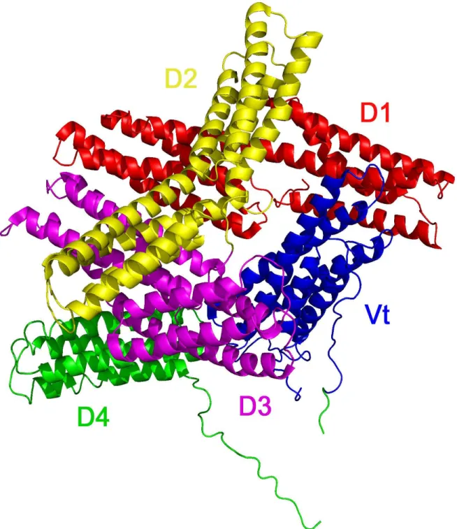
Figure 1.1: The Structure of Full Length Vinculin………...…………..17
Figure 1.2: Structural Features of Vinculin Tail………...…….18
Figure 2.1: Backbone NMR Assignments of Vinculin Tail……….…..28
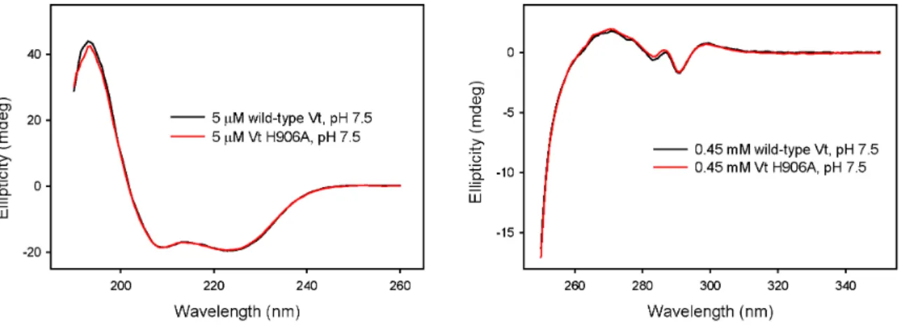
Figure 3.1: Circular Dichroism of Wild-type Vt and Vt H906A………...………46
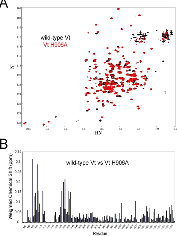
Figure 3.2: NMR HSQC Spectra of Vt H906A……….….47
Figure 3.3: NMR Chemical Shift Perturbations in Vt H906A………..………..…..…….48
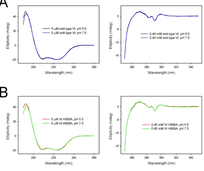
Figure 3.4: Circular Dichroism of Wild-type Vt and Vt H906A at pH 5.5 and 7.5……...49
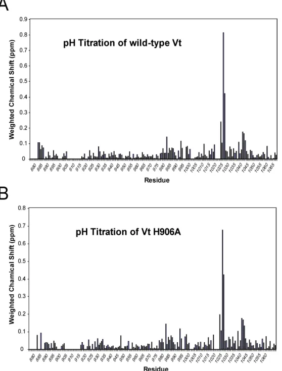
Figure 3.5: NMR Chemical Shift Perturbations Between pH 5.5 and 7.5…………...…..50
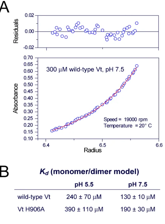
Figure 3.6: Analytical Ultracentrifugation of Vinculin Tail……….……….51
Figure 4.1: Co-sedimentation of Vt with PIP2 Containing Vesicles………..………70
Figure 4.2: Co-sedimentation of Vt with PI and PS Containing Vesicles……….……….71
Figure 4.3: Vt in the presence of DPC Micelles………...………..72
Figure 4.4: Circular Dichroism and HSQC Spectra of Vt∆C………...………..73
Figure 4.5: NMR HSQC Spectra of Vt∆C5 and Vt∆C7………74
Figure 4.6: Interactions of the N-terminal Strap of Vt………...……75
List of Abbreviations and Symbols
2D Two Dimensional
3D Three Dimensional
Arp 2/3 Actin-related protein complex AUC Analytical Ultracentrifuge BME β-mercaptoethanol
BMRB Biological Magnetic Resonance Data Bank CD Circular Dichroism
cDNA Complementary Deoxyribonucleic Acid D2O Deuterium Oxide
DNA Deoxyribonucleic Acid DPC Dodecylphosphocholine DTT Dithiothreitol
ε Molar extinction coefficient
ECM Extracellular Matrix
EDTA Ethylenediaminetetraacetic Acid ERK Extracellular signal-regulated kinase F-actin Filamentous actin
FAK Focal Adhesion Kinase GdmCl Guanidinium Chloride
IpaA A Shigella flexneri virulence factor IPTG isopropyl β-D-1-thiogalactopyranoside
Kd Dissociation constant
kDa kilodalton
LD Paxillin-Leucine and Aspartate Rich Motif
LIM Protein domain named due to their discovery in Lin11, Isl-1 and Mec-3 LPPC 1-palmitoyl-2-hydroxy-sn-glycero-3-[phospho-RAC-(1-glycerol)] MHz Megahertz
MVt MetaVinculin Tail
NMR Nuclear Magnetic Resonance
NOESY Nuclear Overhauser Enhancement Spectroscopy PC Phosphatidylcholine
PDB ID Protein Data Bank Identification Number PE Phosphatidylethanolamine
PI Phosphatidylinositol
PIP2 Phosphatidylinositol 4,5-bisphosphate
PKCa Protein Kinase C α PPI Phosphoinositide ppm Part Per Million PS Phosphatidylserine SDS Sodium Dodecyl Sulfate
Tris Tris(hydroxymethyl)aminomethane
UV Ultraviolet
VASP Vasodilator-stimulated Phosphoprotein VBS Vinculin Binding Site
Vh Vinculin Head
Vt Vinculin Tail
Vt∆C A mutant form of vinculin tail, lacking the C-terminal 15 residues (1052-1066)
Chapter I.
Introduction
A. Vinculin
Vinculin is a ubiquitously expressed, abundant, and highly conserved 116 kDa protein that localizes to membrane-associated complexes and couples the actin cytoskeleton to the membrane at sites of cellular attachment. These sites of attachment include adherence junctions and focal adhesions which link cells to neighboring cells and cells to extracellular matrix (ECM), respectively[1]. A larger isoform of vinculin, called metavinculin, is found exclusively in smooth and cardiac muscle[2]. Both vinculin and metavinculin are found in costameres, protein complexes that link the force generating sacromeres to the cell
membrane, and in cardiac muscle both isoforms are also localized to intercalated discs, a specialized linkage between adjacent cells[2, 3]. In all of these locations vinculin plays a role in connecting the actin cytoskeleton to the membrane and therefore located at primary sites of contractile force transmission. In addition to the obvious importance for muscle contraction, transmission of contractile forces is also critical for cell motility and migration. Not too surprising, vinculin plays an important role in processes that require regulated cell movement, including development and wound healing, as well as pathological processes including invasion and metastasis[4, 5]. Dynamic regulation of cell adhesions and actin cytoskeletal morphology is also important for processes involved in cell growth,
to a number of human diseases, including cancer. Loss of normal vinculin function, in particular, has been associated with cancer phenotypes, cardiovascular disease, and lethal errors in embryogenesis[5-9].
B. The Role of Vinculin in Development, Cell Motility, Cell Survival, and Cancer
Cell adhesions, whether they are between adjacent cells or with the extracellular matrix (ECM), play a critical role in development and survival. Ordered cellular motility requires adhesive structures to be dynamic and precisely controlled. In addition, these sites of adhesion are also involved in the sensing and transduction of a multitude of signals both from the extracellular and intracellular environment. Vinculin plays an important role in the regulation and function of both focal adhesion and adherence junctions and, consequently, affects many cellular processes that are mediated by cellular attachment. Vinculin null mice die in early embryogenesis, with mice embryos exhibiting brain and heart defects[5]. Vinculin null cells also exhibit tumor suppressor properties, including: decreased focal adhesion, increased motility, resistance to apoptosis and anoikis, ability to grow in soft agar, and altered signaling through extracellular signal-regulated kinase (ERK)[4, 5, 7, 10-12]. Additional support for vinculin’s role in tumor suppression comes from studies showing decreased vinculin protein levels that are correlated with highly metastatic, invasive cancers. Moreover, transfection of vinculin cDNA into tumor cells lines with decreased vinculin expression drastically reduces their tumorigenic ability[13], further solidifying vinculin’s role as a tumor suppressor.
Although vinculin is a widely expressed protein, it is particularly abundant in muscle cells where it localizes to costameres, which link sacromeres to the cell membrane[3]. In contrast, a larger isoform of vinculin, metavinculin, is exclusively expressed in smooth and cardiac muscle and contains a 68 amino acid insertion in the tail domain[2, 14, 15]. Both isoforms are found in intercalated discs of cardiac tissue[2]. Although no ligand specific to the metavinculin insert has been found, there is some evidence suggesting that metavinculin possesses distinct properties. In addition to the tissue specific expression, metavinculin tail (MVt) promotes distinct F-actin organization from the vinculin tail domain (Vt). Both Vt and MVt bind F-actin, however Vt bundles F-actin into parallel filaments, while MVt generates “highly viscous F-actin webs”[15].
Vinculin null mice and nematodes both die in development with cardiac and muscle defects, respectively[5, 16]. While it is possible that these developmental abnormalities are due solely to defects in cell migration, it is also possible that vinculin has a specific
developmental role in these tissues. Regardless of its role in development, both vinculin and metavinculin have been implicated in cardiovascular function, as separate mutations in each have been associated with human cardiomyopathy. A leucine to methionine mutation at position 277 in vinculin is believed to confer susceptibility to hypertrophic cardiomyopathy, while an arginine to tryptophan substitution at position 975 in metavinculin is associated with both hypertrophic and dilated cardiomyopathy[17, 18]. The metavinculin mutation (R975W) has also been shown to alter F-actin organization in vitro as cardiac myocyte samples
aortic stenosis, show a consistent defect in vinculin/metavinculin expression in intercalated discs suggesting an important and distinct role for vinculin/metavinculin in cardiac tissue[8].
D. Vinculin Domains and Their Ligands
Early studies using electron microscopy and proteolytic cleavage approaches indicated that vinculin consists of three domains; a globular head domain (Vh), a flexible neck, and a tail domain (Vt)[20, 21]. Each of these domains has been shown to bind multiple ligands. The head domain has been shown to interact with talin, α-actinin, α-catenin, β -catenin, and the bacterial virulence factor IpaA. The neck domain interacts with vinexin, ponsin, the Arp 2/3 complex, and the vasodilator-stimulated phosphoprotein (VASP). The tail domain binds to F-actin, paxillin, protein kinase C-α (PKCα), acidic phospholipids and
α-synemin (reviewed by Ziegler, et al., 2006)[22, 23].
E. Vinculin Structure and Auto-inhibition
to Vt in the context of full length vinculin[24, 25]. Multiple sites of interaction between Vh and Vt occur, giving rise to a compact, auto-inhibitory complex. While the majority of contacts are observed between D1 and Vt, additional interactions occur between Vt and D3 and D4. The numerous interactions between Vh and Vt, give rise to a high affinity
intramolecular interaction, with an estimated Kd of ~1 nM [24]. Activation of vinculin
requires release of auto-inhibitory Vh and Vt interactions to expose binding sites for interactions with α-actinin, VASP, talin, F-actin, PKCα, acidic phospholipids, and the
Arp2/3 complex[10, 30-35].
F. Vinculin Activation
The tight auto-inhibitory interaction between the head and tail domains of vinculin is possible due to three distinct interaction surfaces between Vh and Vt (D1-Vt, D3-Vt, D4-Vt)[24, 36]. The apparent affinity is further increased due to the intramolecular nature of the interaction (once separated Vt does not diffuse away from Vh). Notably, binding cell
actin[37]. Given the number of ligands that bind to both the head and tail domains of vinculin, it is possible that additional ligand pairs may also be capable of activating vinculin or that subsequent binding of additional ligands after activation could help maintain vinculin in an activated state. Additional ligands interacting with the head, neck, or tail could
conceivably play such roles. Interestingly the Shigella flexneri virulence factor, IpaA, appears to activate vinculin in the absence of other ligands[37]. Although IpaA contains two vinculin-binding sites (VBS), their binding to vinculin is mutually exclusive. Crystal
structures of D1 of vinculin with either IpaA VBS have been solved and appear to mimic the binding of vinculin-binding sites from talin and α-actinin[28]. IpaA-VBS has been reported to have at least 10-fold higher affinity for Vh-D1 relative to other VBSs, which may account for its ability to activate vinculin[28]. However, it cannot be ruled out that an additional interaction between IpaA and vinculin could be involved in disrupting the Vt-D3 and Vt-D4 interactions.
G. The Tail Domain of Vinculin and its Ligands
1. Vinculin Tail Structure
interaction between Vt and the D1 domain of Vh in the auto-inhibited structure[24]. In addition to the main helical bundle of Vt, there are two structural features of interest; an N-terminal strap and C-N-terminal extension (Figure 1.2).
Residues 879-893 form the N-terminal strap, and adopt an extended conformation. In the crystal structure of the isolated tail domain, this strap is seen in multiple conformations, suggesting some conformational mobility. In the more ordered conformation, this N-terminal strap packs against helix 1 and 2. In the second conformation, only weak electron density was seen prior to residue 890, and crystal contacts were seen at the position the strap occupied in the first conformation. Due to a more ordered electron density and the lack of crystal contacts, the conformation with the strap packing against helix 1 and 2 was assumed to be a better model of the structure in solution[25]. In support of this idea, in the full length vinculin structure the N-terminal strap of Vt is seen in a very similar conformation, packed along helix 1 and 2. Residues F885 and D882 of the strap form interactions with the helix bundle, and are likely important to maintaining the extended conformation along the helix 1-2 interface. In both the isolated tail structure and the full length vinculin structure, the side chain of F885 packs in a hydrophobic crevice between helix 1 and 2, making interaction with H906 of helix 1. Interactions made by D882 vary some between crystal structures, but they include interactions with S914 and K924, located in the helix 1-2 loop and helix 2
respectively, and K1061 and Y1065, located in the C-terminal extension[24, 25].
loop and these interactions are thought to be important for the structural stability of Vt[25, 38]. The final C-terminal residues form a “hydrophobic hairpin” (TPWYQ) that emerges close to the N-terminal strap. It has been postulated that this hydrophobic hairpin is important for insertion into the membrane.
The crystal structure of Vt also aided in identifying two solvent-accessible surface features that have been postulated to be important for lipid binding; the “basic collar” and “basic ladder” (Figure 1.2)[25]. The basic collar consists of residues from helix 1, helix 5, and the C-terminal extension, including R910, K911, R1039, K1049, R1060, and K1061. These residues surround the hydrophobic hairpin, at the bottom of the helical bundle. The basic ladder consists of residues located along the length of helix 3 and at the base of the helical bundle, including K944, R945, K952, K956, R963, K966, K970, R978, R1008, and R1049.
2. Actin Interactions
F-actin[42], however, in the context of the crystal structure of Vt neither of these fragments correspond to a canonical actin binding site. Changes in protease sensitivity and tryptophan fluorescence upon F-actin association have been reported, providing evidence of a possible conformational change in Vt upon actin binding[25], further complicating clear identification of the binding site or sites.
Three-dimensional models of Vt, both bound to and crosslinking F-actin, were created using a combination of electron microscopy, image analysis and computational docking[43]. Crystal structures of Vt, alone and in the context of full length vinculin, were docked into 3D difference maps produced from images of F-actin decorated with Vt.
However, in contrast to earlier observations[25], the docked protein models did not support a large scale conformational change in Vt upon binding F-actin, as a high correlation of the docked structure with the corresponding reconstructions was obtained with no major clashes observed between the docked proteins [43]. This model supports Vt binding to two separate actin monomers along the actin filament via two distinct sites. The upper interaction
interface includes residues at the top of helices 2 and 3, along with residues contained in the N-terminal strap (883-890). The corresponding interface on actin is located at the bottom of subdomain 1, and contains a large hydrophobic pocket implicated in interactions with several actin binding proteins[43]. The lower F-actin/Vt interaction interface is more polar, is
located at the base of helix 3, and includes residues close to the C-terminus. This Vt interface interacts with an acidic region at the top of subdomain 1 of a second actin
monomer. Superimposition of the full length structure of vinculin with the tail domain in the actin bound model exhibits severe clashes between Vh and actin, consistent with the
three-dimensional model of actin bundling by Vt was produced by computationally docking the F-actin-Vt model into tomographic reconstructions of Vt-crosslinked actin arrays. This model shows an asymmetric dimer with the C-terminal residues of one monomer (located at the bottom of the helical bundle) contacting the N-terminal strap along the side of the second monomer. However, as severe steric clashes are observed between the C-terminus of one monomer and the N-terminus of the other, it was speculated that either one or both of these regions of Vt may undergo a conformational change to promote F-actin bundling[43]. The overall 5-helix bundle of Vt is believed to remain largely unaltered by either F-actin binding or bundling. Currently, no specific point mutations in Vt have been identified that effectively block its F-actin binding and/or bundling activity. The production and characterization of such mutants would be beneficial in elucidating the activation, role, and function of vinculin
in vivo.
3. Lipid Interactions
The tail domain of vinculin has been shown by multiple groups to bind acidic
phospholipids[25, 31, 44, 45]. Among the various types of acidic phospholipids, Vt has been shown to have an increased affinity for phosphatidylinositol 4,5-bisphosphate (PIP2),
although the affinity and degree of specificity has varied in the literature (possibly due to varying methods used to assay binding)[24, 25, 31, 46, 47]. PIP2 has been shown to be a key
to vinculin was proposed to release interactions between Vh and Vt, thereby leading to vinculin activation[24, 31, 35, 46, 50]. However, subsequent work indicated that
phospholipid binding to full length vinculin is significantly reduced relative to Vt, and that this interaction is unable release Vh-Vt interaction. Thus, phospholipid binding to vinculin, alone, does not lead to vinculin activation[31, 44]. Instead of a role of phospholipids in vinculin activation and localization to sites of adhesion, multiple reports have now suggested that vinculin-lipid interactions are important in regulating focal adhesion disassembly and turnover, a process critical to cell motility[38, 44].
with the lipid membrane and possibly additional ligands[25]. Further research, however, indicated that deletion of 15 C-terminal residues in Vt, significantly destabilizes the Vt tertiary fold due to removal of a hydrophobic interactions between the C-terminus and the base of the helical bundle. Hence, the reduction in lipid binding to Vt∆C is likely to result
from large scale conformational/dynamic change in Vt[38]. Two remaining basic collar residues, K911 and K924 were also proposed as key determinants of PIP2 binding [24, 38].
A separate study produced a “lipid binding deficient” variant of vinculin by introducing a series of mutations into the basic collar and basic ladder; mutation of 4 basic residues (K952, K956, R963, and K966) in the basic ladder and 2 residues (R1060 K1016) within the basic collar and contained in the C-terminal extension. Mutation of either group of residues alone led to only a partial loss in lipid binding[44]. However, the effects of these multiple
mutations on ligand binding were not fully characterization. Moreover, no studies were conducted to determine whether mutations of 6 residues within Vt alters Vt structure. Given the 3D disposition of the basic ladder and collar, it is unlikely that vinculin binds both sites, unless more than one site of interaction occurs upon interaction with phospholipids. Hence, with multiple mutations in disparate surfaces of the protein, the complete picture of lipid binding remains unclear. It is also still unknown whether these mutations have effected the conformation of Vt causing confusion in data interpretation, as appears to have happened with Vt∆C.
4. Paxillin Interactions
Rous sarcoma virus[51, 52]. Paxillin was identified as a vinculin binding protein that associates with a C-terminal cleavage fragment of vinculin containing the tail domain[51]. The domain structure of paxillin consists of 5 leucine-rich ‘LD’ motifs and 4 double zinc finger LIM domains. Paxillin is believed to function as a molecular scaffold, with both the LD and LIM domains participating in protein-protein interactions [53, 54]. Three of the LD motifs, LD1, LD2, and LD4, were shown to bind to the tail domain of vinculin[55]. Deletion mutagenesis studies suggested that paxillin binds to a Vt fragment comprising residues 979-1000[56]. Paxillin binding was assessed by an ‘overlay’ assay, by first using denaturing SDS-gel electrophoresis of Vt or Vt fragments, followed by blotting on nitrocellulose filters prior to paxillin binding[25, 56]. It should be noted that this fragment contains the majority of helix 4 of Vt, which is markedly amphipathic. Thus, the conformation of this fragment in overlay binding assays could be significantly different from Vt in solution.
The physiological role of the vinculin-paxillin interaction is currently unclear. Intriguingly, both vinculin and focal adhesion kinase (FAK) bind to the LD2 and LD4 domains of paxillin, suggesting that both of these cell adhesion proteins may compete for paxillin binding. This idea is further supported by findings that alterations in vinculin expression can modulate FAK-paxillin interactions and the activity of extracellular signal-regulated kinase (ERK), leading to changes in cell motility and survival[7]. However, no clear in vivo evidence currently exists, that supports a direct interaction between vinculin and paxillin, due at least in part to a lack of characterized, specific mutations to block the
interaction. Further complicating this matter, recent evidence indicates that vinculin overexpression induces the robust recruitment of paxillin to focal adhesions, and this
in the interpretation of protein overexpression experiments, this would appear to suggest that, in addition to possible direct interactions with Vt, vinculin either possesses additional
paxillin interactions or vinculin is capable of modulating the subcellular localization of paxillin indirectly. As there is no evidence suggesting a secondary paxillin binding site, the latter appears more likely. However, either circumstance may complicate interpretation of data concerning the physiological role of a vinculin-paxillin interaction.
5. Vinculin Tail Phosphorylation
Vinculin is phosphorylated as a result of various stimuli, including calcium, phorbol esters, and leukotriene D(4)[57-59]. In the case of calcium stimulation, phosphorylation of vinculin is observed in connection with both platelet stimulation and formation of tight junctions[59, 60]. Src kinases and protein kinase C-α (PKCα) are both capable of phosphorylating vinculin, although a direct functional role for phosphorylation by either kinase is still not understood[35, 61-63].
During cell spreading, PKCα has been shown to interact with vinculin in vivo via crosslinking and pulldown experiments, and two serine residues in Vt, S1033 and S1045, have been shown to be phosphorylated by PKCα in vitro. The in vitro phosphorylation of Vt by PKCα was promoted by the addition of acidic phospholipids, leading to the speculation that binding of phospholipids may help release of the Vh-Vt complex allowing
phosporylation[35].
Y1065 could inhibit the Vh-Vt interaction without affecting actin-binding[63]. Cells that contain a vinculin mutant with both Src phosphorylation sites (Y100F/Y1065F) mutated exhibited decreased cell spreading relative to wild-type vinculin, indicating a physiological role for Src phosphorylation[63].
6. Vinculin Tail Self-Association
H. Literature Cited
1. Geiger, B., et al., Vinculin, an intracellular protein localized at specialized sites
where microfilament bundles terminate at cell membranes. Proc Natl Acad Sci U S
A, 1980. 77(7): p. 4127-31.
2. Belkin, A.M., et al., Diversity of vinculin/meta-vinculin in human tissues and
cultivated cells. Expression of muscle specific variants of vinculin in human aorta smooth muscle cells. J Biol Chem, 1988. 263(14): p. 6631-5.
3. Pardo, J.V., J.D. Siliciano, and S.W. Craig, A vinculin-containing cortical lattice in
skeletal muscle: transverse lattice elements ("costameres") mark sites of attachment between myofibrils and sarcolemma. Proc Natl Acad Sci U S A, 1983. 80(4): p.
1008-12.
4. Rodriguez Fernandez, J.L., et al., Suppression of vinculin expression by antisense
transfection confers changes in cell morphology, motility, and anchorage-dependent growth of 3T3 cells. J Cell Biol, 1993. 122(6): p. 1285-94.
5. Xu, W., H. Baribault, and E.D. Adamson, Vinculin knockout results in heart and
brain defects during embryonic development. Development, 1998. 125(2): p. 327-37.
6. Coll, J.L., et al., Targeted disruption of vinculin genes in F9 and embryonic stem cells
changes cell morphology, adhesion, and locomotion. Proc Natl Acad Sci USA, 1995.
92(20): p. 9161-5.
7. Subauste, M.C., et al., Vinculin modulation of paxillin-FAK interactions regulates
ERK to control survival and motility. J Cell Biol, 2004. 165(3): p. 371-81.
8. Vasile, V.C., et al., Obstructive hypertrophic cardiomyopathy is associated with
reduced expression of vinculin in the intercalated disc. Biochem Biophys Res
Commun, 2006. 349(2): p. 709-15.
9. Zemljic-Harpf, A.E., et al., Heterozygous inactivation of the vinculin gene
predisposes to stress-induced cardiomyopathy. Am J Pathol, 2004. 165(3): p.
1033-44.
10. DeMali, K.A., C.A. Barlow, and K. Burridge, Recruitment of the Arp2/3 complex to
vinculin: coupling membrane protrusion to matrix adhesion. J Cell Biol, 2002.
159(5): p. 881-91.
11. Goldmann, W.H. and D.E. Ingber, Intact vinculin protein is required for control of
cell shape, cell mechanics, and rac-dependent lamellipodia formation. Biochem
Biophys Res Commun, 2002. 290(2): p. 749-55.
12. Xu, W., J.L. Coll, and E.D. Adamson, Rescue of the mutant phenotype by
13. Rodriguez Fernandez, J.L., et al., Suppression of tumorigenicity in transformed cells
after transfection with vinculin cDNA. J Cell Biol, 1992. 119(2): p. 427-38.
14. Glukhova, M.A., et al., Meta-vinculin distribution in adult human tissues and
cultured cells. FEBS Lett, 1986. 207(1): p. 139-41.
15. Rudiger, M., et al., Differential actin organization by vinculin isoforms: implications
for cell type-specific microfilament anchorage. FEBS Lett, 1998. 431(1): p. 49-54.
16. Barstead, R.J. and R.H. Waterston, Vinculin is essential for muscle function in the
nematode. J Cell Biol, 1991. 114(4): p. 715-24.
17. Vasile, V.C., et al., A missense mutation in a ubiquitously expressed protein, vinculin,
confers susceptibility to hypertrophic cardiomyopathy. Biochem Biophys Res
Commun, 2006. 345(3): p. 998-1003.
18. Vasile, V.C., et al., Identification of a metavinculin missense mutation, R975W,
associated with both hypertrophic and dilated cardiomyopathy. Mol Genet Metab,
2006. 87(2): p. 169-74.
19. Olson, T.M., et al., Metavinculin mutations alter actin interaction in dilated
cardiomyopathy. Circulation, 2002. 105(4): p. 431-7.
20. Molony, L. and K. Burridge, Molecular shape and self-association of vinculin and
metavinculin. J Cell Biochem, 1985. 29(1): p. 31-6.
21. Winkler, J., H. Lunsdorf, and B.M. Jockusch, The ultrastructure of chicken gizzard
vinculin as visualized by high-resolution electron microscopy. J Struct Biol, 1996.
116(2): p. 270-7.
22. Sun, N., et al., Human alpha-synemin interacts directly with vinculin and
metavinculin. Biochem J, 2008. 409(3): p. 657-67.
23. Ziegler, W.H., R.C. Liddington, and D.R. Critchley, The structure and regulation of
vinculin. Trends Cell Biol, 2006. 16(9): p. 453-60.
24. Bakolitsa, C., et al., Structural basis for vinculin activation at sites of cell adhesion. Nature, 2004. 430(6999): p. 583-6.
25. Bakolitsa, C., et al., Crystal structure of the vinculin tail suggests a pathway for
activation. Cell, 1999. 99(6): p. 603-13.
26. Borgon, R.A., et al., Crystal structure of human vinculin. Structure (Camb), 2004. 12(7): p. 1189-97.
28. Izard, T., G. Tran Van Nhieu, and P.R. Bois, Shigella applies molecular mimicry to
subvert vinculin and invade host cells. J Cell Biol, 2006. 175(3): p. 465-75.
29. Izard, T. and C. Vonrhein, Structural basis for amplifying vinculin activation by talin. J Biol Chem, 2004.
30. Johnson, R.P. and S.W. Craig, An intramolecular association between the head and
tail domains of vinculin modulates talin binding. J Biol Chem, 1994. 269(17): p.
12611-9.
31. Johnson, R.P. and S.W. Craig, The carboxy-terminal tail domain of vinculin contains
a cryptic binding site for acidic phospholipids. Biochem Biophys Res Commun,
1995. 210(1): p. 159-64.
32. Johnson, R.P. and S.W. Craig, F-actin binding site masked by the intramolecular
association of vinculin head and tail domains. Nature, 1995. 373(6511): p. 261-4.
33. Miller, G.J., S.D. Dunn, and E.H. Ball, Interaction of the N- and C-terminal domains
of vinculin. Characterization and mapping studies. J Biol Chem, 2001. 276(15): p.
11729-34.
34. Weekes, J., S.T. Barry, and D.R. Critchley, Acidic phospholipids inhibit the
intramolecular association between the N- and C-terminal regions of vinculin, exposing actin-binding and protein kinase C phosphorylation sites. Biochem J, 1996.
314(Pt 3): p. 827-32.
35. Ziegler, W.H., et al., A lipid-regulated docking site on vinculin for protein kinase C. J Biol Chem, 2002. 277(9): p. 7396-404.
36. Cohen, D.M., et al., Two distinct head-tail interfaces cooperate to suppress activation
of vinculin by talin. J Biol Chem, 2005. 280(17): p. 17109-17.
37. Chen, H., D.M. Choudhury, and S.W. Craig, Coincidence of actin filaments and talin
is required to activate vinculin. J Biol Chem, 2006. 281(52): p. 40389-98.
38. Saunders, R.M., et al., Role of vinculin in regulating focal adhesion turnover. Eur J Cell Biol, 2006. 85(6): p. 487-500.
39. Humphries, J.D., et al., Vinculin controls focal adhesion formation by direct
interactions with talin and actin. J Cell Biol, 2007. 179(5): p. 1043-57.
40. Jockusch, B.M. and G. Isenberg, Interaction of alpha-actinin and vinculin with actin:
opposite effects on filament network formation. Proc Natl Acad Sci U S A, 1981.
78(5): p. 3005-9.
41. Johnson, R.P. and S.W. Craig, Actin activates a cryptic dimerization potential of the
42. Huttelmaier, S., et al., Characterization of two F-actin-binding and oligomerization
sites in the cell-contact protein vinculin. Eur J Biochem, 1997. 247(3): p. 1136-42.
43. Janssen, M.E., et al., Three-dimensional structure of vinculin bound to actin
filaments. Mol Cell, 2006. 21(2): p. 271-81.
44. Chandrasekar, I., et al., Vinculin acts as a sensor in lipid regulation of adhesion-site
turnover. J Cell Sci, 2005. 118(Pt 7): p. 1461-72.
45. Fukami, K., et al., alpha-Actinin and vinculin are PIP2-binding proteins involved in
signaling by tyrosine kinase. J Biol Chem, 1994. 269(2): p. 1518-22.
46. Gilmore, A.P. and K. Burridge, Regulation of vinculin binding to talin and actin by
phosphatidyl- inositol-4-5-bisphosphate. Nature, 1996. 381(6582): p. 531-5.
47. Johnson, R.P., et al., A conserved motif in the tail domain of vinculin mediates
association with and insertion into acidic phospholipid bilayers. Biochemistry, 1998.
37(28): p. 10211-22.
48. Sechi, A.S. and J. Wehland, The actin cytoskeleton and plasma membrane
connection: PtdIns(4,5)P(2) influences cytoskeletal protein activity at the plasma membrane. J Cell Sci, 2000. 113 Pt 21: p. 3685-95.
49. Yin, H.L. and P.A. Janmey, Phosphoinositide regulation of the actin cytoskeleton. Annu Rev Physiol, 2003. 65: p. 761-89.
50. Steimle, P.A., et al., Polyphosphoinositides inhibit the interaction of vinculin with
actin filaments. J Biol Chem, 1999. 274(26): p. 18414-20.
51. Turner, C.E., J.R. Glenney, Jr., and K. Burridge, Paxillin: a new vinculin-binding
protein present in focal adhesions. J Cell Biol, 1990. 111(3): p. 1059-68.
52. Burridge, K., C.E. Turner, and L.H. Romer, Tyrosine phosphorylation of paxillin and
pp125FAK accompanies cell adhesion to extracellular matrix: a role in cytoskeletal assembly. J Cell Biol, 1992. 119(4): p. 893-903.
53. Tumbarello, D.A., M.C. Brown, and C.E. Turner, The paxillin LD motifs. FEBS Lett, 2002. 513(1): p. 114-8.
54. Dawid, I.B., J.J. Breen, and R. Toyama, LIM domains: multiple roles as adapters and
functional modifiers in protein interactions. Trends Genet, 1998. 14(4): p. 156-62.
55. Turner, C.E., et al., Paxillin LD4 motif binds PAK and PIX through a novel 95-kD
ankyrin repeat, ARF-GAP protein: A role in cytoskeletal remodeling. J Cell Biol,
1999. 145(4): p. 851-63.
56. Wood, C.K., et al., Characterisation of the paxillin-binding site and the C-terminal
57. Massoumi, R. and A. Sjolander, Leukotriene D(4) affects localisation of vinculin in
intestinal epithelial cells via distinct tyrosine kinase and protein kinase C controlled events. J Cell Sci, 2001. 114(Pt 10): p. 1925-34.
58. Werth, D.K. and I. Pastan, Vinculin phosphorylation in response to calcium and
phorbol esters in intact cells. J Biol Chem, 1984. 259(8): p. 5264-70.
59. Vostal, J.G. and N.R. Shulman, Vinculin is a major platelet protein that undergoes
Ca(2+)-dependent tyrosine phosphorylation. Biochem J, 1993. 294 (Pt 3): p. 675-80.
60. Perez-Moreno, M., et al., Vinculin but not alpha-actinin is a target of PKC
phosphorylation during junctional assembly induced by calcium. J Cell Sci, 1998.
111 (Pt 23): p. 3563-71.
61. Ito, S., et al., Vinculin phosphorylation by the src kinase. Interaction of vinculin with
phospholipid vesicles. J Biol Chem, 1983. 258(23): p. 14626-31.
62. Sefton, B.M., et al., Vinculin: a cytoskeletal target of the transforming protein of
Rous sarcoma virus. Cell, 1981. 24(1): p. 165-74.
63. Zhang, Z., et al., The phosphorylation of vinculin on tyrosine residues 100 and 1065,
mediated by SRC kinases, affects cell spreading. Mol Biol Cell, 2004. 15(9): p.
4234-47.
64. Fringeli, U.P., et al., Structure-activity relationship in vinculin: an IR/attenuated total
reflection spectroscopic and film balance study. Proc Natl Acad Sci U S A, 1986.
83(5): p. 1315-9.
65. Otto, J.J., Detection of vinculin-binding proteins with an 125I-vinculin gel overlay
technique. J Cell Biol, 1983. 97(4): p. 1283-7.
66. Wilkins, J.A., K.Y. Chen, and S. Lin, Detection of high molecular weight vinculin
binding proteins in muscle and nonmuscle tissues with an electroblot-overlay technique. Biochem Biophys Res Commun, 1983. 116(3): p. 1026-32.
67. Huttelmaier, S., et al., The interaction of the cell-contact proteins VASP and vinculin
is regulated by phosphatidylinositol-4,5-bisphosphate. Curr Biol, 1998. 8(9): p.
Chapter II.
Backbone 1H, 13C, and 15N NMR Assignments of the Tail Domain of Vinculin
A. Introduction
Vinculin is a 116 kDa highly conserved cytoskeletal protein that localizes to both cell-matrix and cell-cell contacts and plays an important role in the linkage between
transmembrane receptors (integrins or cadherins) and the actin cytoskeleton [1]. Vinculin is composed of an N-terminal head domain (Vh), a flexible neck domain, and a C-terminal tail domain (Vt). Each domain contains binding sites for multiple ligands. In addition to an important auto-inhibitory interaction with Vh, Vt binds F-actin, paxillin, and acidic phospholipids. However, these interactions are at least partially occluded in the intact protein due to auto-inhibitory interactions with Vh. The activation of vinculin requires release of the head/tail interaction, and is thought to require combinatorial binding of ligands to both Vh and Vt [2].
Vinculin is essential in development, as vinculin null embryos die early in
the vinculin gene have been linked to both hypertrophic and dilated cardiomyopathy, while decreased expression of vinculin is correlated with a predisposition to stress-induced cardiomyopathy [5, 6]. In order to elucidate the role of vinculin in both physiological and pathophysiological states, a detailed understanding of its activation, function, and ligand interactions is needed.
B. Methods and Experiments
1. Expression and Purification
Escherichia coli strain BL21(DE3) was transformed by electroporation with a
pET15b vector (Novagen) containing the tail domain of vinculin (G. gallus, 879-1066), as previously described [7]. Uniformly 15N or 13C/15N isotopically enriched Vt was grown in M9 minimal media containing 1 g/L 15NH4Cl, 2 g/L 13C6-glucose (Spectra) in H2O.
Perdeuterated Vt was grown in M9 minimal media containing 1 g/L 15NH4Cl, 2 g/L 2H7-13C6
-glucose (Spectra) in 99% D2O. Bacterial cultures were grown at 37°C until reaching an
optical density of 0.6, at which point Vt expression was induced by the addition of 0.25 mM IPTG and grown for 4-5 hours. The bacteria were harvested by centrifugation and
resuspended in lysis buffer (20 mM Tris, pH 7.5, 150 mM NaCl, 5 mM Imidizole, 0.1% β-mercaptoethanol) and lysed by sonication. Vt was initially purified using by affinity separation using Ni-NTA Agarose beads (Qiagen). Following elution from the Ni-NTA beads, Vt was exchanged into thrombin cleavage buffer (20 mM Tris, pH 7.5, 500 mM NaCl, 2.5 mM CaCl2, 0.1% BME) by dialysis. The 6-His tag was cleaved by thrombin and removed
by dialysis in the same buffer. Vt was then further purified by cation-exchange
0.05-1 M NaCl gradient at pH 7.5. In some cases insoluble Vt was refolded and purified by resuspending cell pellets in 6M guanidinium chloride (GdmCl) prior to sonication. Vt was bound to Ni-NTA beads under denaturing conditions and refolded by removal of the GdmCl through dialysis. After refolding, thrombin cleavage and cation-exchange chromatography was performed as described above. Perdeuterated Vt was purified under denaturing
conditions to ensure the back-exchange of amide protons to 1H.
2. Nuclear Magnetic Resonance Spectroscopy
NMR spectra of Vt were collected at 37°C on Varian Inova 600, 700, and 800 MHz spectrometers. Select spectra were collected on Varian Inova 700 and 800 MHz
spectrometers equipped with cryogenically cooled probe heads. The NMR buffer contained 10 mM K2HPO4, 50 mM NaCl, 0.01% NaN3, 2 mM DTT at pH 5.5, in 90% H2O and 10%
D2O. Backbone assignments were determined using 1H-15N HSQC, HNCO, HN(CA)CO,
HNCA, HNCACB, HN(CA)CB, HN(CO)CA, and HN(COCA)CB experiments.
Assignments were also verified where possible by HN-HN NOESY cross peaks obtained from 3D 15N-edited NOESY data. NMR data was processed using NMRPipe/NMRDraw [8] and analyzed using NMRView [9].
C. Assignments and Deposition
The Vt domain employed for this study, comprises residues 879-1066 of full length vinculin, and was crystallized as a dimer with the two monomer units packed in an
Consistent with previous observations of Vt dimerization, our NMR, analytical
ultracentrifugation, isothermal titration calorimetry, and fluorescence anisotropy studies all indicated that Vt is significantly dimeric at the concentrations used for NMR (0.3 mM). Standard 3D triple resonance NMR data on Vt showed poor magnetization transfer efficiency, consistent with a 43 kDa dimer at these concentrations. Perdeuteration of Vt improved efficiency of magnetization transfer in triple resonance NMR experiments, making sequential assignment possible. However, concentrations were limited to 0.3 mM. At higher Vt concentrations, magnetization transfer decreased significantly suggesting further
oligomerization likely occurs. We were able to determine sequence specific assignments for 84.24% of the non-proline 15N and amide proton resonances. We were also able to obtain 86.7%, 88.83%, and 82.98% of the 13CO, 13Cα, and 13Cβ resonances assignments,
Figure 2.1: 2D 1H-15N HSQC spectrum of uniformly [2H,15N,13C]-labeled Vt collected on a Varian Inova 700 MHz spectrometer at 37 ºC. The sample contained 0.3 mM Vt, 10 mM K2HPO4, 50 mM NaCl, 0.01% NaN3, and 2 mM DTT at pH 5.5 in 90% H2O and 10% D2O.
D. Literature Cited
1. Jockusch, B.M. and M. Rudiger, Crosstalk between cell adhesion molecules: vinculin
as a paradigm for regulation by conformation. Trends Cell Biol, 1996. 6(8): p. 311-5.
2. Chen, H., D.M. Choudhury, and S.W. Craig, Coincidence of actin filaments and talin
is required to activate vinculin. J Biol Chem, 2006. 281(52): p. 40389-98.
3. Xu, W., H. Baribault, and E.D. Adamson, Vinculin knockout results in heart and
brain defects during embryonic development. Development, 1998. 125(2): p. 327-37.
4. Subauste, M.C., et al., Vinculin modulation of paxillin-FAK interactions regulates
ERK to control survival and motility. J Cell Biol, 2004. 165(3): p. 371-81.
5. Vasile, V.C., et al., Identification of a metavinculin missense mutation, R975W,
associated with both hypertrophic and dilated cardiomyopathy. Mol Genet Metab,
2006. 87(2): p. 169-74.
6. Zemljic-Harpf, A.E., et al., Heterozygous inactivation of the vinculin gene
predisposes to stress-induced cardiomyopathy. Am J Pathol, 2004. 165(3): p.
1033-44.
7. Bakolitsa, C., et al., Crystal structure of the vinculin tail suggests a pathway for
activation. Cell, 1999. 99(6): p. 603-13.
8. Delaglio, F., et al., NMRPipe: a multidimensional spectral processing system based
on UNIX pipes. J Biomol NMR, 1995. 6(3): p. 277-93.
9. Johnson, B.A. and R.A. Blevins, NMR View: A computer program for the
Chapter III.
The Role of pH and Histidine 906
A. Introduction
Contacts between cells (cell-cell) and with the extracellular matrix (cell-matrix) regulate a wide variety of critical cellular processes including cell growth, migration, differentiation, and cell death[1-3]. Aberrant regulation of these processes contributes to a number of human diseases, including cancer[4]. The highly conserved cytoskeletal protein, vinculin, is found in both cell-cell and cell-matrix contacts [5], is essential for
embryogenesis[6], and plays an important role in regulating cell morphology and
migration[7]. Vinculin is a large, approximately 116 kDa protein consisting of an amino-terminal "head" domain (~ 90 kDa) and a carboxy-amino-terminal "tail" domain (~ 21 kDa) connected by a flexible hinge region. The vinculin tail (Vt) domain forms auto-inhibitory contacts with the vinculin head (Vh) domain and binds several ligands including F-actin, paxillin, PKCα and acidic phospholipids [8-15]. Auto-inhibitory contacts between the head and tail domain are believed to downregulate vinculin function by preventing the interaction of multiple ligands [11-13, 16, 17]. On the flip side, vinculin activation requires release of auto-inhibitory contacts. Current models of vinculin activation have proposed that a
activity [18-21]. The binding of these ligands is believed to induce conformational changes in both the head and tail domains causing disruption of auto-inhibitory contacts [18, 19, 22, 23]. One study suggested that a single histidine residue (H906) in the vinculin tail domain (Vt) is critical for both a pH- and lipid-dependent conformational change in Vt, which in turn, can modulate vinculin head/tail interactions [24].
Several crystal structures of vinculin are now available [18, 19, 22, 25]. The Vt domain was solved at pH 5.0, and found to possess an anti-parallel, five helix bundle fold. The tail domain adopts a similar structure in the context of the full length protein, despite differences in the pH at which the structures were solved. In addition to the helix bundle fold, Vt contains an N-terminal “strap” (residues 879 to 893) that exists in an extended conformation and forms interactions with residues along the helix 1-2 face and the C-terminus. In particular, F885 in the strap packs against the H906 side chain in helix 1, and D882 of the strap forms electrostratic interactions with S914, K924, K1061, and Y1065 (located in the loop between helices 1 and 2, helix 2, and the C-terminus respectively). In the isolated Vt domain solved at pH 5.0, the strap is observed in multiple conformations, suggesting conformational mobility.
7. 5. Mutation of H906 to alanine cause localized chemical shift perturbations in helices 1 and 2 and the N-terminal strap, consistent with perturbation of the F885/H906 interaction resulting in release of the strap from the helix 1-2 interface. However, loss of this contact does not appear to cause a large scale conformational change in the Vt five helix bundle fold. As H906 has also been implicated in pH-dependent Vt self-association[24], we employed NMR and analytical ultracentrifugation (AUC) approaches to investigate whether Vt undergoes pH-dependent self-association. The isolated Vt domain was crystallized as a dimer at pH 5.0 with the site of dimerization located in the upper portions of helices 4 and 5[19]. Our NMR data support Vt self-association in the solution state, with the dimerization interface consistent with that observed by crystallography. However, in contrast to a
previous report[24], our AUC and NMR results indicate that Vt does not undergo a monomer to dimer transition between pH 5.5 and pH 7.5. Thus, our overall results indicate that Vt does not undergo a pH-dependent change in structure, and retains the ability to self-associate between pH 5.5 and pH 7.5.
B. Materials and Methods
1. Protein expression and purification
The tail domain of vinculin (Vt) comprising residues 879-1066 of chicken vinculin (98.9% identity with human vinculin) was expressed with a N-terminal His-tag[19]. The Vt construct was transformed into E. coli strain BL21(DE3), and expression of Vt induced upon addition of 0.25 mM IPTG at 37°C. Cells were grown for 5 hrs and lysed by sonication in a buffer containing 20 mM Tris, pH 7.5, 150 mM NaCl, 5 mM Imidizole, 0.1%
25,000g. Vt was initially purified by affinity separation using Ni-NTA Agarose beads (Qiagen). The bound protein was washed and eluted with lysis buffers containing 60 mM and 500 mM Imidizole, respectively. The eluted protein was dialyzed into Thrombin cleavage buffer (20 mM Tris, pH 7.5, 500 mM NaCl, 2.5 mM CaCl2, 0.1% BME) and the
His-tag was cleaved by incubation with thrombin (~1 unit per 5 mg protein) overnight at 37° C. Thrombin was then removed by dialysis in the same buffer. Vt was then further purified by cation-exchange chromatography (HiPrep 16/10 SP XL column from GE Healthcare Life Sciences) in a buffer containing 20 mM Tris (pH 7.5), 2.5 mM ethylenediaminetetraacetic acid (EDTA), and 0.1% BME with a 0.05-1 M NaCl gradient. In some cases insoluble Vt was refolded and purified by resuspending cell pellets in 6M guanidinium chloride (GdmCl) prior to sonication. GdmCl-treated Vt was purified using a similar procedure as for soluble Vt, except that purification using Ni-NTA Agarose beads was carried out under denaturing conditions. Vt was subsequently refolded by removal of the GdmCl by dialysis in a buffer containing 20 mM Tris, pH 7.5, 500 mM NaCl, 0.1% BME. The His-tag was removed and Vt further purified by cation-exchange chromatography, using the procedures described above for the natively folded protein. 1H-15N HSQC spectra were acquired on 15N-enriched refolded Vt and natively folded Vt, to verify similar spectral features and proper refolding. The Vt mutant, Vt H906A, was expressed and purified using procedures described above for wild-type Vt.
2. Nuclear Magnetic Resonance Assignments
The nuclear magnetic resonance (NMR) assignments for the majority of the backbone
1
assignments and experimental details have been deposited in the Biological Magnetic Resonance Data Bank (http://www.bmrb.wisc.edu/), accession number 15653. In brief, NMR assignments were determined using a standard series of triple resonance experiments (1H-15N HSQC, HNCO, HN(CA)CO, HNCA, HNCACB, HN(CA)CB, HN(CO)CA, and HN(COCA)CB)[26] on (2H, 13C, 15N)-enriched Vt in a buffer containing 10 mM K2HPO4,
50 mM NaCl, 0.01% NaN3, 2 mM dithiothreitol (DTT) at pH 5.5, in 90% H2O and 10% D2O.
NMR resonance assignments for Vt H906A were determined using the wild-type
assignments as a starting point, and were confirmed from HNCO, HNCA, and HN(CA)CB spectra on (13C, 15N)-enriched labeled Vt H906A.
3. NMR Samples
Bacteria containing the Vt construct[19] were grown in minimal media containing 1g/L 15N-NH4Cl (Spectra Stable Isotopes). Vt protein was expressed and purified as
described above and exchanged into NMR buffer (10 mM Potassium Phosphate, 50 mM NaCl, 2mM DTT, 0.1% NaN3 and 10% D2O) using an Amicon Ultra centrifugal filter device
(10000-dalton molecular weight cutoff, Millipore). Protein concentration was determined by UV absorbance (280 nm, ε = 17990 M-1 cm-1).
4. NMR Spectroscopy
NMR experiments were conducted on a Varian INOVA 700 MHz spectrometer at 37° C. 1H-15N HSQC spectra were collected on uniformly 15N-enriched wild-type and Vt H906A in NMR buffer (10 mM Potassium Phosphate, 50 mM NaCl, 2 mM DTT, 0.1% NaN3 and
1
H-15N HSQC spectra were collected over a pH range from 5.5 to 7.5 on 150 µM wild-type Vt and Vt H906A protein samples. Weighted chemical shifts were calculated using the following equation:
6
)
(
)
(
2 15 21
N
chemical
shift
shift
chemical
H
shift
chemical
weighted
=
+
5. Circular Dichroism
All circular dichroism (CD) data were collected using an Applied Photophysics Pistar-180 spectrometer. The CD buffer contained 10 mM Potassium Phosphate, 50 mM Na2SO4, 1 mM DTT, at either pH 5.5 or 7.5 and 25° C. Near UV (ultraviolet) CD spectra
(350-250 nm) were collected using 450 µM protein samples. Far UV CD spectra (260-190 nm) were collected using 5 µM protein samples. In both cases, spectra were recorded in 0.5 nm steps, averaging over 100,000 samplings per step. A smoothing function using a three point window was applied to all spectra.
6. Analytical Ultracentrifugation
hours. Due to high absorbance at 280 nm, data were collected at 305 nm (absorbance of all samples was between 0.2 and 0.95). Equilibrium was assumed to have been reached when the difference between two consecutive absorbance profiles was zero. The meniscus-depletion method was used to determine absorbance offsets after centrifugation of the samples at 40,000 rpm for 6 h[29]. Data were analyzed with Beckman XL-A/XL-I Analysis Software Version 4.0, and fit to a monomer/dimer model using a partial specific volume of 0.736.
C. Results
The tail domain of vinculin (Vt) has been reported to undergo an H906-dependent conformational change as a function of pH and acidic phospholipid binding[24].
Intriguingly, H906 has also been linked to pH dependent Vt self-association[24]. Based on these findings, it was speculated that H906 plays a key role in the conformational dynamic properties of Vt and thus in the regulation of vinculin function. To this end, we have
employed NMR, CD, and AUC approaches to better characterize the role of H906 in pH- and lipid-dependent Vt conformational changes.
1. Mutation of Histidine 906 to Alanine does not significantly alter Vt structure
conformation of the peptide bond and therefore secondary structure of proteins and is often used to estimate the percent of secondary structure (i.e., α-helix or β-sheet) present. Near-UV (250-350 nm) CD can be used to detect aromatic side chain packing interactions in proteins, and is thus a useful probe of tertiary structure[30]. We obtained virtually identical near and far-UV CD spectra for both wild-type Vt and the Vt H906A variant at pH 7.5 (Figure 3.1), indicating that both the overall helical content and the tertiary packing of aromatic residues in Vt are not significantly altered by mutation of histidine 906 to alanine.
However, as near and far-UV CD provide information on the average of all
chromophores that absorb at the wavelength of interest in the molecular population, it is not possible to obtain residue specific information. To more specifically characterize site specific spectral perturbations in Vt resulting from mutation of H906, we employed multidimensional heteronuclear Nuclear Magnetic Resonance (NMR) spectroscopy. Uniformly 15N-enriched wild-type Vt and the H906A Vt proteins were expressed and
purified as described in Methods, and 2D NMR heteronuclear correlation spectra collected.
1
H-15N Heteronuclear Single Quantum Coherence (HSQC) spectra detect signals for protons attached to 15N nuclei, and can provide a residue specific probe for each NH pair in wild-type and H906A Vt. The NH resonance is sensitive to changes in its electrochemical
the three dimensional structure, and are displayed in Figure 3.3. NH resonances associated with residues that exhibit changes in either chemical shift or intensity almost all localize to the N-terminal strap of Vt and the surface of helices 1 and 2 that contact the strap. In crystal structures of Vt and full length vinculin, H906 packs against the side chain of the N-terminal strap residue, F885. It is likely that this interaction is disrupted by mutation of the histidine aromatic side chain, resulting in loss of contacts between the strap and helix 1, consistent with the chemical perturbations observed at these sites.
2. The conformation of Vt is largely unaltered between pH 5.5 and 7.5
It was previously reported that Vt undergoes a pH dependent conformational change. However, this study employed far-UV CD, which is sensitive only to secondary
structure[24]. To assess the effects of pH changes on the secondary and tertiary structure of Vt, both near and far-UV CD spectra were acquired on wild type Vt and Vt H906A at pH 5.5 and 7.5. Inspection of Figure 3.4, shows virtually identical near and far-UV CD spectra at pH 5.5 and 7.5 for both Vt and Vt H906A, suggesting that neither the secondary or tertiary structure of Vt or Vt H906A differs between pH 5.5 and pH 7.5. As noted previously, CD does not provide residue specific information. To complement CD analyses, NMR studies were conducted.
1
majority of resonances showed no significant perturbation (< 0.1 ppm). Thus, our NMR and CD data indicate that the neither the conformation of wild-type Vt or Vt H906A is
significantly altered between pH 5.5 and 7.5. Based on the small and localized nature of the chemical shifts around H1025, it is likely that these chemical shift perturbations simply represent the protonation/deprotonation of the histidine side chain rather than a localized conformational change. We could not detect and therefore assign NH resonances near to and including H906 (904-914), presumably due to broadening of these resonances by
conformational exchange on the intermediate NMR time scale. However, our findings that the NH resonances corresponding to E884, K924, A927, L928, which are proximal to this site, do not significantly change as a function of pH, indicates that either H906 does not titrate over this pH range due to interactions with the strap or that deprotonation of this side chain does not cause a large scale conformational change in the helix bundle.
3. Vinculin Tail Self-Association
Evidence for vinculin tail domain dimerization and oligomerization has been observed in both the presence and absence of F-actin and acidic phospholipids[19, 24, 31, 32]. As noted earlier, the vinculin tail domain was crystallized at pH 5.0 as a dimer[19]. Moreover, studies by Miller et al (2001) indicate that Vt undergoes pH-dependent self-association with H906 playing a critical role. To better characterize Vt self-self-association and the role of H906 in the pH dependence, we have conducted analytical ultracentrifugation (AUC) studies.
values and a representative monomer/dimer fit are reported in Figure 3.6. Interestingly, Miller et al (2001) reported that Vt was monomeric at pH 7.0, but self-associated at pH 5.5 as determined by gel filtration chromatography[24]. However, our AUC data indicates that Vt is capable of self-association at both pH 5.5 and 7.5, with a Kd of 243 and 134 µM,
respectively. Moreover, we observed a decrease in the dissociation constant at pH 7.5 relative to pH 5.5, which is reverse of the trend reported by Miller et al[24]. A decrease in self-association was observed with Vt H906A at both pH 5.5 and pH 7.5 relative to wild-type Vt, however all apparent Kd values were >100 µM, suggesting this self-association may not
be biologically relevant.
D. Discussion
Ligand binding to both the vinculin head and tail domain has been reported to induce conformational changes in vinculin that modulate its function[19, 22, 32, 33]. In particular, binding of F-actin and acidic phospholipids to the tail domain has been reported to cause both a change in conformation and oligomerization state[19, 32]. Studies reported by Miller and Ball (2001) supported a H906-dependent conformational change in Vt in response to pH changes and acidic phospholipid binding [24]. In addition, the protonation state of H906 was reported to modulate Vt self-association. To better understand the molecular basis for the role of H906 in pH–dependent Vt conformation and dimerization, we conducted a series of biophysical studies on Vt and a Vt H906A variant.
further characterizing whether mutation of H906 to alanine affects the tertiary structure of Vt. We conducted UV CD (250-350 nm) on wild-type Vt and the H906A variant, as near-UV CD spectra are sensitive to differences in packing interactions associated with aromatic amino acids and therefore tertiary structure. Interestingly, we found both the near and far UV CD spectra of Vt and Vt H906A were similar at pH 7.5 (Figure 3.1). These results indicate that, within the resolution of CD, that mutation of histidine 906 to alanine does not alter the secondary or tertiary fold of Vt at pH 7.5. We also conducted Nuclear Magnetic Resonance (NMR) studies, as NMR spectroscopy can report on site specific differences in the chemical environment of residues within wild-type and Vt H906A. For these studies, 1
H-15
Vt[18, 19]. The N-terminal strap is held in an extended conformation along the interface of helices 1 and 2, mainly via interactions involving F885 and D882. Interactions between the side chains of F885 and H906 are likely to stabilize contacts with the helix 1-2 interface and the extended conformation of the strap. Intriguingly, in the crystal structure of Vt, the strap is observed in two distinct conformations, suggesting conformational flexibility[19]. Disruption of the interaction between H906 and F885 is likely to promote increased
conformational flexibility in the strap, though additional interactions between residues in the strap and the helical bundle should still restrain the conformation of the strap to some degree. Mutation of histidine 906 to alanine should increase the conformational flexibility of the N-terminal strap, allowing increased sampling of alternate strap conformations, due to
disruption of contacts between the N-terminal strap and the helical bundle. These
conformations are likely also sampled in the wild-type structure although at lower frequency. The chemical shift changes observed for NH resonances associated with the strap and the surface of helices 1 and 2 between wild-type Vt and Vt H906A, are consistent with increased conformational flexibility of the strap. The lack of chemical shift changes in the remaining helices of the bundle suggest that there are not significant changes in helices 1 and 2, as structural alterations in these helices are likely to perturb the chemical environment of the other helices within the helical bundle.
H906 was critical for a pH dependent conformational change in Vt[24]. To further
characterize this reported conformational change, we conducted both near and far UV CD of both wild-type Vt and Vt H906A at pH 7.5 and pH 5.5. However, comparison of CD spectra for both Vt and Vt H906A, showed no significant spectral differences at either pH 7.5 or pH 5.5 (Figure 3.4). Thus, our CD results do not support a pH dependent conformational change for either wild-type Vt or Vt H906A. As these results differed from findings of Miller and Ball, we sought to corroborate these data with additional NMR studies. For these studies, 1
H-15

![Figure 2.1: 2D 1 H- 15 N HSQC spectrum of uniformly [ 2 H, 15 N, 13 C]-labeled Vt collected on a Varian Inova 700 MHz spectrometer at 37 ºC](https://thumb-us.123doks.com/thumbv2/123dok_us/8315331.2202961/42.918.149.800.176.857/figure-spectrum-uniformly-labeled-collected-varian-inova-spectrometer.webp)