SEGREGATION OF THE BIOLOGICAL FUNCTIONS OF HEPARAN SULFATE
Courtney Latrice Jones
“A dissertation submitted to the faculty of the University of North Carolina at Chapel Hill in partial fulfillment of the requirements for the degree of Doctor of Philosophy in the School of Pharmacy (Medicinal Chemistry and Natural Products).”
Chapel Hill 2011
ABSTRACT
COURTNEY JONES: Segregation of Biological Functions of Heparan sulfate Structures (Under the direction of Jian Liu)
Heparan sulfate (HS) represents a major class of glycans that perform central
physiological functions. Heparin, a special class of HS polysaccharides, is an anticoagulant that inhibitss factors Xa and IIa via its ability to modulate the antithrombin (AT) mechanism. However, medicinal administration of exogenous heparin has been shown to elicit heparin-induced thrombocytopenia (HIT) in 2-5% of patients. This response results from a Type II hypersensitivity reaction to heparin-platelet factor 4 (PF4) complexes. Studies have shown that the interaction of heparin and PF4 may, in part, be mediated by charge. Emerging HS and glycosaminoglycan microarray techniques are used to interrogate HS‟s structure and
function relationship to develop novel therapeutic agents. However, availability of HS with specific sulfation patterns has been a limiting factor and impedes the accuracy of HS
iii
these glycans by enzymatically modulating the degree of sulfation in order to reduce the binding of these polysaccharides to PF4. By employing a biosynthetic scheme, we generated different species of polysaccharides encompassing varying sulfation patterns. We discovered that modulation of the amount of N-sulfation at the beginning of synthesis is the most
iv
DEDICATION
To my parents, Anderson Jones III and Sitara Jones, and my grandmothers, the late Maude Jones and the late Thelma Marlow, for all of their support, love and advice during my
graduate school experience. Your words of encouragement have been invaluable.
To my fiancé, Anthony Brian Law, for his love and support during the ups and downs of my Ph.D experience. Without you, this journey would have been a lonely one and I thank you
v
ACKNOWLEDGEMENTS
I would like to thank my advisor, Dr. Jian Liu, for aiding in my development as a scientist. He has provided me with excellent training and opportunities during my time in his lab. He has been dedicated to shaping me into a better scientist through his support and advice. He has been an exemplary mentor and scientist and I will cherish this experience.
Additionally, I would like to thank all of my lab colleagues (former and current) Dr. Michael Duncan, Dr. Ronald Copeland, Dr. Jinghua Chen, Dr. Ding Xu, Danying Song, Dr. Miao Chen, Dr. Renpeng Liu, Dr. Yongmei Xu, Dr. Kai Li, Dr. Juzheng Sheng, Dr.
vi
TABLE OF CONTENTS
LIST OF TABLES ... xiii
LIST OF FIGURES ... xiv
LIST OF ABBREVIATIONS ... xviii
CHAPTER I: INTRODUCTION ... 1
Section 1: Biological Functions of Heparan sulfate ... 2
Antiviral Activity... 2
Proliferative Activity ... 3
Inflammation ... 5
Anticoagulant Activity ... 5
Section 2: Side-effects of Medicinal Heparin Administration ... 6
Heparin Induced Thrombocytopenia (HIT) ... 7
Pathology of HIT and Platelet Factor 4‟s Role ... 7
Studies of PF4 and heparin interaction ... 8
Section 3: Structural Diversity of Heparin and Heparan sulfate (HS) Structures ... 9
Heparin versus Heparan sulfate ... 9
Section 4: Biosynthesis of Heparan sulfate (HS) ... 11
Heparan sulfate Proteoglycans ... 11
vii
Synthesis of the building block ... 14
Chain initiation ... 16
Chain polymerization ... 18
Modifications of the Backbone ... 20
N-Deacetylase/N-Sulfotransferase ... 23
Glucuronyl C5 Epimerase ... 26
Uronosyl 2-O-Sulfotransferase ... 28
Glucosaminyl 6-O-Sulfotransferase ... 30
Glucosaminyl 3-O-Sulfotransferase ... 32
Section 5: Chemical and Enzymatic Synthesis of Heparan sulfate ... 35
Chemical Synthesis ... 35
Chemoenzymatic Approach ... 36
Section 6: Structural Analysis of Heparan Sulfate ... 37
Heparin Lyase Degradation ... 38
Nitrous Acid Degradation ... 39
Section 7: Statement of Problem ... 40
CHAPTER II: MATERIALS AND METHODS ... 42
Agarose Gel Electrophoresis ... 42
SDS-PAGE Electrophoresis ... 42
viii
Expression of HS Sulfotransferases in Origami B/OrigamiBchap cell lines ... 43
Origami B (DE3) cells ... 43
Origami Bchap cells ... 43
Expression of HS Sulfotransferases in BL21/BL21star cell lines ... 44
Nickel Sepharose Fast Flow Affinity Chromatography for His6-Tagged Proteins... 44
Amylose Affinity Chromatography for MBP Fusion Proteins ... 44
Glutathione Sepharose 4 Fast Flow Affinity Chromatography for GST fusion Proteins ... 45
Cloning and Purification of mPF4 ... 46
Design of Primers and Polymerase Chain Reaction (PCR) ... 46
TEV Cleavage ... 47
Heparin column ... 47
Hi-Load Superdex 75/200 Gel Filtration Chromatography ... 47
Calculating the concentration of mPF4 ... 48
Expression of HS Biosynthetic Enzymes ... 48
Purification of Heparosan ... 49
Deacetylation of Heparosan with NaOH ... 49
Deacetylation of Heparosan with NDase II ... 49
ix
N-acetylation Reaction of Deacetylated or N-sulfated Heparosan ... 50
Preparation of N-sulfo Heparosan ... 50
Preparation of N-sulfo Oligosaccharides ... 51
Preparation of Polysaccharides with Varying 6-O-sulfation ... 51
Preparation of Polysaccharides with Varying N-sulfation ... 52
Modification of Polysaccharides ... 52
Modification of Oligosaccharides ... 52
PAPS Regeneration System... 53
DEAE Purification ... 53
Heparin Lyase Degradation ... 53
Low pH Nitrous Degradation ... 53
Analyzation of Disaccharides ... 54
Determination of the amount of polysaccharides ... 54
Isothermal titration calorimetry with Artixa and PF4... 54
Affinity Co-Electrophoresis ... 55
AT-Affinity Assay ... 55
Factor Xa and IIa Assays ... 56
x
PF4 Filter Binding Assay ... 57
CHAPTER III: USING AN ENZYMATIC COMBINATORIAL APPROACH TO IDENTIFY ANTICOAGULANT HEPARAN SULFATE STRUCTURES ... 58
Introduction ... 58
Using An Enzymatic Approach To Create A Diverse Structure Library ... 58
Antithrombin (AT) Binding of Structures ... 62
Determining Binding Percentage via Antithrombin Affinity Column ... 62
KD Determination via Affinity Co-Electrophoresis ... 62
Anticoagulant Activity Determined Via Anti-Xa and Anti-IIa Assays ... 63
Structural Analysis of Anticoagulant Heparan sulfate Structures ... 64
Large Scale Preparation of Recomparin ... 66
AT Binding of Heparan sulfate Structures is Size Dependent ... 69
Newly Synthesized Polysaccharides Do Not Exhibit Proliferative Activity ... 73
Conclusions ... 75
CHAPTER IV: IDENTIFYING ANTICOAGULANT HEPARAN SULFATE STRUCTURES WITH DECREASED AFFINITY FOR PLATELET FACTOR 4 (PF4) ... 76
Introduction ... 76
Section 1: Purification and Characterization of Murine Platelet Factor 4 ... 77
xi
Tobacco etch virus (TEV) cleavage ... 78
Maltose binding protein-fusion mPF4 (MBP-mPF4) ... 78
Thioredoxin-fusion mPF4 protein (TRX-mPF4) ... 79
Optimization of Murine Platelet Factor 4 (mPF4) Purification ... 80
Determination of Protein Product ... 82
Compositional analysis via Amino Acid Analysis ... 82
Matrix assisted laser desorption/ionization (MALDI) Mass Spectometry ... 83
KD determination between PF4 and heparin ... 83
Isothermal Titration Calorimetry (ITC) ... 83
Dot blot analysis ... 86
Affinity Co-electrophoresis ... 86
Section 2: Decreasing Binding Affinity of Anticoagulant Heparan sulfates for mPF4 ... 87
The effects of varying 6-O-sulfation of AT binding polysaccharides ... 89
Structural information ... 90
PF4 binding percentage ... 92
The effects of varying N-sulfation of AT binding polysaccharides ... 94
Structural information ... 95
PF4 binding percentage ... 96
AT binding percentage versus PF4 binding percentage ... 99
AT affinity (KD) ... 100
xii
Anticoagulant activity determined via Anti-Xa and Anti-IIa assays ... 102
Conclusions ... 103
CHAPTER V: CONCLUSIONS ... 105
APPENDIX: FIGURES ... 107
APPENDIX: CURRICULUM VITAE ... 114
xiii
LIST OF TABLES Table
1. Summary of cell surface and extracellular matrix HS proteoglycans. ... 12
2. Primers usd for murine Platelet Factor 4 Cloning ... 46
3. PCR conditions for cloning of mPF4 ... 46
4. Summary of the Synthetic Polysaccharides and the Results of Their Binding to Antithrombin ... 61
5. Anti-Xa and Anti-IIa activities of the biosynthetic polysaccharides. ... 64
6. Compositional analysis of family 2 and family 8 ... 66
7. Summary of the disaccharide analysis of N-sulfo heparosan 6-sulfate ... 68
8. Table of Murine Platelet Factor 4 constructs. ... 78
9. KD‟s for Group 2B species binding to antithrombin. ... 101
xiv
LIST OF FIGURES
Figure
1. FGF2:FGF1:heparin models.. ... 4
2. A schematic of the consequences of unwanted interactions between platelet factor 4 and heparin. ... 8
3. Critical residues in PF4 for interaction with heparin ... 9
4. Disaccharide repeating units of HS... 10
5. Schematic view of the HSPG.. ... 13
6. Schematic view of HS backbone synthesis. ... 15
7. Schematic for the use of PAPS in sulfation via the sulfotransferases. ... 16
8. Schematic view of the HS modifications ... 22
9. Schematic of enzymatic function of NDST. ... 23
10. Schematic of C5-epimerase activity. ... 26
xv
12. Schematic of 6-OST activity. ... 30
13. Substrate specificities of 3-OSTs. ... 33
14. Schematic of the PAPS regeneration system. ... 37
15. Schematic of the substrate specificity of the heparin lyases. ... 39
16. Schematic of low and high pH nitrous degradation. ... 40
17. Biosynthetic Pathway of HS.. ... 59
18. Synthetic Scheme of Polysaccharide Variants. ... 60
19. ACE gel and Scatchard Plot for Family 2 Bound to AT... 63
20. RPIP-HPLC Chromatograms of the Disaccharide Analysis of Families 8 and 2.. ... 65
21. Synthetic scheme of Recomparin... 67
22. Inhibition effects of heparin and recomparin on Xa activity ... 69
xvi
24. Enzymatic Synthesis of AT-Binding Oligosaccharides ... 72
25. The Effect of the Synthetic Polysaccharides on FGF2-Dependent BaF3 FGFR1c Cell Proliferation. ... 74
26. Purification of mPF4 ... 81
27. ITC chromatogram for PF4 and Arixtra. ... 85
28. Affinity Co-electrophoresis is a general method for determining the
binding affinity of polysaccharides. ... 87
29. Electrostatic map of murine Platelet Factor 4 ... 88
30. Flow chart of the strategy for developing heparan sulfate structures with anticoagulant activity with decreased affinity for PF4.. ... 89
31. Schematic for generating Group A heparan sulfate species with varied amounts of 6-O-sulfation ... 90
32. The number of picomoles transferred per microgram of heparan sulfate polysaccharide.. ... 91
33. Percent binding of Group 1A (top panel) and Group 1B (lower panel) with varied 6-O-sulfation to PF4 as determined by dot blot analysis.. ... 93
xvii
35. The number of picomoles of sulfo groups transferred to heparan sulfate polysaccharides calculated using the specific activity of 6-O-sulfotransferase. ... 96
36. Percentage binding of heparan sulfate species with varying N-sulfation to PF4 as determined by the dot blot.. ... 98
37. The qualitative binding percentage of the biosynthetic heparan
sulfates and heparin to AT. ... 99
38. Graph of the percentage of Group 2B samples that bound AT versus the percentage of samples that bound PF4. ... 100
39. Anti-Xa and Anti-IIa activities of Group 2 biosynthetic heparan
sulfates compared to medicinal heparin and heparan sulfate (negative control). ... 103
xviii
LIST OF ABBREVIATIONS
2-OST 2-O-sulfotransferase
3-OST 3-O-sulfotransferase
6-OST 6-O-sulfotransferase
AAA Amino acid analysis
ACE Affinity co-electrophoresis
AnMan Anhydro-D-mannose
AT Antithrombin
AST-IV Arylsulfotransferase IV
BaF3 Mouse pro-B cell line
C5-epi C5-epimerase
C. elegans Caenorhabditis elegans
ConA Concanavalin A
CS Chondroitin sulfate
CSPG Chondroitin sulfate proteoglycan
xix ER Endoplasmic reticulum
EXT Exotosin
EXLT1 Exotoses (multiple)-like 1
EXTL2 Exotoses (multiple)-like 2
EXTL3 Exotoses (multiple)-like 3
FGF Fibroblast growth factor
FGFR Fibroblast growth factor receptor
Fz Frizzled
Gal Galactose
GalTI Galactosyltransferase I
GalTII Galactosyltransferase II
GalNAc N-acetylated galactose
GlcUATI Glucuronylsyltransferase I
GlcUAT-P Promiscuous glucuronyltransferase
GAG Glycosaminoglycan
GlcNAc N-acetylated glucosamine
xx
GlcNAcTI Heparan sulfate N-acetylated glucosamine transferase I
GlcNAcTII Heparan sulfate N-acetylated glucosamine transferase II
GlcNH23S6S 3-O-sulfated, 6-O-sulfated N-unsubstituted glucosamine
GlcNS N-sulfated glucosamine
GlcNS6S 6-O-sulfated N-sulfated glucosamine
GlcUA Glucuronic acid
GlcUA2S 2-O-sulfated glucuronic acid
GPI Glycophosphatidylinositol
gB Glycoprotein B
gC Glycoprotein C
gD Glycoprotein D
HS Heparan sulfate
HSPG Heparan sulfate proteoglycans
HIT Heparin induced thrombocytopenia
HSV-1 Herpes simplex virus 1
HA Hyaluronan
xxi IdoUA2S 2-O-sulfated iduronic acid
IgG Immunoglobulin G
IL-3 Interleukin 3
IPTG isopropyl-β-D-galactopyranoside
ITC Isothermal titration calorimetry
Kal-1 Kallmann syndrome 1
KS Keratan sulfate
LMWH Low molecular weight heparin
MBP Maltose binding protein
MALDI-MS Matrix assisted laser desorption ionization – mass spectometry
mPF4 Murine platelet factor 4
NA N-acetylated
NDST N-deacetylase/N-sulfotransferase
NS N-sulfated
PAGE Polyacrylaminde gel electrophoresis
PAP 3‟-phosphoadenosine 5‟-phosphate
xxii
PNP p-nitrophenol
PNPS p-nitrophenol sulfate
PF4 Platelet factor 4
RPIP-HPLC Reverse phase ion paring high performance liquid chromatography
SDS Sodium dodecyl sulfate
SAR Structure activity relationship
TCF4 Transcription clotting factor 4
TEV Tobacco etch virus
Trx Thioredoxin
UDP Uridine diphosphate
UDP-Gal Uridine diphosphate-galactose
UDP-GlcNAc Uridine diphosphate-N-acetylated glucosamine
UDP-Glc Uridine-phosphate glucose
UDP-GlcUA Uridine diphosphate-glucuronic acid
UDP-Xyl Uridine diphosphate-xylose
VEGF Vascular enodothelial growth factor
xxiii
CHAPTER I:
INTRODUCTION
Three major classes of biopolymers exist in nature: proteins, nucleic acids, and polysaccharides. Unlike proteins and nucleic acids, our understanding of polysaccharides is limited largely because of the structural complexity and the absence of methods for
replicating polysaccharide molecules with high fidelity. The glycosaminoglycan (GAG) represents an important type of polysaccharide in mammals because of the wide range of biological functions encompassed by GAGs. The GAG family includes chondroitin sulfate (CS), keratan sulfate (KS), hyaluronic acid (or hyaluronan, HA), and heparan sulfate (HS). These GAGs differ in disaccharide structure, possible structural modifications, and biological function.
Heparin, a special form of HS, is a commonly used anticoagulant drug with a worldwide annual sale of more than $3 billion. In addition to anticoagulant activity,
exploration of antiviral, anticancer, and anti-inflammatory activities for therapeutic purposes has been expanded in recent years. However, the structure activity relationship (SAR) of these polysaccharides has been difficult to study due to the difficulty to synthesize precise heparan sulfate structures. Without SAR studies, it becomes impossible to systematically synthesize heparins with specific biological activities. Within this chapter, the
2
Section 1: Biological Functions of Heparan sulfate
Heparan sulfate (HS) is an ubiquitously expressed endogenous polysaccharide that intrinsically possesses various biologic functions ranging from viral infection to
anticoagulation. Its ability to affect these pathways is why heparin (an analog of HS) drug research remains popular.
HS is prevalent throughout the body and has been shown to be essential in many regulatory functions. HS exists on cell surfaces in the form of heparan sulfate proteoglycans (HSPGs) and it is these HSPGs that have been proven to function as co-receptors for various signaling molecules. HSPGs also play roles in cell to matrix adhesion, lipoprotein
metabolism, both viral and parasitic infections, modulation of protease activity, and
modulation in early embryonic development (1,2). There is also suggestion that binding to the HSPG scaffold can protect proteins from proteolytic degradation (3). There are a few main pathways where HS/heparin exhibits a key regulatory function, such as, viral entry, cell proliferation, the anticoagulation pathway, and inflammation. HS‟s function is determined by its sulfation pattern and size.
Antiviral Activity
HSPGs can aid in herpes simplex viral entry (HSV-1) via the interaction with HS side chain. Thus, using a soluble HS or heparin to compete with cell surface HSPG could block HSV-1 infection. Although the exact mechanism of HSV-1 entry is not fully understood, it is known that entry via viral glycoproteins B, C, and D (gB, gC and gD, respectively) is due to an interaction with cell surface HS. Viral gB and gC‟s interaction with HS is important in
3
as an entry receptor for HSV-1. Given the fact that 3-O-sulfated glucosamine is rare in HS, this unique 3-O-sulfated HS could serve as a specific compound to inhibit HSV-1 infection. A glycoprotein D binding octasaccharide has been characterized as
ΔUA-GlcNSIdoUA2S-GlcNAc-UA2S-GlcNS-IdoUA2S-GlcNH23S6S and has a KD of 19 μM (9). Further,
Copeland and colleagues demonstrated that the inhibition of HSV-1 entry was achieved by using a 3-O-sulfated heparin octasaccharide that mimics the structure of the gD-binding octasaccharide. This demonstrated for the first time that a specific HS oligosaccharide can be used to inhibit HSV-1 infection (9).
Proliferative Activity
4
Figure 1: FGF2:FGF1:heparin models. (Adapted from Mohamadi, et. al (10)) (A) Panel
A shows the 2:2:2 model. (B) Panel B shows the 2:2:1 model. FGFR is represented in green, FGF is represented in yellow, and heparin is represented in the red spherical model.
5
for binding. But, the incorporation of 6-O-sulfation within the polysaccharide is required for activity (11).
Inflammation
In response to tissue injury or pathogen infections, the multi-step process that is involved in recruiting chemokines is initiated. Endothelial cell surface HSPG has been demonstrated to play an essential role in the inflammatory response (12). It has been shown that chemokines bind to heparin because most chemokines are highly positively-charged polypeptides which contain patches of basic residues at heparin and HS binding sites (13). The chemokine and HS interaction are believed to be essential for the presence of
chemokines on the luminal surface of endothelial cells. Furthermore, HS binds to cell adhesion molecules (such as L- and P-selectins), growth factors and growth factor receptors to regulate nearly every stage of leukocyte migrations through the blood vessel wall. The sulfation patterns of endothelial HS direct the movement of neutrophil via the interactions of L-selectin and chemokines (14). The heparin carrying 6-O-sulfo groups exhibit
anti-inflammatory effects by blocking the binding of HS and L- and P-selectins (15). It is conceivable to develop HS-based anti-inflammatory agents (12).
Anticoagulant Activity
6
the treatment of arterial and venous thrombotic disorders (16). The major side effect of heparin drug is heparin-induced thrombocytopenia (HIT) because the binding of heparin to platelet factor 4 (PF4) leads to the aggregation of platelets.
Clinical studies have clearly demonstrated the benefits of treating cancer patients using heparin, especially LMW heparin (17). The association between thrombotic diseases and cancer has been known to oncologists for a long time. Administration of LMW heparin has improved the survival of the patients with small cell lung cancer from 29.5% to 51.3%, while warfarin (another commonly used anticoagulant drug) did not exhibit the anticancer activity (18). In addition, treatment of advanced breast cancer patients with a combined chemotherapy with LMW heparin has reversed or stabilized the progression of the tumors (19). Although the molecular mechanism for the link of cancer and coagulation is not
completely clear, it is believed that the saccharide structures exhibiting the anticancer activity are different from those carrying anticoagulant activity as determined in a mice model
(20,21). The activities of these saccharides are a key example for how specific structures can be used for specific functions.
Section 2: Side-effects of Medicinal Heparin Administration
Although heparin is the number one drug used for anticoagulation, there are some side-effects associated with its use. These side-effects are due to the highly negatively charged heparin‟s ability to interact with various protein targets by charge-charge
7 Heparin Induced Thrombocytopenia (HIT)
Heparin induced thrombocytopenia occurs in two clinical forms, HIT type I and HIT type II. HIT type I is reversible, while type II remains the most detrimental side-effect of heparin administration. Although it only occurs in 0.2-5% of patients who receive heparin, this hypersensitivity reaction can lead to the potential loss of limbs or death (22,23). The exact mechanism of how heparin mediates this response remains unclear, but there have been great strides made into this study.
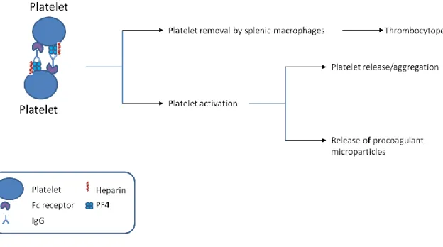
Pathology of HIT and Platelet Factor 4’s Role
When platelets become activated due to vascular injury, they release platelet factor 4. The type II form of HIT occurs when platelet factor 4 becomes bound to heparin. This
complex attracts immunoglobulins, such as IgG and initiates the immune response (Figure 2). Platelet factor 4 is a highly positively charged protein released by platelets during
8
Figure 2: A schematic of the consequences of unwanted interactions between platelet factor 4 and heparin.
Studies of PF4 and heparin interaction
Protein-polysaccharide interactions have been studied in various ways, including mass spectrometry, nuclear magnetic resonance and other biophysical techniques. In
particular, the interaction between PF4 and heparin have been studied from two perspectives: 1. The perturbation of PF4 structure due to heparin binding and 2. The formation of
9
Figure 3: Critical residues in PF4 for interaction with heparin. This figure shows the arginines and lysines (in green) responsible for interaction with heparin.
Also, continued studies by Arepally and collaborators suggest that the heparin:PF4 cluster size can affect its interaction with antibodies, wherein the cluster size is determined by the ratio of heparin to PF4. This ratio is important in order to neutralize the overall surface charge of the macromolecule (26).
Section 3: Structural Diversity of Heparin and Heparan sulfate (HS) Structures
Heparin versus Heparan sulfate
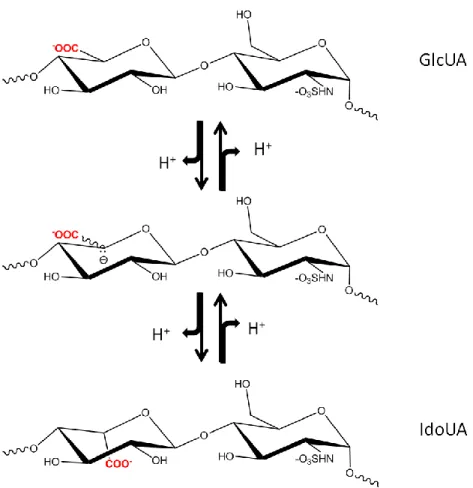
10
surface of cells, known as proteoglycans, or pericellularly linked to the basement membrane in the extracellular matrix (29). The anticoagulant heparin is an exclusive product of mast cells and, as previously mentioned, contains higher levels of sulfo groups and more iduronic acid than HS(16). Both heparin and HS consist of a repeating disaccharide unit of
glucosamine and either glucuronic or iduronic acid with various sulfations as shown in Figure 4.
Figure 4: Disaccharide repeating units of HS. Sulfation (R=-SO3) at Carbon 6 (known as
6-O-sulfo glucosamine) of glucosamine is common. Sulfation at Carbon 2 of iduronic acid (known as IdoUA2S) is common. Sulfation at Carbon 3 of glucosamine (known as 3-O-sulfo glucosamine) is rare. Both N-acetyl (R‟=acetyl, GlcNAc) and N-sulfo glucosamine (R‟=-SO3, GlcNS) are common. N-unsubstituted glucosamine (R‟=-H, GlcNH2) is a low
abundance component. IdoUA±2S is present in both 4C1 and 2S0 conformations. Both
11
Various sulfation patterns confer the functional selectivities of HS and heparin. These sulfations occur at the NH2, 3-OH, and 6-OH positions of glucosamine residue. The hexuronic acid residues, including both iduronic and glucuronic acid, can include sulfo groups at the 2-OH position. To further complicate the structure, the N-position of the glucosamine residue can also exist in the acetylated or unsubstituted form. Different substitutions at the saccharide unit result in up to 32 variable structures for the disaccharide unit, which serves as the building block for the polysaccharide (31). For a polysaccharide with a size of 20 disaccharide units, the theoretical number of permutations within the chain is to 1032. However, it is believed that the actual number of variable polysaccharides are less than this value because of the substrate specificity restrictions of HS biosynthetic enzymes. Nevertheless, the existing permutations lead to the structure of HS from natural sources which exhibit vast structural heterogeneity and diversity. Dissecting the relationship of structures and functions of has a large impact on understanding the physiological functions of HS, as well as, the development of HS-based therapeutic reagents.
Section 4: Biosynthesis of Heparan sulfate (HS)
Heparan sulfate Proteoglycans
HS can be found in almost all animal cells (27,32). The lowest organisms where HS has been found are the metazoans ctenophora and cnidaria or jellyfish (33-35). HS chains are attached to HS proteoglycans (HSPGs) either on the cell surface or within the extracellular matrix. The amount and presence of HSPGs varies from cell to cell (33-35). A HSPG consists of a core protein to which the HS chains are attached. The core proteins are
membrane-12
spanning domain. HSPGs found in the extracellular matrix come from the core proteins that are secreted during the biosynthesis (36).
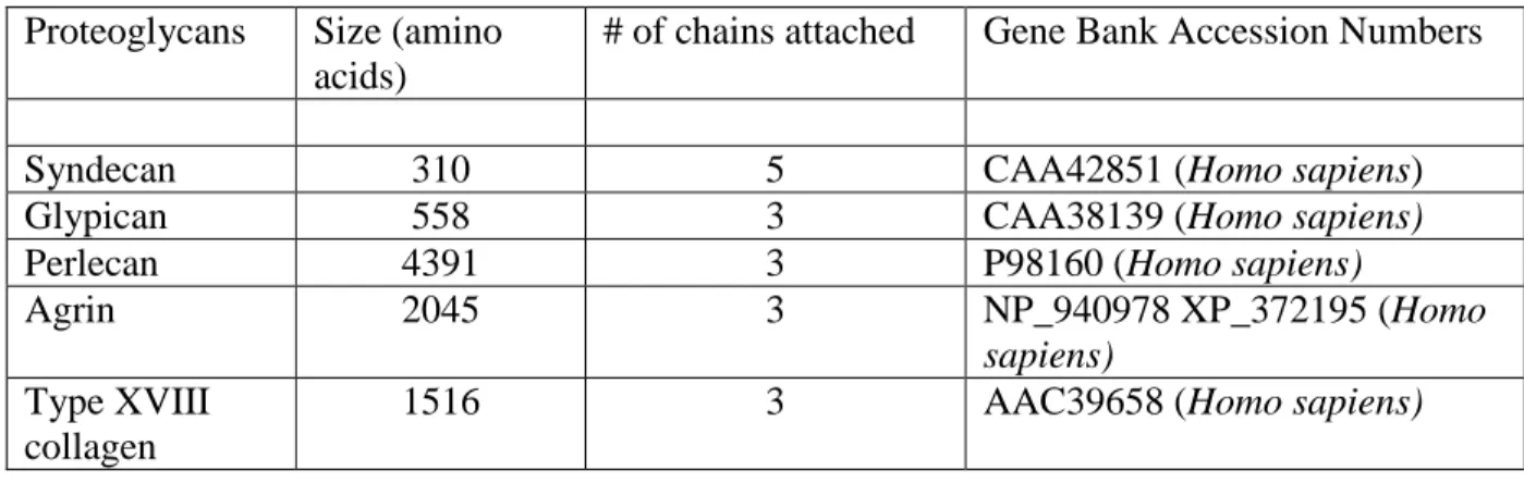
HSPGs can be classified in two ways: part-time HSPGs and full-time HSPGs. Part-time HSPGs are proteins that have the ability to carry HS side chains, but can exist without HS side chains, also known as the non-proteoglycan form. CD44, betaglycan and testican are a few examples of these proteoglycans. Full-time HSPGs are either directly membrane-bound or are linked to components within the extracellular matrix. Full-time HSPGs include syndecans, glypicans, perlecan, agrin and type XVIII collagen (12,29,37,38). A total of 13 genes encode the full-time HSPGs: four syndecans (syndecan-1 to -4), six glypicans
(glypican-1 to -6), and one isoform of each perlecan, agrin and type XVIII collagen although perlecan, agrin and type XVIII also have splice variants. The average size of these protein cores range from about 32 to almost 500 kDa (Table 1).
Table 1: Summary of cell surface and extracellular matrix HS proteoglycans. Proteoglycans Size (amino
acids)
# of chains attached Gene Bank Accession Numbers
Syndecan 310 5 CAA42851 (Homo sapiens)
Glypican 558 3 CAA38139 (Homo sapiens)
Perlecan 4391 3 P98160 (Homo sapiens)
Agrin 2045 3 NP_940978 XP_372195 (Homo
sapiens) Type XVIII
collagen
1516 3 AAC39658 (Homo sapiens)
glycosyl-13
phosphatidylinositol (GPI). Perlecan, agrin and type XVIII collagen, although not directly associated with the plasma membrane, can be associated via integrins or other cell surface receptors (29).
Figure 5: Schematic view of the HSPG. The core proteins are show in colored tubes, while the HS chains are shown in blue lines. The core protein of syndecan is a transmembrane protein, with 3-5 attachment sites in the extracellular domain. The core protein of glypican attaches to the membrane through a glycosyl-phosphatidylinositol (GPI) anchor, with 1-3 HS attachment sites. The core protein of collagen is not directly attached to the cell membrane; it has three potential HS attachment sites.
Building the backbone
14
our knowledge of this essential biological process. It should be noted that the availability of HS biosynthetic enzymes also provided the opportunity for conducting enzyme-based synthesis of HS polysaccharides (39,40).
Synthesis of the building block
HS biosynthesis takes place in the lumen of Golgi apparatus, where the sugar residues are assembled and modified by the actions of multiple Golgi membrane bound enzymes. However, the initial synthesis of building blocks occurs in the cytoplasm, not in the Golgi. These blocks are then transported into the Golgi lumen via an unknown mechanism for the subsequent modifications described below. The synthesis requires access to various
activated sugar donors including xylose (Xyl), galactose (Gal), UDP-N-acetyl glucosamine (UDP-GlcNAc) and UDP-glucuronic acid (UDP-GlcUA), as well as, the sulfo donor 3‟-phosphoadenosine 5‟-phosphosulfate (PAPS) for the sulfation reactions.
The UDP-Xyl, UDP-Gal and UDP-GlcUA are all derived from the activated glucose, namely UDP-glucose (UDP-Glc). UDP-Gal arises from epimerization at the C4 position of UDP-Glc; while UDP-GlcUA arises from UDP-Glc via UDP-Glc 6-dehydrogenase. The UDP-Xyl is synthesized from UDP-GlcUA in the Golgi apparatus by UDP-GlcUA (Figure 6). The biosynthesis of UDP-GlcNAc is more complex. It starts from fructose 6-phosphate, and requires three enzymes, a transaminase, an N-acetyltransferase and a mutase, to generate GlcNAc 1-phosphate, which is then converted to UDP-GlcNAc by UDP-GlcNAc
15
Figure 6: Schematic view of HS backbone synthesis. For simplicity, the synthesis of UDP-GlcNAc is not shown. The EXT family member of EXTL1 is also absent in the figure, which was shown to possess GlcNAcTII activity.
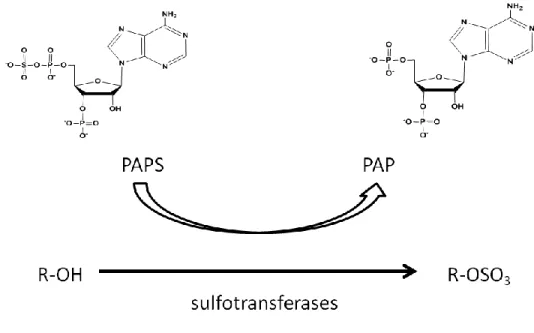
16
The general schematic of sulfation of the HS chain by PAPS is shown in Figure 7. The biosynthesis of PAPS occurs in both the nucleus and the cytoplasm. Then PAPS is transported to the Golgi by PAPS transporter for HS biosynthesis (42).
Figure 7: Schematic for the use of PAPS in sulfation via the sulfotransferases.
Chain initiation
The biosynthesis of HS is initiated by formation of a tetrasaccharide linkage to the core proteins: GlcUA β1-3Gal β1-3Gal β1-4Xyl β1-O-Ser, which also serves as the linkage
for the biosynthesis of CS. The formation of the tetrasaccharide linkage is catalyzed by the sequential actions of four glycosyltransferases, namely xylotransferase (XT),
galactosyltransferase I (GalTI), galactosyltransferase II (GalTII) and glucuronosyltransferase I (GlcUAT1) (Figure 6) (43).
17
have been identified in vertebrates; however, only XT1 has been determined unambiguously to have XT activity (44,45). The full length human XT1 has recently been expressed in mammalian cells in the active form and was shown to localize in the early cis-Golgi (46). Mechanisms of core protein attachment sites recognition by XT have attracted considerable attention, given the pivotal role of this enzyme in initiating synthesis of HSPG. A Ser-Gly repeating dipeptide seems to be the minimum structural requirement for the xylosylation to occur, and a Glu or Asp residue of the core protein is often present in the vicinity of the Ser-Gly sequence (47). Nevertheless, how XT recognizes the substrate core proteins and selects the Ser residue is still an open question.
Once the xylose is transferred to the core protein, the glycan moiety is elongated by two galactosyltransferases, GalTI and GalTII. GalTI, belonging to the β1,4 GalT
superfamily, transfers a galactose onto xylose to form Galβ1-4Xylβ1-O-Ser; while GalTII belongs to the β1-3 GalT superfamily, and subsequently adds another galactose to form Galβ1-3Galβ1-4Xylβ1-O-Ser. Both GalTs were cloned and expressed, and their substrate
specificities have been described. They have been shown to be located in the early-medial cis-Golgi (48-50).
Synthesis of the tetrasaccharide linkage is finalized by GlcUAT1, which is localized in the medial cis-Golgi (50). It transfers GlcUA from UDP-GlcUA to the existing glycan chain to form GlcUAβ1-3Galβ1-3Galβ1-4Xylβ1-O-Ser. The crystal structure of GlcUAT1
18
a regulatory mechanism for proteoglycan biosynthesis (52). In addition, sulfation of the galactose in the linkage region has only been seen in CSPG but not in HSPG, suggesting it may serve as a signal to direct subsequent GAG synthesis (53). Moreover, evidence suggests that the synthesis of the tetrasaccharide linkage can be carried out by GlcUAT-P, a
promiscuous glucuronosyltransferase that normally works on glycoproteins, thus providing a potential redundant mechanism for the linkage region synthesis (54).
Chain polymerization
The tetrasaccharide linkage to the core protein provides a common precursor for the biosynthesis of both HS and CS polysaccharide chains. The critical step that separates polymerization of HS from that of CS is the addition of a GlcNAcβ1- unit instead of the GalNAcβ1- unit to the nonreducing end of the tetrasaccharide. This step should not be
confused with the polymerization reaction, where the GlcNAc unit is also incorporated due to the action of a different enzyme (55). For clarity, the enzyme that adds GlcNAc to the
linkage region is HS GlcNAc transferase I (GlcNAcT1), while the enzyme that adds GlcNAc to the growing HS chain is HS GlcNAc transferase II (GlcNAcTII) (Figure 6). Two
mammalian proteins encoded by EXTL2 and EXTL3, belong to the EXT (exostosin) gene family, have been proved to carry the GlcNAcT-I activity (56,57). Their activities have been demonstrated in vitro using a synthetic tetrasaccharide linkage analogue, GlcUAβ1-3Galβ1-naphthalenemethanol, as the acceptor. However, the GlcNAcT-1 activity could not be detected using the natural tetrasaccharide linkage GlcUAβ1-3Galβ1-3Galβ1-4Xylβ1-O-Ser
19
(56). Nevertheless, biochemical studies directly using HSPG core proteins as the acceptors of EXTL2 and EXTL3 are necessary to test this hypothesis. In addition, genetic studies targeting EXTL2 and EXTL3 are essential to establish the specific roles of these two isozymes in initiation of HS biosynthesis. It is important to note that a recent study of the boxer
(EXTL3) mutant in zebra fish suggested that abolishing EXTL3 function causes severe reduction of HS biosynthesis, supporting the role of EXTL3 in initiating the synthesis of HS (58).
20
GlcNAcT-II activity of EXTL1 and EXTL3 correlates to that of EXT1 and EXT2 is an intriguing question, especially considering that they do not possess any GlcUAT-II activity. One hypothesis is that EXTL1 and EXTL3 may play regulatory roles in HS biosynthesis since they may compete with the EXT1/EXT2 complex (56). However, direct evidence supporting this hypothesis has been lacking, and the true functions of EXTL1 and EXTL3 in HS polymerization must wait for future genetic studies targeting these two enzymes.
The crystal structure of EXTL2 has been solved in complex with the sugar donor UDP-GlcNAc, and in complex with UDP and the acceptor analogue GlcUAβ1-3Galβ1-O-naphthalenemethanol, providing an excellent model for understanding the mechanism of action of EXT family members (63). However, since EXTL2 is the shortest isozyme of the family and it aligns to the C-terminal GlcNAcT domains of other EXT members, its structure only helps to understand the GlcNAcT activity of EXT(L)s. Detailed biochemical studies are needed to elucidate the functions of the N-terminal domain of EXT(L)s. For some enzymes, more specifically EXT1 and EXT2, the N-terminal domain harbors the GlcUAT activity (64), and for others, EXTL1 and EXTL3, its function is still unknown.
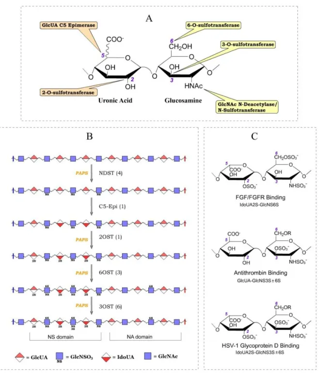
Modifications of the Backbone
21
22
23 N-Deacetylase/N-Sulfotransferase
NDST is the enzyme that initiates modification of N-acetyl heparosan. N-sulfation of the glucosamine unit has been demonstrated to be essential for most of the subsequent modifications on HS, thus representing a critical step in HS biosynthesis (Figure 9) (43). NDST does not modify HS uniformly: instead, the N-sulfated glucosamine units are
clustered, giving rise to the NS domain of HS, whereas the NA domain contains mostly the unreacted N-acetylated glucosamine (Figure 8) (28).
24
NDST can be divided into two domains: the N-terminal deacetylase domain (within ~600 amino acid residues of the N-terminus) and the C-terminal sulfotransferase domain (including about 280 amino acid residues). Both domains have been expressed and proved to contain the anticipated activities (65,66). In addition, NDST is a typical type II membrane bound protein with the transmembrane domain at the N-terminus.
Four isoforms of NDST have been identified in human and mouse (67-70). The isoforms can be divided into two subgroups: NDST-1 and NDST-2 with 70% identities in amino acid sequence, and NDST-3 and NDST-4 with 80.4% identity. Those four NDST isoforms have distinct tissue expression patterns. NDST-1 and NDST-2 are expressed quite uniformly in different tissues, while the expression of NDST-3 and NDST-4 occurs
predominantly in the brain (71). Each NDST isoforms appears to have distinct deacetylase and sulfotransferase activities. For example, NDST-3 has good deacetylase activity but very poor sulfotransferase activity, and NDST-4 shows the opposite with poor deacetylase activity and excellent sulfotransferase activity. The biological significance of the variations of the enzymatic activities of NDST isoforms is not fully understood, but it has been proposed that it may help generate unique sulfation patterns on HS (71).
A series of genetic studies of NDST-1 and NDST-2 have provided great insights onto the physiological functions of N-sulfation in model animals. The Ndst-1 null mice die shortly after birth due to severe lung failure (43,72). In addition to prominent defects in lung development, detailed phenotypic study of Ndst-1 deficient mice embryos at various
25
widespread effects of the Ndst-1 mutant; the only obvious defect is in connective tissue-type mast cell (74,75). These cells are generally active during inflammatory processes, and they release various inflammation mediators such as heparin, histamine and mast cell protease. However, in the mast cells of the NDST-2 deficient mice, production of heparin is aborted, the amount of histamine release is greatly reduced, and mast cell proteases are lacking. These data suggest that NDST-2 plays a predominant role in mast cells in terms of HS sulfation. Conversely, since NDST-2 deficient mice develop normally; otherwise, it seems that NDST-2 is dispensable in other tissues, where NDST-1 may play more dominant roles. Recently, NDST-1 has been knocked out in mice in a tissue-specific manner in endothelial cells and leukocytes, which helped demonstrate the essential roles of HS in inflammatory responses (14).
In comparison to the largely elucidated structure and function of the
N-sulfotransferase domain, the function of the N-terminal region of NDSTs is apparently understudied. The N-terminal region, about 600 amino acid residues in length, contains the N-deacetylase domain (65). However, we have extremely limited knowledge of the
deacetylase domain because it has no homology with the other carbohydrate deacetylases. Thus, the mechanism for deacetylation is still elusive. Understanding the deacetylation mechanism of NDSTs would be very informative, considering that varying NDST isoforms have distinct N-deacetylase activities. The action of different NDSTs could implant
26 Glucuronyl C5 Epimerase
The first modification that happens on the GlcUA residue is catalyzed by HS C5-epi, which converts the configuration of proton of the C5 position, thus generating IdoUA unit (Figure 10). An N-sulfated glucosamine (GlcNS) is required at the non-reducing end of the GlcUA for the action of C5-epi, suggesting the C5-epimerization rigorously follows N-sulfation (Figure 8) (28). Switching from GlcUA to IdoUA has been suggested to give HS a more flexible structure because it is known that IdoUA adopts several pyranose ring
conformations, including 1C4, 4C1 and 2S0, while GlcUA usually exists in 4C1 conformation (76). In addition, IdoUA is a much more favorable substrate for HS 2OST, and the resultant IdoUA2S is essential for many biological functions (77-80).
27
Different from most other HS modification enzymes, only a single isoform of C5-epi is present in almost all species examined (with the exception of fish). C5-epi is a fairly large protein with over 600 amino acids. The epimerase catalytic domain has been tentatively assigned to the 220-230 residues of the C-terminus based on alignment to homologous proteins and biochemical studies (81).
The C5-epi has been shown to form a complex with HS 2OST in vivo, which is indispensable for correct Golgi localization of C5-epi (50). Formation of the complex may have a substantial effect on the enzymatic activity of epi. The common substrate for C5-epi and HS 2OST, GlcUA, may be channeled through the enzyme complex to achieve a higher efficiency of modification, as it is known that IdoUA2S is no longer a substrate for C5-epi (50,82). The final ratio of the GlcUA/IdoUA presented in the HS may be actively regulated by this reaction.
The knockout mouse model of C5-epi has provided significant information about the physiological functions of the enzyme (83). C5-epi deficient mice died immediately after birth due to respiratory failure, apparently as a result of poorly developed lungs. Other evident phenotypes include undeveloped kidneys, eye defects, various skeletal abnormalities such as shorter body length and excessive mineralization. Close examination of HS from C5-epi-/- mice shows that production of IdoUA is totally abrogated, while the proportion of GlcUA2S increased 3-fold and the 6-O-sulfation increased 5-fold. The phenotypic
28
A recent study pointed out that human C5-epi is transcriptionally regulated by the β-catenin-TCF4 transactivation complex, the terminal effector of the Wnt/β-catenin signaling pathway (84). This study showed that there are two β-catenin-TCF4 cis-acting elements located in the enhancer region of the promoter; it also showed that ectopic expression of
β-catenin and TCF4 together significantly enhances the cellular level of C5-epi transcript along with its enzymatic activity. Based on the evidence that HS is an important regulator of the interaction between Wnt and its cell surface receptor Fz (Frizzled), HS biosynthesis and Wnt signaling may be independent. It raised the possibility that a feedback loop is present
between an essential signaling pathway and specific cell surface HS structures, which in turn may help achieve the final control of the animal development. Further study is needed to test the generality of this phenomenon in different cell types and in different species.
Uronosyl 2-O-Sulfotransferase
2-O-sulfation is the only sulfation that occurs on the uronic acid units (Figure 8), and it is observed in HS originating from all metazoans. For instance, more than half of the IdoUA unit found it mouse embryo are 2-O-sulfated, while only a minute portion of the GlcUA are 2-O-sulfated (83). In the adult tissues of Drosophila and C. elegans, about 20% and 30% uronic acid units are 2-O-sulfated (85). 2OST is capable of reacting with both GlcUA and IdoUA units, but prefers the latter under most conditions (Figure 12) (86).
29
Very much like C5-epi, 2OST has only one isoform in most species with the exception of fish. 2OST contains a type II transmembrane domain and is localized in the Golgi apparatus. As described above, 2OST forms a complex with C5-epi in vivo, which is essential for the Golgi localization of C5-epi (50). However, whether or not formation of this complex is critical for the Golgi localization of 2OST remains unclear. The complex may also influence the substrate specificities of 2OST in HS biosynthesis in vivo. One can imagine that when C5-epi and 2OST complex is formed, the majority of the resultant modification products would contain IdoUA2S. When C5-epi is dissociated from 2OST, 2OST may be allowed to generate GlcUA2S, and C5-epi is able to generate IdoUA without sulfation. Thus, a possible role of C5-epi is to regulate the type of uronic acid that is subjected to 2OST modification. Definitive demonstration of this possibility will require structural information from both 2OST and C5-epi, and the biological significance of the interaction should be tested in animal model systems.
30
positions of HS may attenuate the adverse effect imposed by the ablation of 2-O-sulfation, thus disguising some physiological functions associated with 2-O-sulfation.
The 2OST null mutant has been described in C. elegans as well (88). Surprisingly, 2OST deficient mutants are viable and fertile without any locomotory defect or embryonic paralysis as observed in perlecan deficient worms (89). The phenotypes of 2OST null worms are only detectable in nervous system development by displaying axonal guidance and
migration defects in a neuron-type specific manner. These findings suggest that the 2-O-sulfation of HS plays very specific roles in metazoan development. These roles may vary in the developmental schemes of different species.
Glucosaminyl 6-O-Sulfotransferase
6-O-sulfation of glucosamine residues is a prevalent modification of HS. The percentage of 6-O-sulfated disaccharide units represents 20 to 30% of total disaccharides in the HS from different species (83,85). The 6-O-sulfation occurs most at the NS domain, generating a GlcNS6S moiety, but it can also occur at the NA domain, generating a
GlcNAc6S moiety (90) (Figure 12). It should be noted that 6-O-sulfation is the only type of sulfation that occurs at the NA domain as both 2OST and 3OSTs depend on the N-sulfation of glucosamine (Figure 8) (43,91).
31
Three isoforms of 6OST have been identified in human and they exhibit
approximately 60% identity (90). These isoforms have highly conserved sulfotransferase domains, while varying in both N- and C-termini. Several in vitro substrate specificity assays suggest that 6OST isoforms have distinct substrate preferences (90,92). 6OST1 appears to favor the IdoUA-GlcNS unit significantly more than the GlcUA-GlcNS unit, while 6OST2 prefers GlcUA-GlcNS over IdoUA-GlcNS, and 6OST3 has almost equal activities toward both disaccharide structures. Three isoforms of 6OST are also present in other mammals and birds as well as amphibians, while invertebrates normally have only a single isoform of 6OST.
Unlike the previously mentioned HS modification enzymes, a mouse knockout model for 6OST has not been reported. Nonetheless, evidence from 6OST null Drosophila and C. elegans suggests that 6OST plays significant roles in animal developmental processes
(77,88,93,94). Knocking down 6OST in Drosophila disrupts the development of the tracheal system, their analog of the vertebrate vascular system. Strong evidence indicates that this phenotype is directly related to the essential function of 6-O-sulfated HS in fibroblast growth factor (FGF) signaling (77). Interestingly, C. elegans 6OST null mutants are viable and fertile, suggesting that 6-O-sulfation of HS is dispensable for worm FGF signaling.
However, very specific axonal guidance defects were seen in a neuron-type specific manner. The effects of worm 6OST are believed to be related to several essential pathways in nervous system development, such as the Slit/Robo system and the Kal-1 system (88,93).
32
6-O-sulfation level by merely 20%, yet several obvious phenotypes were observed. The most notable defects of 6OST knockdown zebrafish embryo were seen in somite
development, as the embryo showed undifferentiated muscles and muscle degeneration. Additional defects caused by 6OST knockdown were also identified in the brain and the fins. It is now known that zebrafish have at least one other 6OST isoform, that is very homologous to human 6OST1 (81% identity), in addition to the 6OST that is knocked down in this study, which suggests that observed phenotypes may only represent a small fraction of the real impact of 6-O-sulfation of HS on zebrafish embryonic development.
Glucosaminyl 3-O-Sulfotransferase
33
Figure 13: Substrate specificities of 3-OSTs. R groups are either N-SO3 or NH2 groups of
the glucosamine.
34
two 3-OST isoforms were found in C. elegans. These observations suggest that it is likely that 3-OST diverged into two branches early in the evolution to perform separate functions, and both branches expanded in vertebrates to carry out more diverse and specific roles.
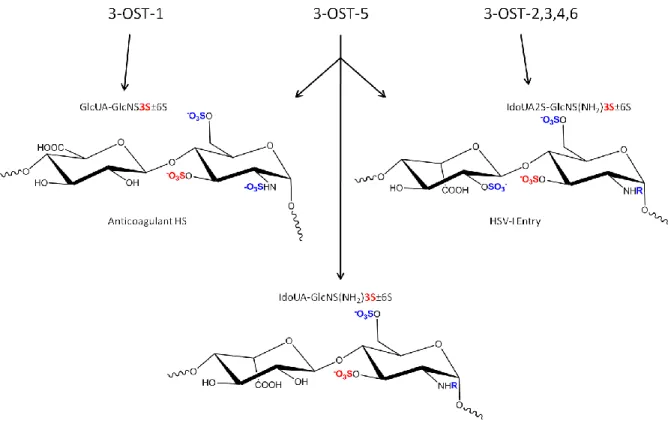
In comparison to the promiscuous substrate specificities of 6OSTs, members of 3-OST family have distinctive substrate specificities. 33-OST1 synthesizes a characteristic disaccharide unit with the structure of GlcUA-GlcNS3S±6S (Figure 8) (101), which is a critical modification step for synthesizing anticoagulant HS. 3OST2, 3OST3, 3OST4 and 3OST6 all generate a characteristic disaccharide unit with a structure of IdoUA2S-GlcNS (or NH2)3S6S (Figure 13) (102-104), which is part of the structure of HS serving as an entry
receptor for herpes simplex virus 1. 3OST5 is unique in that it enables to synthesize these two disaccharide units (105). The distinct disaccharide units generated by 3-OST confer the functions of HS.
Among seven isoforms of 3-OST, only 3-OST-1 has been knocked out in mice (106). The study was originally aimed to determine the effect of the level of anticoagulant HS on the blood coagulation process, provided that 3-OST-1 is a critical enzyme for synthesizing anticoagulant HS. However, the 3-OST-1-/- mice exhibit a surprisingly normal
35
some profound roles in other biological processes although the details of this involvement remain unclear.
Studies in Drosophila provided the first direct evidence of the critical roles played by 3OST in animal development (94). When Drosophila Hs3st-B was silenced by RNAi
treatment, several morphological defects were observed. These included server rough eye phenotype, extra sensory bristles at multiple tissues and profound defects in wing
morphology such as reduced wing size, abnormal wing veins and notching at the wing margin. The phenotypic effects of Hs3st-B silencing have been related to Notch signaling directly, as Hs3st-B was show to influence the protein level of Notch (cell surface receptor in Notch signaling). The exact role of Hs3st-B modified HS in the Notch signaling pathway remains to be determined.
Section 5: Chemical and Enzymatic Synthesis of Heparan sulfate
The study of heparin has been limited due to the difficulty in its synthesis. There have been two approaches to synthesizing these polysaccharides for study: chemical and enzymatic. Chemical synthesis has been proven to be successful for oligosaccharides and strides have been made to make this synthesis easier and more diverse. The enzymatic synthetic approach is now being exploited for the synthesis of polysaccharides.
Chemical Synthesis
36
occurs in different ways. Building an oligosaccharide backbone is difficult because you must maintain the stereochemistry of the glycosidic bonds. Also, chemical sulfation is difficult to control. Unwanted O-sulfations can arise (108-110).
Chemoenzymatic Approach
37
Figure 14: Schematic of the PAPS regeneration system. PAPS is regenerated using PNPS as a donor. The sulfate group is removed from PNPS and transferred to PAP via
arylsulfotransferase IV. The generated PAPS can then be used by the HS sulfotransferases for transfer of the sulfate group to the HS chain.
Section 6: Structural Analysis of Heparan sulfate
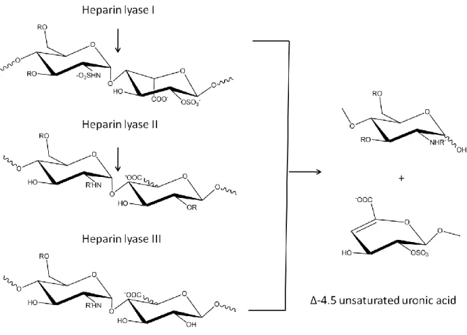
38 Heparin Lyase Degradation
The heparan sulfate lyases are which are able to cleave heparan sulfate
polysaccharides down to disaccharides using β-elimination. The use of lyases has become a popular tool to identify the composition of heparan sulfate polysaccharides. There are three heparin lyases, named heparin lyase I, heparin lyase II and heparin lyase III and each has it‟s
own substrate specificity (Figure 15). Heparin lyase I cleaves at GlcNS(±6S)( ±3S)α(1 →4)IdoA(±2S). Heparin lyase II cleaves at either iduronic or glucuronic residues. Heparin
39
Figure 15: Schematic of the substrate specificity of the heparin lyases.
Nitrous Acid Degradation
40
Figure 16: Schematic of low and high pH nitrous degradation. R groups represent either –OSO3- or –H.
Section 7: Statement of Problem
The study of heparin and heparan sulfate function has grown vastly over the past 40 years. These structures have been implicated in various biological activities including, proliferaction, viral and bacterial infection and anticoagulation. Study of the structure and activity relationship of HS is useful to tailoring these molecules for specific functions. For instance, currently, HS-like structures are being studied for their ability to inhibit cancer (17,116). The most significant structurally designed heparin is the anticoagulant
41
medically. But, the cost of production of Arixtra is more than the cost of the production of heparin, thus, the use of heparin has remained popular in the hospital. Medical
CHAPTER II:
MATERIALS AND METHODS
Agarose Gel Electrophoresis
A 1% agarose gel (in 0.5x TAE buffer) containing ethidium bromide (1 µL of 5 mg/mL solution) was used to analyze PCR products and DNA plasmid constructs. The gel was run at 120 mA and analyzed with UV light.
SDS-PAGE Electrophoresis
For analysis of the HS biosynthetic enzymes, a 10% SDS-PAGE gel (Bio-Rad) was used. For analysis of PF4 enzyme, a 12% Bis-Tris gel (Invitrogen) was used. Samples analyzed via the SDS page gel were mixed with β-mercaptoethanol and loading dye and
boiled at 100°C for 5 minutes before adding to lanes. Samples analyzed via the Bis-Tris gel were mixed with a sample reducing agent and loading buffer before boiling them at 70°C for 10 minutes. The SDS gel was run using SDS buffer and the Bis-Tris gel was run using MES buffer (supplemented with an antioxidant agent immediately before running of the gel). Both gels were run at 120mV for about 45-60 minutes. Both gels were analyzed via staining.
Transformation of Competent Cells
43
1 mL of LB broth. Incubate these cells at 37°C for 2-4 hours while shaking at 220 rpm. Plate 100-100 µL of cells on LB plates containing the appropriate antibiotics and incubate the plate at 37°C overnight.
Expression of HS Sulfotransferases in Origami B/OrigamiBchap cell lines
Origami B (DE3) or Origami Bchap (Origami B cells that also express chaperonin
proteins GroEL/GroES) were transformed as previously described.
Origami B (DE3) cells
A single colony was used to inoculate a 3mL starter culture comprised of LB broth containing 12.5 µg/mL tetracycline, 50 µg/mL carbenicillin (if the plasmid had this resistance), and 15 µg/mL kanamycin. This starter culture was incubated at 37°C with shaking at 220 rpm overnight. The following day, 750 µL of the starter culture was added to a 100 mL culture containing the appropriate antibiotics and incubated for another 3-4 hours. The cells were then cooled to 22°C after the OD600 reached between 0.6-0.8 and induced with isopropyl-β-D-galactopyranoside (IPTG) of final concentration 200 µM. These cells were then shaken overnight at 22°C before harvesting.
Origami Bchap cells
These cells were expressed using the aforementioned protocol for Origami B cells with a few minor additions. Cultures were incubated with 3-4 antibiotics, 12.5 µg/mL tetracycline, 50 µg/mL carbenicillin (if the plasmid had this resistance), 15 µg/mL
44
Expression of HS Sulfotransferases in BL21/BL21star cell lines
BL21 and BL21star (BL21 cells with a mutation in RNase) were transformed as previously described. Both cell lines were expressed as described above, however, antibiotic resistance solely depended upon the resistance of the plasmid. These cells were induced with only IPTG.
Nickel Sepharose Fast Flow Affinity Chromatography for His6-Tagged Proteins
Cells were harvested at 7000 rpm for 30 minutes. The cell pellet was then
resuspended in 25 mLs (per liter of culture) of Nickel Buffer A (25 mM Tris, 500 mM NaCl, 30 mM imidazole, pH 7.5). Once resuspended, the cells were sonicated in a 50 mL tube for 3 x 45 seconds (duty cycle 50%, output 7) for lysing. These cells were then pelleted at 10,000 rpm for 30 minutes. The cell lysate was then filtered via a 1.5 micron filter (Whatman) prior to loading to a pre-equilibrate 7 mL Nickel Sepharose 6 Fast Flow column (GE Healthcare) at a rate of 3 mL/min. The column was then washed with 7 column volumes of Ni buffer A to remove all unbound sample. Then the protein of interest was collected in 3 mL fractions following elution with Ni buffer B (25 mM Tris, 500 mM NaCl, 300 mM imidazole, pH 7.5) during a 4 column volume gradient length followed by a 60mL elution with 100%B. The protein was typically eluted in 35-40 mL total volume.
Amylose Affinity Chromatography for MBP Fusion Proteins
pre-45
equilibrated 10 mL Amylose column (New England Biolabs) at a rate of 2vmL/min. The column was then washed with 10 column volumes of amylose buffer A to remove all unbound sample. Then the protein of interest was collected in 3vmL fractions following elution with amylose buffer B (25 mM Tris, 500 mM NaCl, 30 mM maltose, pH 7.5) during a 3 column volume gradient length followed by a 40 mL elution with 100%B. The protein was typically eluted in 20-25 mL total volume.
Glutathione Sepharose 4 Fast Flow Affinity Chromatography for GST fusion Proteins These cells were harvested as previously described. The cell pellet was then resuspended in 25 mLs (per liter of culture) of GST Buffer A (20 mM NaH2PO4, 500 mM
NaCl, pH 7.2). Once resuspended, the cells were sonicated in a 50 mL tube for 3 x 45 seconds (duty cycle 50%, output 7) for lysing. These cells were then pelleted at 10,000 rpm for 30 minutes. The cell lysate was then filtered via a 1.5 micron filter (Whatman) prior to loading to a pre-equilibrate 10 mL Glutathione Sepharose 4 Fast Flow column (GE
Healthcare) at a rate of 2 mL/min. The column was then washed with 10 column volumes of amylose buffer A to remove all unbound sample. Then the protein of interest was collected in 3 mL fractions following elution with glutathione buffer B (20 mM NaH2PO4, 500 mM
46 Cloning and Purification of mPF4
Design of Primers and Polymerase Chain Reaction (PCR)
The forward and reverse primers were designed to contain the cut sites in the plasmid of interest (Table 2).
Table 2: Primers usd for murine Platelet Factor 4 Cloning
Primer name Primer sequence
mouse PF4 forward primer with EcoRI for pMalc2x/Tev
ATATATAAGAATTCGCTGGTCCCGAAGAAAGC GAT
mouse PF4 reverse primer with HindIII for pMalc2x/Tev and PET32/TEV
ATTAAATAAAGCTTTACCTAACTCTCCAGGATT TTC
mouse PF4 forward primer with NcoI for PET32/TEV
ATTAATTACCATGGCGCTGGTCCCGAAGAAAG CGATG
The PCR reaction contained the following: 5 µL 10X Pfu buffer, 1 µL 10 mM dNTP mix (New England Biolabs), 50-100 ng plasmid DNA, 1.5 µL of forward and 1.5 µL of reverse primers (25 µM each), 1µL Pfu turbo DNA polymerase (Stratagene), and water to a final volume of 50 µL. The PCR protocol followed is shown in Table 3.
Table 3: PCR conditions for cloning of mPF4
Step Number of Cycles Temperature (°C) Time
Initialization 1 94 3 min
Denaturing 16 94 1 min
Annealing 52 1 min
Elongation 68 1 min/kilobase
Final Elongation 1 68 1 hour
47
the digested product was used for transformation into 50 µL of XL10 gold competent cells. These plasmids were then transformed into the following expression systems: Origami B (DE3). BL21 and BL21star cell lines.
Purification of mPF4 was completed in four steps, purification with an affinity column, cleavage of the tag, purification with the heparin column and finally purification with the Sephadex 75. Each of these steps was performed as described below.
TEV Cleavage
Affinity affinity column purification (as previously described), cleavage of mPF4 with TEV was carried out at 4°C. The milligram amount of TEV added to the reaction was 1/25th of the milligram amount of mPF4. The reaction was rotated for 16-18 hours at 4°C.
Heparin column
The heparin column was used to purify mPF4 from its cleaved tag. Once the TEV cleavage reaction was complete, the reaction was spun at 5000rpm and filtered with a 1.5 micron (Whatman) filter. The filtered sample was loaded at 3 mL/min to a pre-equilibrated 10 mL Heparin column (GE Healthcare). The column was washed with 10 mL of heparin buffer A (500 mM NaCl, 20 mM Tris, pH 8). PF4 was eluted into 2mL fractions during a gradient elution over 4 column volumes with heparin buffer B (2000 mM NaCl, 20 mM Tris, pH 8) and a delay of 40 mLs. The protein typically eluted in 20-30 mLs.
Hi-Load Superdex 75/200 Gel Filtration Chromatography
48
concentrated protein was then injected into the 10 mL sample loop and loaded at 1 mL/min onto the pre-equilibrated (with 20 mM MOPS, 400 mM NaCl, pH 7) 100 mL Superdex 75 Gel Filtration column. The column was then washed with the buffer at 1mL/min while collecting 2 mL fractions. Molecular mass standards (cytochrome c – 12.4 kDa, carbonic anhydrase – 29 kDa, BSA – 66 kDa, and β-amylase – 200 kDa) purchased from Sigma Aldrich were used to determine the protein peak of interest.
Calculating the concentration of mPF4
The concentration of mPF4 was calculated using the amino acid analysis results received from the University of California at Davis molecular structure facility. The concentration of the salt concentration of mPF4 was decreased to 100 mM NaCl and about 100 ug of sample was sent for analysis. Initial mPF4 concentration was determined by multiplying the concentration given by the UV detection at 280 by 5. Amino acid results were used to determine the molar amount of each amino acid. Tyrosine was used as the internal standard since there is only one per monomer. The molar amount of tyrosine was used to calculate the molar amount of protein.
Expression of HS Biosynthetic Enzymes
49
supplemented with 2 mg/mL glucose, 12.5 mg/mL tetracycline, 15 mg/mL kanamycin, 35 mg/mL chloramphenicol, and 50 mg/mL carbenicillin at 37°C. When the A600 reached 0.4– 0.7, isopropyl-thiogalactopyranoside (0.15 mM) and L-arabinose (1 mg/mL) were added to induce the expression of 6-OST-3 and chaperonin proteins, respectively. The cells were allowed to shake overnight at 22°C. Purification was carried out with an amylose-agarose (New England BioLab) column by following the protocols provided by manufacturer.
Purification of Heparosan
K5 capsular polysaccharide, or heparosan, is purified from E. coli containing the K5 strain. These cells are grown at 37°C for 16-18 hours. Heparosan is purified from the cells after centrifugation at 7000 rpm. The supernatant is collected, filtered and titrated to pH 5. This supernatant is then mixed at a 1:1 ratio with loading buffer (20 mM NaAcO, 50 mM NaCl, pH 5). Heparosan is further purified via ion-exchange chromatography (DEAE purification). After elution with high salt, the purified heparosan is ethanol precipitated with 3:1 ethanol to heparosan. Final purification is performed using ammonium sulfate
precipitation.
Deacetylation of Heparosan with NaOH
Forty milligrams of heparosan is lyophilized. This sample was resuspended in 4 mL of 2000 mM NaOH and rotated at 68°C for another 3 hours. This reaction was neutralized with 2000 mM HCl and dialyzed and lyophilized.
Deacetylation of Heparosan with NDase II
50
reaction was incubated at 37°C for 1 hour. The efficiency of this reaction was checked by the efficiency of a subsequent N-sulfation reaction.
Chemical N-sulfation of Deacetylated Heparosan
Dissolve 2 mg of deacetylated K5 in 1 mL of water. Add 0.1 M NaOH dropwise until the pH reaches between 9.5-10. Calculate the number of free amine groups and multiply by 15 to get the amount of sulfur trioxide pyridine that needs to be added. Add about 1/5th of the amount of sulfur trioxide pyridine to the mixture. Re-titrate the sample with 0.1 M NaOH to pH 9.5 after 30 minutes. Then add another 1/5th of the sulfur trioxide pyridine. Repeat the procedure until all of the sulfur trioxide pyridine is added. When the pH is stable at 9.5 for about an hour, dialyze the sample for 16 hours in 0.1 N ammonium bicarbonate buffer.
N-acetylation Reaction of Deacetylated or N-sulfated Heparosan
About 72 pmols of lyophilized polysaccharide was dissolved in 20 µl of a solvent containing N,N-dimethylformamide and triethylamine (1:1, v/v) and 5 µl of acetic anhydride, and incubated on ice for 1 hr. Tris (20 ul of 50 mM) was then added and the reaction mixture was incubated on ice for an additional hour. The sample was then diluted with 10 volumes of water and dialyzed against 50 mM ammonium bicarbonate.
Preparation of N-sulfo Heparosan
51
purified AST-IV was incubated with 40 mM PAP and 1 mM p-nitrophenol sulfate (PNPS) in 40 mL 50 mM MES (pH 7.0), 1% Triton X-100, 1 mM MgCl2, and 1 mM MnCl2 at 25°C for
15 min. The reaction mixture was mixed with N-sulfotransferase (10 mg), 5 mg deacetylated heparosan was added, and the mixture was rotated at 25°C for 24 hr. The N-sulfo heparosan was recovered by a DEAE column. The resultant product was subjected to a mixture of heparin lyases, followed by a disaccharide analysis to assess the level of N-sulfation. After two rounds of the modifications, the N-sulfation level reached 80% as determined by HPLC analysis.
Preparation of N-sulfo Oligosaccharides
N-35S-labeled sulfo heparosan (1 mg, 1.5 x 10⁶ cpm) was mixed with 20 ng purified heparin lyase III in 1 mL 50 mM sodium phosphate (pH 7.0). The digestion lasted for 18 hr and was terminated by heating at 100°C for 2 min. The digested polysaccharide was resolved on a Bio- Gel P-10, which was eluted with a buffer containing 25 mM Tris and 1000 mM NaCl (pH 7.4) at a flow rate of 2 mL/hr.
Preparation of Polysaccharides with Varying 6-O-sulfation