THE ACUTE EFFECTS OF A HIGH GLYCEMIC INDEX MEAL COMBINED WITH PROLONGED SITTING ON VASCULAR FUNCTION: A RANDOMIZED CROSSOVER TRIAL
Elizabeth Kelsch
A thesis submitted to the faculty at the University of North Carolina at Chapel Hill in partial fulfillment of the requirements for the degree of Masters of Arts in the Department of Exercise
and Sports Science (Exercise Physiology).
Chapel Hill 2019
Approved by:
Lee Stoner
Erik Hanson
ABSTRACT
Elizabeth Kelsch: The Acute Effects of a High Glycemic Index Meal Combined with Prolonged Sitting on Vascular Function: A Randomized Crossover Trial
(Under the direction of Lee Stoner)
Chronic prolonged sitting and consumption of high glycemic index (GI) meals are known to increase the risk of cardiovascular diseases. However, it is currently unknown whether prolonged sitting and consumption of a high GI meal have a combined negative acute effect on local and central arterial stiffness (AS). Therefore, 18 young, healthy participants (21.7±2.5 y, 70% F, 25.5±6.1 kg/m2) were randomized to: 3h sitting with a high GI beverage (HGI) or a low GI beverage (LGI). Prolonged sitting (3 hours) and consumption of a HGI beverage increased central (brachial-femoral) AS, in the LGI (0.27 m/s) and the HGI (0.45 m/s) condition (p=0.027). Peripheral (femoral-ankle) PWV increased (0.211 m/s) regardless of the level of GI consumed. Carotid-femoral PWV was not influenced by prolonged sitting or HGI meal consumption. Sitting increases central and peripheral AS in young, healthy individuals. Consuming a HGI meal has a moderate detrimental effect on central (brachial-femoral) PWV.
KEY WORDS: Arterial stiffness; pulse wave velocity; high glycemic; near infra-red
ACKNOWLEDGEMENTS
TABLE OF CONTENTS
LIST OF TABLES ... ix
LIST OF FIGURES ... x
LIST OF ABBREVIATIONS ... xi
DEFINITION OF TERMS ... xiii
CHAPTER I: INTRODUCTION ... 1
ASSUMPTION AND DELIMITATIONS ... 3
Delimitations: ... 3
Limitations ... 3
CHAPTER II - REVIEW OF LITERATURE ... 4
INTRODUCTION TO TOPIC ... 4
CONSIDERATION 1: VASCULAR RESPONSES TO PROLONGED SITTING ... 4
Chronic effects of prolonged sitting ... 4
Acute effects of prolonged sitting ... 5
Blood Flow ... 5
Vascular Function ... 6
CONSIDERATION 2: ACUTE RESPONSES TO A HIGH GLYCEMIC INDEX MEAL ... 7
Local cardiovascular effects from glucose ... 8
Local cardiovascular effects from insulin ... 8
Central CV ... 8
IMPLICATIONS/WHY IS THIS STUDY NEEDED? ...10
METHODOLOGICAL CONSIDERATIONS ...10
STUDY DESIGN CONSIDERATIONS ...10
MEASUREMENT CONSIDERATIONS ...11
POPULATION/SAMPLING ...14
STATISTICAL CONSIDERATIONS ...15
OTHER CONSIDERATIONS ...15
Sex As A Biological Factor ...15
Ethnicity/Race ...15
Generalizability ...16
Potential Challenges & Alternative Strategies ...16
SUMMARY ...16
CHAPTER III: METHODOLOGY ...18
PARTICIPANTS ...18
EXPERIMENTAL DESIGN ...18
Familiarization Visit ...19
Visit 2 and 3 ...20
Primary And Secondary Outcome: Central And Peripheral PWV ...21
Control Measures ...22
RANDOMIZATION ...23
SAMPLE SIZE ...24
QUALITY CONTROL ...24
DATA MANAGEMENT AND STATISTICAL ANALYSIS ...24
CHAPTER IV: RESULTS ...26
PARTICIPANTS ...26
BASELINE DATA ...27
STIMULUS: CONTINUOUS BLOOD GLUCOSE ...28
PRIMARY OUTCOME: CENTRAL PULSE WAVE VELOCITY ...29
SECONDARY OUTCOME: PERIPHERAL PWV ...30
ANCILLARY ANALYSIS ...31
SUPPLEMENTAL ANALYSIS ...31
CHAPTER V: DISCUSSION ...33
LIMITATIONS AND STRENGTHS ...33
COMPARISON TO LITERATURE ...34
IMPLICATIONS ...37
CONCLUSIONS ...38
APPENDIX A – ETHICAL APPROVAL ...39
APPENDIX C – PARTICIPANT LOGS ...45
LIST OF TABLES
Table 1.Low and High GI meals used in literature………14
Table 2. Participant Characteristics and Hemodynamic Measures at Baseline………30
Table 3. Supine Arterial Stiffness Measures………32
Table 4. Supine Hemodynamic and NIRS Measures……….………33
LIST OF FIGURES
Figure 1.The proposed mechanism for the individual and combined
effects of prolonged sitting and high GI meal on the cardiovascular system………….…10 Figure 2. Protocol timeline for experimental measures………..20 Figure 3. CONSORT diagram……….28 Figure 4. Average blood glucose response to high and low GI
beverage over 3 hours of sitting………30 Figure 5. Mean brachial-femoral PWV supine measurements
before and after three hours of uninterrupted sitting………..30 Figure 6. Mean femoral-ankle PWV supine measurements before
and after three hours of uninterrupted sitting………..31 Figure 7. Correlation between Glucose Variability and bfPWB (A)
LIST OF ABBREVIATIONS
AIx Augmentation Index
ANS Autonomic Nervous System
APL Applied Physiology Laboratory
AS Arterial Stiffness
AUC Area under the curve
BF Blood Flow
BP Blood Pressure
CGM Continuous Glucose Monitor
CO Cardiac Output
cSBP Central Systolic Blood Pressure
CV Cardiovascular
CVD Cardiovascular disease
ET-1 Endothelin-1
GI Glycemic index
Hb Hemoglobin
HbO2 Oxygenated Hemoglobin
H-GI High glycemic index
HHb Deoxygenated Hemoglobin
HR Heart Rate
HRV Heart Rate Variability
MAP Mean Arterial Pressure NIRS Near Infrared Spectroscopy
NO Nitric Oxide
PRU Peripheral Resistance Units
PWA Pulse Wave Analysis
PWV Pulse Wave Velocity
SB Sedentary Behavior
SRS Spatially Resolved Spectroscopy
SV Stroke Volume
T2D Type 2 diabetes
tHb Total Hemoglobin
DEFINITION OF TERMS
Arterial Stiffness Describes distensibility, compliance, and elastic modulus of the arterial vascular system
Flow Mediated Dilation Any vasodilatation of an artery following an increase in luminal blood flow and internal-wall shear stress
Glycemic Index A relative ranking of carbohydrate in foods according to how they affect blood glucose levels
High Glycemic Index Simple carbohydrate; in the present study, GI of 100
Low Glycemic Index Index Complex carbohydrate; in the present study, GI of 19 Postprandial period The period of time following consumption of a meal
Prolonged Sitting Acute bout of sitting lasting for >30 minutes
CHAPTER I: INTRODUCTION
Chronic sedentary behavior, defined as very low intensity behaviors (≤1.5 metabolic equivalents) in a seated, reclined or supine posture[122] has been directly tied to the development of cardiovascular disease (CVD). This is a concern because the majority of American adults are sitting for 70% of their day[1], and CVD cost the US $555 billion in medical bills, prescriptions, home-care, and other services in 2016[2]. While the chronic associations between repeated exposure to prolonged sitting and CVD are well known, the mechanism by which an acute bout of sitting leads to cardiovascular decline is less clear. Additionally, sitting is typically accompanied by other poor lifestyle choices, such as the consumption of high glycemic index (GI) meals[124]. Regular dietary exposure to high GI meals has also been linked to the development of CVD[66,107]. Prolonged sitting following consumption of a high GI food is common in today’s society[1,66]; however, the combined acute effect of both prolonged sitting and consumption of a high GI meal on the cardiovascular system is unclear.
It has been shown that sitting for three hours results in decreased blood flow to the legs and subsequent leg endothelial dysfunction[3,4]. Current unpublished findings from our
laboratory demonstrate that this local impairment translates to modest (0.3 m/s) central
The long-term goal from this study is to elucidate the acute effects of prolonged sitting and consumption of a high GI meal, which will allow for a better understanding of the
mechanism linking these chronic exposures to CVD. This will either lead to the need for
research of strategies to reduce the dose of sedentary behavior and high GI consumption, or a need for greater understanding surrounding the specific positive contributions healthy lifestyle choices have on “protecting” against these stimuli. While both sedentary behavior and repeated consumption of HGI foods are associated with metabolic, vascular, and cognition dysfunction, this study focused on central and peripheral vascular function, via pulse wave velocity (PWV). To achieve this, this study compared a HGI meal versus a low glycemic index (LGI) meal during a bout of 3 hours of sitting and explored the effects on central and peripheral PWV in a young, active, healthy population. We also looked at perfusion to the medium gastrocnemius to assess blood pooling in the lower extremities.
Aim: To examine the acute effects of prolonged sitting with consumption of a low or high
glycemic index meal on local and central cardiovascular health
Twenty healthy, young and physically active (>90 min/week) participants were recruited. Using a randomized cross-over design, each participant completed two conditions: prolonged sitting (3 hours) plus low GI meal consumption (L-GI), and prolonged sitting plus high GI meal consumption (H-GI). The primary outcome was central arterial stiffness (AS), and the secondary outcome was local (leg) arterial stiffness.
1. Compared to L-GI, does H-GI result in greater increase in central AS? 2. Compared to L-GI, does H-GI result in a greater increase in leg AS? Research Hypotheses:
ASSUMPTION AND DELIMITATIONS
Delimitations:
1. Women were studied during the follicular phase of their menstrual cycle in order to control for fluctuations in reproductive hormones.
2. Participants were matched for risk factors to avoid confounding effects.
3. All participants had similar dietary intake prior to and during both testing sessions 4. All participants were between the ages of 18 and 35.
5. All participants engaged in regular physical activity (>90 min/week Mod-Vig intensity)
6. Researchers encouraged no movement throughout the protocol.
Limitations
1. Only two NIRS probes were available for use, and were only able to measure the Hb in the tissue directly beneath the probe.
CHAPTER II - REVIEW OF LITERATURE
INTRODUCTION TO TOPIC
Adults in the U.S. spend approximately 50-70% of their waking hours sitting[110]. The American workplace has seen an 83% increase in sedentary desk jobs since 1950 111]. Recent findings have shown that prolonged sitting is linked to vascular impairment, via endothelial dysfunction and arterial stiffness, and has been associated with cardiovascular diseases (CVD), metabolic syndrome, and all-cause mortality[3,11,72]. Additionally, the Western diet, which commonly includes highly refined sugars with a high GI, has been shown to increase risk for developing CVD[42,99].
In order to understand the association between chronic exposure to prolonged sitting and high GI foods and the acute response, we need to assess the known acute effects of these two stimuli separately. The following review will outline the current findings in the literature concerning prolonged sitting and high GI meals. The effects of prolonged sitting will be broken down by blood flow, vascular function, and central cardiovascular effects. The effects of a high GI meal will be broken down by hormone response, the effect of glucose and insulin on the local vasculature, and central CV effects. A proposed mechanism for the combined acute effects of sitting and consumption of a high GI meal will then be explored (figure 1).
CONSIDERATION 1: VASCULAR RESPONSES TO PROLONGED SITTING
Chronic effects of prolonged sitting
sitting, common to many professions, has been associated with the development of
CVD[3,4,17–20]. Many studies have linked chronic prolonged sitting with the development of type II diabetes, due to the acute effect of decreased insulin sensitivity[21]. There is now enough evidence to conduct meta-analyses exploring the association between sedentary time and disease incidence, concluding that prolonged sedentary time is independently associated with deleterious health outcomes regardless of physical activity[11]. More specifically, these deleterious health outcomes include impaired physical function[112], CVD risk-factors[113,114], T2DM risk-factors[115], and mortality[11,116].
Acute effects of prolonged sitting
During a period of prolonged sitting, blood will pool in the lower limbs due to the lack of muscular contractions facilitating venous return to the heart[3,4,25]. The lack of muscle pump will impair the uptake of insulin by the muscle, and could lead to insulin resistance. Blood pooling will lead to reduced overall leg blood flow, and in turn, reduced shear stress. Since shear stress in a vessel is essential for maintaining endothelial health[22], impaired shear stress leads to impaired endothelial function. Additionally, the biomechanics of the sitting position has been shown to reduce shear stress by 45% when compared to straight legs in a supine
position[4]. Overall, prolonged sitting is associated with an acute impairment in aortic shear stress and subsequently aortic endothelial function leading to aortic arterial stiffness, which could potentially lead to cardiometabolic disease in the future.
Blood Flow
In the context of sitting, it is known that long bouts of uninterrupted sitting leads to a decrease in lower limb blood flow[3]. There are several proposed mechanisms for this
mechanism is adrenergic vasoconstriction due to the increased muscle sympathetic nerve activity that is elicited in the seated position[23]. It was also theorized that circulating
vasoconstrictor factors may increase leg vascular resistance leading to decreased blood flow[3]. One study found that after 6 hours of sitting, the ability of resistance vessels in the leg to
vasodilate was impaired by 40%[3]. This suggests that nitric oxide (NO), a vasodilator, may be contributing to decreased lower limb blood flow.
Vascular Function
All blood vessels have an inner layer composed of endothelial cells that secrete nitric oxide (NO), among other molecules[24]. NO is secreted in response to shear stress in the vessel[3]. Shear stress is the force produced by blood physically moving along the endothelial cells and is proportional to blood flow. Shear stress contributes to endothelial health[3]. As shear stress increases, via increased blood flow, NO is released causing the vessel to dilate, which leads to increased blood flow and shear stress, maintaining endothelial health.
Decreased shear stress has been noted as the source of vascular dysfunction during prolonged sitting[3,4,25]. A decrease in shear stress has been linked to the production of
endothelin-1 (ET-1)[26]. Endothelial cells express ET-1, which leads to the formation of vascular reactive oxygen species, reduced NO availability, and impaired endothelium-dependent
dilation[22,27–31]. Therefore, it is known that prolonged sitting leads to decreased blood flow to the legs, decreased leg vascular shear stress, impaired vasodilator abilities, and impaired endothelial function. However, these local impairments also translate to whole-body cardiovascular effects and other systems of the body due to the interaction between the cardiovascular system and the central nervous system and endocrine system.
in the aorta, so it is considered a measure of central cardiovascular health. AS has been shown to increase with sedentary behavior independent of age and disease[33], as well as after a short bout of sitting[34–36,108,120]. Studies have also shown that endothelial NO and shear stress are related to arterial stiffness (AS)[37,38]. Since AS is dependent on structure and function of the vessel, if endothelial function is impaired, there will be subsequent effects on AS. Therefore, we conclude that prolonged sitting has a negative effect on central cardiovascular health in addition to local vascular health.
CONSIDERATION 2: ACUTE RESPONSES TO A HIGH GLYCEMIC INDEX MEAL
Other lifestyle factors influence CVD disease risk, including the consumption of high glycemic index meals. Glycemic index (GI) quantifies the glycemic response following the consumption of a carbohydrate-rich food. Foods with a high GI are accompanied with an elevation of postprandial plasma glucose levels, rather than the gradual glucose response that is seen after consuming a low GI meal[42,43]. Hyperglycemia in the postprandial state has been linked to oxidative stress, which, in conjunction with nitric oxide (NO) synthesis disruption, leads to macrovascular endothelial dysfunction[44,45]. There is increasing evidence that the
postprandial state is a contributing risk factor for the development of atherosclerosis in health adults[46,47].
Hormone Responses
Blood glucose begins to rise approximately 10 minutes after the start of a meal[47]. Following consumption of a high GI meal or food, blood glucose rises significantly and puts the body in a state of postprandial hyperglycemia. The beta cells of the pancreas respond by releasing insulin into the bloodstream. The release of insulin leads to the mobilization of GLUT4 receptors from inside cells to the cellular membrane to allow for glucose uptake. The
males[48,49]. This oscillation in blood glucose levels has been associated with oxidative stress and endothelial dysfunction[50,51], and was found to have more negative effects of endothelial function than constant hyperglycemia in both normal and type 2 diabetic populations[51].
Local cardiovascular effects from glucose
Acute hyperglycemia has been shown to cause endothelial dysfunction within minutes or hours[47,53–55]. The proposed mechanism for this impaired endothelial function is that acute hyperglycemia increases the production of free radicals, which reduces NO availability, and inhibits vasodilation[53,55–57]. Endothelium-derived NO is considered the most potent vasodilator[58]. The production of free radicals from hyperglycemia that leads to endothelial dysfunction is known as oxidative stress. Oxidative stress can elicit changes in DNA, proteins, lipids, and give rise to various disease states[59].
Local cardiovascular effects from insulin
Insulin is an endothelial-dependent vasodilator in skeletal muscle vasculature[60–63]. This has been shown to aid in insulin-mediated glucose uptake in skeletal muscle[61,64]. When insulin is secreted by the beta cells of the pancreas, it facilitates blood flow to skeletal muscle via capillary recruitment and vasodilation to allow glucose to be taken up more readily. The mechanism behind insulin-mediated vasodilation is via stimulation of nitric oxide production in endothelial cells[65]. After the consumption of a high GI meal, postprandial hyperglycemia triggers hyperinsulinemia[66]. Hyperinsulinemia is a higher-than-normal blood insulin concentration, and is linked to the development of coronary heart disease[67–69].
Central CV
The acute effects of consuming a high glycemic meal are harmful on local
direct result from consumption of a high GI meal, doubles the risk for cardiovascular
mortality[15]. Since chronic consumption of high GI meals significantly contribute to CVD risk and mortality, the acute response in the central CV system should be studied in order to identify potential mechanisms to link acute response to chronic disease.
Figure 1. The proposed mechanism for the individual and combined effects of prolonged sitting and high GI meal on the cardiovascular system
Prolonged Sitting
No muscular contraction (muscle pump) to facilitate venous return
Blood pooling in lower extremeties
Decreased central blood volume & reduced blood flow to lower limbs
Reduced endothelial shear stress in lower limbs
Endothelial dysfunction
COMBINED
EFFECT
Postprandial hyperglycemia
Beta cells of pancrease secrete insulin
Lack of muscle pump reduces glucose uptake & insulin induces
vasodilation
Sustained hyperglycemia; mainly in lower limb tissues due to lack of
venous return via muscle pump
Signal to Beta cells of pancreas to secrete a greater amount of insulin
Increased blood flow to tissues via insulin-mediated vasodilation
Greater hypoglycemia response
Greater circulating insulin and glucose due to lack of uptake via
muscles
Oxidative stress
Greater endothelial dysfunction & central vascular impairments
High GI Meal
Postprandial hyperglycemia
Beta cells of pancreas secrete insulin
Decreased blood glucose (hypoglycemia) & insulin-mediated
vasodilation
Rapid changes in blood glucose and insulin levels lead to endothelial
IMPLICATIONS/WHY IS THIS STUDY NEEDED?
It is known that both prolonged sitting and consumption of a high GI meal have acute negative effects on local cardiovascular measures. However, it is unknown how these two stimuli interact and what their combined effect on local vasculature is, and it is unknown how the central cardiovascular system is affected as a result. Currently, no studies have observed the effects of a high GI meal and prolonged sitting on the cardiovascular system in young, healthy adults. If this gap in knowledge is elucidated, it will contribute to the larger picture of how sedentary behavior interacts with other lifestyle factors and how healthy adults are affected. This will either lead to the need for research of strategies to reduce the dose of sedentary behavior and high GI consumption, or a need for greater understanding surrounding the specific positive contributions healthy lifestyle choices have on “protecting” against these stimuli.
METHODOLOGICAL CONSIDERATIONS
The following section will outline proposed methodology in order to test the proposed mechanism. To date, there has been no research looking into the effects of prolonged sitting and consumption of a high glycemic index meal on the local and central cardiovascular measures. Two major considerations in the study design were external validity and internal validity. External validity refers to the extent to which study results can be generalized to a population. Internal validity refers to how well a study controlled for confounding variables, and allows a cause-effect relationship to be established. The appropriate participants and study design were used to control external validity, and the appropriate instrumentation was used to control internal validity.
STUDY DESIGN CONSIDERATIONS
also be appropriate, but we would not be able to deem causality, so it is not the most optimal. Therefore, the best study design for the desired outcome was a double-blind randomized crossover design with two experimental conditions (L-GI and H-GI) and the primary outcome being central cardiovascular health via aortic pulse velocity (PWV). A crossover design allows participants to act as their own control, which reduces the chance for confounders.
MEASUREMENT CONSIDERATIONS
Vascular Function
Vascular health can be measured using endothelial function, central blood pressure, pulse wave analysis, and arterial stiffness. Endothelial function can be measured using
ultrasound flow mediated dilation (FMD) of the brachial artery, but this requires a highly trained ultrasound technician. Additionally, previous research has found that prolonged sitting plays a role in the impairments of lower limb vascular health (via arterial stiffness), but it does not translate to brachial FMD[3,22,72]. Therefore, measuring endothelial function is not feasible for this experimental design.
risk factors for coronary artery disease [83]. PWA is a reliable and valid measure for arterial stiffness and endothelial function [77,84,85]. According to Vlachopoulos et al., a >1 m/s change in PWV correlated to a 14% increase in CV events, 15% increase in CV mortality, and a 15% increase in all-cause mortality[86]. Similarly, a 10% increase of Aix correlated to a 32% increase in CV mortality, and a 38% increase in all-cause mortality[86].
Blood Pooling
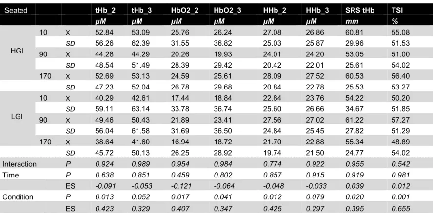
Throughout a bout of sitting, blood pools in the lower limbs. It is necessary to measure this pooling to ensure we are getting the predicted response from sitting, as well as be able to contextualize pooling among the other vascular responses. As time spent sitting increases, hemoglobin is expected to rise. Previous work from this lab showed a 24% increase in tHb from 10 to 90 minutes of sitting in a young, sedentary population[121]. Blood pooling in the calf was measured by looking at tissue perfusion assessed using near infrared spectroscopy (NIRS) (PortaMon, Artinis, Medical Systems). NIRS is a reliable and valid method for assessing tissue perfusion [89]. A NIRS probe was placed on the medial gastrocnemius of the right leg to determine the total amount of hemoglobin, as well as oxygenated and deoxygenated, in the tissue directly underneath.
Blood Glucose
day to measure real-time interstitial glucose. The CGM provided relevant information on hyper and hypoglycemia, as well as glucose variability [88].
Meal Intervention
The literature was searched extensively to determine the most appropriate high and low GI beverage choices. This is represented in Table 1. Most studies matched for macronutrient content and the average low GI used was ~36 and the high GI ~80 [16,49,51,93–104].
Table 1. Low and High GI meals used in literature
Author Low GI meal GI High GI meal GI Matched
Kaviani 2016 Boiled lentils, tomato
sauce and Canola oil 26
Instant mashed potato, egg whites
and white bread 76
Macronutrie nt content
Ludwig 1999 Veggie omelet & fruit N/ A
Instant oatmeal with sweetener (glucose plus artificial sweetener) + milk treated with 2 drops of lactase to increase the GI of the milk sugar
N/
A N/A
Nilsson 2007 Spaghetti 58 White wheat bread 10
0
Starch (g/serving)
Clark 2006 Psyllium ready-to-eat
cereal 56 Farina 64 N/A
Vega-Lopez
2015 N/A/
N/ A
500 mL of glucose OR 96g of commercial white bread
(pepperidge farm)
N/ A
50g carbs each
Bailey 2017 milk, apple and peaches Muesli, semi-skimmed 47 Cornflakes, skimmed milk, white bread and strawberry jam 78
Macronutrie nt content, energy, and
fluid
Little 2010 Lentils 26 Instant mashed potato, egg whites and white bread 76 Macronutrient content
Cocate 2011
All Bran cereal, fat-free strawberry yogurt, grape
juice, multi-grain bread, margarine, and apple
28
Corn flake cereal, whole milk, sports drink (high carbohydrate,
water, and electrolytes replenisher), white bread, and
margarine
79 Macronutrie nt content
Jamurtas
2011 Dried apricots 30 White bread with strawberry jam 70
1.5 g of carbohydrat e per 1 kg of
body mass Keller 2016 37 g isomaltulose 32 28g maltodextrin + 9g sucrose 90 N/A
Kaplan 2000
300 mL lemon beverage (290 mL water and 10 mL
lemon juice) sweetened with 23.7 mg sodium saccharin) (Hermesetas Original; JL Freeman Inc,
Boucherville, Canada) N/
A
Glucose: 300 mL lemon beverage (290 mL water and 10 mL lemon
juice) containing 50 g glucose (Dextrose monohydrate; Bio-Health, Dawson Traders Ltd,
Toronto)
N/
A N/A
Nilsson 2009
Glucose solution (50 g glucose suspended in 450
ml water): divided into 6 portions over 150minutes
N/ A
Glucose solution (50 g glucose suspended in 450 ml water): consumed in 10-12 minutes prior to
protocol
N/
2003 0
Moore 2009, 2010
Bran flakes cereal, semi-skimmed milk, water 30
Corn flakes cereal, semi-skimmed
milk, water 72
1g/kg body weight of carbohydrat e Visvanathan 2004
379 ml Apple & Cherry Juice, a commercially available preservative-free fruit juice (Wild about Fruit
Company Pty Ltd, Wandin, Victoria, Australia; (Foster-Powell
et al. 2002))
43
50g glucose, 359mL water & 20 ml bottled lemon squeeze (Berri Ltd,
Victoria, Australia; 99·9% lemon juice with 25 mg carbohydrate/L)
10
0 N/A
POPULATION/SAMPLING
Research participants were young adults (18-35), free of disease and similar in physical activity participation status (>90 minutes/week of moderate-vigorous exercise). A young, healthy population avoids the potential for age-related cardiovascular impairments. Using young
STATISTICAL CONSIDERATIONS
The independent variables for the study were time (pre, 10, 90, 170, post) and high or low GI intervention. The main dependent variables were PWV, AIx, and TSI%. Potential covariates for PWV was MAP, and accelerometer fidgeting data was a covariate for blood pooling data. For this study, a repeated measures analysis was performed. First, we considered a multivariate test, but with many time points for assessing PWV, it is risky to run a multivariate approach. This is because if a subject is missing one time point, they will be dropped from the entire analysis through listwise deletion. However, we wanted a statistical test that will only drop that one time point. Thus, we considered use of a mixed linear model. A mixed linear model handles repeated measures and missing data (by dropping that one missing time point). Additionally, we recognized that many subjects will start at different intercepts. The best statistical test that can handle these considerations is a mixed linear model. T-tests were performed to compare across time points or condition for all variables.
OTHER CONSIDERATIONS
Sex As A Biological Factor
Men and women have different trajectories for acquisition of CVD risk factors over the course of a lifetime. We are not adequately powered to draw sex-specific conclusions from these results. However, the sexes can be stratified for sensitivity analysis.
Ethnicity/Race
Generalizability
We were not able to generalize our results, as we selected a homogenous sample in order to minimize confounding variables. However, we would be able to apply our results to young, healthy, active individuals.
Potential Challenges & Alternative Strategies
As with any research study, there may be challenges with proper functioning of equipment and recruitment of participants. For 1+ year prior to commencement of data collection, all research assistants involved became well versed with the usage of laboratory equipment. Additionally, all channels of media were used to recruit participants in an attempt to complete scheduling in advance of data collection commencement.
Since sensitive cardiovascular and metabolic measures were taken in this study, participation in exercise should be controlled. Current literature is inconsistent, but was summarized to find that most pre-testing guidelines included a 24-hour refrain from exercise. Therefore, this approach was taken for this study.
SUMMARY
It is known that both prolonged sitting and consumption of a high GI meal have acute negative effects on local cardiovascular structure and function. However, it is unknown how these two stimuli interact and what their combined effect on central and local vasculature in young health adults. Currently, no studies have observed the effects of a high GI meal and prolonged sitting on the cardiovascular system in young healthy individuals.
As previously stated, prolonged sitting acutely elicits decreased blood flow to the legs, decreased leg vascular shear stress, impaired vasodilator abilities, impaired endothelial
dysfunction, oxidative stress, and hyperinsulinemia. The response of the combined stimuli is unknown, but the predicted response is augmented arterial stiffness and vascular dysfunction.
CHAPTER III: METHODOLOGY
This study is reported in accordance with CONSORT (Consolidated Standards of
Reporting Trials) guidelines. Ethical approval was obtained from the University of North Carolina at Chapel Hill institutional review board, and all participants provided written informed consent prior to participating in the study.
PARTICIPANTS
Participants were recruited from the University of North Carolina at Chapel Hill and the surrounding area and screened prior to enrollment in the study. Recruitment took place between October and February. Testing took place between November and March. Twenty participants, between 18 and 35 years old, were identified and scheduled for participation. Exclusion criteria included: less than 90 minutes of structured exercise per week, any known cardio-metabolic disorders, pregnant women, smokers, currently taking medication known to affect
cardiovascular function. Healthy, moderately active individuals were recruited to ensure a homogenous sample size and enhance the internal validity of the study. Participants signed an informed consent form prior to participation in the study.
EXPERIMENTAL DESIGN
~2 hours post-meal consumption[92]. Three hours ensures that we capture the peak postprandial glucose response. Testing began between 6:00 and 10:00 AM in the Cardiometabolic Lab. Refer to figure 1 for a complete overview of the study design.
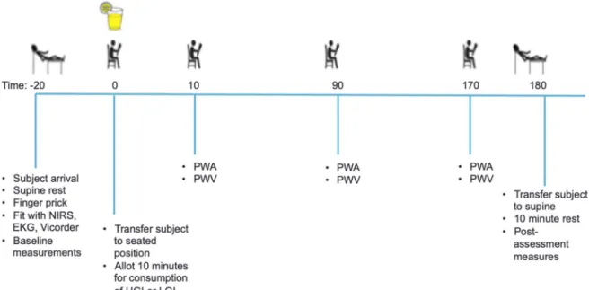
Figure 2. Protocol timeline for experimental measures
Familiarization Visit
Participants reported to the Cardiometabolic Lab 12-24 hours prior to their first
experimental day. Upon arrival, they signed the informed consent document and completed a medical history questionnaire. Subsequently, height, via stadiometer (Perspective Enterprises, Portage, Michigan), and weight, via calibrated scale (Health o meter; Pelstar LLC, McCook, IL) were measured. Participants were then given a food log for them to complete for the 24 hours leading up to each experimental day. Each participant was fitted with an accelerometer (ActiSleep +; ActiGraph LLC, Fort Walton Beach, FL) on their right ankle to covary for
completion of the study. A continuous glucose monitor (iPro2, Medtronic, Northridge CA) was then inserted into the participant’s site of choice: abdomen (approximately 5cm lateral from the umbilicus), lower back above left or right hip, or the posterior side of the upper arm. The
continuous glucose monitor (CGM) was worn 12-24 hours prior to the first experimental day until their completion of the study. Participants were instructed to perform a finger prick to calibrate the CGM one hour after leaving the lab and once before bed (glucometer: Contour Next One; Ascensia Diabetes Care US Inc., Parsippany, NJ). Before leaving, the participant was given a pre-determined frozen meal to be consumed 12 hours prior to their experimental visits (Lean Cuisine; Nestle, Vevey, Switzerland).
Visit 2 and 3
Participants arrived to the Cardiometabolic lab (22.36 ±2°C, 26.09 ± 8.94% humidity)
between 6:00 and 10:00 AM, with 2-7 days between experimental visits. Participants arrived overnight fasted from food, alcohol, and caffeine (at least 12 hours) and having abstained from exercise for at least 24 hours prior to the visit. Upon arrival, height and weight were recorded and a finger prick was performed followed by 20 minutes of quiet rest in the supine position. During the 20 minute rest, the participant was fitted with a NIRS probe on the medial
gastrocnemius of the right leg, 3-lead ECG, a non-invasive blood pressure device (NIBP; ADInstruments) and the Vicorder device. After 20 minutes, baseline measurements were attained in the supine position. ECG and NIRS data were collected continuously until the completion of the study visit. Vicorder measurements were taken in triplicate: brachial blood pressure, central PWA, carotid-femoral PWV, brachial-femoral PWV, brachial-ankle PWV, and femoral-ankle PWV.
visit. Following re-positioning, participants were allotted 10 minutes to consume either a high GI (glucose; GI: 100) or low GI (fructose; GI: 19) beverage (1g/kg BW + 300mL water) flavored with lemon juice ad libitum. The 3-hour sitting protocol commenced following the consumption of the beverage [4,22,90,91]. During the 3 hours, participants were asked to sit completely still while watching a low stimulus documentary (pre-screened by EK and KB). PWV and PWA measurements were recorded in triplicate at 10, 90, and 170 minutes. After 3 hours (180 minutes) of sitting, the participants were passively transferred to the supine position and PWV and PWA was reassessed after 10 minutes of rest. Blood volume may change throughout the course of the sitting protocol due to filtration of the blood in the kidneys and insensible water loss through perspiration and respiration. This may cause between 100-250 ml of water loss in a period of 3 hours. For this purpose, water intake was monitored during both testing sessions. In addition, participants were instructed to refrain from using the restroom during the study
because standing and walking to the restroom would alter CV mechanisms. There were no instances of participants getting up to use the restroom at any point during the study.
EXPERIMENTAL MEASURES
Primary And Secondary Outcome: Central And Peripheral PWV
and a 15% increase in all-cause mortality[86]. A clinically meaningful change for the lower limb is currently unknown.
Blood pressure cuffs were placed on the dominant arm, upper thigh, and ankle for PWV and PWA measurements. The bottom of each cuff was marked with a permanent marker on the skin in the event of cuff movement. A specialized device was used to measure the distance for PWV measurements: all measurements were recorded in cm. For carotid-femoral PWV, the distance was measured from the sternal notch to the top of the femoral cuff. An additional 2.5cm was added to the distance to represent the distance as if measured to the middle of the cuff (this was to decrease measurement error as the middle of the cuff in not marked and would be difficult to guess by simply looking). For femoral-ankle PWV, the distance was measured from the top of the femoral cuff to the top of the ankle cuff. For brachial-ankle PWV, the distance from the top of the brachial cuff to the top of the ankle cuff was measured. When transferred to a seated position, all cuffs were checked to ensure exact placement remained from the supine position. All PWV and PWA measurements were taken in triplicate in the supine position and repeated at minute 10, minute 90, and minute 170 of the sitting protocol. When the participant was transferred to the supine position after 180 minutes, PWV and PWA measurements were repeated following a 10-minute supine rest.
Control Measures
ECG, NIBP, NIRS, and the continuous glucose monitor (CGM) collected measurements continuously throughout the three hours. ECG and NIBP measures allowed for continuous monitoring of hemodynamics. NIRS was used in order to assess blood pooling in the calf because this is the site for blood pooling and the gastrocnemius has been shown to be as sensitive as the soleus[125]. The CGM, which had been inserted 12-24 hours prior, was
value for each subject over 3 hours). For NIRS placement, the participant was asked to perform a calf raise in order for the researcher to identify the largest muscle belly circumference of the right medial gastrocnemius. This place was marked and ultrasound was used to find the thickest muscle belly with no large vasculature for the placement of a NIRS probe. The placement was measured from the popliteal fossa to the heel to ensure the same placement for the second visit. The NIRS probe was secured to the skin with double-sided tape and a light shield. SRS tHb and TSI% data were extracted from the NIRS output. NIRS SRS tHb is a validated estimate of total hemoglobin under the area of interest[125]. A 3-lead ECG was then attached to the participant at the right arm, left arm, and left lower sites. The pointer and middle fingers of the dominant hand were then measured to determine sizing for proper finger cuffs for the non-invasive blood pressure (NIBP) device. The cuffs were secured on the middle phalanx and attached to the NIBP wrist unit. The device was set to sample for alternating 30 minute periods on each cuff, beginning with the pointer finger. This was to diminish the discomfort for the participant. Although NIBP data was collected continuously over the 3 hours, 30 second averages were taken at each time point of interest for analysis purposes.
RANDOMIZATION
SAMPLE SIZE
Calculations to determine minimum sample size were based on the primary central vascular health outcome, aortic pulse wave velocity (PWV). Previous research has reported that prolonged sitting reduces lower limb vascular health and endothelial function between
57-80%[25]. Based on a PWV of 6.2 m/s, which was predicted for healthy participants <30 y[105], a 57% decrease in PWV would be 3.5 m/s. For the current study, we opted to sample based on a conservative change score of 1 m/s. We also used a conservative usual error of 1 m/s. Using magnitude-based inference, to estimate the sample size required to detect the smallest detrimental (or beneficial) effect in a cross-over study, with the maximum chances of a type 1 and 2 error set at 5% (i.e. very unlikely), approximately seventeen participants were required. In order to account for drop out or missing data, twenty participants were recruited for the study. Participants were recruited from undergraduate classes at UNC as well as through email, flyer posting, and word of mouth. In order to improve adherence to intervention protocols, specifically the pre-testing guidelines, we informed the participant that the accelerometer and CGM would provide information concerning all activity and food/beverage consumption.
QUALITY CONTROL
At the conclusion of the study a random selection of 10% of the Vicorder data sets (e.g. from 3 participants) were re-entered by an independent observer. No data points differed, so no further action was taken.
DATA MANAGEMENT AND STATISTICAL ANALYSIS
Data sheets were stored in a locked drawer and in digital form on two study computers. All statistical analyses were performed using Jamovi (2018, Version 0.9). The α level was set a priori for all statistical procedures at α=0.05. Raw data are presented as mean [standard
CHAPTER IV: RESULTS
PARTICIPANTS
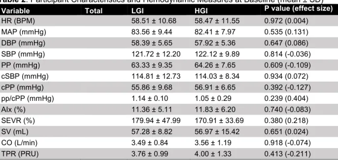
Participants were young (23 ± 3 years), moderately active (280 ± 175 minutes/week), normal height and weight (172.6 ± 8.7 cm; 72.8 ± 15 kg; BMI: 24.3 ± 3.7 kg/m2), and otherwise healthy men and women (33% female). Participants self-identified as Caucasian (n=14), African American (n=2), and Middle-Eastern (n=4). All participants conformed to pretesting guidelines. Participant characteristics are illustrated in table 2.
Figure 3. CONSORT diagram. NIRS: near infrared spectroscopy
BASELINE DATA
Baseline data is reported in Table 2. There were no significant differences in baseline hemodynamic measures between testing days (table 2). Baseline tHb data are not reported as values as they are expressed relative to baseline. In order to achieve the most precise results, baseline measures were set as covariates when analyzing all supine primary, secondary, and ancillary measures. Adherence to pre-assessment guidelines were confirmed through
accelerometer and blood glucose data. There was no significant difference between total MVPA (moderate-vigorous physical activity) during the 12-24 hours prior to experimental visits (HGI: 42.2, LGI: 52.4 minutes, p=0.079). Additionally, the AUC for blood glucose data 12 hours prior to experimental visits was similar between LGI and HGI (64,330 ± 19.9 and 65,936 ± 14.3, respectively, p<0.05).
Participants recruited (n=20)
Participants analyzed (n=18)
NIRS (17/18)
Sickness n=1 Time n=1 Total drop-outs n=2 Potential participants emailed
(n=1500)
Table 2. Participant Characteristics and Hemodynamic Measures at Baseline (mean ± SD)
Variable Total LGI HGI P value (effect size)
HR (BPM) 58.51 ± 10.68 58.47 ± 11.55 0.972 (0.004)
MAP (mmHg) 83.56 ± 9.44 82.41 ± 7.97 0.535 (0.131)
DBP (mmHg) 58.39 ± 5.65 57.92 ± 5.36 0.647 (0.086)
SBP (mmHg) 121.72 ± 12.20 122.12 ± 9.89 0.814 (-0.036)
PP (mmHg) 63.33 ± 9.35 64.26 ± 7.65 0.609 (-0.109)
cSBP (mmHg) 114.81 ± 12.73 114.03 ± 8.34 0.934 (0.072)
cPP (mmHg) 55.86 ± 9.68 56.91 ± 6.65 0.392 (-0.127)
pp/cPP (mmHg) 1.14 ± 0.10 1.05 ± 0.29 0.239 (0.404)
AIx (%) 11.36 ± 5.11 11.83 ± 6.20 0.740 (-0.083)
SEVR (%) 179.94 ± 47.99 170.91 ± 33.69 0.380 (0.218)
SV (mL) 57.28 ± 8.82 56.97 ± 15.42 0.651 (0.024)
CO (L/min) 3.49 ± 0.84 3.56 ± 1.19 0.918 (-0.074)
TPR (PRU) 3.76 ± 0.99 4.00 ± 1.33 0.413 (-0.211)
PA: physical activity; HR: heart rate; MAP: mean arterial pressure; DBP: diastolic blood pressure; SBP: systolic blood pressure; PP: pulse pressure; cSBP: central systolic blood pressure; cPP: central pulse pressure; pp/cPP: pulse pressure/central pulse pressure; AIx: augmentation index; SEVR: subendocardial viability ratio; SV: stroke volume; CO: cardiac output; TPR: total peripheral resistance; PRU: peripheral resistance units
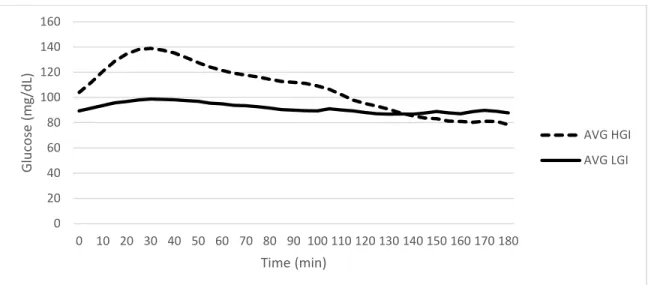
STIMULUS: CONTINUOUS BLOOD GLUCOSE
Figure 4. Average blood glucose response to high and low GI beverage for all subjects over 3 hours of sitting
PRIMARY OUTCOME: CENTRAL PULSE WAVE VELOCITY
There was no significant time x condition interaction for central (carotid-femoral) PWV in the supine position (table 3). However, there was a significant time x condition interaction for bfPWV (p=0.027) (figure 5). PWV increased by 0.27 m/s in the LGI condition (95% CI: 0.2517, 0.669) and 0.45 m/s in the HGI condition (95% CI: 0.068, 0.485).
Figure 5. Mean brachial-femoral PWV supine measurements before and after three hours of uninterrupted sitting (bars represent SD). bfPWV: brachial-femoral pulse wave velocity; HGI: high glycemic index; LGI: low glycemic index
0 20 40 60 80 100 120 140 160
0 10 20 30 40 50 60 70 80 90 100 110 120 130 140 150 160 170 180
Table 3. Supine Arterial Stiffness Measures
cfPWV bfPWV faPWV
Time Condition m/s m/s m/s
Pre HGI X 6.08 8.63 6.75
SD 0.95 0.66 0.83
LGI X 6.11 9.00 6.96
SD 1.08 0.75 0.77
Post HGI X 6.13 9.08 7.13
SD 0.97 0.63 1.11
LGI X 5.98 9.27 7.67
SD 1.24 0.95 1.15
Interaction P 0.223 0.027 0.178
Time P 0.628 0.001 0.004
ES -0.060 0.470 0.380
Condition P 0.254 0.078 0.180
ES -0.190 -0.300 0.230
cfPWV: carotid-femoral pulse wave velocity; bfPWV: brachial-femoral pulse wave velocity; faPWV: femoral-ankle pulse wave velocity; HGI: high glycemic index; LGI: low glycemic index
SECONDARY OUTCOME: PERIPHERAL PWV
There was no significant time x condition interaction for lower limb (femoral-ankle) PWV in the supine (Table 2) or seated position. However, there was a significant effect for time (p=0.004, d=0.38; 95% CI: 0.219, 0.897) in the supine condition (figure 6).
Figure 6. Mean femoral-ankle PWV supine measurements before and after three hours of uninterrupted sitting (bars represent SD). faPWV: femoral-ankle pulse wave velocity; HGI: high glycemic index; LGI: low glycemic index
ANCILLARY ANALYSIS
There were no significant interaction or condition effects for AIx or cSBP. There was a significant time effect for augmentation index (AIx). AIx decreased by 2.93% (p=0.02, d=-0.32; 95% CI: -5.05, -0.814) in the supine position (table 4).
Table 4. Hemodynamic measures and NIRS output. cSBP: central systolic blood pressure; Aix: augmentation index; CO: cardiac output; TPR: total peripheral resistance; PRU: peripheral resistance units; HR: heart rate; BPM: beats per minute; SRS tHb: spatially resolved
spectroscopy total hemoglobin; TSI: tissue saturation index; HGI: high glycemic index; LGI: low glycemic index
cSBP AIx CO TPR HR SRS tHb TSI
mm Hg % L PRU BPM mm %
Pre HGI X 114.03 11.83 3.56 4.00 61.70 49.61 57.98
SD 8.34 6.20 1.19 1.33 11.51 22.94 54.41
LGI X 114.81 11.36 3.49 3.76 60.71 56.84 58.68
SD 12.73 5.11 1.19 1.33 11.51 22.94 54.41
Post HGI X 112.69 8.58 3.62 3.85 61.74 52.16 52.29
SD 8.22 4.52 1.00 1.17 11.58 16.36 52.96
LGI X 114.72 8.75 3.68 3.90 68.75 55.92 58.09
SD 10.90 3.21 0.89 0.89 16.20 20.66 51.29
Interaction P 0.615 0.710 0.710 0.421 0.050 0.586 0.226
Time P 0.581 0.015 0.501 0.976 1.000 0.818 0.138
ES -0.066 -0.320 0.120 -0.005 0.308 0.028 -0.179
Condition P 0.387 0.859 0.982 0.644 0.233 0.074 0.123
ES 0.148 -0.030 0.006 -0.116 0.304 0.305 0.262
There were no significant condition x time interaction effects or time effects in the NIRS output. There were significant condition effects for SRS tHb (p=0.02), and TSI% (p=0.001) (table 4).
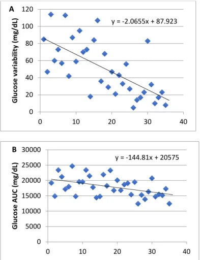
SUPPLEMENTAL ANALYSIS
and a weak-moderate correlation between glucose variability and changes in bfPWV (r=0.400, p=0.017; 95% CI: 0.647, 0.077) (figure 7).
Figure 7. Correlation between glucose variability bfPWV (A) and glucose AUC and
bfPWV (B). AUC: area under the curve; HGI: high glycemic index; LGI: low glycemic index y = -2.0655x + 87.923
0 20 40 60 80 100 120
0 10 20 30 40
G lu co se v a ri a b il it y ( m g /d L) A
y = -144.81x + 20575
0 5000 10000 15000 20000 25000 30000
0 10 20 30 40
CHAPTER V: DISCUSSION
The purpose of this study was to examine the peripheral and central cardiovascular response to prolonged sitting following the consumption of a low and high GI meal. It was hypothesized that prolonged sitting and the consumption of a high GI meal would elicit greater detriments in both central and peripheral PWV. The expected glucose stimulus was achieved, with the LGI meal eliciting a small, gradual increase in blood glucose, and the HGI meal eliciting a large increase in blood glucose for ~30 minutes following consumption. Prolonged sitting (3 hours) and consumption of a high GI beverage increased central (brachial-femoral) arterial stiffness, with a slightly greater detriment in the HGI condition. However, carotid-femoral PWV was not influenced by prolonged sitting or HGI meal consumption. Peripheral (femoral-ankle) PWV increased regardless of the level of GI consumed. Collectively, this data demonstrates that in young health adults a single bout of prolonged uninterrupted sitting combined with a high glycemic index beverage can negatively affect markers of central and peripheral cardiovascular function, but there are some inconsistencies in the findings.
LIMITATIONS AND STRENGTHS
To better contextualize the discussion, the limitations and strengths of the current study must be stated. First, our study results lack generalizability as we used a group of healthy, active, young adults. However, we elected to recruit a homogenous group of healthy, active, young adults to minimize the confounding influence of age and CVD. Although the sample was healthy, it is unknown how much previous exposure they had to these stimuli. Second,
accelerometer and food log data on participants for an extended period of time in order to gather more data concerning lifestyle choices. Third, we were unable to determine sex interactions due to being inadequately powered, but we are confident that the experimental conditions were well controlled in that fluctuations in estrogen in females would not affect our results.
Although this study has a few limitations, it also has several strengths including the use of the Vicorder device. The Vicorder measures PWV and PWA in a non-intrusive manner with little operator training, ease of use, and low user error. This also allowed for a comprehensive assessment of cardiovascular measures. Second, we were adequately powered to conduct this study. Third, trial conditions were standardized for potential confounders such as diet, physical activity, and baseline values. Finally, this study is a double blind crossover design, which reduces bias in our results.
COMPARISON TO LITERATURE
This study found that prolonged sitting (3 hours) and consumption of a high GI beverage increased central (brachial-femoral) arterial stiffness, with a moderately (d=0.50) greater
detriment in the HGI condition. However, carotid-femoral PWV was not influenced by prolonged sitting or HGI meal consumption. There was a moderate (d=0.41) increase in peripheral
(femoral-ankle) PWV slightly regardless of the level of GI consumed. Although the results were not clinically significant (>1m/s), it does support the current literature[108,120-121]. That is, the established <1m/s clinical value is for chronic changes, repeatedly exposing the aorta to a 0.45 m/s increase in stiffness following a high GI meal may accumulate to significant cardiovascular burden over time[123]. This offers an additional insight into a young, healthy person’s ability to respond to a high dose of sugar.
one additional unpublished paper [personal communication] have reported increased central PWV with an acute bout of prolonged sitting [108,120]. The current study is the first to
investigate changes in central PWV with prolonged sitting and a high glycemic index meal. The findings indicate that high GI consumption exacerbates the detriments from prolonged sitting, and should be further explored. However, the carotid-femoral PWV was not significantly impacted by prolonged sitting or a high GI meal, which was predicted based on previous literature [3, 108]. A potential reason is that the brachial-femoral measurement incorporates the subclavian artery. Since the subclavian artery is slightly smaller than the aorta, it may be more responsive to change in a healthy population, which could explain the differences in
measurement responses. While no studies have directly compared the reliability of brachial-femoral PWV versus carotid-brachial-femoral PWV, it is plausible that the brachial-brachial-femoral PWV confers greater reliability and sensitivity. For the current study we did consistently find greater signal strength for the brachial-femoral measurement. The responsiveness of carotid-femoral and brachial-femoral PWV should be explored further to understand their reliability in this population.
Prolonged siting resulted in a significant increase in peripheral (femoral-ankle) PWV, regardless of GI beverage. These results contribute to the existing literature that prolonged sitting acutely impairs the structure and function of lower limb vascular health [3-4,22,25], with a decrease in shear stress being the proposed mechanism [22]. Shear stress has been shown to decrease across a bout of prolonged uninterrupted sitting due to a decrease in blood flow to the limb caused by blood pooling in the legs (shown by the increase in perfusion). However, in the current study, tHb did not increase (indicative of blood pooling) in the LGI condition. This could be attributed to the subjective feeling of cold many subjects experienced during the bout of sitting, potentially causing local vasodilation, facilitating venous return. This may not have been seen in the HGI condition, because HGI consumption is associated with greater insulin
were no significant changes in CO. The potential blood pooling is caused by lack of venous return due to lack of muscle pump and gravitational forces. A decrease in shear stress is associated with endothelial function impairment. Findings from this study, an increase in lower limb arterial stiffness due to sitting, are in line with current literature. Current literature assessing the effects of acute prolonged sitting (3-8 h) on endothelial function, using flow-mediated dilation (FMD), reported a 48-71% decline in leg FMD [3-4,25,72,117-119]. Impairment to the
vasculature of the leg due to prolonged sitting is of clinical relevance since evidence supports that the leg vasculature is vulnerable to atherosclerosis [4]. However, consumption of a high GI meal did not further impair peripheral vasculature. This suggests that not all vasculature
responds the same to the same stimulus, and this should be explores in other areas of the body.
Prolonged sitting resulted in a significant reduction in AIx, regardless of GI beverage. AIx is a measure of systemic arterial stiffness derived from the ascending aortic pressure waveform [106]. A reduction in AIx after prolonged uninterrupted sitting could be attributed to the increase in blood pooling, leading to a reduction in venous return and therefore a reduction in stroke volume and subsequently AIx. These results have been found previously in our lab[121]. The results showed a slight reduction in stroke volume from minute 10 to minute 170 in the high GI beverage condition, but a fluctuation in stroke volume between minute 10 and 90 and minute 90 and 170 in the low GI beverage condition. These findings suggest that AIx may not be telling us about vascular health during sitting, but rather autonomic function. It also suggests that the blood may in fact be pooling, and the NIRS measurement is not adequately reflecting this pooling.
IMPLICATIONS
Prior to this study, it was understood that prolonged sitting and high GI consumption have detrimental chronic and acute effects on the cardiovascular system[3-4,108]. However, it was unknown how the two stimuli interacted acutely to effect the central and local
cardiovascular system. The data herein supports the interaction between prolonged sitting and high GI consumption and their effects on the cardiovascular system in a young, healthy
population. There seem to be greater changes in arterial stiffness at the level of the heart rather than the local leg vasculature. Due to the common nature of prolonged sitting and high GI consumption in everyday life, particularly in offices, this information is useful and pertinent. If the mechanism behind acute exposure to prolonged sitting and high GI consumption and
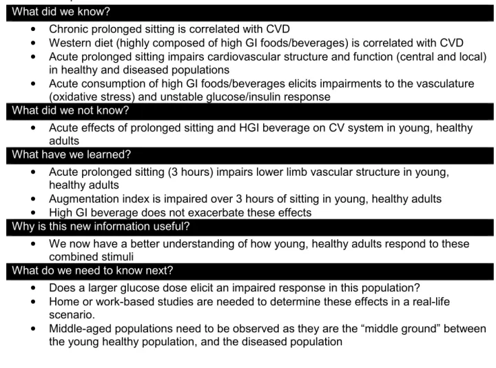
Table 5. Implications
What did we know?
• Chronic prolonged sitting is correlated with CVD
• Western diet (highly composed of high GI foods/beverages) is correlated with CVD • Acute prolonged sitting impairs cardiovascular structure and function (central and local)
in healthy and diseased populations
• Acute consumption of high GI foods/beverages elicits impairments to the vasculature
(oxidative stress) and unstable glucose/insulin response
What did we not know?
• Acute effects of prolonged sitting and HGI beverage on CV system in young, healthy
adults
What have we learned?
• Acute prolonged sitting (3 hours) impairs lower limb vascular structure in young,
healthy adults
• Augmentation index is impaired over 3 hours of sitting in young, healthy adults • High GI beverage does not exacerbate these effects
Why is this new information useful?
• We now have a better understanding of how young, healthy adults respond to these
combined stimuli
What do we need to know next?
• Does a larger glucose dose elicit an impaired response in this population? • Home or work-based studies are needed to determine these effects in a real-life
scenario.
• Middle-aged populations need to be observed as they are the “middle ground” between
the young healthy population, and the diseased population
CONCLUSIONS
APPENDIX A – ETHICAL APPROVAL
17-0745 Adult Consent Form Page 1 of 4
University of North Carolina at Chapel Hill Consent to Participate in a Research Study Adult Participants
Consent Form Version Date: _7/26/2018_____________ IRB Study # 17-2532
Title of Study: Prolonged Sitting With or Without a High Glycemic Index Meal: Acute Effects on Vascular and Cerebrovascular Function in Healthy Adults
Principal Investigator: Elizabeth Kelsch
Principal Investigator Department: Exercise and Sport Science Principal Investigator Phone number: (919) 962-0396 Principal Investigator Email Address: [email protected] Co-Investigators: Katie Burnet, Dr. Erik Hanson
Faculty Advisor: Lee Stoner
Faculty Advisor Phone Number: (919) 962-0534 Faculty Advisor Email Address: [email protected]
________________________________________________ _________________
What are some general things you should know about research studies? You are being asked to take part in a research study. To join the study is voluntary.
You may choose not to participate, or you may withdraw your consent to be in the study, for any reason, without penalty.
Research studies are designed to obtain new knowledge. This new information may help people in the future. You may not receive any direct benefit from being in the research study. There also may be risks to being in research studies. Deciding not to be in the study or leaving the study before it is done will not affect your relationship with the researcher, your health care provider, or the University of North Carolina-Chapel Hill. If you are a patient with an illness, you do not have to be in the research study in order to receive health care.
Details about this study are discussed below. It is important that you understand this information so that you can make an informed choice about being in this research study.
You will be given a copy of this consent form. You should ask the researchers named above, or staff members who may assist them, any questions you have about this study at any time.
What is the purpose of this study?
To examine the acute effects of sitting and consumption of a high glycemic index (HGI) meal on cardiovascular health, cerebrovascular health, cognition, and the proposed mechanisms within the body that may lead to the observed effects. Findings from this study may contribute to public health policy concerning sedentary behavior and the Western diet.
You are being asked to be in the study because you are between the ages of 18-40 years.
17-0745 AdultConsentForm Page2of4
You should not be in this study if you have a BMI greater than 30 kg/m2, you have known cardiovascular or metabolic diseases, you smoke tobacco, or you are pregnant (urine pregnancy test will be given).
How many people will take part in this study?
There will be approximately 20 people in this research study.
How long will your part in this study last?
Should you wish to participate in the study, you will be required to attend the Applied Physiology Laboratory at University of North Carolina at Chapel Hill on three occasions. The first visit will last approximately 30 minutes, and the following visits approximately 4 hours.
What will happen if you take part in the study?
Visit One:Participants will complete report to the UNC Applied Physiology Laboratory where informed consent will be obtained. Participants will be screened for participation in the study, which will include a medical history questionnaire. During this familiarization day, participants will be given a food log for them to complete for the 24 hours leading up to the first
experimental day until their completion of the study. Each subject will be fitted with an accelerometer (ActiSleep +; ActiGraph LLC, Fort Walton Beach, FL) on their ankle to covary for spontaneous movement as well ensure abstinence from exercise 24h prior to the experimental visits. The accelerometer will be worn at least 24 hours prior to the first experimental day until their completion of the study. A continuous glucose monitor (iPro2, Medtronic, Northridge CA) will then be inserted into the participant’s abdomen, approximately 5cm lateral from the umbilicus. The continuous glucose monitor (CGM) will be worn the day prior to each
experimental visit, until the cessation of each experimental visit. The participants will receive a standardized frozen meal to consume 12 hours prior to each experimental visit.
17-0745 AdultConsentForm Page3of4 Visit Three: Participants will return for follow up testing within 10 days after visit two. The experimental procedures will be identical to visit 2. The participant will be randomly assigned to either HGI or CON for visits two and three. The beverage will be administered by an
undergraduate research assistant to allow for double blindedness.
What are the possible benefits from being in this study? There is no direct benefit to participants.
What are the possible risks or discomforts involved from being in this study? While in this study, a finger prink will need to administered to obtain blood glucose levels, which may be uncomfortable. The insertion of a continuous glucose monitor could cause discomfort. A Urine Pregnancy test provided by the study will be obtained for all women of child-bearing potential, which could also cause some discomfort. The device we will use to monitor blood flowing to your calf and brain also possesses a small risk of eye damage/irritation, and skin heating/irritation. There may be uncommon or previously unknown risks. You should report any problems to the researcher.
How will information about you be protected?
Hard copies of any identifiable information will be stored in a locked file cabinet within an access-controlled laboratory in Fetzer Hall (Applied Physiology Lab) at the University of North Carolina at Chapel Hill campus. Only members of the research team will have access to the cabinet. Any electronic files with identifiable information will be kept separate in password-protected files on password-password-protected computers that will be accessible to only members of the research team. Upon completion of the study, all data will be transferred to an electronic storage device, files will become password protected, and all hard copies will be shredded.
Participants will not be identified in any report or publication about this study. Although every effort will be made to keep research records private, there may be times when federal or state law requires the disclosure of such records, including personal information. This is very unlikely, but if disclosure is ever required, UNC-Chapel Hill will take steps allowable by law to protect the privacy of personal information. In some cases, your information in this research study could be reviewed by representatives of the University, research sponsors, or government agencies (for example, the FDA) for purposes such as quality control or safety.
What if you want to stop before your part in the study is complete?
You can withdraw from this study at any time, without penalty. The investigators also have the right to stop your participation at any time. This could be because you have had an unexpected reaction, or have failed to follow instructions, or because the entire study has been stopped.
Will you receive anything for being in this study?
You will be receiving a vascular health report for taking part in this study.
Will it cost you anything to be in this study? It will not cost you anything to be in this study.
What if you are a UNC student?