Journal of Global Pharma Technology
Available Online at: www.jgpt.co.in
RESEARCH ARTICLE
Ethanolic Extracts of Young Papaya Seed (
Carica papaya, Linn
)
Decrease Spermatocytes, Leydig Cells, and Androgen Receptor of
Leydig Cells of Adult Mice (Mus musculus)
Bagus Komang Satriyasa
1,Agung Wiwiek Indrayani
1,I Gusti Kamasan
Nyoman Arijana
2,Dewa Made Ruspawan
3,Christopher Ryalino
4*1. Departments of Clinical Pharmacology Faculty of Medicine Udayana University, Indonesia.
2. Histology Faculty of Medicine Udayana University, Indonesia.
3. Post-Graduate Student of Medical Sciences Faculty of Medicine Udayana University, Indonesia.
4. Anesthesiology Faculty of Medicine Udayana University, Indonesia.
*Corresponding Author: Christopher Ryalino
Abstract
Background: Uncontrolled population growth is one of the problems in Indonesia. Among the various types of wild plants that can be used as anti-fertility drugs are papaya plants. This study aims to determine the effect of extracts on the number of spermatozoa, Leydig cells and androgen receptor (AR) expression on Leydig cells as an anti-fertility mechanism. Materials and Methods: This was an experimental, randomized posttest-only controlled group study. We used a total of 36 Wistar rats as the subjects of this study. The rats were classified equally into two groups by simple random sampling method. Group E rats received 0.5 mL ethanol extract of papaya seeds for 50 days (orally), and the rats in Group C (control) received 0.5 cc aquabidest for 50 days. Results: The treatment group showed a decrease in the number of spermatozoa (201.86 vs. 94.3, p <0.001), a decrease in the number of Leydig cells (7.61 vs. 3.32, p <0.001), and decreased AR expression of Leydig cells (46.19 vs. 18.48, p <0.001) compared to the control group. Conclusion: ethanol extracts of young papaya seeds significantly reduce the number of spermatocytes, Leydig cells, and the AR expression of the Leydig cells.
Keywords: Young papaya seed, Male anti-fertility, Androgen receptor, Leydig cells.
Introduction
Indonesia is a country with a very high-dense population. The population in Indonesia is currently around 250 million people and is the fourth most populous country in the world. If projections are made using scientific calculations, this country will have a population of more than 270 million people in 2025, more than 285 million in 2035 and 290 million in 2045 [1].
The welfare of the nation depends on the quality and competence of its human resources to sustain the nation's future. To avoid an explosion in population, the family planning program has become a national program in Indonesia. In Indonesia, the participation in family planning program is still dominated by women, while men have not participated as many since male
contraception is very limited and have some drawbacks [2,3].
It is essential to strive for the development of an ideal male contraceptive drug. An alternative is by finding ingredients from plants that have anti-fertility property. One plant-source ingeredient that is known to possess antifertility property is papaya seeds. Papaya seed extract can cause damage to the germinal epithelium, and degeneration of spermatosides and spermatids [4].
Young papaya seeds are one of the natural ingredients that have antifertility properties. This research was conducted to find out if the local Balinese papaya ethanol extract inhibits spermatogenesis by decreasing spermatozoa cells, Leydig cells and testosterone receptors.
Materials and Methods
This was an experimental, randomized
posttest-only controlled group study
conducted at Udayana University, Bali, Indonesia in 2018-2019. We used a total of 36 Wistar rats (12-weeks old, 150-200 g weight) as the subjects of this study. The study protocol was approved the Committee of Ethical Research of Udayana University. The rats were classified equally into two groups by simple random sampling method. Group E rats received 0.5 mL ethanol extract of papaya seeds for 50 days (orally), and the rats in Group C (control) received 0.5 cc aquabidest for 50 days.
The Making of Ethanol Extract of Papaya Seeds
The young papaya fruit is peeled-off and the seeds are collected. The seeds were then put in hot water until they are submerged for about one minute in order to stop the process of cell metabolism and enzymatic reactions. Papaya seeds are drained and then dried in an open room with good air circulation, avoiding any direct contact to the sunlight. The drying process was 2-weeks long. The dried papaya was then mashed into a powder. A total of 5 kg of papaya seeds
(dried powder) was macerated with 70% ethanol (in a 1:10 mixture) in a dark extractor bottle at room temperature for 24 hours. After that, 1,000 grams of simplicia and 7.5 liters of solvent were put into the mixture. The mixture was soaked for five days while stirring every 6 hours, filtered, and then we macerated the pulp again with 2.5 liters of solvent, and macerated again with the similar protocol. The filtrate obtained was evaporated with freshdryer to
obtain concentrated ethanol extract.
Concentrated papaya seed extract is then ready to be used for this study.
Preparation and Treatment of
Experimental Animals
Thirty-six adult male Wistar rats, aged 12 weeks, weighing 150-200 grams were then adapted into a similar environment and food for two weeks. Food provided in the form of rat food and drinks (in ad libitum dose). Papaya ethanol extract was administrated orally using a straw.
Making Testicular Histology
Preparations
The rats were put down by intracardial injection of Pentothal. The testes were then mutilated, then were put in a 10% formalin buffer for 24 hours. A dehydration procedure using alcohol followed, and then we put in toluene and blocked with paraffin. Histology incision was made with microtomes, 3-5
microns thick, and stained with
Hematoxyilin Eosin (Figure 1).
Figure 1: Examples of FOVs for the sperm counts from out study: Group E (left) and Group C (right)
Counting the Number of Androgen Receptors of Leydig Cells
The number of androgen receptors on leydig cells is measured by immunohistochemical method using the Rabbit Anti-phospho-
Data Analysis
Descriptive analysis was used to present data from each group such as age, sex, number of spermatozoa cells, number of Leydig cells, and testosterone receptors in Leydig cells. Data distribution of rats from each experimental group was tested with Shapiro-Wilks test. The data variants were then tested for homogeneity.
If the data is homogeneous and has a normal distribution then a t-test is performed. The Mann-Whitney test is performed otherwise. We used SPSS 24.0 (IBM Corp. Released 2016. IBM SPSS Statistics for Windows, Version 24.0. Armonk, NY: IBM Corp.) For all statistical analysis. A p-value of <0.05 was considered statistically significant.
Figure 2: AR expression examples in our study: E group (left) and C group (right)
Results
Thirty-six Wistar rats were used in this study according to the study protocol. The Shapiro-Wilk test in this study resulted in a normally distributed data.
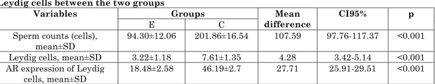
Therefore, we continued to the parametric test, using the unpaired t-test (Table 1). We found significant differences between the two groups in terms of sperm coutns, Leydig cell counts, and AR expressions on the Leydig cells.
Table 1: The comparison of sperm counts, Leydig cell counts, and AR expression on the Leydig cells between the two groups
Variables Groups Mean
difference CI95% p
E C
Sperm counts (cells),
mean±SD 94.30±12.06 201.86±16.54 107.59 97.76-117.37 <0.001
Leydig cells, mean±SD 3.22±1.18 7.61±1.35 4.28 3.42-5.14 <0.001
AR expression of Leydig
cells, mean±SD 18.48±2.58 46.19±2.7 27.71 25.91-29.51 <0.001
AR: androgen expression; CI: confidence interval; SD: standard deviation
Discussion
The results showed the average number of spermatozoa in the treatment group was 42.72±3.33 cells/FOV, compared to control’s 75.89±4.71 cells/FOV. The low count of spermatozoa cells in this study are probably caused by active substances contained in ethanol extracts of young papaya seeds (steroid, triterpenoid, and alkaloid). These substances are thought to be antifertility
agent that possess cytotoxic, anti
androgen/estrogenic effects [7].The decrease in the number of spermatozoa is also thought to be due to the estradiol and progesterone hormones contained in papaya seed extract
[8]. The hormone progesterone or estradiol will cause disruption of FSH and LH secretion.
Estradiol will cause suppression of the hypothalamus and anterior pituitary, causing GnRH and gonadotropin hormones to be inhibited [9].The estrogen hormone will inhibit the process of spermatogenesis because it results in degeneration of the seminiferous tubular epithelium. While progesterone will inhibit FSH secretion
which results in disruption of
disrupted. Disruption of the spermatogonia
cells will cause the process of
spermatogenesis to be disrupted as well. FSH and testosterone act as survival factors for the survival of spermatocytes and spermatids through the regulation of intrinsic and
extrinsic apoptotic pathways, where
spermatid cells will become spermatozoa [11].The number of spermatozoa is one of the parameters that is widely used to test fertility. It is also used to test the antifertility effect of a compound. Previously, papaya seeds have been widely studied in their effect as one of the natural ingredients which has antifertility properties.
Papaya seed extract can inhibit the population of spermatogonia and primary spermatocytes in male Rattus norvegicusc rats. Another research shows that papaya
seed’s methanol extract can inhibit
spermatogenesis causing necrosis of the eptelial germinal testes [12].The number of Leydig cells in the treatment group was lower than in the control group. The decrease in the number of Leydig cells is probably caused by the active substances contained in
papaya seed extract (steriod and
triterpenoids, alkaloids) which are
antifertility.
The active substances contained in papaya seeds can have cytotoxic, anti-androgenic or estrogenic effects. The decrease in the number of Leydig cells is also thought to be due to the estradiol and progesterone contained in the extract. The estradiol and progesterone will suppress the secretion of FSH and LH. If LH is inhibited, the function of Leydig cells to produce testosterone will be disrupted because on the surface of Leydig cells there are many LH receptors [13].Progesterone will also inhibit FSH secretion which results in disruption of spermatogenesis [14].
A decrease in Leydig cells will cause a decrease in the production of the hormone testosterone, because these Leydig cells produce about 95% of the hormone testosterone in the body. In the process of spermatogenesis in addition to testosterone, FSH is also needed. FSH stimulates Sertoli cells to form androgen binding protein (ABP) [15].The testosterone hormone produced by Leydig cells will be transported by ABP with very high concentrations to the site of
spermatogenesis.
In this study, the average expression of androgen receptors in the Leydig cells in the treatment group was 28.11±3.06%, compared to 55.07±2.49% that we found in the control group. The mechanism that may be involved in the low expression of androgen receptors (AR) is the active alkaloids contained in the extract of young papaya seeds. Previous research has shown that the alkaloids contained in pepper (Piper longum) have been reported to reduce the amount of AR in prostate cancer cell culture (Pca).
The study shows that alkaloids can reduce the full-length expression of AR in the ligand-binding domain (LBD) [16].The low number of AR expression due to alkaloids is likely due to the increased degradation of AR through the ubiquitin-proteasome pathway [17].ARs are a type of nuclear receptor that is
activated by testosterone or
dihydrotestosterone in the cytoplasm and translocated into the nucleus [18].
Androgen receptors (RA) are involved primarily in the development of male-specific
phenotypes during embryogenesis, in
spermatogenesis, sexual behavior, and fertility during adult life. Activation of autocrine RA by testosterone is important for the maturation and function of Leydig cells and regulation of stereogenic enzymes [19].Furthermore, RA autocrine signaling in Leydig cells protects the seminiferous tubular epithelium and protects Leydig cell apoptosis in rats.
The number of receptors in the cell largely determines its reaction to hormones. If enough hormones and receptors are in the cell, the ligand (testosterone) binds and activates the receptor [18].As a result of the
reduction in AR in leydig cells,
steroidogenesis was inhibited so that spermatogenesis stops at the spermatid stage.
Conclusion
References
1. McDonald P (2014) A Population
Projection for Indonesia, 2010-2035. Bull Indones. Econ. Stud., 50(1): 123-129. doi:10.1080/00074918.2014.896240
2. Wilopo SA, Setyawan A, Pinandari AW,
Prihyugiarto T, Juliaan F, Magnani RJ (2017) Levels, trends and correlates of unmet need for family planning among postpartum women in Indonesia: 2007-2015. BMC Womens Health, 17: 120. doi:10.1186/s12905-017-0476-x
3. Irawaty DK, Pratomo H (2019) Spousal
communication on family planning and contraceptive adoption in Indonesia. Indian J. Public Heal. Res Dev., 10(3): 372-376. doi:10.5958/0976-5506.2019.00521.7
4. D’Cruz SC, Vaithinathan S, Jubendradass
R, Mathur PP (2010) Effects of plants and plant products on the testis. Asian J.
Androl., 12(4):468-479.
doi:10.1038/aja.2010.43
5. Vasudeva N, Vats M (2011)
Anti-spermatogenic Activity of Ethanol Extract of Dalbergia sissoo Roxb. Stem Bark. JAMS J. Acupunct Meridian Stud.,
4(2):116-122.
doi:10.1016/S2005-2901(11)60017-4
6. Manivannan B, Mittal R, Goyal S, Ansari
AS, Lohiya NK (2009) Sperm
characteristics and ultrastructure of testes of rats after long-term treatment with the methanol subfraction of Carica papaya seeds. Asian J. Androl., 11(5):583-599. doi:10.1038/aja.2009.25
7. Lohiya NK, Manivannan B, Mishra PK, et
al (2002) Chloroform extract of Carica papaya seeds induces long-term reversible azoospermia in langur monkey. Asian J. Androl., 4(1):17-26.
8. Satriyasa BK, Mahendra AN, Gusti
Kamasan Arijana I, Ruspawan DM (2018) Unripe papaya seed ethanol extract (Carica papaya, Linn.) inhibits FSH and LH of male mice (Mus musculus). Biomed
Pharmacol. J., 11(2).
doi:10.13005/bpj/1457
9. Shaw ND, Histed SN, Srouji SS, Yang J,
Lee H, Hall JE (2010) Estrogen negative feedback on gonadotropin secretion: Evidence for a direct pituitary effect in
women. J. Clin Endocrinol. Metab, 95(4): 1955-1961. doi:10.1210/jc.2009-2108
10.Ramaswamy S, Weinbauer GF (2014)
Endocrine control of spermatogenesis: Role
of FSH and LH/ testosterone.
Spermatogenesis, 4(2):e996-025.
doi:10.1080/21565562.2014.996025
11.Shaha C, Tripathi R, Prasad Mishra D
(2010) Male germ cell apoptosis:
Regulation and biology. Philos Trans. R Soc. B Biol. Sci., 365(1546):1501-1515. doi:10.1098/rstb.2009.0124
12.Nkeiruka UE, Chinaka NO (2013)
Anti-fertility effects of Carica papaya linn: Methanol leaf extracts in male wistar rats.
J. Pharmacol. Toxicol., 8(1):35-41.
doi:10.3923/jpt.2013.35.41
13.Joseph DN, Whirledge S (2017) Stress and
the HPA axis: Balancing homeostasis and fertility. Int. J. Mol. Sci., 18(10):E2224. doi:10.3390/ijms18102224
14.Whirledge S, Cidlowski JA(2010)
Glucocorticoids, stress, and fertility. Minerva Endocrinol., 35(2):109-125.
15.Walker WH, Cheng J (2005) FSH and
testosterone signaling in Sertoli cells.
Reproduction, 130(1):15-28.
doi:10.1530/rep.1.00358
16.Armstrong CM, Gao AC (2019) Current
strategies for targeting the activity of androgen receptor variants. Asian J. Urol., 6(1):42-49. doi:10.1016/j.ajur.2018.07.003
17.Monaghan AE, McEwan IJ (2016) A sting
in the tail: The N-terminal domain of the androgen receptor as a drug target. Asian
J. Androl., 18(5): 687-694.
doi:10.4103/1008-682X.181081
18.Davey RA, Grossmann M (2016) Androgen
Receptor Structure, Function and Biology: From Bench to Bedside. Clin Biochem Rev., 37(1):3-15.
19.O’Hara L, McInnes K, Simitsidellis I, et al
(2015) Autocrine androgen action is essential for Leydig cell maturation and function, and protects against late-onset Leydig cell apoptosis in both mice and
men. FASEB J., 29(3): 894-910.