Address for correspondence Dr. Vishalakshi S. Pandit
Department of Dermatology, Venereology and Leprosy,
Koppal Institute of Medical Sciences, Koppal, Karnataka, India
Email: [email protected]
Original Article
A hospital-based cross-sectional clinicomycological
study of dermatophytoses in a tertiary care centre
Introduction
Superficial fungal infections due to dermatophytes are one of the most common skin infections in the world. The dermatophytes are a group of fungi that invade the superficial layer of the epidermis and degrade the keratinized tissues of skin, hair and nails in living animals
including man, causing ringworm.1
Cutaneous dermatophyte infections are common in the general population; up to 20% of people are infected at any time.2 The prevalence of
dermatophytosis in Croatia was reported to be 26% in 1986 and it reached to 73% in 2001. This significant rise in the frequency of this infection was also reported in the other countries.3 There
is a higher incidence of dermatophytoses in males than in females which has been reported both in India and abroad.4 Neglecting the patient
for treatment and the lack of knowledge of the general physician about of this infection may
Vishalakshi S Pandit, Hita Mehta*
Department of Dermatology, Venereology and Leprosy, Koppal Institute of Medical Sciences, Koppal, Karnataka, India
* Department of Dermatology, Venereology and Leprosy, Government Medical College, Bhavnagar University, Bhavnagar, Gujarat
Abstract
Objective To determine the clinicomycological pattern of dermatophytosis in patients attending tertiary care dermatology centre.Methods All patients attending Dermatology OPD, Sir. T. General Hospital, Bhavnagar, clinically diagnosed of dermatophytosis, were subjected to direct microscopy in KOH solution and culture on Sabouraud’s dextrose agar medium. Fungal species were identified on the basis of cultural characteristics, pigment production and microscopic examination in lactophenol cotton blue preparation.
Results Out of the 530 cases, majority of patients were in the age group of 30-40 years and male to female ratio of 1.2:1. Majority of the patients were housewives, followed by laborers. 70.9% patients were diagnosed of having tinea corporis, followed by tinea cruris (17.7%) and 52.3% of patients showed positivity in KOH smear and culture was positive in 32.1% cases. Most commonly isolated species was Trichophyton rubrum, followed by T. mentagrophytes and least was T. tonsurans (0.2%).
Conclusion Tinea corporis was the commonest clinical type of all dermatophytoses. Trichophyton rubrum was the most common isolate, thus concluding that it is the most common cause of superficial dermatophytic infection.
Key words
increase the frequency of disease. Knowing the frequency of this disease and its etiologic agents are important factors for providing the control measures.
The purpose of this study was to determine the clinicomycological pattern of dermatophytosis in the southern part of Gujarat
Methods
The study population included 530 patients, diagnosed clinically as having dermatophytosis
attending the outpatient Department of
Dermatology, Venereology and Leprosy of tertiary care hospital from August 2008 - August 2010 after approval by the institutional ethical committee.
Patients of either sex, infected with
dermatophytes on any part of the body, diagnosed clinically and willing to give written informed consent were included in the study. Patients treated with oral or topical antifungal agents in the last 3 months and on topical and systemic steroid therapy were excluded from the study.
A detailed history was taken from all patients. It
included age, sex, economic conditions,
occupation, and duration of disease, history of recurrence, habits and associated diseases. History of similar illness in family members was also elicited.
A thorough clinical examination was made regarding general condition of patient and morphology and distribution of lesions. Relevant systemic examination was also done and the findings were recorded. A written consent was obtained from all the participants.
The patients were classified according to the sites of involvement. The affected area with
maximum activity was selected for examination. The scrapings were taken from the edge of the lesion with the blunt edge of a cleaned slide, and the affected nail clippings using cleaned nail cutter/disposable blade. In cases involving hair, the affected hairs were plucked. The specimen was collected in an autoclaved paper which permitted storage of the material. After collection of clinical specimen, the patients were treated with appropriate local and / or oral antifungal on the basis of clinical diagnosis.
The scraping was placed on a clean sterilized glass slide and 1-2 drops of 10% KOH solution were put and covered with a coverslip using a gentle pressure. For hair and nail sample, 20% KOH solution was used. The slides were examined under low and high power for evidence of fungal hyphae and spores.
For culture, Sabouraud’s dextrose agar medium with chloramphenicol and gentamicin to prevent growth of contaminants was used. The scrapings were inoculated into the agar slant using a sterile chromium wire spud on to the centre of the slope and were incubated at 37°C and 25°C for up to four weeks. The identification of the fungal colonies was done based on their gross and microscopic morphology. The tubes that did not show evidence of fungal growth at the end of 4 weeks were considered negative and discarded. Microscopic examination was done by preparing teased mounts from the isolates with a drop of lactophenol cotton blue (LCB) stain. Urease test was performed for species identification of
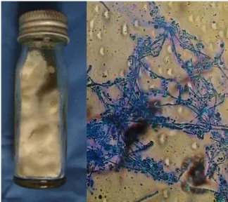
Figure 1 The front and reverse of Trichophyton rubrum colony
Figure 2 A microscopic feature of the lactophenol cotton blue-stained smear of Trichophyton rubrum is shown in Figure 2.
Results
In this study of 530 cases, majority of patients (31.6%) belonged to the age group of 30-40 years, followed by 22.8% in 20-30 years, 20.7% in 40-50 years, 14.5% in less than 20 years and 10.1% in more than 50 years group. 295 (55.7%) cases were male and rest 235 (44.3%) were females showed making a ratio of 1.2:1.
Figure 3T. mentagrophytes: White-colored granular colony and LCB smear showing round microconidia arranged in clusters along spiral hyphae. Few thin-walled cylindrical macroconidia are also seen.
Figure 4 Grey waxy colony of T. verrucosum and LCB smear of elongated chlamydoconidia with clavate microconidia seen along the sides of hyphae.
Residential distribution of cases showed preponderance of cases from urban areas (58%) than in rural area (41%).
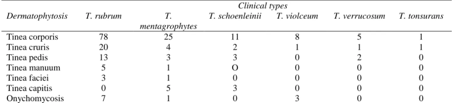
Table 1 Frequency of dermatophytes isolated (according to the site of infection), n=169. Dermatophytosis
Clinical types T. rubrum T.
mentagrophytes
T. schoenleinii T. violceum T. verrucosum T. tonsurans
Tinea corporis 78 25 11 8 5 1
Tinea cruris 20 4 2 1 1 1
Tinea pedis 13 3 3 0 2 0
Tinea manuum 5 1 O 0 0 0
Tinea faciei 3 1 0 0 0 0
Tinea capitis 0 5 3 0 0 0
Onychomycosis 7 1 0 3 0 0
government employees and unemployed (4.9%) each.
Most common clinical presentation was tinea corporis (70.9%) followed by tinea cruris (17.7%), tinea pedis (12.8%), tinea unguium (10.7%), tinea manuum (8.7%), tinea capitis (5.3%) and least number of patients were in tinea faciei (4.7%) group. In this study, most of the body sites were affected such as buttocks (24%), abdomen (22%), nails (9%), hands and feet (7% each), back, groin and legs (6% each), scalp, chest and thighs (3%), arms (2%), forearms (1%), face, neck etc. in their decreasing order of frequency. 61 (11.5%) patients had family history of disease.
Out of 530 cases enrolled, 275 (52.26%) cases were positive by direct microscopy and 169 (32.1%) cases were culture positive. Mainly
Trichophyton species such as T. rubrum, T. mentagrophytes (Figure 3), T. schoenleinii, T. vioalceum, T. verrucosum (Figure 4) and T. tonsurans were isolated.
The frequency of these species according to the site of infection is shown in Table 1.
Discussion
Despite extensive development in medical science, dermatophytosis is the most prevalent skin disease that involves people of any age group, sex and profession and causes enormous financial and emotional disturbances.
Dermatophytosis is still being considered as one of the major public health problems in many parts of the world. To identify the prevalence and etiological agents of dermatophytosis, this study was carried out in a tertiary care hospital for two years.
In our study, most cases belonged to the age group of 31-40 years which is similar to the other studies4,5,6 and the reason being this is the
age of maximum outdoor activity. Our study showed more number of dermatophytosis in males (55.7%) as compared to females. A higher incidence of dermatophytosis in males than in females has been reported both in India and abroad.4,7,8 The reason is the greater contact of
males with the contaminating sources in their working places. The higher incidence in young males could be due to greater physical activity and increased sweating. In our study male to female ratio is 1.2:1 as compared to 2:1 ratio of study conducted in western Rajasthan.9 In
contrast, some other earlier studies have recorded a higher prevalence of dermatophytes in females than in males.6,10
Occupation-wise, the incidence was more in housewives followed by businessmen and students compared to other study11 which shows
The predominant clinical manifestations of dermatophytosis vary considerably in different studies reported in literature. The commonest clinical types of dermatophytosis seen in this study were tinea corporis (70.9%), followed by tinea cruris (17.7%), which is in conformity with reports from other parts of India.4,7,12 A study
carried out by Teklebirhan et al.6 reported tinea
unguium as the dominant clinical manifestation (51.1%) followed by tinea capitis. The incidence of tinea capitis was 5.3% in our study which is comparable with reports from other workers.13,14
Tinea capitis is less common in India than in other countries.15,16,17 This may be attributable to
the use of hair oils which are customarily used by Indians and have been shown to have an inhibitory effect on dermatophytes in vitro.18,19
We found an isolation rate of 32% with culture, which is quite comparable with study carried out in Iran (38%).20 Isolation rate of dermatophytes
in different studies13,14,21,22 varied from 7% to
49%. Table 2 shows KOH and culture positivity rate in different studies conducted in various places.
Variations in isolation rates in our study and other studies may be due to the difference in selection of cases, geographical region, socioeconomic status and cultural factors. Although we could detect fungal hyphae, on direct microscopy in 52.3% cases, we could culture the fungus in only 32.1% of cases. This discrepancy among the 2 methods of fungal detection has been noticed by many other workers and could possibly be the result of various contributory factors involved in collection, transport, inoculation and incubation of specimen. The small sample material, loss during transport, absence of the infectious agent in the sample and difference in pH and sensitivity of media used may be some of responsible factors.
Table 2 Comparison of results of present with different studies.
Study No. of
patients
KOH +ve (%)
Culture +ve (%)
Baroda14 260 60 44
Iran20 17, 573 - 38
Jaipur28 120 65 55
Bijapur5 102 76 64
Rajasthan9 250 86 40
Present Study 530 52.26 32.08
In the present study 9.1% of cases showed a negative KOH, but a positive culture result. The reason for this could be due to the fungal hyphae being missed in KOH smear.
Trichophyton was the predominant genus among the three genera of dermatophytes in our study which concurs the observation in previous studies performed worldwide.23,24 T. rubrum is
the main dermatophyte reported from India and other countries.4 Contrast to the most studies, a
study carried out between 2004 and 2006 in Qazvin, Iran showed Epidermophyton floccosum
as the most frequently isolated species representing 32.8% of isolates, followed by T.
rubrum, T. verrucosum and T. mentagrophytes.25
E. floccosum and T. rubrum were reported to be
the most common causative agents in Tehran from 2000-2005.20 In a study conducted in
Northeast India, T. rubrum was the predominant fungus followed by T. mentagrophytes, T.
violaceum and Epidermophyton spp.12 Many
other species of dermatophytes like T.
schoenleinii, T. tonsurans, T. verrucosum, T.
ferrugineum, T. concentricum and M. audouinii
have been isolated by other workers6,25-27 but we
could isolate only T. rubrum, T.
mentagrophytes, T. violaceum, T. schoenleinii,
T. verrucosum and T. tonsurans.
Conclusion
rubrum was the predominant fungus isolated followed by T. mentagrophytes, T. schoenleinii
and T. violaceum. Consideration of current
epidemiological trends is of key importance in the diagnosis and treatment of fungal infection.
References
1. Peres NTA, Maranhão FCA, Rossi A, Martinez-Rossi. Dermatophytes: host-pathogen interaction and antifungal resistance. An Bras Dermatol. 2010;85:657-67.
2. Vander Straten MR, Hossain MA, Ghannoum MA. Cutaneous infections: dermatophytosis, onychomycosis, and tinea versicolor. Infect Dis Clin North Am. 2003;17:87-112.
3. Barisic Drusko V, Rucevic I, Bilijan D, Jukić Z. Epidemiology of dermatomycosis in the eastern Croatia today and yesterday. Coll Antropol. 2003;27:11-7.
4. Kanwar AJ, Mamta, Chander J. Superficial fungal infections. In: Valia RG, Valia AR, editors. IADVL Textbook and Atlas of dermatology. 2nd edn. Mumbai: Bhalani Publishing House; 2001. P: 215-58.
5. Peerapur BV, Inamadar AC, Pushpa PV, Srikant B. Clinicomycological study of dermatophytosis in Bijapur. Indian J Med Microbiol. 2004;22:126-7.
6. Teklebirhan G, Bitew A. Prevalence of dermatophytic infection and the spectrum of dermatophytes in patients attending a tertiary hospital in Addis Ababa, Ethiopia. Int J Microbiol. 2015;653419.
7. Lakshmanan A, Ganeshkumar P, Mohan SR, Hemamalini M, Madhavan R. Epidemiological and clinical pattern of dermatomycoses in rural India. Indian J Med Microbiol. 2015;33 :S134-6.
8. Bassiri-Jahromi S. Epidemiological trends in zoophilic and geophilic fungi in Iran. Clin Exp Dermatol. 2012;38:13-9.
9. Karmakar S, Kalla G, Joshi KR, Karmakar S. Dermatophytoses in a desert district of Western Rajasthan. Indian J Dermatol Venereol Leprol. 1995;61:280-3.
10. Rassai S, Feily A, Sina N, Derakhshanmehr F. Some epidemiological aspects of dermatophyte infections in southwest Iran. Acta Dermatovenerol Croat. 2011;19:13-5. 11. Sarma S, Borthakur AK. A
clinico-epidemiological study of dermatophytosis in north-east India. Indian J Dermatol Venereol. 2007;73:427-8.
12. Goldstein AO, Smith KM, Ives TJ, Goldstein B. Mycotic infection. Effective management of
condition involving skin, hair and nails. Geriatrics. 2000;55:40-2, 45-7, 51-2.
13. Bindu V, Pavithran K. Clinico-mycological study of dermatophytosis in Calicut. Indian J Dermatol Venereol Leprol. 2002;68:259-61. 14. Singh S, Beena PM. Profile of dermatophyte
infections in Baroda. Indian J Dermatol Venereol Leprol. 2003;69:281-3.
15. Kaur S. Incidence of dermatophytosis in Chandigarh and surrounding areas. Indian J Dermatol Venereol. 1970;36:143-5.
16. Vasu DRBH. Incidence of dermatophytosis in Warangal, Andhra Pradesh. India. Indian J Med Res. 1966;54:468-74.
17. Malik AK, Chugh TD, Prakash K. Dermatophytosis in North India. Indian J Pathol Microbiol. 1978;21:53-9.
18. Hajini GH, Kandhari KC, Mohapatra LN. Effect of hair oils and fatty acids on the growth of dermatophytes and their in vitro penetration of human scalp hair. Sabouradia. 1970;8:174. 19. Garg AP, Muller J. Inhibition of growth of
dermatophytes by Indian hair oils. Mycoses. 1992;35:363-9.
20. Bassiri-Jahromi S, Khaksari AA. Epidemiological survey of dermatophytosis in Tehran, Iran, from 2000 to 2005. Indian J Dermatol Venereol Leprol. 2009;75:142-7. 21. Gupta RN, Shome SK. Dermatomycoses in
Uttar Pradesh - an analysis of 620 cases. Indian J Med Assoc. 1959;33:39-43.
22. Bhaskaran CS, Rao PS, Krishnamoorthy T, Tarachand P. Dermatophytoses in Tirupati (Andhra Pradesh). Indian J Pathol Microbiol. 1977;20:251-5.
23. Havlickova B, Czaika VA, Friedrich M. Epidemiological trends in skin mycoses worldwide. Mycoses. 2008;51:2-15.
24. Costa-Orlandi CB, Magalhães GM, Oliveira MB, Taylor EL, Marques CR, de Resende-Stoianoff MA. Prevalence of dermatomycosis in a Brazilian Tertiary Care Hospital. Mycopathologia. 2012;174:489-97.
25. Aghamirian MR, Ghiasian SA. Dermtophytoses in outpatients attending the dermatology, Centre of Avicenna Hospital in Qazvin, Iran. Mycoses. 2008;51:155-60. 26. Woldeamanuel Y, Mengitsu Y, Chryssanthou
E, Petrini B. Dermatophytosis in Tulugudu Island, Ethiopia. Med Mycol. 2005;43:79-82. 27. Agrawalla A, Jacob M, Sethi M, Parija SC,
Singh NP. A clinico-mycological study of dermatophytosis in Nepal. J Dermatol. 2001;28:16-21.