Regulation of the Epithelial Sodium Channel (ENaC) by the Short Palate, Lung, and Nasal Epithelial Clone (SPLUNC1)
Brett Rollins
A thesis submitted to the faculty of the University of North Carolina at Chapel Hill in partial fulfillment of the requirements for the degree of Master of Science in the
Department of Cell and Molecular Physiology
Chapel Hill 2010
Approved by
© 2010
Abstract
Brett Rollins: Regulation of the Epithelial Sodium Channel (ENaC) by the Short Palate, Lung, and Nasal Epithelial Clone (SPLUNC1)
(Under the direction of Robert Tarran)
The airways rely on mucociliary clearance (MCC) to remove inhaled particulates.
Table of Contents
List of Abbreviations……….vii
List of Figures………..……...ix
List of Tables……….….xi
Chapter I. REGULATION OF MUCOCILIARY CLEARANCE (MCC) ………..………...…...…..….1
Introduction……….…….1
Healthy State MCC: Mucus Regulation……….…………2
Heatlhy State MCC: Ion Transport………...………..8
Healthy State MCC: Cilia………...………..12
Disease State MCC………13
Summary………17
II. SPLUNC1 EXPRESSION REDUCES SURFACE LEVELS OF THE EPITHELIAL SODIUM CHANNEL (ENAC) IN XENOPUS LAEVIS OOCYTES………...……..20
Abstract………..……20
Discussion……….………..……...25
Acknowledgments………..28
Methods………..………...….28
Figures………..………….….32
III. THE ACTIVE SITE OF SPLUNC1 SHARES 40% HOMOLOGY WITH THE ENAC INHIBITORY PEPTIDE………..….35
Abstract………..……35
Introduction……….…………..…….36
Results……….………..……….38
Discussion……….………..……...40
Methods………..………...….43
Figures………..………….….46
IV. ACIDIC ENVIRONEMNTS, LIKE THOSE IN CYSTIC FIBROSIS AIRWAYS, PREVENT SPLUNC1’S INHIBITION OF ENAC………..……53
Abstract………..……53
Introduction……….…………..…….54
Results……….………..……….56
Discussion……….………..……...58
Methods………..………...….28
Figures………..………….….64
V. Summary………....72
List of Abbreviations
ASL Airway Surface Liquid ATP Adeonsine Tri-phosphate CAP Channel Activating Protease CBF Ciliary Beating Frequency CF Cystic Fibrosis
CFTR Cystic Fibrosis Transmembrane Conductance Regulator ENaC Epithelial Sodium Channel
g Single Channel Conductance
HBECs Primary Human Bronchial Epithelial Cultures
I Current
LPLUNC1 Long Palate, Lung, and Nasal Epithelial Clone 1
MTSET sulfhydryl-reactive reagent [2-(trimethylammonium)ethyl methaneth iosulfonate, bromide]
MCC Mucociliary Clearance
N Number of Channels
PCD Primary Ciliary Dyskinesis PCL Periciliary Liquid
PHA Pseudohypoaldosteronism Po Open Probability of Channels
List of Figure Chapter I
Figure 1.1: Model of Airway Ion Transport……….18 Figure 1.2: Model for the inhibition of channel activating
proteases (CAPs)………..………....19 Chapter II
Figure 2.1: The effect of PLUNC family members on ENaC activity…………...…..32 Figure 2.2: SPLUNC1 is not a protease inhibitor………..……….………..33 Figure 2.3. SPLUNC1 decreases the number of αENaC subunits
in the plasma membrane………..…………..……….……..……..34 Chapter III
Figure 3.1: Alignment of SPLUNC1 to inhibitory peptides, α26
and γ43, cleaved from ENaC’s extracellular loops………..………...…..46
Figure 3.2: SPLUNC1 and SPLUNC1 mutants examined for
ability to inhibit ENaC...……….…47 Figure 3.3: Comparison of ENaC inhibition by full-length and
truncated SPLUNC1 at pH 7.4………..………..………..….……48 Figure 3.4: SPLUNC1 truncates lacking the active site fail to
inhibit ENaC………..………..………..…..……….…49 Figure 3.5: Proline point mutations within SPLUNC1’s active site
reduce its inhibitory effects….…………..………..………50 Figure 3.6: Inhibition of ENaC current by addition of S18 peptide………….….…51 Figure 3.7: No inhibition of ENaC current by acute addition of
Chapter IV
Figure 4.1: pH-sensitive ENaC inhibition by SPLUNC1………..………..64 Figure 4.2: Comparison of ENaC inhibition by Full-length
and truncated SPLUNC1 at pH 6.0………..………..……....……65 Figure 4.3: Correction of acidic ASL pH in CF HBECS
increases ASL Height………..………..………..…….67 Figure 4.4: Basically buffered HBECs exhibit spontaneous
ENaC inhibition over time………..………..………..……68 Figure 4.5: Combined solution of pH-sensitive fluorophores,
SNARF-1 and FITC detects a broad range of pHs………..……….……69 Figure 4.6: Reduced acidity of CF HBEC ASL in buffer
treated cultures. ………..………..………..………….…70 Figure 4.7: Unchanged pH measurements of buffer solution
List of Tables Chapter 4
Table 4.1 List of Biological Buffers applied to the ASL
Chapter 1
REGULATION OF MUCOCILIARY CLEARANCE
Introduction
(PCD). This chapter will explain current understandings of the MCC components in the normal condition as well as in these diseased states.
I. Healthy State Mucociliary Clearance
Under healthy conditions, the three mucus components are individually regulated and properly coordinated with one another. This includes entrapment of inhaled
particulates in an adequately hydrated mucus layer that rests above the periciliary liquid layer, which combined comprise the airway surface liquid (ASL). Ion transport
regulation defines the correct height of this layer so that cilia originating from underlying epithelial cells may be outstretched. Functioning in an ASL of correct composition allows the cilia to beat against the mucus layer to propel the inhaled particulates away from the airway.
1. Mucus Regulation 1.1 Mucus
are responsible for the non-newtonian, viscoelastic properties that allow for efficient translation of ciliary beating force as well as the bacterial recognition abilities of this layer (Chen and Dulfano 1978).
1.2 Mucins
chromosome 11 can have broad consequences on MUC expression (Vincent, Perrais et al. 2007; Thai, Loukoianov et al. 2008).
Within the airways, the major secreted and gel-forming mucins are MUC5AC and MUC5B. The PCL is a grafted polyanionic gel composed of mucins 1, 4, 13, and 16, which are different than those found in the mucus layer. Here, these mucins are tethered to the cell surface. The concentrations of mucins differ between these two layers. These repulsive forces generated between the polyanionic mucins are proportional to their concentration and generates an osmotic pressure. As a result, the osmotic pressures between the two ASL components differ. Under normal condition, the PCL has a greater osmotic pressure than the mucus layer. New insight into this field will advance
understanding of mucus hydration.
1.3 Mucin gene expression
Baseline levels of mucin expression and secretion are beneficial for airways. Overproduction and hypersecretion of mucins, however, must be paired with adequate levels of hydration and clearance to maintain its protective role. Furthermore, a loss of coordination between mucus secretion, ion transport, and cilia function contributes to pathogenesis as infectious or noxious agents fail to be cleared from the now static mucus layer.
production to be a responsive defense mechanism to threats presented to the host. For instance, bacterial products such as LPS or LTA can trigger MAP-kinase pathways to activate NF-kB, which subsequently is capable of promoting mucin expression such as MUC5AC (Li, Gallup et al. 1998). Inflammation responses can further influence mucin gene expression. Activated neutrophils, for instance, trigger TGF-α pathways to increase
expression (Song, Cho et al. 2005). Conversely, anti-inflammatory pharmacological agents are able to inhibit mucin transcription (Shimizu, Shimizu et al. 2003). Mucin expression is responsive to other environmental challenges such as oxidative agents. Nitric oxide has been shown to induce airway epithelial MUC5AC via a PKC and ERK dependent pathway (Song, Kang et al. 2007). Dual oxidase 1 (Duox 1) is also found in airway epithelium, and via ROS generation, activates TACE. This enzyme then converts TGF-α to its soluble form that triggers mucin expression (Shao and Nadel 2005). As an
example, cigarette smoke is a common source of such oxidative agents and likely triggers these pathways and affects such pathways (Chow 1993).
1.4 Mucus Secretion
The hydration and subsequent decompression of mucins, follows controlled exocytosis and secretion. As has previously been reviewed, this regulation involves actin filament disruption and remodeling, as well as mucin secretory granule positioning, docking, priming, and exocytosis (Davis and Dickey 2008).
release the vesicular content into the airway the soluble N- ethyl sensitive factor attachment protein receptor (SNARE) complex system is utilized. Mucin containing vesicles are labeled with v-SNARE and become associated with the t-SNARE labeled membrane. Another important component of exocytosis is actin/myosin contraction. This contraction is regulated by MARCKS phosphorylation by protein kinase C, allowing cytoplasmic relocation and interaction with mucin vesicles. This leads to actin/myosin contraction so that the vesicle fuses with the plasmalemma (Park, Fang et al. 2008). Furthermore, another set of exocytosis regulatory proteins know as Munc proteins
prepare mucin vesicles. Munc 18 is required for syntaxin binding and Munc 13-4 primes vesicles for fusion-competence (Brose, Rosenmund et al. 2000; Martin 2002; Rizo and Sudhof 2002; Brose, Betz et al. 2004).
reactive oxygen species (ROS). TNF-α increases EGFR expression in airway epithelium,
which can be triggered by ROS. In addition, neutrophil elastase can also activate EGFR, both of which lead to significant goblet cell degranulation. This is accompanied by further chemokine secretions, which perpetuate the inflammatory response. Lipid
mediators, platelet activating factor (PAF), and arachidonic acid have also been shown to stimulate mucin secretions (Davis and Dickey 2008).
Another critical component that regulates mucus production is mucus cell hyperplasia. This process can occur through cellular differentiation and is triggered by challenges presented to the cells. These signals include inflammatory responses, such as IL-1β, that converge onto TNFα pathways (Burgel, Escudier et al. 2000). Mucus cell
hyperplasia occurs at the expense of progenitor cells, including serous and Clara cells. Progenitors produce anti-inflammation, anti-bacterial molecules, and immunomodulatory molecules. Consequently, hyperplasia leads to a loss of this defense in addition to mucus hyperproduction. Prolonged hypersecretory mucin state or other regulatory imbalance posses a threat to proper MCC and leads to pathogenesis.
1.5 Mucus Hydration
decreased hydration increases mucus concentration, decreases the slip layer height, and slows mucus transport. As hydration is dependent on ion transport, it becomes clear how the interaction of MCC components is required for maintaining homeostasis. Likewise, the velocity of the mucus layer is contingent on the efficiency of cilia beating.
In addition to defining osmotic pressures between ASL components, mucins may provide a source of lubrication due to their polyanionic charges. Raviv et al. have demonstrated that hydrated ions can act as effective lubricants between sliding charged substances (Raviv, Giasson et al. 2003). An application of this system is likely found in the airway. Charged mucins tethered to the cell surface may repel the charges exerted by those found in the mucus layer. As the charged polymers attached to rubbing surfaces demonstrated superior lubrication, the mucin distribution may provide the ASL with a means of efficient movement that facilitates clearance. Such lubrication would also increase the productivity of cough clearance (Basser, McMahon et al. 1989).
Mucus production and the resulting percent solids of the ASL determine fluid movement. A percent solid of 6% defines a threshold where the osmotic pressure
between the PCL and mucus layer are equal. Once this threshold is exceeded in the PCL, water travels from the PCL into the mucus layer. Cilia beating will be inhibited and velocity of mucus slowed. Mucus percent solids may then serve as a predictor of the ability to clear mucus.
2. Ion Transport
correctly regulate NaCl and water movement in order to maintain the correct environment for MCC. The volume hypothesis predicts active ion transport to maintain an isotonic ASL at a volume that facilitates MCC as a means of defense (Matsui, Grubb et al. 1998; Boucher 1999; Matsui, Davis et al. 2000). Recent studies have found evidence for the ASL volume hypothesis (Tarran, Grubb et al. 2001). Furthermore, these studies
illustrated a feedback mechanism between the ASL and airway epithelia that regulate ion transport and volume absorption. The visionary Kilburn hypothesis explained the upward movement of mucus through a stark airway gradient of larger to smaller surface area (Kilburn 1968). Maintenance of a consistent ASL volume along the airway would require fluid absorption, which is possible though regulated ion transport.
Regulation of ion transport includes autocrine and paracrine signaling, mechanical stimulation, and proteases (Vallet, Chraibi et al. 1997; Chraibi, Vallet et al. 1998; Rossier 2004; Tarran 2004; Tarran, Button et al. 2005). One molecule that regulates ASL height indirectly is HCO3-, which is secreted into the ASL at low concentrations (<10mM) (Coakley, Grubb et al. 2003). ASL may be altered by HCO3- levels as it affects the local pH and therefore ion channel or ecto-enzyme function. Among the most potent ASL regulators are nucleotides and nucleosides. ATP is capable of triggering apical P2Y2 receptors (Mason, Paradiso et al. 1991). This stimulation inhibits PKC-mediated ENaC Na+ absorption while increasing Cl- secretion through Ca2+ mediated, calcium-activated chloride channels (CaCC) (Mason, Paradiso et al. 1991). Autocrine/paracrine regulation of CFTR may also occur as ATP and its metabolites are processed by ecto-nucleotidases and apyrases. For instance, adenosine has been shown to regulate ASL height through stimulation of CFTR via the adenosine receptor subtype, A2b (Hentchel-Franks, Lozano et al. 2004; Huang, Gilmore et al. 2004; Chambers, Constable et al. 2006; Rollins, Burn et al. 2008). The role of soluble mediators, such as adenosine, is speculated as a major means of regulating ASL volume (Tarran, Trout et al. 2006).
One source of nucleotides is a mechanical stress-dependent release (Burnstock 2002; Guyot and Hanrahan 2002). Air movement across the epithelial cells is
Intracellular signaling is also an important source of ion transport regulation (Rossier 2004). The second messenger cAMP has been shown to inhibit ENaC and activate CFTR. This coordination facilitates Cl- transport as ENaC inhibition hyperpolarizes the membrane, which then allows Cl- exit against an unfavorable
concentration gradient into the ASL. However, in CF cAMP has been shown to activate ENaC. It would seem that CFTR thus negatively regulates ENaC but through a yet defined pathway. Such interactions may be direct or indirect. Intracellular domain interactions between the two channels have been identified (Kunzelmann, Kiser et al. 1997). It is also possible that alterations in intracellular ion concentration and
cytoskeleton arrangement alter ENaC and CFTR interaction (Ismailov, Berdiev et al. 1997; Berdiev, Latorre et al. 2001; Okiyoneda and Lukacs 2007).
Another important means of regulating ion transport is by directly altering ion channel function. As an example, serine proteases such as trypsin and channel activating protease, CAP1, have been shown to activate ENaC (Vallet, Chraibi et al. 1997; Chraibi, Vallet et al. 1998). Accordingly, serine proteases such as aprotinin are capable of
blocking such stimulation (Vuagniaux, Vallet et al. 2000). Recent evidence suggests that CAPs are tethered to the membrane, proximal to ENaC whereas CAP inhibitors are soluble within the ASL (Tarran, Trout et al. 2006). SPLUNC1 was identified as an endogenously secreted protein in the airways, which inhibits CAP activated ENaC
that act as ASL sensors. Such molecules may be responsible for the feedback of ASL volume to ion transport regulation.
3. Cilia
complex signaling system (Nakahari 2007). This signaling molecule also influence ion transport that influence the environment into which the cilia extend (Mason, Paradiso et al. 1991). Furthermore, the effectiveness of cilia beating is also dependent on mucus composition. Increased viscosity has been shown to disrupt CBF as well as coordinated beating (Gheber, Korngreen et al. 1998). Regulated MCC component coordination is essential for proper cilia beating and is required for homeostasis.
II. Disease State Mucociliary Clearance
Disrupted regulation and coordination of mucociliary components lead to impaired mucus clearance and increased susceptibility to infection. Malfunctioning MCC components are observed in diseases such as Cystic Fibrosis, Chronic Obstructive Pulmonary Disorder (COPD), and Primary Dyskinesis (PCD) (Imbrie 1981; Quinton 1983; Mullen, Wright et al. 1985; Saetta, Turato et al. 1997). Each disorder may arise from abnormalities primarily affecting one MCC component but due to the regulatory interaction of each component results in impairing the entire MCC system.
Chronic Obstructive Pulmonary Disorder (COPD)
COPD is characterized by reduced airflow into and out of the lungs (Hogg, Chu et al. 2004). Exposure to inhaled particulates such as allergens or cigarette smoke initially triggers an inflammatory response. The reduction in airflow is not completely reversible and likely due to chronically abnormal inflammatory responses, although the
Gadgil et al. 2008). Furthermore, such a disorder may have genetic predispositions as a higher percent of smokers who are related develop COPD than those who are unrelated (Silverman, Chapman et al. 1998). Following increased inflammatory responses, heavy mucus secretion occurs. The mucus layer consists of higher percent solids relative to healthy individuals yet lower than those of CF patients.
Inflammatory pathways trigger increased mucus production and eventually cause mucus plugging. Mucus cell hyperplasia and metaplasia have been observed in COPD. (Bergeron and Boulet 2006) The abnormal regulation of mucus production and section represents an MCC response initially elicited to combat the inhaled challenges but once misregulated results in harm to the host. Infiltration of macrophage and neutrophils, for instance, follows mucus build up and releases harmful inflammatory contents into the airway, as they cannot be properly cleared (Worlitzsch, Tarran et al. 2002; Chmiel and Davis 2003; Gibson, Burns et al. 2003).
Cystic Fibrosis
Cystic Fibrosis is the most common genetic COPD and illustrates the complication arising from abnormal ion transport. This genetic disease results in a mutated chloride channel known as the Cystic Fibrosis Transmembrane Regulator (CFTR) (Kerem, Rommens et al. 1989). The most common mutation is a deleted
lost in CF (Mall, Bleich et al. 1998; Knowles and Boucher 2002; Chmiel and Davis 2003). The still elusive molecular mechanism of hyperactive ENaC in CF depends on cAMP activation while the second messenger serves to inhibit ENaC in normal cells (Knowles, Gatzy et al. 1983; Knowles, Stutts et al. 1983; Boucher, Stutts et al. 1986). The combination of both ion transport abnormalities exaggerates the ASL depletion, which results in collapsed cilia and a mucus layer of high percent solids(Matsui, Grubb et al. 1998). Mucus stasis ensues and provides opportunities for infection.
Chloride transport still occurs in CF epithelial cells through a secondary channel, calcium activated chloride channel (CaCC) (Mason, Paradiso et al. 1991). ATP is a known activator of this channel, via a P2Y2 receptor dependent pathway, and becomes important for ASL regulation in CF airways (Mason, Paradiso et al. 1991). Alteration in ATPase activity has a large impact in CF airways, which are heavily dependent on this secondary chloride channel. Viral infections have been shown to increase ATPase activity and further dehydrate the diseased airway (Tarran, Button et al. 2005).
Another consequence of altered CFTR activity is an abnormal pH of the ASL. In addition to chloride conductance, CFTR can conduct bicarbonate and therefore affects the pH of the ASL. The activity of enzymes in the ASL that effect channel activity is
undoubtedly affected by alterations in the pH of their working environment. Of particular interest channel activating proteins (CAPS) have been shown to regulate ENaC. Understanding the interaction of CAPS and their inhibitors in various pH conditions will shed light on other areas of interest for ion transport regulation.
inflammation and infection were not observed. This may be explained as chloride secretion in lower airways was compensated by CaCC (Mall 2008). A mouse model that demonstrates ASL dehydration is the beta-ENaC overexpressing mouse (Mall, Grubb et al. 2004). Studies with these mice have recently found airway surface dehydration leads to airway inflammation. Neutrophilic inflammation was observed with chronic airway mucus obstruction in addition to the emphysema and transient eosinophilic inflammation observed. This model will be useful for studies of airway dehydration and its
contribution to COPD (Mall, Harkema et al. 2008).
In contradiction to the hydration defects of CF, type I pseudohypoaldosteronism (PHA) is a disorder with excess fluid in the airway. Here, Na+ absorption through ENaC is impaired and, consequently, fluid is not absorbed from the ASL. Interestingly, in the face of this impaired ion transport the mucus layer is able to regulate the excess fluid. This low percent solid mucus remains in contact with the underlying cilia. The abnormal ion transport in PHA leads to an increased mucous transport rate when compared to normal (Kerem, Bistritzer et al. 1999; Tarran 2004).
Primary Ciliary Dyskinesia (PCD)
4. Summary
CHAPTER 1 FIGURES
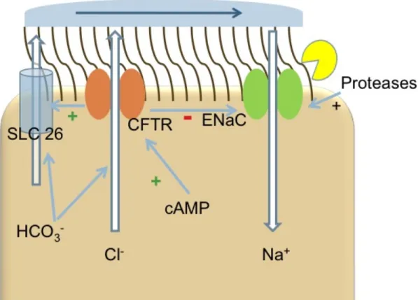
Figure 1. Model of Airway Ion Transport. Appropriate hydration of the airway surface liquid (ASL) is achieved, as a balance between Cl- efflux and Na+ influx is struck at the apical surface of airway epithelial cells. In addition to Cl- secretion, CFTR is coupled to HCO3- secretion. Together with SLC26, these channels largely influence the pH of the ASL. Once ENaC is cleaved by proteases, the opened channel allows
Chapter 2
SPLUNC1 EXPRESSION REDUCES SURFACE LEVELS OF THE EPITHELIAL SODIUM CHANNEL (ENAC) IN XENOPUS LAEVIS OOCYTES
Abstract
Throughout the body, the epithelial Na+ channel (ENaC) plays a critical role in salt and liquid homeostasis. In cystic fibrosis airways, for instance, improper regulation of ENaC results in hyperabsorption of sodium that causes dehydration of airway surface liquid. This dysregulation then contributes to mucus stasis and chronic lung infections. ENaC is known to undergo proteolytic cleavage, which is required for its ability to conduct Na+ ions. We have previously shown that the short, palate lung and nasal epithelial clone (SPLUNC1) binds to and inhibits ENaC in both airway epithelia and in Xenopus laevis oocytes. In this study, we found that SPLUNC1 was more potent at inhibiting ENaC than either SPLUNC2 or long PLUNC1 (LPLUNC1), two other PLUNC family proteins that are also expressed in airway epithelia. Furthermore, we were able to shed light on the potential mechanism of SPLUNC1’s inhibition of ENaC. While
currents by reducing the number of ENaCs in the plasma membrane. A better understanding of ENaC’s regulation by endogenous inhibitors may aid in the
development of novel therapies designed to inhibit hyperactive ENaC in cystic fibrosis epithelia.
Introduction
ENaC is the rate-limiting step for Na+ absorption in the airways (Gaillard, Kota et al. ; Kunzelmann and Mall 2002; Rossier and Stutts 2008). The importance of
appropriately regulated ion transport is illustrated by the pathogenesis that results when this channel fails to function correctly. For example, hyperabsorption of Na+ through ENaC has been proposed as an initiating event in cystic fibrosis (CF) lung disease (Knowles and Boucher 2002; Chmiel and Davis 2003). This likely occurs because Na+ hyperabsorption contributes to mucus dehydration and mucus stasis, which prevents clearance of inhaled pathogens (Knowles and Boucher 2002), (Chmiel and Davis 2003), (Matsui, Grubb et al. 1998). Furthermore, Na+ hyperabsorption, volume depletion, and inflammation have recently been demonstrated in transgenic mice overexpressing ENaC, thus directly linking ENaC and the initiation of chronic lung disease (Mall, Grubb et al. 2004). In contrast, as ENaC is down regulated in the monogenic disorder,
pseudohypoaldosteronism, it leads to an abundance of airway surface liquid and mucus transport rates that are significantly increased above normal levels (Kerem, Bistritzer et al. 1999).
proteases also regulate ENaC (Hughey, Carattino et al. 2007; Rossier and Stutts 2008). Rossier and colleagues initially identified a membrane-bound serine protease that acts as a channel activating protease (CAP) which they termed CAP1 (Vallet, Chraibi et al. 1997). The activation of ENaC by CAP1 can be mimicked by external addition of trypsin and the effects are not additive, indicating that CAP1 and trypsin act via the same
pathway. Additional CAPs, termed CAP2 (TMPRSS4) and CAP3 (matriptase) have also been identified as being able to activate ENaC by increasing its open probability (Po), without any significant change in channel density or conductance (Vuagniaux, Vallet et al. 2000; Rossier 2004). The effect of these CAPs can be blocked by the Kuitz-type serine protease inhibitor aprotinin (Vallet, Chraibi et al. 1997; Vuagniaux, Vallet et al. 2000) which prevents the cleavage of ENaC and the subsequent conduction of Na+ in both primary human bronchial epithelial cultures (HBECs) and Xenopus oocytes
(Bridges, Newton et al. 2001)(Donaldson, Hirsh et al. 2002; Myerburg, Butterworth et al. 2006).
proteins and incited theories that SPLUNC1 would similarly function in a direct innate host defense capacity (Bingle, LeClair et al. 2004). To date, however, this theory remains uncertain. Investigators have reported minimal anti-microbial function of SPLUNC1 (Bartlett, Hicks et al. 2008) and no binding to lipopolysaccaride, as might be expected from its strong homology to lipopolysaccaride binding proteins (Campos, Abreu et al. 2004). In contrast to its putative anti-microbial actions, we identified SPLUNC1 as a potent inhibitor of ENaC in both Xenopus oocytes and in human airway epithelia (Garcia-Caballero, Rasmussen et al. 2009). We also found that SPLUNC1 binds to ENaC and prevents its proteolytic cleavage by serine proteases including trypsin, CAP1 and CAP2 (Garcia-Caballero, Rasmussen et al. 2009). Since little is known about the SPLUNC1-ENaC interaction, we further explored this relationship and here we report that among the other PLUNC family members expressed in HBECs, SPLUNC1 alone significantly inhibits ENaC. We also demonstrate that SPLUNC1 exerts its effects on ENaC by lowering the number of ENaCs available for cleavage at the plasma membrane.
Results
We have previously demonstrated that SPLUNC1 is secreted into the media of SPLUNC1-injected oocytes where it binds to the extracellular loops of the α, β, γ ENaC
subunits, resulting in ENaC inhibition (Garcia-Caballero, Rasmussen et al. 2009). Data-mining of existing gene array data revealed that other PLUNC family members are expressed in our HBEC system, suggesting that other PLUNCs may be secreted into the airway surface liquid and could also regulate ENaC (Ribeiro, Hurd et al. 2009).
expressed under basal conditions. After exposing HBECs to supernatant of mucopurulent material (SMM), which is derived from CF airway secretions and acts as a powerful pro-inflammatory agent (Ribeiro, Paradiso et al. 2005), LPLUNC1 and SPLUNC1 expression were not altered. However, SPLUNC2 expression increased by 2.4 and 1.5 fold at 6 and 24 h post-SMM addition, respectively (Ribeiro, Hurd et al. 2009). To test whether these other PLUNC family members could also affect ENaC activity, we expressed
α,β,γ ENaC in Xenopus oocytes and measured their subsequent amiloride-sensitive
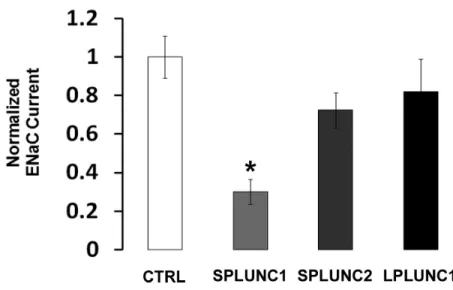
current under control conditions and following co-injection with either SPLUNC1, SPLUNC2, or LPLUNC1. ENaC currents were reduced by ~70% when SPLUNC1 was co-injected into the oocytes (Figure 1). In contrast, co-injection of SPLUNC2, which has 24% amino acid homology to SPLUNC1, or LPLUNC1, which has 2 domains rather than the single domain of the SPLUNC subgroup, and has 30% amino acid homology to SPLUNC1, had no significant effect on ENaC activity (Figure 1).
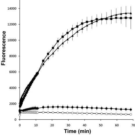
To understand how SPLUNC1 regulated ENaC, we examined whether SPLUNC1 could alter the proteolytic activity of trypsin. We have previously observed significant inhibition of ENaC by SPLUNC1 at 50 ng/ml, which is equivalent to 1.79 nM. At this same concentration, SPLUNC1 did not alter the ability of trypsin to cleave a serine protease-specific fluorogenic substrate, while the trypsin-specific inhibitor aprotinin, also administered at 1.79 nM, abolished proteolytic cleavage (Figure 2). Thus, we conclude that the regulation of ENaC by SPLUNC1 does not result from direct protease inhibition.
We have previously shown that SPLUNC1 can inhibit both basal and trypsin-stimulated ENaC currents (Garcia-Caballero, Rasmussen et al. 2009). The most
plasma membrane had been reduced by SPLUNC1. To test this hypothesis directly, we surface biotinylated oocytes and then probed for αENaC surface expression levels in the
presence and absence of SPLUNC1. SPLUNC1 indeed decreased the total amount of
αENaC in the plasma membrane, as compared to controls that did not express SPLUNC1
(Figure 3A). In contrast SPLUNC1 did not affect whole-cell ENaC protein levels (Figure 3B).
In order to confirm SPLUNC1’s ability to reduce ENaC levels in the plasma membrane, we used the sulfhydryl-reactive reagent [2-(trimethylammonium)ethyl
methanethiosulfonate, bromide] (MTSET), which locks ENaC containing a S518C mutation in the β subunit (βS518C) into the fully open position and thus yields an electrical
approximation of the number of active channels in the plasma membrane (Anantharam, Tian et al. 2006). With this system, a reduction in α, βS518C, γ-ENaC current in the
presence of MTSET would indicate fewer ENaCs in the plasma membrane, which we observed in oocytes coinjected with SPLUNC1 (Figure 3C). Moreover, the fold-stimulation of α, βS518C, γ-ENaC currents by MTSET was 5.32 ± 0.45 while groups
co-expressing SPLUNC1 had an 8.05 ± 0.48 fold increase. This indicates that ENaC resident on the cell surface in SPLUNC1 expressing oocytes reside in a low open probability state. Together with the enhanced fold-stimulation by trypsin
Discussion
We have previously reported that SPLUNC1 inhibits ENaC, likely via direct, extracellular binding to all three ENaC subunits (Garcia-Caballero, Rasmussen et al. 2009). Following on from these findings, we were curious to know whether the other PLUNC family members that are expressed in our HBEC system would produce similar inhibitory effects. While SPLUNC1 inhibits ENaC by ~70%, SPLUNC2 and LPLUNC1 do not significantly inhibit ENaC (Figure 1). These data are in keeping with our previous observations that the knockdown of SPLUNC1 to greater than 90% by shRNA
upregulated ENaC activity in HBECs and led to ASL volume hyperabsorption, despite expression of other PLUNC family members in these epithelia(Garcia-Caballero, Rasmussen et al. 2009). Interestingly, SPLUNC1 is able to reduce ENaC currents by 70% when applied at 50 ng/ml (1.79 nM) either in the Xenopus oocytes or in HBECs lacking endogenous SPLUNC1(Garcia-Caballero, Rasmussen et al. 2009), while this concentration of SPLUNC1 produces negligible direct effects on the proteolytic activity of trypsin, suggesting that SPLUNC1 does not act as a protease inhibitor (Figure 2). Thus, based on this and our previous data (Garcia-Caballero, Rasmussen et al. 2009), we conclude that SPLUNC1 acts by directly interacting with ENaC and not by direct
inhibition of extracellular proteases.
channel conductance (g) multiplied by both the open probability (Po) and the number of channels in the plasma membrane (N). Proteolytically-induced changes in I have been mainly attributed to altered Po and can be rapidly reversed by trypsin exposure (Vallet, Chraibi et al. 1997). In contrast, we found that acute trypsin exposure failed to fully reverse the effects of SPLUNC1 (Garcia-Caballero, Rasmussen et al. 2009), suggesting that the effects of SPLUNC1 are not exclusively limited to changes in Po. Our present data indicate that SPLUNC1 reduces the number of ENaCs subunits at the plasma
membrane, without affecting overall cellular ENaC levels (Figure 3A, B). These data are consistent with the effects of SPLUNC1 on ENaC currents (Figure 3C). For example, such a reduction in N would serve to lower basal ENaC currents in addition to preventing MTSET from further activating ENaC. While we have demonstrated this mechanism exclusively in oocytes (Figure 3), we have previously shown that SPLUNC1 decreases the ENaC/trypsin-sensitive potential difference in HBECs, suggesting that SPLUNC1 inhibits ENaC in a similar mechanism in airway epithelia (Garcia-Caballero, Rasmussen et al. 2009).
blocker, in a fashion analogous to amiloride, though with less reversible binding (Schild, Schneeberger et al. 1997). Such behavior could lead to a reduction in both basal and protease-activated ENaC currents and cannot be formerly excluded although this mode of inhibition alone is inconsistent with the observed decrease in N (Figure 3).
In summary, we have shown that SPLUNC1, but not other PLUNC family members, induces a potent inhibition of macroscopic ENaC current. While the
mechanism of SPLUNC1’s inhibition of ENaC is not fully understood, we have found that SPLUNC1 is capable of reducing the number of ENaC multimers at the plasma membrane. This is the first report of an endogenously secreted protein that can facilitate removal of ENaC from the plasma membrane.
Acknowledgements
Methods
SPLUNC1 protein purification. V5/6His-tagged SPLUNC1 was stably expressed in HEK293 cells and purified from their media as previously described using ethanol and acetone precipitation21.Media collected from HEK293 cells not expressing SPLUNC1 was purified using the same ethanol/acetone precipitation method and was used as a control.
Oocyte studies. Xenopus laevis oocytes were harvested and injected as described(Garcia-Caballero, Rasmussen et al. 2009). In brief, defolliculated healthy stage V–VI oocytes were injected with 0.3 ng of cRNA of each ENaC subunit. Injected oocytes were kept in modified Barth’s saline (in mM: 88 NaCl, 1 KCl, 2.4 NaHCO3, 0.3 Ca(NO3)2, 0.41 CaCl2, 0.82 MgSO4, and 15 HEPES, adjusted to pH 7.35 with Tris). Oocytes were
studied 24 h after injection using the two-electrode voltage clamp technique as previously described(Donaldson, Hirsh et al. 2002; Garcia-Caballero, Rasmussen et al. 2009).
Oocytes were clamped at a holding potential of −60 mV. The change in
amiloride-sensitive whole cell current as an indicator of ENaC activity was determined by
subtracting the corresponding current value measured in the presence of 10 µM amiloride
Western Blotting. Protein was harvested from Xenopus oocytes, resolved using SDS-PAGE and transferred to PVDF. The membrane was then probed using a anti-αENaC or
anti-V5 monoclonal antibodies (Invitrogen)) as previously described (Garcia-Caballero, Rasmussen et al. 2009).
Surface labeling. Xenopus oocytes were injected with V5-CT(C-terminus)-tagged
α, untagged−β,γ rat ENaC subunits (0.3 ng each) ± V5/6His-tagged SPLUNC1 (1 ng).
After 24 h, eggs (70 per experimental condition) were pre-chilled on ice for 30 minutes and labeled with 0.7 mg/ml sulfo-NHS-biotin in MBS-Ca++ (mM), 85 NaCl, 1 KCl, 2.4 NaHCO3, 0.82 MgSO4, 0.41 CaCl2, 0.33 Ca(NO3), 16.3 hepes titrated to pH 8.0 with NaOH, while tumbling gently for 20 min at 4°C. Oocytes were washed twice with chilled MBS-Ca++ buffer and incubated in MBS-Ca++ buffer with 100 mM glycine for 10 min at 4°C to quench free biotin. Eggs were washed again three times with chilled MBS-Ca2+ buffer. Proteins were extracted as previously described(Garcia-Caballero,
Rasmussen et al. 2009). Total inputs were taken from whole cell samples representing 4% of total protein. Solubilized proteins were incubated with 100 µl of neutravidin beads
Trypsin MCA Assay. The trypsin fluorogenic substrate assay was performed using 96 well black plates (Corning Costar). Boc-Gln-Ala-Arg-MCA (100 µM; Peptides
International) was added to each well in 100 µl Ringer total volume. Aprotinin
(Sigma-Aldrich) or SPLUNC1 as appropriate were then combined with the substrate, and trypsin (Sigma-Aldrich) was then added at 1 U/ml to initiate the reaction. Boc-Gln-Ala-Arg-MCA was excited at 380 ± 5 nm and emission was collected at timed intervals at 460 ± 15 nm using a Tecan Infinite multi-plate reader.
CHAPTER 2 FIGURES
Figure 1. The effect of PLUNC family members on ENaC activity. Current is displayed relative to amiloride-sensitive current from α,β,γ ENaC-expressing oocytes
Figure 3. SPLUNC1 decreases the number of αENaC subunits in the plasma
membrane. A, Surface biotinylation of αENaC shows that plasma membrane ENaC is
decreased following coexpression with SPLUNC1 in Xenopus oocytes. Samples were analyzed by Western blot using a anti-V5 (for V5/6His-tagged SPLUNC1 and for V5-CT-tagged αENaC) monoclonal antibody Lane 1, control; 2, αβγENaC, 3, αβγENaC &
SPLUNC1. Total lysate per lane was equivalent to 3-4 oocytes and was run on a 10% Gel. B, Whole cell western blot of oocytes probing for αENaC (top), SPLUNC1 and
actin (bottom) shows that total ENaC levels are not decreased by SPLUNC1. Total lysate per lane was equivalent to 3-4 oocytes and was run on a 12% Gel. C, Addition of MTSET to ENaC containing the βS518C mutant increases ENaC Po to 1.0 when
Chapter 3
THE ACTIVE SITE OF SPLUNC1 SHARES 40% HOMOLOGY WITH THE ENAC INHIBITORY PEPTIDE
Abstract
Hyperabsorption of Na+ through the epithelial Na+ channel (ENaC) contributes to cystic fibrosis (CF) lung disease and thus emphasizes the need to further understand the regulation of this channel. The short, palate lung and nasal epithelial clone
(SPLUNC1) is expressed in the submucosal glands and surface epithelia of the conducting airways and was recently identified as secreted protein, which negatively regulates ENaC. Since SPLUNC1 is a small, secreted protein (256 amino acids), identifying its active site may enable us to design a therapeutic peptide that inhibits ENaC. In this study we examined the ability of SPLUNC1 truncants to inhibit ENaC in Xenopus oocytes. The peptide sequence of functional mutants revealed that residues 22 to 39 were ~40% homologous to the ENaC inhibitory peptides, α26 and γ43, that are
We applied this labeled peptide to primary human bronchial epithelial cultures (HBECs) and observed increased the airway surface liquid (ASL) height, as compared to controls. Further understanding the regulation of ENaC by this region of SPLUNC1 and the S18 peptide may provide insight for new therapeutic avenues for CF patients.
Introduction
As the airways are constantly exposed to inhaled particulates that may be
infectious or noxious, they rely on efficient mucociliary clearance (MCC) to remove such threats. MCC is dependent on three coordinated components including ion transport, mucin secretion, and ciliary beating. Improper regulation of any component disrupts MCC, leaving the lung vulnerable to infection. Among these components, ion transport is critical for the hydration of the airway surface liquid (ASL) to volumes sufficient for ciliary extension and beating. Hydration also regulates the appropriate percent solids within the mucus layer that rests above the ASL, which is critical for efficient mucus clearance. The importance of precise regulation and coordination of these pathways becomes evident, as initiating events in chronic airway diseases, such as cystic fibrosis (CF), have been traced back to the failure of ion transport. Specifically ENaC is known to be hyperabsorptive in the airway epithelia of CF patients and contributes to dehydrated ASL, collapsed cilia, and high percent solid mucus. Collectively, as the MCC defense mechanism fails under these misregulated circumstances, CF patients are unable to clear inhaled pathogens and face life-threatening infections.
Currently, secondary messenger signals such as cAMP and PIP2 are known to regulate the function of ENaC (Rossier 2004). More recently, ENaC is known to be cleaved and activated by extracellular serine proteases, which are termed channel-activating proteases (CAPs) (Vallet, Chraibi et al. 1997). In CF, protease levels are also known to be
upregulated, leading to increased ENaC activity. Inhibition of these CAPs results in decreased ENaC conductance and increased MCC. Accordingly, further elucidation of ENaC regulation by protease cleavage is required so that novel therapies can be targeted to inhibit cleavage and activation of hyperactive ENaC.
Previous studies have identified several membrane bound CAPs and have indicated that CAP inhibitors are soluble(Garcia-Caballero, Rasmussen et al. 2009). Among these potential soluble inhibitors SPLUNC1 was identified, which proved to be a potent inhibitor of ENaC and thus a candidate volume-sensing molecule
(Garcia-Caballero, Rasmussen et al. 2009). As SPLUNC1 is a small protein (256 peptides) that is secreted into the ASL (Campos, Abreu et al. 2004), identifying its active site may provide insight for development of a yet smaller peptide inhibitor of hyperactive ENaC. In hopes of developing a therapeutic peptide for CF patients, we examined the ability of full-length and truncated SPLUNC1 to inhibit ENaC current. In this study we analyzed the peptide sequence of functional SPLUNC1 truncants to those of inhibitory peptides that originate from the cleavage of the extracellular loops of α and γ ENaC (Carattino,
Sheng et al. 2006; Carattino, Hughey et al. 2008). These peptides are referred to as α26
and γ43, which indicate the subunit of ENaC from which they are derived and the number
peptides. The homology between these 18 amino acids of SPLUNC1 and α26 is 40%
while the homology to γ43 is 35%. Cumulatively, this region of SPLUNC1 shares 78%
homology with both α26 and γ43. We hypothesized that this region of SPLUNC1 may
be responsible for the inhibition of ENaC current. Further, four proline residues in this 18 amino acid region of SPLUNC1 are homologous to proline residue that were
necessary for the function of the ENaC inhibitory peptides. We speculate that these prolines (P25, 27, 33, and 38) may be necessary for ENaC inhibitions by SPLUNC1. We observed a reduction in ENaC inhibition in SPLUNC1 truncants containing proline point mutations. Finally, we designed a mimetic peptide to the proposed active site of
SPLUNC1 termed, S18. We found this peptide to be functional, as it decreased ENaC activity in Xenopus laevis oocytes and increased HBEC ASL height following S18 administration.
Results
It was recently show that peptide fragments released from the extracellular domains of ENaC following proteolytic cleavage, termed α26 and γ43, have inhibitory
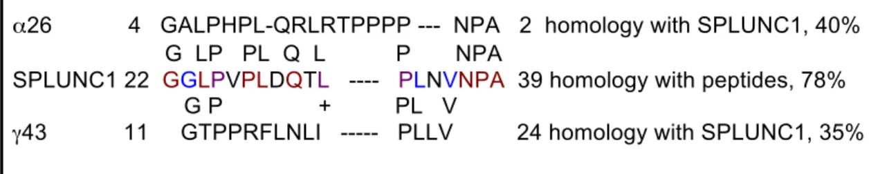
effects on ENaC (Carattino, Sheng et al. 2006; Carattino, Hughey et al. 2008). As SPLUNC1 is a small, secreted protein that also inhibits ENaC, we searched for any sequence similarities between SPLUNC1 and these peptides. Amino acid sequence alignment revealed a striking similarity. From residues 22 to 39, SPLUNC1 shares 40% homology with α26 and 35% homology with γ43 (Figure 1). Additionally, within this
function of the ENaC inhibitory peptide fragments (Carattino, Sheng et al. 2006; Carattino, Hughey et al. 2008). We proposed that this high homology region of SPLUNC1’s sequence might be the active site for its inhibition of ENaC.
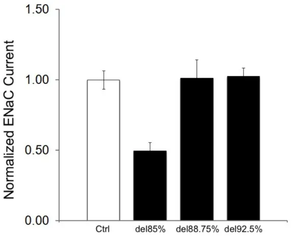
To test this hypothesis, we designed several SPLUNC1 truncants (Figure 2). Deletions up to 85% of full length SPLUNC1 from the C-terminal retained the putative active site while deletions of 88.75% bisected this site and 92.5% removed the site. These SPLUNC1 truncants were then co-injected with α, β, γ-ENaC subunits into
Xenopus oocytes and current was recorded by voltage clamp technique. Currents from control oocytes injected with α, β, γ-ENaC and water were set to 1 nA/nA and compared
to those co-expressing SPLUNC1 truncants. As seen in Figures 3 and 4, significant ENaC inhibition was observed for SPLUNC1 deletions up to 85%, which retained the putative active site, but this inhibition was lost for deletions that disrupted the site (control 1 ± 0.04, SPLUNC1 0.52 ± 0.03, SPLUNC1del30% 0.389± 0.09, SPLUNC1 del60%
0.45 ± 0.06, SPLUNC1del85% 0.49 ± 0.06, SPLUNC1del88.75% 1.01 ± 0.13,
SPLUNC1del92.5% 1.03
± 0.06; n=12-18 per group, p<0.05). Interestingly, point mutations
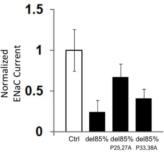
of proline residues P25, 27A and P33, 38A within the putative active site of the SPLUNC1del85% truncant reduced the efficiency of ENaC inhibition (Figure 5). This suggests that these proline residues are important for SPLUNC1’s function and that point mutations of all residues, P25, 27, 33, 38A may abolish its ENaC inhibitory effects.
voltage clamp technique. Incubation for 90 minutes of 10µM S18 significantly reduced baseline current by 51% and trypsin-activated current by 52% (Figure 6). Of note, acute addition at the same concentration rendered no effect on ENaC current (Figure 7).
We then questioned if the S18 peptide could effect ASL height regulation in normal primary human bronchial epithelial cultures (HBECs). During preliminary work, S18 was applied at 100µM at the start of a 24-hour time course and ASL height was measured by confocal microscopy. As compared to cultures treated with vehicle alone, HBECs exposed to S18 exhibited a higher ASL height over time (Appendix, Figure 1). These pilot studies suggest that the S18 peptide could increase ASL height by inhibiting Na+ absorption through ENaC and strongly encourage further investigations with S18.
Discussion
As SPLUNC1 is an endogenously secreted protein in the airways, which has known ENaC inhibitory properties (Garcia-Caballero, Rasmussen et al. 2009; Rollins, Garcia-Caballero et al. 2010), characterizing the region of this protein with strong homology to the ENaC inhibitory peptides, α26 and γ43 is an important step for
identifying the active site. We engineered SPLUNC1 truncants in hopes of isolating the region of the protein that was responsible for ENaC inhibition. We considered residues 22 to 39 of SPLUNC1 to be the putative active site, as it held strong homology to the ENaC inhibitory peptide fragments. Voltage clamp current recordings of oocytes co-injected with α, β, γ-ENaC and these truncants revealed significant ENaC inhibition by
truncates deleting as much as 85% of the full-length protein from the C-terminus, deletions of 88.75 and 92.5% bisected and omitted the putative active site, respectively, and had no inhibitory effect on ENaC current. From these findings we concluded that this region of SPLUNC1 was necessary for its inhibition of ENaC.
A span of 8 residues from positions 211 to 218 of the α26 ENaC inhibitory
peptide was identified as the key inhibitory region (Carattino, Passero et al. 2008). Residues L1, P2, H3, P4, and L8 were specifically required for inhibitory activity. Interestingly, from residues 24 to 32 of the proposed active site of SPLUNC1 a 75%
homology can be found with the 8 residue key inhibitory domain of α26. Further, critical
residues L1, P2, P4, and L8 of α26 are conserved in the putative active site of SPLUNC1.
We point mutated resides P25 and P27 of SPLUNC1, which correspond to P2 and P4 of
the key inhibitory region of α26. We also point mutated the remaining prolines of the
SPLUNC1 active site P33, 38 to alanines. Setting the normalized ENaC current for α, β
,γ-ENaC expressing control oocytes to 1 nA/nA, we compared the current of oocytes
co-expressing SPLUNC1del85%, P25, 27A SPLUNC1del85%, and P33, 38A SPLUNC1del85% to
controls. Co-expression of SPLUNC1del85% reduced ENaC current by 76%, while
co-expression of P25, 27A SPLUNC1del85% reduced ENaC current by only 32%.
Co-expression with P33, 38A SPLUNC1del85% still reduced current by 59%. This suggests
that P25 and P27 of SPLUNC1, which correlate to P2 and P4 of the 8-residue key
inhibitory domain of α26 are important for efficient ENaC inhibition.
Based on our findings that the 18 amino acids spanning residues 22 to 39 of
SPLUNC1 play an important role in the protein’s ability to inhibit ENaC, we designed a
the sequence, GGLPVPLDQTLPLNVNPA. Oocytes expressing α, β, γ-ENaC that were
incubated for 90 minutes with 10µM S18 peptide exhibited a significant, 51% reduction in current, as compared to these oocytes in the presence of vehicle alone (Figure 6).
Acute addition of 10µM S18 peptide, however, did not reduce ENaC current (Figure 7). These data may provide clues into the mechanism of action of S18. As we previously demonstrated that SPLUNC1 reduces the number of ENaCs in the membrane as a means of reducing macroscopic current (Rollins, Garcia-Caballero et al. 2010), the required incubation time for S18 to function may indicate a similar means of ENaC inhibition. Further, the 52% reduction in trypsin-activated ENaC current by S18 suggests that the number of membrane localized channels available for cleavage-dependant activation is reduced. Characterizing how SPLUNC1 and the S18 peptide inhibits ENaC remain important areas of research. It may be found that full-length SPLUNC1 and the S18 peptide have different mechanisms for inhibiting ENaC. Perhaps through the proposed active site, SPLUNC1 and the S18 peptide can affect conformational changes in ENaC, which stimulates internalization. Specifically, the ENaC inhibitory peptides have been proposed to prevent conformational changes that facilitate Na+ conductance and
blocking CAP access to ENaC (Garcia-Caballero, Rasmussen et al. 2009). Analysis of ENaC cleavage assays in the presence of the S18 peptide will be useful tools for addressing this important question.
Our pilot studies with S18 effects on ASL height regulation in HBECs also encourage further investigations with this peptide. We found that normal HBECs treated with 100µM S18 maintained a higher ASL height over a 24-hour time course, as
compared to untreated cultures (Appendix, Figure 1). We speculate that S18 inhibits ENaC in HBECs, as in our studies using injected oocytes. Reducing Na+ conductance through ENaC would slow HBEC’s ability to absorb excess ASL over time, as we observed. This insight of S18’s function in HBECs raises further questions, though. Within the complex environment of the HBEC system, it will be important to know if S18’s effect on ASL height regulation is ENaC-specific. Understanding S18’s ability to affect the Po and N of ENaCs within HBECs should also be addressed. In addition to these investigations, testing for the ability of S18 to work in CF airways will provide important insight for a potentially novel therapeutic tool.
Methods
Sequence Analysis: Homologous sequences between SPLUNC1 and peptides, α26 and
γ43, were identified using protein BLAST searches.
C-terminus at various lengths. Resulting constructs included, bisected, or removed the region homologous to the ENaC inhibitory peptide. Additionally, four prolines were mutated to alanines within this region of homology to α26 and γ43. This point mutations
were engineered within the SPLUNC1del85% truncant and termed SPLUNC1del85% P25,27A and SPLUNC1del85% P33, and 38A.
Oocyte studies. Xenopus laevis oocytes were harvested and injected as described (Garcia-Caballero, Rasmussen et al. 2009). In brief, defolliculated healthy stage V–VI oocytes were injected with 0.3 ng of cRNA of each ENaC subunit. Injected oocytes were kept in modified Barth’s saline (in mM: 88 NaCl, 1 KCl, 2.4 NaHCO3, 0.3 Ca(NO3)2, 0.41 CaCl2, 0.82 MgSO4, and 15 HEPES, adjusted to pH 7.35 with Tris). Oocytes were studied 24 hours after injection using the two-electrode voltage clamp technique as previously described (Donaldson, Hirsh et al. 2002; Garcia-Caballero, Rasmussen et al. 2009). Oocytes were clamped at a holding potential of -60 mV. The change in amiloride-sensitive whole cell current indicated of ENaC activity and was determined by
Peptide Design: The S18 peptide was synthesized and purified by the UNC Peptide Synthesis Facility with sequence, GGLPVPLDQTLPLNVNPA. The peptide was amino-terminal acetylated and caroboxy-amino-terminal amidated
Chapter 3 Figures
Figure 1: Alignment of SPLUNC1 to inhibitory peptides, α26 and γ43, cleaved
from ENaC’s extracellular loops. Alignment to α26 residues are shown in red and
those to γ43 are shown in blue while alignments to both are shown in purple.
α26 4 GALPHPL-QRLRTPPPP --- NPA 2 homology with SPLUNC1, 40%
G LP PL Q L P NPA
SPLUNC1 22 GGLPVPLDQTL ---- PLNVNPA 39 homology with peptides, 78% G P + PL V
Figure 4: SPLUNC1 truncates lacking the active site fail to inhibit ENaC. While the SPLUNC1del85% contains the active site and inhibits ENaC, the SPLUNC1del88.75% and SPLUNC1del92.5% truncants, which bisect or omit the active site of SPLUNC1,
Figure 5: Proline point mutations within SPLUNC1’s Active Site reduce its
Figure 6: Inhibition of ENaC current by addition of S18 peptide. Significant ENaC inhibition ( n=6, p<0.0001) is seen following 90 minute incubation with 10µM of the S18 peptide (Black Bars) reduces baseline and trypsin-activated ENaC current in Xenopus Oocytes expressing α, β, γ-ENaC subunits., as compared to ENaC-expressing oocytes in
Chapter 4
ACIDIC ENVIRONEMNTS, LIKE THOSE IN CYSTIC FIBROSIS AIRWAYS, PREVENT SPLUNC1’S INHIBITION OF ENAC
Abstract
Ion transport is a key component of mucociliary clearance (MCC), which the airways rely upon for successful removal of inhaled pathogens. Abnormal ion transport occurs in Cystic Fibrosis, as CFTR fails to secrete Cl- and as ENaC’s absorption of Na+ is hyperactive. This results in dehydrated airway surface liquid (ASL) that leads to higher percent solid mucus and collapsed cilia. Under these conditions, MCC fails and CF patients face life-threatening infections. Recent investigations have uncovered that impaired bicarbonate secretion due to the loss of functional CFTR affects the pH of CF ASL. As CFTR contributes to NaHCO3 secretions, its absence in CF leads to an acidic environment. In this report, we show that the endogenously secreted inhibitor of ENaC, SPLUNC1, fails to impede Na+ absorption in acidic environments. We propose that this pH-dependency could explain the loss of SPLUNC1 function in CF, despite its
present a novel approach to thin-film pH measurements, as a powerful tool for investigating pH effects on airway cultures. Together, this work describes important consequences of misregulated pH in CF airways and highlights the need for further investigations to develop corrective therapies for CF patients.
Introduction
As a genetic disorder, CF is characterized by a dysfunctional CFTR anion channel (Welsh and Smith 1993). One of the initiating events in the disease, however, has been linked to the hyperabsorption of sodium through ENaC, as this channel is misregulated in the absence of CFTR (Knowles and Boucher 2002). This imbalance of ion transport contributes to a dehydrated airway surface liquid (ASL) to levels insufficient for cilia extension. Once collapsed, ciliary beating is inhibited and inhaled pathogens cannot be cleared. Furthermore, the loss of functional CFTR adversely alters the pH of the ASL, as CFTR is capable of conducting HCO3- and affects Cl- coupled NaHCO3 transport
(Poulsen, Fischer et al. 1994; Choi, Lee et al. 2001). As a consequence CF airways become more acidic than normal airways (Wine 2006).
While dehydrated ASL and increased percent solids of mucus are well
therapies. SPLUNC1, for instance, has recently been identified as an endogenously secreted inhibitor of ENaC in the normal airways (Garcia-Caballero, Rasmussen et al. 2009; Rollins, Garcia-Caballero et al. 2010). Despite the abundance of SPLUNC1 protein in the ASL of CF HBECS ENaC remains hyperactive. As exogenous protease inhibitors such as aprotinin can still inhibit ENaC in CF airways, we propose that SPLUNC1’s action may be defective, rather than ENaC’s. To test this hypothesis, we examined the ability of SPLUNC1 to inhibit ENaC over a range of pH’s. Here, we report maximal inhibition of ENaC by SPLUNC1 in physiological pH ranges and a loss of inhibition in acidic environments. In Chapter 3, we demonstrated ENaC inhibition by SPLUNC1 truncants that contain the putative active site between residues 22 to 39. For this study, we examined their effects in the acidic conditions of CF ASL. Like full-length SPLUNC1, these truncants lose their ability to inhibit ENaC in acidic conditions. These findings of pH dependency provide a possible explanation for the loss of SPLUNC1 function in CF airways, despite its abundance at the protein level.
As pH affects this important component of airway physiology, we then questioned if reducing the acidic environment of CF HBECs could restore more normal ASL volume regulation. Towards these efforts, we selected biological buffers with alkaline pH buffering ranges for administration to CF HBECs. Indeed, CF HBECs treated with buffer solutions maintained a higher ASL, as compared to untreated CF HBECs. Additionally, CF HBECs with reduced acidity demonstrated spontaneous ENaC
While pH measurements of lavaged ASL from normal and CF HBECs reveal an acidic environment in CF cultures (Coakley, Grubb et al. 2003), we aimed to take thin-film pH measurements from these cultures. This approach would provide a more
accurate understanding of the pH in native conditions and during treatment with alkaline buffers. This work establishes a novel approach for thin-film pH measurements over a broad pH range by combining two pH-sensitive fluorophores. Using this technique, we drew a correlation to the increase in ASL height to the reduced acidity of CF HBEC ASL.
Results
Previous studies have reported an abnormal acidic environment in CF airways (Coakley, Grubb et al. 2003). As exogenous inhibitors, such as aprotinin, can still inhibit ENaC in CF HBECs we speculated that endogenous ENaC inhibitors are malfunctioning in the acidic enviorment. As an endogenous regulator of ENaC, SPLUNC1 activity was examined under various pH conditions. Xenopus oocytes were co0injected with α, β, γ
-ENaC and SPLUNC1 or H2O, as a control, and incubated in a range of buffered solutions. We found a pH-dependent inhibition of ENaC (Figure 1). Maximal ENaC inhibition by SPLUNC1 occurred in physiological pH conditions mimicking those of normal HBECs, but was lost in the acidic environment like those found in CF HBECs. We also tested for any pH-dependent activity of SPLUNC1 mutants that are deleted from the C-terminus, but include the previously identified active site of SPLUNC1. Consistent with the behavior of full-length SPLUNC1, these truncants SPLUNC1del30%, SPLUNC1 del60%, and SPLUNC1del85% maintain the ability to inhibit ENaC at pH 7.4 but were
Having identified a potential explanation for the loss of ENaC inhibition in the acidic environment of CF airways, we attempted to correct the abnormal pH in CF HBECs by buffer administration. Biological buffers were selected with alkaline
buffering ranges in hopes of reducing the acidity of CF HBECs (Table 1). Proposing that larger molecules would be retained longer in the ASL to sustain their buffering capacity, we also considered the molecular weight of each buffer. Basically buffered solutions were apically administered to CF HBECs in efforts to reduce ASL acidity and to
potentially improve the ability of CF cultures to regulate ASL height under these altered conditions. As an indicator of ion transport function in these cultures, ASL height was measured by confocal microscopy over the time course of buffer administration. We found that biological buffers POPSO, AMPSO, and CAPSO initially set to pH 8.5 were effective in preventing hypersaborption. Compared to control cultures in pH 7.4 PBS, treated CF HBECs maintained a higher ASL of the 24-hour time course (Figure 3).
To address how this buffer treatment affected ENaC activity we measured the thin-film potential difference (PD) of CF HBECs. PD measurements on normal cultures reveal that ENaC activity can be inhibited spontaneously, as endogenous inhibitors accumulate over time (Garcia-Caballero, Rasmussen et al. 2009). Here, we show that this spontaneous inhibition over time does not occur in untreated CF cultures, but is recovered in CF HBECs treated with a solution buffered to pH 8.2 (Figure 4).
appear to be maximally active. Treatment with the basically buffered solution, however, reestablished ENaC activation by the exogenous trypsin administration (Figure 4).
In order to more accurately describe the pH environment in CF cultures in native conditions and during buffer treatment, we attempted to take thin-film pH measurements on these cultures. This was accomplished by combining two pH-sensitive fluorophores, SNARF-1 and FITC (Figure 5), and by recording the appropriate excitation and emission wavelength ratios of these dyes while on the HBECs. Over a 24-hour time course, the pH was calculated from these measurements (Figure 6). We found a correlation between decreased ASL acidity and ASL height. The buffer with the largest molecular weight, POPSO, also sustained buffering effects and ASL height increases for longer periods of time. Additionally, we noticed that the thin-film pH measurement in CF HBECs at the 0 hour time point was already more acidic than the initial pH 8.5 solution. To address this discrepancy, we asked if pH measurements from a lavage of the buffer solution would reveal its acidification. Surprisingly, however, there was no change in pH between pre- and post-application of the buffer solution (Figure 7). This suggested, perhaps, that fixed components on the HBECs surface were contributing to the acidic environment. One possible source of fixed charges is the collection of mucins, which are tethered to airway epithelia (Raynal, Hardingham et al. 2003).
Discussion
SPLUNC1 inhibits ENaC current. Specifically, the ASL where SPLUNC1 attempts to bind and inhibit ENaC is subject to abnormal bicarbonate transport in CF airways and becomes more acidic than normal airways (Coakley, Grubb et al. 2003). Our data suggest that the acidic ASL of CF airways prevent the proper inhibition of ENaC by SPLUNC1.
Our voltage clamp data studies showed that SPLUNC1 inhibits ENaC in normal physiological pH conditions, but fails to do so in acidic environments, which mimic those of CF airways (Figure 1). The abnormal pH environment could alter the confirmation of SPLUNC1 to prevent its binding to ENaC. As we found SPLUNC1 truncants to be functional in normal pH conditions with as much as 85% of the protein deleted from the C-terminus, there are few possibilities for pH-sensitive SPLUNC1 conformational changes. Additionally, the functional 18 amino acid SPLUNC1 peptide, S18, has only one charged residue, D29. The extracellular loops of ENaC, though, are also exposed to the acidic environment and may have pH-induced conformational changes that prevent SPLUNC1 binding. Within the α-ENaC subunit, three histidine residues (H283, 326, and
327), for instance, are subject to protonation in acidic conditions, which could perhaps mediate SPLUNC1 interactions. Utilizing point mutants at specific pH sensitive residues within SPLUNC1 and ENaC to record current in various pH conditions may shed light on the binding site of these proteins.
included POPSO, CAPSO, and AMPSO. Initially set to a pH of 8.5, these buffers increased the ASL height of CF HBECs over a 24-hour time course, as compared to cultures treated with PBS buffered to pH 7.4 (Figure 4). While ASL height regulation is highly complex, this finding is encouraging for a new approach in restoring mucociliary clearance for CF patients. Identifying how these regulatory components are affected by buffer treatment will be key studies for this goal. Here, we demonstrated that recovered responsiveness of ENaC to endogenous inhibitors during buffer treatment (Figure 4). This is likely a critical component of this recovered homeostasis, as ASL height also increased during buffer treatment (Figure 3). Thin-film measurements show that freshly washed normal HBECs have high ENaC activity, which can be spontaneously inactivated over time as endogenous inhibitors, such as SPLUNC1, accumulate (Garcia-Caballero, Rasmussen et al. 2009). These measurements in CF HBECs reveal that the accumulation of inhibitors fails to reduce ENaC activity in the natively acidic environments (Figure 4). CF HBECs treated with HEPES buffered solution to pH 8.2, however, regained
responsiveness to inhibitors over time. Additionally, ENaC activity was maximal in CF cultures, as exogenous trypsin administration could not increase thin-film PD
measurements. Trypsin-activated current was also recovered in buffer-treated cultures (Figure 4). Though further investigations are required to describe buffer effects on ion secretion, for instance, this work demonstrates that sodium hyperabsorption through ENaC my be halted in less acidic conditions.
the largest molecular weight, POPSO, established a larger ASL height increase and sustained its buffering effects for longer. Potentially, buffers with larger molecular could be retained longer in the ASL, as absorption through the monolayer is impeded based on size exclusion.
This work describes important effects of pH on components of ASL height regulation. Pursuit of this phenomenon appears to be a fruitful area of research for recovering ASL height homeostasis. Our data suggest that therapies aimed to
reconstitute normal physiological pH conditions for CF patients could improve MCC. Further understanding of pH-induced conformational changes in ENaC or its inhibitors, such as SPLUNC1, may allow for the development of compounds that bind and inhibit ENaC in these abnormal conditions. Additionally, as channel activating proteases (CAPs) have recently been identified as a major regulatory component of ENaC activity, it will also be necessary to characterize the role of acidic pH in these specific interaction. Ultimately, work in this field could lead to a new approach for treating impaired MCC in CF.
Acknoledgments
We thank Dr. Robert Tarran for his data presented in Figure 4.
Methods
Biological Buffer Treatment: Buffer solutions set to 290 mOsm were composed of either Piperazine-1,4-bis(2-hydroxypropanesulfonic acid) dihydrate (POPSO),