State of the Art
Diagnosis, Monitoring, and Treatment of Primary
Ciliary Dyskinesia: PCD Foundation Consensus
Recommendations Based on State of the Art Review
Adam J. Shapiro,
MD,1* Maimoona A. Zariwala,
PhD,2Thomas Ferkol,
MD,3Stephanie D. Davis,
MD,4Scott D. Sagel,
MD, PhD,5Sharon D. Dell,
MD,6Margaret Rosenfeld,
MD,7Kenneth N. Olivier,
MD,8§Carlos Milla,
MD,9Sam J. Daniel,
MD,10Adam J. Kimple,
MD,11Michele Manion,
12Michael R. Knowles,
MD,13and Margaret W. Leigh,
MD,14for the Genetic Disorders of Mucociliary Clearance Consortium
Summary. Primary ciliary dyskinesia (PCD) is a genetically heterogeneous, rare lung disease resulting in chronic oto-sino-pulmonary disease in both children and adults. Many physicians incorrectly diagnose PCD or eliminate PCD from their differential diagnosis due to inexperience with diagnostic testing methods. Thus far, all therapies used for PCD are unproven through large clinical trials. This review article outlines consensus recommendations from PCD physicians in North America who have been engaged in a PCD centered research consortium for the last 10 years. These recommendations have been adopted by the governing board of the PCD Foundation to provide guidance for PCD clinical centers for diagnostic testing, monitoring, and appropriate short and long-term therapeutics in PCD patients.Pediatr Pulmonol. 2016;51:115– 132. ß2015 The Authors.Pediatric PulmonologyPublished by Wiley Periodicals, Inc.
This is an open access article under the terms of the Creative Commons Attribution-NonCommercial-NoDerivs License, which permits use and distribution in any medium, provided the original work is properly cited, the use is non-commercial and no modifications or adaptations are made.
1
Department of Pediatrics, Montreal Children’s Hospital, McGill Universi-ty, Quebec, Canada.
2
Department of Pathology and Laboratory Medicine, University of North Carolina School of Medicine, Marsico Lung Institute, Chapel Hill, North Carolina.
3
Department of Pediatrics, Washington University School of Medicine, St. Louis, Missouri.
4
Department of Pediatrics, Riley Hospital for Children, Indiana University, Indianapolis, Indiana.
5
Department of Pediatrics, Children’s Hospital Colorado and University of Colorado School of Medicine, Aurora, Colorado.
6
Department of Pediatrics, The Hospital for Sick Children and University of Toronto, Toronto, Ontario, Canada.
7
Department of Pediatrics, Seattle Children’s Hospital and University of Washington, Seattle, Washington.
8
National Heart, Lung, and Blood Institute, Bethesda, Maryland.
9Department of Pediatrics, Stanford University, Palo Alto, California. 10
Department of Otolaryngology, Montreal Children’s Hospital, McGill University, Montreal, Quebec, Canada.
11
Department of Otolaryngology—Head and Neck Surgery, University of North Carolina School of Medicine, Chapel Hill, North Carolina.
12
The PCD Foundation—President, Minneapolis, Minnesota.
13
Department of Medicine, University of North Carolina, Marsico Lung Institute, Chapel Hill, North Carolina.
14
Department of Pediatrics, University of North Carolina, Marsico Lung Institute, Chapel Hill, North Carolina.
§Supported in part by the intramural research program of the NHLBI, NIH.
Michael R. Knowles and Margaret W. Leigh are co-senior who contributed equally to this statement as senior authors.
Correspondence to: A.J. Shapiro, MD, Department of Pediatrics, Montreal
Children’s Hospital, McGill University, 2300 Tupper, D-380, Montreal, Quebec, Canada, H3H 1P3. E-mail: [email protected]
Received 1 May 2015; Revised 30 June 2015; Accepted 21 August 2015.
DOI 10.1002/ppul.23304
Published online 29 September 2015 in Wiley Online Library (wileyonlinelibrary.com).
Key words: primary ciliary dyskinesia; PCD, kartagener; consensus statement; PCD Foundation.
Funding source: National Institutes of Health (NIH), Number: U54HL096458, 5R01HL071798; The Genetic Disorders of Mucociliary Clearance (U54HL096458) is a part of the NCATS Rare Diseases Clinical Research Network (RDCRN). RDCRN is an initiative of the Office of Rare Diseases Research (ORDR), NCATS, funded through a collaboration between NCATS and NHLBI; CTSA NIH/NCATS UNC ULTR000083; CTSA NIH/NCATS Colorado UL1TR000154; Intramural Research Program of NIH/NIAID.
INTRODUCTION
Primary ciliary dyskinesia (PCD) is a genetically
heterogeneous, rare lung disease causing chronic
oto-sino-pulmonary disease and irreversible lung damage that
may progress to respiratory failure.
1–3Recently,
signifi-cant progress has been made in PCD diagnosis,
4yet few
physicians outside of highly experienced PCD centers are
skilled in recognizing the characteristic clinical
pheno-type and interpreting diagnostic tests.
5–9Patients often
receive false-positive or false-negative PCD diagnoses, as
physicians are unaware of the pitfalls commonly
encountered with ciliary electron microscopy,
10,11PCD
molecular genetic panels,
12,13ciliary motility
stud-ies,
14–16and nasal nitric oxide testing.
17,18Furthermore,
PCD is often missed when respiratory symptoms are
present in patients with other complex diseases involving
cilia, such as heterotaxy and various genetic
syn-dromes.
19–22From a therapeutic perspective, there are
no prospective, randomized clinical trials on monitoring
or treating PCD. Thus, physicians treating PCD adapt
therapeutic approaches used for other chronic respiratory
diseases, such as cystic fibrosis (CF) and non-CF
bronchiectasis. Differences in various phenotypic
param-eters among PCD, CF, and non-CF bronchiectasis suggest
that extrapolating therapies may not be appropriate for
PCD management in some circumstances.
23–26Because of the uncertainty surrounding diagnosis and
management of PCD, physicians from the Genetic
Disorders
of
Mucociliary
Clearance
Consortium
(GDMCC) created this consensus statement to guide
new North American PCD clinical centers endorsed by
the PCD Foundation. The GDMCC includes clinicians at
nine academic centers in North America that have
systematically evaluated over 1,000 patients suspected
of having PCD and performed longitudinal studies of
pediatric patients with a confirmed diagnosis of PCD. The
GDMCC also works closely with the PCD Foundation on
research and clinical PCD projects. This consensus
statement is evidence based where possible, and addresses
key clinical PCD issues, but it is not the product of
GRADE recommendations.
27Through telephone
confer-ences, email communications, and in person meetings,
eight pediatric pulmonologists, two adult pulmonologists,
and two otolaryngologists from North America undertook
to: (1) describe the PCD clinical phenotype, (2) establish
standard PCD diagnostic recommendations, (3)
recom-mend PCD clinical care and long-term monitoring
schedules, and (4) outline clinical therapies used to
manage PCD. After a literature review (using Pubmed and
Embase), drafts were created and circulated iteratively to
participating physicians with discussion of feedback and
suggestions over sequential telephone conferences and
electronic communications. Participating GDMCC
physi-cians and the PCD Foundation governing board
unani-mously approved this consensus statement.
PCD CLINICAL PHENOTYPE
Clinical symptoms in PCD affect the entire respiratory
tract; the majority of symptoms occur on a chronic, daily
basis and start soon after birth (Table 1). At least 80% of
newborn babies with PCD develop neonatal respiratory
distress despite a full-term gestation, with increased work
of breathing, tachypnea, and prevalence of upper and
middle lobe atelectasis on chest radiographs.
28Most PCD
patients are well immediately after birth, but develop
ABBREVIATIONS:
ABPA allergic bronchopulmonary aspergillosis CF cystic fibrosis
CFTR cystic fibrosis transmembrane regulator CRS chronic rhinosinusitis
CT computed tomography EM electron microscopy ESS endoscopic sinus surgery
GDMCC Genetic Disorders of Mucociliary Clearance Consortium IDA inner dynein arm
IF immunofluorescence testing IVIG intravenous immunoglobulin MTD microtubule disorganization N-DRC nexin-dynein regulatory complex NGS next generation sequencing nNO nasal nitric oxide
NTM non-tuberculosis mycobacterium ODA outer dynein arm
OME otitis media with effusion PBB protracted bacterial bronchitis PCD primary ciliary dyskinesia PET pressure equalization tubes ROM recurrent otitis media SIT situs inversus totalis SA situs ambiguus
respiratory distress at 12–24 hr of life (as opposed to other
causes of respiratory distress in term neonates (e.g.,
transient tachypnea of the newborn—TTN), which often
present in the first few hours after birth). A small
proportion of PCD patients are discharged home on day 1
of life but are then hospitalized with respiratory distress
within the first few weeks of life. Often misdiagnosed
with TTN or pneumonia, PCD infants frequently require
supplemental oxygen for days to weeks. When neonatal
respiratory distress appears, particularly with situs
inversus totalis or other situs anomalies, PCD should be
investigated.
At least 80% of PCD patients also have year-round,
daily nasal congestion (or chronic sinusitis in older
children and adults), which appears in early infancy and
does not resolve with changes of season or between viral
infections. Nasal polyps can occur in PCD,
29and nearly
all PCD patients demonstrate severe pansinusitis on
computed tomography (CT) scan.
4Persistent, year-round, daily cough from early infancy
is present in nearly 100% of PCD patients.
4The cough is
usually wet and productive, even in infancy, yet
occasionally patients report dry cough. The cough can
partially improve with antibiotic therapy, but does not
resolve with therapy or changes of season. Conversely,
episodic cough alternating with symptom-free periods is
unlikely to be from PCD.
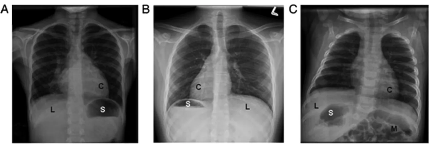
A spectrum of organ laterality defects occur with PCD,
including situs inversus totalis (SIT—mirror-image
arrangement) and situs ambiguus (SA—arrangement
falls somewhere between normal and mirror image;
Fig. 1). SA may be associated with complex congenital
heart disease (known as heterotaxy), yet mild cardiac
septal defects can also occur with PCD. SIT occurs in
slightly less than 50% of PCD patients,
30whereas SA
occurs in at least 12% of PCD.
19Subtle laterality defects
(e.g., intestinal malrotation, interrupted inferior vena
cava, or polysplenia) may be undetected in PCD patients
without further imaging studies, such as abdominal
ultrasound, spleen scan, or echocardiogram. In patients
with chronic oto-sino-pulmonary disease and any organ
laterality or cardiac defect, PCD should be considered.
Recurrent otitis media (ROM) with chronic middle ear
effusion affects at least 80% of children with PCD,
particularly in the first year of life.
31Complications of
ROM may include multiple sets of pressure equalization
tubes, conductive hearing loss, speech/language delay, or
need for hearing aids.
32Chronic middle ear disease is
quite common in the general pediatric population;
thereby, ROM alone is insufficient to warrant further
PCD testing. The absence of ROM goes against, but does
not rule out, a diagnosis of PCD.
Recurrent pneumonia or bronchitis is common in
PCD; however, some infants will lack this history due
to frequent antibiotics for nasal discharge and otitis
media. By preschool age, up to 80% of PCD patients
have recurrent lower respiratory tract infections.
4Bronchiectasis, predominantly affecting the middle
and lower lobes,
33is an age-related finding in PCD,
with 50% of children having bronchiectasis by 8 years
of age and nearly universal presence in adults.
34,35In
adults with PCD, the combination of bronchiectasis and
chronic sinusitis may be the most readily identifiable
PCD-related features because adults with PCD may not
be able to recall age of onset of early childhood
symptoms.
Finally, infertility occurs in nearly 100% of adult males
with PCD, while females with PCD also have reduced
fertility. The structure in both sperm tails and the fimbriae
of fallopian tubes are almost identical to those in
respiratory cilia. Thus, males with PCD have diminished
fertility through reduced sperm motility,
36while females
with PCD have increased risk of ectopic pregnancy from
abnormal fallopian transit of oocytes.
37TABLE 1— Age-Related Prevalence of Clinical Features in Primary Ciliary Dyskinesia1
PCD clinical feature
Youngest age when feature present in>50% of PCD
Youngest age when feature present in>80% of PCD
Neonatal respiratory distress 12 hr of life2 24 hr of life2
Organ laterality defects (SIT or SA) Neonatal to school age —
Recurrent otitis media with effusion Infancy Infancy
Year-round, daily cough Infancy Infancy
Year-round, daily nasal congestion Infancy Infancy
Chronic pansinusitis Preschool3 School age
Recurrent lower respiratory infections Infancy Preschool
Bronchiectasis School age Adult
Male infertility — Adult
SIT, situs inversus totalis; SA, situs ambiguus.
1Adapted from Knowles et al.4
2Reference.28
All of the above features may not be seen in each
individual patient with PCD; however, most patients have
3 or more of the above features. The combination of
multiple distinct clinical features of PCD (neonatal
respiratory distress, chronic wet cough with recurrent
lower respiratory infections and bronchiectasis, chronic
nasal drainage with pansinusitis, recurrent otitis media
particularly in childhood, laterality defect, and male
infertility) markedly increases the likelihood of a PCD
diagnosis.
APPROACH TO DIAGNOSING PCD
Diagnostic Tests
The first step in diagnosing PCD is evaluation for
clinical features of PCD as outlined in the prior section.
The diagnosis of PCD requires clinical phenotypic
features in conjunction with diagnostic testing. A number
of tests can be used to support the diagnosis of PCD, and
often a panel of tests are required to confirm a PCD
diagnosis (Table 2). As PCD can result from various
defects in ciliary biogenesis, structure, function, or
organization, no single test captures all PCD defects.
For instance, patients with biallelic
DNAH11
mutations
have a classic clinical phenotype and low nasal nitric
oxide levels, but normal electron microscopy (EM)
ultrastructure with only subtle changes on ciliary
waveform analysis.
10Patients with biallelic mutations
in
RSPH1
demonstrate later onset of clinical symptoms,
subtle EM defects, and slight changes in ciliary
waveform, with borderline (and in some cases normal)
nasal nitric oxide levels.
38Consequently, a panel of the
following PCD diagnostic tests are recommended, and
with a greater number of positive tests, there is a higher
likelihood of definite PCD.
Diagnostic PCD algorithms will differ per patient
location (where some tests will not be readily accessible)
and upon local expertise of the institution performing the
PCD investigation. Furthermore, age of the patient may
dictate which PCD testing should be initially pursued. In
neonates and children
<
5 years old, nasal nitric oxide
values are not as reliable; thus diagnostic testing in this
age group usually includes ciliary biopsy for electron
microscopy and/or genetic studies in North America,
versus ciliary biopsy for high speed videomicroscopy
analysis in Europe. In children over 5 years old and adults,
who can cooperate with the required maneuvers for nasal
nitric oxide measurement, a low nasal nitric oxide value
coupled with an appropriate clinical phenotype may be
adequate for a clinical diagnosis of PCD, followed by
ciliary biopsy for electron microscopy or high speed
videomicroscopy and/or genetic studies, as needed.
Minimal PCD diagnostic criteria have been proposed
by the GDMCC (Table 3). For all patients given a
diagnosis of PCD by clinicians outside of PCD
Founda-tion Clinical Centers, at least one visit to a PCD
Foundation Clinical Center is recommended to officially
confirm the diagnosis. For patients followed in centers
without PCD expertise, a PCD Foundation Clinical
Center referral for diagnostic investigation is highly
recommended.
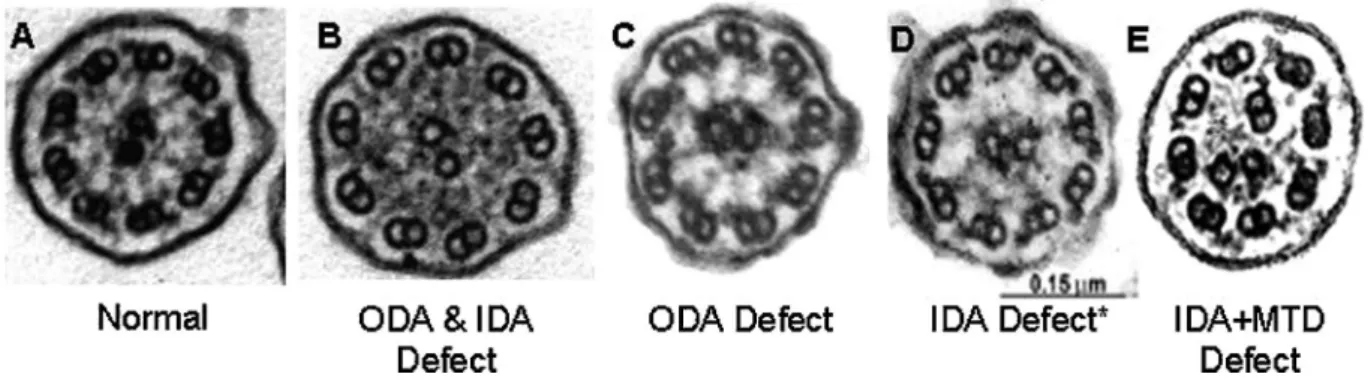
Respiratory Epithelial Biopsy With Electron Microscopy
Respiratory epithelial biopsy with EM processing for
ultrastructural examination of ciliary axonemes is a proven
technique for PCD diagnosis
39and is recommended as part
Fig. 1. Examples of various laterality defects on radiology imaging in PCD. Different situsof a panel of diagnostic tests for PCD. Disease causing EM
defects in the outer dynein arms,
40outer and inner dynein
arms,
41inner dynein arms with microtubule
disorganiza-tion,
42radial spokes,
43or central apparatus
44,45provide
confirmation of PCD diagnosis (Fig. 2). However, EM
studies with normal ciliary ultrastructure do not rule out
PCD, as certain PCD gene mutations can result in normal
ultrastructure,
10,38,46or subtle abnormalities (particularly
those involving the central apparatus and radial spokes)
that are not readily recognized on EM.
47,48Additionally,
repeat biopsies that fail to demonstrate any respiratory cilia
could represent an oligociliary defect causing PCD.
49,50It is estimated that EM will detect approximately 70%
of all PCD cases,
4but in centers inexperienced with EM
processing and interpretation, this percentage will be
notably less. Centers lacking extensive experience with
ciliary EM processing and interpretation should strongly
consider referring patients to a PCD Foundation clinical
TABLE 2— Recommended Diagnostic Testing Methods for Primary Ciliary DyskinesiaTest recommended for PCD diagnosis
Potential for false positive results
Potential for false negative results
Nasal nitric oxide measurement Low1 Low2
Ciliary biopsy with electron microscopy Variable3 Variable4
PCD genetic testing panels Low5 Moderate6
Functional ciliary beat/waveform analysis with high speed videomicroscopy Variable7 Moderate8
Immunofluorescence testing Unknown Unknown
1As long as cystic fibrosis has been excluded. Risk of false positive result is increased during viral respiratory infection, epistaxis, and non-atopic
sinusitis. Testing should be performed at baseline health status and repeated if there is any question about health status.
2Reference.18
3The risk of false positive result is moderately increased with secondary changes from infectious processes or pollutant exposures, improper
specimen handling and processing, or inexperience with electron microscopy interpretation.4
4Several PCD-causing genetic mutations can result in normal electron microscopy10or subtle changes which are not readily apparent.38
5Misinterpretation of genetic panel result (e.g., variants of unknown significance or single mutations in two different PCD genes interpreted as
“diagnostic”).
6Genetic panel testing may miss large insertions, deletions, and mutations in novel genes, since approximately 30% of PCD do not have identifiable
mutations in the currently known PCD associated genes, but this risk should decrease with broader range of genetic analysis provided by NGS panels.
7With a high risk for false positive results from secondary insults on a single test. To limit this risk, many centers now perform three ciliary biopsies
at separate clinical visits for repeat high speed videomicroscopy analysis.
8Subtle waveform defects will be missed in centers without extensive experience.
TABLE 3— Recommended PCD Diagnostic Criteria by Age
Newborns (0–1 month of age)
Situs inversus totalis and unexplained neonatal respiratory distress at term birth plus at least one of the following: Diagnostic ciliary ultrastructure on electron micrographs
Biallelic mutations in one PCD-associated gene
Persistent and diagnostic ciliary waveform abnormalities on high-speed videomicroscopy, on multiple occasions Children (1 month to 5 years)
Two or more major PCD clinical criteria (see below) plus at least one of the following (nasal nitric oxide not included in this age group, since it is not yet sufficiently tested):
Diagnostic ciliary ultrastructure on electron micrographs Biallelic mutations in one PCD-associated gene
Persistent and diagnostic ciliary waveform abnormalities on high-speed videomicroscopy, on multiple occasions Children 5–18 years of age and adults
Two or more major PCD clinical criteria (see below) plus at least one of the following:
Nasal nitric oxide during plateau<77 nl/min on 2 occasions,>2 months apart, with cystic fibrosis excluded Diagnostic ciliary ultrastructure on electron micrographs
Biallelic mutations in one PCD-associated gene
Persistent and diagnostic ciliary waveform abnormalities on high-speed videomicroscopy, on multiple occasions
Major clinical criteria for PCD diagnosis
1) Unexplained neonatal respiratory distress (at term birth) with lobar collapse and/or need for respiratory support with CPAP and/or oxygen for
>24 hr.
2) Any organ laterality defect—situs inversus totalis, situs ambiguous, or heterotaxy. 3) Daily, year-round wet cough starting in first year of life or bronchiectasis on chest CT. 4) Daily, year-round nasal congestion starting in first year of life or pansinusitis on sinus CT.
center for PCD investigations. At least 20–50 clear ciliary
cross-sections are required for a diagnostic EM study, and
diagnostic abnormalities should be consistently
demon-strated on cross sectional images from multiple different
cilia to be considered disease causing. Physicians may try
nasal corticosteroids, nasal saline lavages, or systemic
antibiotics for persistent nasal symptoms interfering with
biopsies, but these practices are unproven and may not
improve biopsy yield. Furthermore, it is essential that
biopsies are collected when patients are at their baseline
health, as secondary changes in ciliary ultrastructure can
occur during respiratory exacerbations.
15Thus, biopsies
should be delayed until at least 2 weeks after full recovery
from an illness. For absence of inner dynein arms in
isolation, repeat biopsy and EM studies are always
required to verify that this pathologic change persists and
therefore is more likely genetic (primary) and not from
secondary causes.
11One may also consider repeat
biopsies to verify the universality and permanence of
findings suggestive of central apparatus, radial spoke, or
inner dynein arm with microtubule disorganization
defects. Patients with EM studies consistent with PCD
should be referred to a PCD Foundation Clinical Center
for confirmation.
Nasal Nitric Oxide Measurement
Measurement of nasal nitric oxide (nNO) by
chemilu-minescence analyzer is recommended as part of a panel of
diagnostic tests for PCD in adults and children
5 years
old.
18This test is sensitive, rapid, non-invasive, and
results are immediately available. Nasal NO values are
more reliable in school aged children and adults because
these patients can cooperate with blowing into a resistor.
Tidal breathing techniques for nNO measurement in
children
<
5 years old are currently being investigated,
17but PCD diagnostic cutoff values for tidal techniques are
not currently available. Unfortunately,
chemilumines-cence devices are limited to research settings in North
America, but they are gaining acceptance as a clinical tool
in various countries across Europe, through efforts by the
BESTCILIA PCD consortium.
51Handheld
electrochem-ical nNO analyzers are affordable and portable, but with
only limited prospective study in PCD,
52,53these devices
are not currently recommended for PCD testing.
Nasal nitric oxide values are extremely low in
PCD.
54,55Using a nNO cutoff value
<
77 nl/min, one
will detect PCD, resulting from ciliary axonemal defects
or mutations in DNAH11, with sensitivity and specificity
of 98% and
>
99%, respectively, if CF has been ruled out
(Figure 3).
18Values well above this cutoff level
significantly decrease the likelihood of PCD. However,
clinicians still must consider PCD when confronted with
an appropriate clinical phenotype for PCD and nNO
values above 77 nl/min, as forms of PCD with nNO values
above this cutoff have rarely been reported.
56,57Very low
nNO levels (below 77 nl/min) can occur during acute viral
respiratory infections and in approximately 30% of
patients with cystic fibrosis; therefore, nNO testing
must be performed when the patient has fully recovered
from a viral illness and after diagnostic testing to rule out
cystic fibrosis.
58Other conditions can also result in nNO
levels below PCD cutoff values (i.e., HIV,
59panbron-chiolitis,
60non-atopic sinusitis
61). Lastly, nNO device
operators must be well trained and use standard operating
protocols to avoid false results.
18Functional Ciliary Beat/Waveform Analysis With High Speed Videomicroscopy
Ciliary biopsy with examination of cilia waveform by
high speed videomicroscopy can provide confirmation of
PCD, and this test is recommended as part of a panel of
PCD diagnostic tests, but only in centers highly
experienced with this technology.
62Functional ciliary
analysis is difficult to perform correctly, and considerable
experience is necessary to avoid positive and
false-negative results. Biopsies should only be performed when
Fig. 2. Electron microscopy findings in primary ciliary dyskinesia. Diagnostic ciliary electronpatients are in their baseline state of health. Repeat
biopsies are required to assure abnormal beat patterns are
not due to secondary processes, such as viral illness,
63tobacco or environmental exposures,
64poor biopsy
specimen,
16or improper biopsy processing.
14Some
European centers also maintain biopsied epithelial cells
in culture for weeks, at an air-liquid interface, to remove
influence of secondary insults.
15There are no prospective
studies examining inter-rater agreement for functional
ciliary analysis. Currently, there are no American centers
that can reliably perform this testing, yet several skilled
European centers regularly employ this test.
Immunofluorescence Testing for Ciliary Proteins
Immunofluorescence testing (IF) using antibodies to
detect missing dynein arm proteins along the ciliary
axoneme can help confirm PCD as part of a panel of PCD
diagnostic tests.
65,66Through staining of specific ciliary
proteins (DNAH5, DNAI2, DNALI1, and RSPH4A/
RSPH1/RSPH9), which are essential for overall dynein
arm and radial spoke head assembly, IF can detect various
outer dynein arm, inner dynein arm, and radial spoke
defects, even when other (often less integral) ciliary
protein deficiencies are the primary cause of PCD.
42,67–72Although IF is currently limited to a few centers, it has
been shown equivalent to EM analysis for detecting outer
dynein arm defects, caused by DNAH5, in a small
(n
¼
16), blinded study.
66Additionally, IF diagnostic
results do not seem to be affected by secondary insults.
73Further investigations are required to evaluate the
sensitivity and specificity of IF against other PCD
diagnostic tests.
PCD Genetic Testing
Genetic testing for disease-causing mutations
associ-ated with PCD is recommended as part of a panel of
diagnostic PCD tests. There are currently 33 known genes
associated with PCD (Table 4), with new genes being
discovered at a rapid pace.
8,12,13,74Almost all of these
genes follow autosomal recessive inheritance (with
exception of two rare, X-linked syndromic genes
RPGR
and
OFD1—see section on “Diseases that co-exist with
PCD”); therefore, two disease-causing mutations must
occur in the same PCD gene for a diagnosis. No
documented cases of digenic inheritance (heterozygous
mutations in two different PCD genes), unequivocally
associated with human PCD, exist thus far. Currently, the
most comprehensive commercial PCD genetic panel tests
19 PCD genes through next generation sequencing
(NGS), at a cost of $1,990, and detects approximately
50% of PCD cases.
75Genetic testing costs for other
commercial NGS panels range from $1,500 to $4,500 and
often include full cystic fibrosis transmembrane regulator
(CFTR) protein analysis.
76–78Results may contain
genetic variants of unknown significance, and a genetic
diagnosis may not be clearly established. Thus, genetic
counselling is recommended. Any patients with genetic
studies that provide unclear diagnostic information
should be referred to a PCD Foundation Clinical Center
for further investigations.
Tests Not Recommended for PCD Diagnosis
Several older diagnostic tests are no longer
recom-mended for PCD evaluation (Table 5), including nasal
saccharin testing,
79ciliary beat frequency
calcula-tion,
62,80and visual assessment of ciliary motion without
high speed recording devices. Each of these tests has
significant limitations, which can lead to frequent false
positive or false negative results, especially in
uncooper-ative children; thus, these tests are not appropriate for
PCD diagnosis. Radioaerosol mucociliary clearance
testing is potentially useful to rule out PCD.
81,82Although
this test remains limited to a few expert centers, requires a
level of patient cooperation suitable for children
>
7 years
old, and cannot distinguish secondary ciliary dysfunction,
it may help to rule out PCD with a normal result.
Other Chronic Respiratory Conditions to Consider
The clinical symptoms associated with PCD often
overlap
with
other
common
pediatric
and
adult
Fig. 3. Nasal nitric oxide in primary ciliary dyskinesia and healthyrespiratory diseases (Table 6). Each of these other
diseases should be considered in patients with chronic
oto-sino-pulmonary symptoms; however, investigations
should only be pursued when the clinical picture suggests
their presence. Thus, PCD is not a diagnosis of exclusion.
Sweat testing or cystic fibrosis genetic testing are
recommended when evaluating patients for PCD, as both
diseases can present with similar phenotypes
83and
produce nNO levels below the PCD diagnostic cutoff of
77 nl/min.
58Immunodeficiency can also present similarly
TABLE 4— PCD GeneticsPCD genes Prevalence in PCD Ciliary structural defect
Detected on current commercial PCD NGS panels
NME8 þ Partial ODA defect Yes
DNAH5 þþþþ ODA defect Yes
DNAI1 þþþ ODA defect Yes
DNAI2 þþ ODA defect Yes
DNAL1 þ ODA defect Yes
CCDC114 þþ ODA defect Yes
CCDC103 þþ ODAdefect Yes
DNAAF1 þþ ODA and IDA defect Yes
DNAAF2 þþ ODA and IDA defect Yes
DNAAF3 þ ODA and IDA defect Yes
LRRC6 þþ ODA and IDA defect Yes
HEATR2 þ ODA and IDA defect Yes
RPGR þ Normal Yes
OFD1 þ Normal Yes
DNAH11 þþþ Normal Yes
CCDC39 þþþ IDA defectþMTD defect Yes
CCDC40 þþþ IDA defectþMTD defect Yes
RSPH9 þ Central pair defect or normal Yes
RSPH4A þþ Central pair defect or normal Yes
RSPH1 þþ Central pair defect or normal
RSPH3 þ Central pair defect or normal
CCNO þ Oligocilia (residual axoneme normal)
MCIDAS þ Oligocilia (residual axoneme abnormal)
DNAH8 þ Not available
CCDC151 þþ ODA defect
ARMC4 þþ ODA defect
DYX1C1 þ ODA and IDA defect
C21orf59 þ ODA and IDA defect
ZMYND10 þþ ODA and IDA defect
SPAG1 þþ ODA and IDA defect
HYDIN þ Normal
CCDC164 (DRC1) þ Mostly normal (N-DRC defect)
CCDC65 (DRC2) þ Mostly normal (N-DRC defect)
þ, genetic mutations causing<1% of all PCD;þþ, genetic mutations causing 1–4% of all PCD;þþþ, genetic mutations causing 4–10% of all PCD;þþþþ, genetic mutations causing>15% of all PCD; IDA, inner dynein arm; IDAþMTD, inner dynein arm defect with microtubule disorganization; N-DRC, nexin-dynein regulatory complex; ODA, outer dynein arm.
TABLE 5— Tests NOT Recommended for Diagnosing Primary Ciliary Dyskinesia
Tests NOT recommended for primary ciliary dyskinesia diagnosis Potential for false positive results Potential for false negative results
Nasal saccharin testing Very high1 Very high1
Radioaerosol mucociliary clearance tests High2 —
Ciliary beat frequency alone (CBF) High3 High3
Ciliary motion analysis without high speed videomicroscopy Very high4 Very high4
1This test is subjective and involves a high degree of cooperation by the patient. In children<5–7 years old, the feasibility of this test will be low
due to poor patient cooperation.
2This test can result in false positive PCD diagnoses, as detected abnormalities in mucociliary clearance lack specificity, and may be due to
secondary causes.
3In proven cases of PCD, CBF can be low, normal, or high, leading to false positive and false negative results.80
to PCD,
84and in patients with suspected PCD, laboratory
studies investigating immunodeficiency are necessary.
Preliminary study of nNO in certain humoral
immuno-deficiencies has shown normal values well above 77 nl/
min,
85but further study is required to know if all forms of
immunodeficiency produce normal nNO levels.
Pulmonary aspiration, with or without
gastroesopha-geal reflux, can cause chronic respiratory symptoms in
adults and children, including cough, wheeze, bronchitis,
or pneumonia.
86,87Thus in patients with possible PCD, a
thorough feeding history is essential. A history of chronic
cough from asthma can also resemble PCD in young
children, especially with frequent viral infections from
daycare exposures. Additionally, chronic nasal
conges-tion from allergic rhinitis can seem similar to PCD
rhinosinusitis. However, PCD nasal disease is present
year-round and does not resolve with seasonal change, as
often occurs with allergic rhinitis. Lastly, protracted
bacterial bronchitis (PBB) is a disorder of preschool aged
children causing
>
3 weeks of wet cough with lower
airway bacterial infection and airway neutrophilia.
88In
general though, the characteristic, year-round, daily, often
wet or productive cough of children with PCD usually
distinguishes them from these other conditions.
Diseases that Co-Exist With PCD
PCD can rarely co-exist with other rare disorders
(Table 7). Retinitis Pigmentosa (an inherited cause of
blindness from retinal ciliary dysfunction) and
Orofacio-digital Syndrome (including mental retardation,
cranio-facial abnormalities, macrocephaly, digital anomalies,
and cystic kidneys) are X-linked disorders involving
ciliary genes,
RPGR
and
OFD1, respectively.
21,89Although these account for a very small minority of
PCD cases, there may be further overlap of retinal and
respiratory cilia.
90,91Thus, retinal examination is
recommended in individuals with PCD due to gene
mutations in
RPGR, clinical visual disturbances, or a
family history of Retinitis Pigmentosa, whereas PCD
patients with
OFD1
phenotypes should be referred for
genetic consultation.
Various diseases caused by genetic disorders of
non-motile cilia can result in cystic kidneys, cystic or
cholestatic liver, skeletal malformations, developmental
delay, hydrocephalus, blindness, or deafness. These
include Joubert Syndrome, Bardet–Biedl syndrome,
Usher Syndrome, Jeune Syndrome, polycystic kidney
disease, and others. The overlap of these non-motile
ciliopathies with respiratory cilia dysfunction is unusual,
and poorly understood at present,
90,92,93but increased
rates of bronchiectasis are found in polycystic kidney
disease.
94Therefore, consultation with a geneticist or
other subspecialists is recommended when patients with
possible PCD have features of non-motile ciliary
dysfunction.
PCD can also co-exist with other rare diseases through
close proximity of disease causing mutations at the same
chromosomal locus (Table 7). Cri du Chat syndrome can
occur with PCD due to a large deletion on chromosome 5p
and a point mutation in
DNAH5
on the remaining
chromosome.
22Glanzmann Thrombasthenia (associated
with
ITGB3) can occur with PCD (associated with
CCDC103) through mutations in the neighboring genes
on chromosome 17.
95Alternatively, PCD can co-exist
with other rare diseases through disease-causing
muta-tions which are not in close genetic proximity; such as
cystic fibrosis due to mutations in
CFTR
(Chr7q) with
PCD due to mutations in
DNAH11
(Chr7p),
96and Miller
Syndrome due to mutations in
DHODH
(chr 16) with
PCD due to mutations in
DNAH5
(Chr5).
97Recent publications have also shown respiratory ciliary
dysfunction in patients with mild forms of congenital
heart disease, not meeting cardiology definitions for SA or
TABLE 6— Other Chronic Respiratory Conditions to Consider When Considering a Diagnosis of PCDChronic condition Methods of evaluation
Cystic fibrosis Sweat chloride testing or cystic fibrosis genetic testing
Immunodeficiency Quantitative measurement of immunoglobulins, lymphocytes, complement levels, antibody responses to vaccines, and complete blood counts. Consultation with a board certified Immunologist is also recommended
Asthma Clinical history, pulmonary function testing, and asthma medication trials. Although one normally expects asthma-related cough to be dry in nature, it can seem wet to parents when accompanied by viral respiratory infections. Obstructive defects on pulmonary function testing can be seen with both PCD and asthma, and bronchodilator responsiveness is not exclusive to asthma and does not exclude a diagnosis of PCD
Pulmonary aspiration Clinical feeding history followed by swallowing assessment and intervention only when pulmonary aspiration seems likely
Allergic rhinitis Clinical history of seasonal symptoms, allergy testing, and trials of nasal corticosteroids and antihistamines, which should greatly improve allergic rhinitis symptoms. PCD nasal disease shows minimal (if any) improvement with these interventions
Protracted bacterial bronchitis
heterotaxy.
98Thus, physicians should ask about chronic
oto-sino-pulmonary symptoms in all patients with
congenital heart disease to screen for possible PCD and
test as indicated.
CLINICAL CARE AND LONG-TERM MONITORING
Pulmonary Care and Monitoring
Long-term follow-up should be in a PCD Foundation
clinical center or an accredited cystic fibrosis center that
has a comprehensive, multidisciplinary team approach to
care. Outpatient visits with a pulmonologist experienced
in management of chronic suppurative lung disease, such
as cystic fibrosis, are recommended 2–4 times annually
(Table 8). Surveillance cultures of expectorated sputum or
oropharyngeal cough swabs are recommended two to four
times annually in all PCD patients.
1Although the most
common airway pathogens in children with PCD are
Streptococcus pneumoniae,
Haemophilus influenzae, and
Moraxella catarrhalis, surveillance cultures should be
processed in the same manner as cystic fibrosis cultures,
including examination for
Pseudomonas aeruginosa
and
other Gram negative organisms, as well as
non-tuberculosis mycobacterial (NTM) organisms.
1,4Culture
results should guide antibiotic therapy during future
respiratory exacerbations. When PCD patients are not
responding to culture-directed antibiotics, physicians
should consider additional NTM and fungal cultures,
allergic bronchopulmonary aspergillosis testing (ABPA)
testing (IgE levels
evidence of aspergillus specificity)
and bronchoscopy with bronchoalveolar lavage fluid
cultures to guide antimicrobial therapy.
Spirometry using ATS/ERS criteria
99is suggested two
to four times annually to follow disease progression in
PCD. Although spirometry may not be the most sensitive
test of pulmonary function in PCD, it is the most available
testing method in pediatric and adult centers. With further
validation, other tests of pulmonary function, such as
multiple breath washout, may be useful in PCD.
100,101Chest radiography should be performed at diagnosis
and during respiratory exacerbations, as indicated.
Otherwise, chest radiography should be performed every
TABLE 7— Other Diseases Co-Segregating With PCDAssociated rare disorder Level of PCD association Method of PCD overlap Specific gene affected
Situs ambiguus and heterotaxy At least 12% of PCD Shared common genes Any PCD gene encoding for ODA, IDA, or ODAþIDA proteins (ex: DNAH5, DNAH11, CCDC39/40, LRRC6, DNAAF1/2/3)19
Retinitis pigmentosa Multiple unrelated cases reported;<1% of PCD
Shared common gene mutation RPGR21,91,161,162
Orofaciodigital syndrome 1 sibling-pair reported; Shared common gene mutation OFD189 (sibling males with mental
retardation and macrocephaly)
<1% of PCD
Cri du chat syndrome 2 unrelated cases reported Mutation in close proximity to PCD gene
Chr 5p deletion including DNAH522
Glanzmann thrombasthenia 1 sibling-pair reported Mutation in close proximity
to PCD gene
Chr 17q haplotype that includes CCDC103 and ITGB395
Cystic fibrosis 1 case reported Mutations in two different genes Chr 7 including region with DNAH11 and CFTR via uniparental isodisomy96 Miller syndrome 1 sibling-pair reported Mutations in two different genes Biallelic mutations in DNAH5 (Chr 5)
for PCD and DHODH (Chr 16) for Miller syndrome97
Common variable immunodeficiency
1 sibling-pair and 2 unrelated cases reported
Unknown 1 sibling-pair with homozygous DNAH11 mutations and low IgM and S.pneumoniae titers after booster.1572 unrelated cases; 1 with outer dynein arm defect and low IgG titers157, and 1 with Kartagener Syndrome, abnormal ciliary ultrastructure, and low IgG, IgM, Tetanus and S.pneumoniae titers after booster.158
Polycystic kidney disease 1 case reported Unknown Unknown92
Familial mediterranean fever 1 case reported Unknown MEFV-R202Q polymorphism on Chr 16p13.3 and unknown PCD gene163 Other non-motile ciliopathies No definite cases reported Unknown Unknown
2–4 years in stable patients, in order to monitor disease
progression. The decision to use serial CT scans for
monitoring PCD disease progression should be decided
on a case by case basis, and the lowest possible radiation
doses should be used. However, a chest CT scan is
generally recommended at least once after diagnosis to
detect bronchiectasis, which may encourage better
compliance to airway clearance in patients and parents
who are aware of this finding. Chest CT can be considered
when children are old enough to cooperate (and avoid
sedation), and images will be of sufficient quality to
diagnose bronchiectasis, or sooner depending on clinical
symptoms.
33,34Some centers perform chest CT scans on
PCD patients every 5 years, but there is no evidence that
this improves clinical outcomes,
102and cumulative
radiation doses need to be considered for PCD patients.
Infection control policy is essential for clinical care in
PCD, and general hospital infection control policies
should be followed where PCD patients receive care.
Patients with resistant organisms on sputum culture should
be specifically targeted for infection control in all clinical
areas. Although there is no evidence for cross
contamina-tion of respiratory organisms among PCD patients, it is
logical to assume this may occur, as it does in similar
diseases.
103More stringent infection control policies have
the potential to cause psychosocial harm to patients and
families,
104and thus should be avoided in PCD. However,
this recommendation may be adjusted if there is clear
evidence for risks that outweigh potential harm.
Otolaryngology Care and Monitoring
Pediatric PCD patients should visit a pediatric
otolaryn-gologist at least once to twice annually, while adult patients
should have otolaryngology care, as needed. An initial
audiology assessment in all PCD patients is suggested at
diagnosis, with subsequent evaluations coordinated
through their otolaryngologist. The major otolaryngology
concern in PCD patients is the nearly universal conductive
hearing loss due to persistent otitis media with effusion
(OME).
105Hearing abnormalities often improve in
adolescence, but in some cases, continue into adulthood.
Pressure equalization tubes (PET) are advocated for
children with PCD who have hearing deficits or speech
delay and middle ear effusions. Although several
systematic reviews have cast doubt on the utility of PET
in OME,
106,107these studies are not necessarily
generaliz-able to a PCD population, where individuals are expected
to have greater portion of their prelingual life with
conductive hearing loss. In studies assessing hearing in
children with PCD post-PET placement, hearing
normal-ized in 80–100% of participants.
32,108,109In another study
examining surgical treatment with PET versus medical
management alone in PCD, children with PET had larger
hearing improvements post-operatively than those treated
with medical therapy.
108All patients undergoing PET insertion should be
counselled on the likelihood of multiple insertions,
post-operative otorrhea, and the possibility of a permanent
TABLE 8— Suggested Schedule of Investigations and Clinical Care in Primary Ciliary DyskinesiaClinical visits
Pulmonology: 2–4 times/year
Otolaryngology: 1-2 time/year in children, as needed in adults Audiology: at diagnosis and as needed per otolaryngology Reproductive medicine: As clinically needed
Long-term surveillance
Chest radiography: every 2–4 years
Chest computed tomography: consider at least once after 5–7 years old (when sedation not required and images are of highest quality)1 Airway microbiology cultures: 2–4 times/year
Non-tuberculosis mycobacterial cultures: every 2 years (and with unexplained clinical decline) Pulmonary function testing: 2–4 times/year
ABPA testing: IgE levelsevidence of aspergillus specificity at diagnosis, with new onset wheezing, unexplained clinical decline Preventative therapies
Airway clearance: daily
Nasal sinus lavage: daily (when pertinent) Standard vaccinations: per local schedule Influenza vaccine: annually2
13-valent pneumococcal vaccine: per ACIP guidelines3 23-valent pneumococcal vaccine: per ACIP guidelines4 RSV immunoprophylaxis: consider monthly in first winter5
1And as clinically indicated on a case by case basis.
2After 6 months old, including household members.
3ACIP guidelines.
4ACIP guidelines.
5Specifically consider in infants with complicated respiratory courses, including prematurity, prolonged mechanical ventilation, prolonged need
tympanic membrane perforation (up to 50% in one
study).
110Additionally, patients with PET are typically
seen by their otolarygologist every 3–6 months while the
tubes remain in place.
107Although some physicians avoid
PET in PCD for fear of prolonged post-operative otorrhea,
studies show that post-operative otorrhea in PCD is no
worse than the general population
111and is easily
controlled with topical therapies.
109Persistent otorrhea
can be attributed to biofilm formation, especially in
children with longer lasting PET
112; however, given the
poor eustachian tube function and multiple PET insertions,
acquired cholesteatoma should also be considered as a
potential cause of persistent otorrhea in PCD.
Otolaryngologists should also monitor for chronic
rhinosinusitis (CRS) in PCD patients. CRS is estimated to
affect over 50% of patients with PCD
31and nasal
endoscopy (as permitted by age) can be used to identify
polyps which may be exacerbating already poor
muco-ciliary clearance. Nasal polyposis has been observed in up
to 15% of PCD patients.
29,113Although CRS is not
generally life threatening, it substantially affects quality
of life.
114Daily saline irrigation has been demonstrated as
safe and beneficial in patients with CRS.
115Anecdotally,
in PCD patients, saline nasal irrigations are beneficial, but
studies demonstrating their efficacy are lacking. Given the
minimal side effect profile and likelihood for benefit,
nasal irrigations are generally encouraged for
symptom-atic CRS relief in PCD. The effects of saline irrigation are
likely increased after functional endoscopic sinus surgery
(ESS), as the saline solution will more easily reach the
sinus mucosa through post-surgical ostia. Thus, ESS is
often performed in PCD patients and may improve lower
respiratory tract disease in some patients.
116Antibiotics
and nasal steroids may be used in acute on chronic
exacerbations of rhinosinusitis; however, a recent review
showed lack of consensus on the treatment of CRS in
children with PCD,
113and there are no randomized,
controlled, or long-term prospective CRS studies in PCD.
PRINCIPLES OF TREATMENT
Routine Therapies in PCD
Airway clearance through daily chest physiotherapy is
highly recommended in PCD.
117Unlike cystic fibrosis,
cough clearance is preserved in PCD.
118Thus, airway
clearance is expected to be quite beneficial in PCD and
should be a cornerstone of long-term therapy. Daily
cardiovascular exercise should also be strongly
encour-aged, as poor exercise capacity is linked to decreased
pulmonary function in PCD,
119and exercise may improve
mucus clearance.
120Antibiotics should be given for acute respiratory
exacerbations in PCD. Acute changes in cough, sputum
production, respiratory rate, or work of breathing are
likely reliable markers of a respiratory exacerbation in
PCD (as demonstrated in non-CF bronchiectasis
121), and
oral antibiotics are recommended for mild exacerbations.
Most physicians use a broad-spectrum oral antibiotic
(amoxicillin plus clavulinic acid or an equivalent
cephalosporin) to target the common respiratory
patho-gens in children with PCD. Typically, at least 2–3 weeks
of oral antibiotics are recommended in PCD, based upon
other disorders with similar pathophysiology (protracted
bacterial bronchitis,
122cystic fibrosis,
123and non-CF
bronchiectasis
124). More severe exacerbations, or those
failing oral therapy, may require parenteral antibiotics.
Antibiotic choice should be guided by past respiratory
cultures. Despite a lack of published evidence, inhaled
antibiotics are also an option for acute PCD respiratory
exacerbations, but these are usually reserved for patients
with
Pseudomonas aeruginosa
infection. Eradication of
initial positive Pseudomonas airway culture also seems
prudent in PCD, although no evidence supports this
practice. Non-CF bronchiectasis guidelines make similar
suggestions for Pseudomonas eradication.
125,126Al-though
Burkholderia cepacia
has not been reported in
PCD, recovery of this organism should prompt
eradica-tion practices.
Finally, PCD patients should receive recommended
vaccinations per local schedules. Annual influenza
127and
pneumococcal vaccinations (per the Advisory Committee
on Immunization Practices)
128,129are recommended in
PCD. In the first year of life, monthly (seasonal)
immunoprophylaxis against respiratory synctial virus
can be considered for infants with PCD, and more
specifically for infants with complicated respiratory
courses requiring prolonged oxygen supplementation.
Therapies to Consider on a Case by Case Basis in
PCD
Chronic suppressive inhaled antibiotics can be used on
an individual basis in PCD patients. Inhaled
aminoglyco-side and beta-lactam antibiotics are recommended for
chronic respiratory infections (particularly those
associat-ed with
Pseudomonas aeruginosa) in non-CF
bronchiec-tasis,
125,130,131and
several
months
of
inhaled
aminoglycosides or colistin in Pseudomonas colonized
adults with non-CF bronchiectasis result in decreased
hospitalization and improved respiratory symptoms.
132–134However, there are no studies of inhaled antibiotics in
children with non-CF bronchiectasis or PCD.
macrolides in adults and children with non-CF
bronchi-ectasis shows decreased respiratory exacerbations and
improved lung function,
136–138but increased emergence
of macrolide resistant respiratory organisms. The
long-term significance of macrolide resistance is unclear.
139Small case reports of chronic macrolide therapy in PCD
also demonstrate some benefits, although not as robust as
those in non-CF bronchiectasis.
140–143Remote studies on
trimethoprim-sulfamethoxazole in chronic bronchitis also
suggest benefit, but this agent has not been studied in
PCD.
144,145Inhaled hyperosmolar agents can be used on a
case-by-case basis in PCD. These agents promote cough clearance
and alter mucus rheology to favor increased cough
clearance. However, a recent meta-analysis reported
unclear long-term benefits of hyperosmolar agents in
non-CF bronchiectasis.
146Hypertonic saline (3% to 7%
concentration) has not been studied in PCD. Trials
comparing inhaled hypertonic saline to isotonic saline
show limited positive effects in non-CF bronchiectasis.
147When physicians use inhaled hypertonic saline in PCD, it
is essential that they instruct patients in proper equipment
sterilization. Inhaled dry powder mannitol has also been
studied in non-CF bronchiectasis, but outcomes are
inconclusive.
146,148Mannitol has not been studied in
PCD.
DNase (dornase-alfa or Pulmozyme
1) can be used on
an individual basis in PCD. Although there are no
prospective trials of DNase in PCD, studies of DNase in
adults with non-CF bronchiectasis show no clinical
benefits in one study
149and increased frequency of
respiratory exacerbations with worsened lung function in
another study.
150Several case reports of DNase in PCD
suggest possible benefit when used for both short and
long-term periods.
151–153Larger, prospective clinical
studies of DNase in children and young adults with
PCD are required before the potential negative effects of
this medication can be dismissed.
Lastly, inhaled bronchodilators can be used on a
case-by-case basis in PCD. In limited study, long-acting
bronchodilators (with inhaled corticosteroids) in non-CF
bronchiectasis do not show clinical efficacy. In PCD,
bronchodilators show mixed results, with one study
demonstrating significant improvement in lung function
after a single bronchodilator dose,
154whereas another
study showed unchanged lung function after 6 weeks of
regular bronchodilators.
155Therapies Not Routinely Recommended in PCD
Inhaled corticosteroids are not routinely recommend in
PCD and should be reserved for PCD patients with
associated asthma or airway reactivity. Inhaled
cortico-steroids are also discouraged in non-CF bronchiectasis
without airway reactivity.
156Similarly, intravenous
immunoglobulin (IVIG) is not recommended for routine
use in patients with PCD. Immunodeficiency rarely exists
with PCD,
157,158and most PCD patients have normal
immune function. PCD patients with documented
dysfunction of vaccine responses or other aspects of
humoral immunity may benefit from IVIG therapy.
Isolated IgA or IgG subclass disorders do not justify
IVIG therapy.
Lobectomy is not routinely suggested as therapy in
PCD. The decision to perform lobectomy in PCD requires
multi-disciplinary discussion between pulmonologists,
intensivists, and surgeons. In the post-operative period,
airway clearance is limited by pain and immobility, and
PCD patients are at risk of pulmonary deterioration.
Although lobectomy may be beneficial in rare cases of
PCD with severe, localized bronchiectasis, it should be
considered with caution. Similarly, lung transplantation
can be considered in PCD patients with advanced
pulmonary disease, but situs anomalies may surgically
complicate this procedure.
159,160Summary
PCD is a rare disorder; consequently, only a limited
number of centers have extensive experience in the
diagnosis and management of PCD. Research over the
past decade has led to a revolution in diagnostic
approaches, including nNO and genetic testing.
Never-theless, many PCD patients are still undiagnosed or
misdiagnosed. To date, only limited studies have
addressed management of PCD, and there have been no
large, randomized clinical trials to direct therapy.
Therefore, this review article includes consensus
recom-mendations from PCD physicians in North America for
diagnosis, monitoring and management of PCD.
ACKNOWLEDGMENTS
drafts of this document and provided input in areas
needing clarification.
REFERENCES
1. Davis SD, Ferkol TW, Rosenfeld M, Lee HS, Dell SD, Sagel SD, Milla C, Zariwala MA, Pittman JE, Shapiro AJ, et al. Clinical features of childhood primary ciliary dyskinesia by genotype and ultrastructural phenotype. Am J Respir Crit Care Med 2015;191:316–324.
2. Magnin ML, Cros P, Beydon N, Mahloul M, Tamalet A, Escudier E, Clement A, Le Pointe HD, Blanchon S. Longitudinal lung function and structural changes in children with primary ciliary dyskinesia. Pediatr Pulmonol 2012;47:816–825. 3. Marthin JK, Petersen N, Skovgaard LT, Nielsen KG. Lung
function in patients with primary ciliary dyskinesia: a cross-sectional and 3-decade longitudinal study. Am J Respir Crit Care Med 2010;181:1262–1268.
4. Knowles MR, Daniels LA, Davis SD, Zariwala MA, Leigh MW. Primary ciliary dyskinesia. Recent advances in diagnostics, genetics, and characterization of clinical disease. Am J Respir Crit Care Med 2013;188:913–922.
5. Leigh MW, O’Callaghan C, Knowles MR. The challenges of diagnosing primary ciliary dyskinesia. Proc Am Thorac Soc 2011;8:434–437.
6. Hosie P, Fitzgerald DA, Jaffe A, Birman CS, Morgan L. Primary ciliary dyskinesia: overlooked and undertreated in children. J Paediatr Child Health 2014;50:952–958.
7. Lucas JS, Leigh MW. Diagnosis of primary ciliary dyskinesia: searching for a gold standard. Eur Respir J 2014;44:1418–1422. 8. Zariwala MA, Knowles MR, Leigh MW. Primary ciliary dyskinesia. 2007 Jan24 [updated 2015 Sep 3] In: Pagon RA, Adam MP, Amemiya A, Bird TD, et al., editors. GeneReviewsTM. Seattle (WA): University of Washington, Seattle; 1993–2015. Available from: http://www.ncbi.nlm.nih.gov/books/NBK1122/.) 9. Boon M, Jorissen M, Proesmans M, De Boeck K. Primary ciliary dyskinesia, an orphan disease. Eur J Pediatr 2013;172:151–162. 10. Knowles MR, Leigh MW, Carson JL, Davis SD, Dell SD, Ferkol TW, Olivier KN, Sagel SD, Rosenfeld M, Burns KA, et al. Mutations of DNAH11 in patients with primary ciliary dyskinesia with normal ciliary ultrastructure. Thorax 2012;67:433–441. 11. O’Callaghan C, Rutman A, Williams GM, Hirst RA. Inner dynein
arm defects causing primary ciliary dyskinesia: repeat testing required. Eur Respir J 2011;38:603–607.
12. Kim RH, A Hall D, Cutz E, Knowles MR, Nelligan KA, Nykamp K, Zariwala MA, Dell SD. The role of molecular genetic analysis in the diagnosis of primary ciliary dyskinesia. Ann Am Thorac Soc 2014;11:351–359.
13. Berg JS, Evans JP, Leigh MW, Omran H, Bizon C, Mane K, Knowles MR, Weck KE, Zariwala MA. Next generation massively parallel sequencing of targeted exomes to identify genetic mutations in primary ciliary dyskinesia: implications for application to clinical testing. Genet Med 2011;13:218–229. 14. Jackson CL, Goggin PM, Lucas JS. Ciliary beat pattern analysis
below 37 degrees C may increase risk of primary ciliary dyskinesia misdiagnosis. Chest 2012;142:543–544; author reply 544–5.
15. Hirst RA, Rutman A, Williams G, O’Callaghan C. Ciliated air-liquid cultures as an aid to diagnostic testing of primary ciliary dyskinesia. Chest 2010;138:1441–1447.
16. Thomas B, Rutman A, O’Callaghan C. Disrupted ciliated epithelium shows slower ciliary beat frequency and increased dyskinesia. Eur Respir J 2009;34:401–404.
17. Mateos-Corral D, Coombs R, Grasemann H, Ratjen F, Dell SD. Diagnostic value of nasal nitric oxide measured with non-velum
closure techniques for children with primary ciliary dyskinesia. J Pediatr 2011;159:420–424.
18. Leigh MW, Hazucha MJ, Chawla KK, Baker BR, Shapiro AJ, Brown DE, Lavange LM, Horton BJ, Qaqish B, Carson JL, et al. Standardizing nasal nitric oxide measurement as a test for primary ciliary dyskinesia. Ann Am Thorac Soc 2013;10: 574–581.
19. Shapiro AJ, Davis SD, Ferkol T, Dell SD, Rosenfeld M, Olivier KN, Sagel SD, Milla C, Zariwala MA, Wolf W, et al. Laterality defects other than situs inversus totalis in primary ciliary dyskinesia: insights into situs ambiguus and heterotaxy. Chest 2014. 146:1176–86.
20. Shapiro AJ, Tolleson-Rinehart S, Zariwala MA, Knowles MR, Leigh MW. The prevalence of clinical features associated with primary ciliary dyskinesia in a heterotaxy population: results of a web-based survey. Cardiol Young 2014;25:752–9.
21. Moore A, Escudier E, Roger G, Tamalet A, Pelosse B, Marlin S, Clement A, Geremek M, Delaisi B, Bridoux AM, et al. RPGR is mutated in patients with a complex X linked phenotype combining primary ciliary dyskinesia and retinitis pigmentosa. J Med Genet 2006;43:326–333.
22. Shapiro AJ, Weck KE, Chao KC, Rosenfeld M, Nygren AO, Knowles MR, Leigh MW, Zariwala MA. Cri du chat syndrome and primary ciliary dyskinesia: a common genetic cause on chromosome 5p. J Pediatr 2014;165:858–861.
23. Lucas JS, Carroll M. Primary ciliary dyskinesia and cystic fibrosis: different diseases require different treatment. Chest 2014;145:674–676.
24. Cohen-Cymberknoh M, Simanovsky N, Hiller N, Gileles Hillel A, Shoseyov D, Kerem E. Differences in disease expression between primary ciliary dyskinesia and cystic fibrosis with and without pancreatic insufficiency. Chest 2014;145:738–744. 25. Paff T, van der Schee MP, Daniels JM, Pals G, Postmus PE,
Sterk PJ, Haarman EG. Exhaled molecular profiles in the assessment of cystic fibrosis and primary ciliary dyskinesia. J Cyst Fibros 2013;12:454–460.
26. Horvath I, Loukides S, Wodehouse T, Csiszer E, Cole PJ, Kharitonov SA, Barnes PJ. Comparison of exhaled and nasal nitric oxide and exhaled carbon monoxide levels in bronchiectatic patients with and without primary ciliary dyskinesia. Thorax 2003;58:68–72.
27. Guyatt GH, Oxman AD, Vist GE, Kunz R, Falck-Ytter Y, Alonso-Coello P, Schunemann HJ, Group GW. GRADE: an emerging consensus on rating quality of evidence and strength of recommendations. BMJ 2008;336:924–926.
28. Mullowney T, Manson D, Kim R, Stephens D, Shah V, Dell S. Primary ciliary dyskinesia and neonatal respiratory distress. Pediatrics 2014;134:1160–1166.
29. Campbell R. Managing upper respiratory tract complications of primary ciliary dyskinesia in children. Curr Opin Allergy Clin Immunol 2012;12:32–38.
30. Afzelius BA. A human syndrome caused by immotile cilia. Science 1976;193:317–319.
31. Sommer JU, Schafer K, Omran H, Olbrich H, Wallmeier J, Blum A, Hormann K, Stuck BA. ENT manifestations in patients with primary ciliary dyskinesia: prevalence and significance of otorhinolaryngologic co-morbidities. Eur Arch Otorhinolaryngol 2011;268:383–388.
32. el-Sayed Y, al-Sarhani A, al-Essa AR. Otological manifestations of primary ciliary dyskinesia. Clin Otolaryngol Allied Sci 1997;22:266–270.