COMBINED BACTERICIDAL/BACTERIAL ADHESION-RESISTANT COATINGS THROUGH NITRIC OXIDE RELEASE
Wesley Langdon Storm
A dissertation submitted to the faculty of the University of North Carolina at Chapel Hill in partial fulfillment of the requirements for the degree of Doctor of Philosophy in the
Department of Chemistry (Analytical Chemistry)
Chapel Hill 2013
ii © 2013
iii ABSTRACT
Wesley Langdon Storm: Combined Bactericidal/Bacterial Adhesion-Resistant Coatings through Nitric Oxide Release
(Under the direction of Mark H. Schoenfisch)
In response to the health and economic burdens associated with implant-related infections, researchers have developed coatings that resist bacterial adhesion and/or kill bacteria. Herein, the synthesis of coatings that release nitric oxide (NO) to inhibit bacterial adhesion and kill bacteria are described.
In order to expand the clinical utility of NO-releasing surfaces, xerogels were synthesized from N-diazeniumdiolate-modified silane precursors. Release kinetics and totals were tunable through careful selection of the silane precursor and its concentration, respectively. To demonstrate the versatility of this approach, NO-releasing xerogels were cast as outer membrane on glucose sensors. The sensors exhibited a linear response towards glucose and maintained glucose sensitivity in phosphate buffered saline for up to one week.
iv
v
To Tabitha and Tara,
vi
ACKNOWLEDGEMENTS
The work presented in this dissertation was only possible with the help of many colleagues, friends, and family members. First, I’d like to thank my advisor, Mark Schoenfisch, for helping me grow as a scientist and person, for providing professional and personal support, and for always accommodating my unexpected fatherhood-related schedule changes without complaint.
I would also like to give special thanks to some particular graduate students in the Schoenfisch lab who have given much of their time and effort to help me succeed—Justin Johnson for synthesizing thousands of silver-releasing xerogels, Katey Reighard for carrying out countless bacteria assays in the superhydrophobic/NO project, Danielle Slomberg for helping with confocal microscopy, Robert Soto for synthesizing electrospun fibers, and Brittany Worley for doing—at some point over this final year— every conceivable laboratory task that I can possibly think of at the moment. I’m lucky to consider all of you friends as well as colleagues. I would also like to thank some of my other lab colleagues, namely Jonghae Youn and Professor Jae Ho Shin, for their help and guidance in the superhydrophobic/NO project.
vii
mentors. Both of you were a source of inspiration to me long after your defense dates. Most of all, I thank both of you for always believing in me.
I’ve also had the pleasure of working with a number of great undergraduate students. Hetali Lodaya developed many superhydrophobic materials that were not included in this dissertation, Shalini Chudasama helped in the early stages of the NO-releasing dendrimer/polyurethane project, and Chris Chouinard helped me operate the ICP-OES when it was first purchased. As the cliché goes, I learned more from each of you than you probably ever learned from me. Thank you for giving me the opportunity to work with you.
viii
TABLE OF CONTENTS
LIST OF TABLES ... xiii
LIST OF FIGURES ...xv
LIST OF SCHEMES...xx
LIST OF ABBREVIATIONS AND SYMBOLS ... xxi
CHAPTER 1. THE DEVELOPMENT OF ANTIFOULING AND BIOCIDAL COATINGS FOR BIOMEDICAL IMPLANTS...1
1.1 Microbial colonization on medical devices ...1
1.2 Passive surface modifications for reducing infection ...5
1.2.1 Non-fouling surfaces ...5
1.2.2 Superhydrophobic materials ...8
1.2.3 Superhydrophobic materials for drug release ...14
1.2.4 Polycationic antimicrobial surfaces ...15
1.3 Active release strategies for antimicrobial surfaces...17
1.3.1 Release of antibiotics ...18
1.3.2 Nitric oxide-releasing surfaces ...19
1.3.3 Antimicrobial silver-releasing materials ...27
1.4 Summary of dissertation research ...31
ix
CHAPTER 2. NITRIC OXIDE-RELEASING XEROGELS
SYNTHESIZED FROM
N-DIAZENIUMDIOLATE-MODIFIED SILANE PRECURSORS ...45
2.1 Introduction ...45
2.2 Materials and methods ...48
2.2.1 Synthesis of N-diazeniumdiolate-modified silanes and xerogels ...50
2.2.2 Characterization of N-diazeniumdiolate-modified silanes and xerogels ...52
2.2.3 Cytotoxicity...54
2.2.4 Fabrication and performance of NO-releasing glucose sensors...54
2.3 Results and discussion ...55
2.3.1 N-diazeniumdiolate formation on aminosilanes ...55
2.3.2 Xerogel synthesis ...59
2.3.3 Xerogel NO-release characterization ...63
2.3.4 Xerogel stability ...73
2.3.5 Electrochemical glucose sensor membranes ...79
2.4 Conclusions ...84
2.5 References ...86
CHAPTER 3. SUPERHYDROPHOBIC NITRIC OXIDE-RELEASING XEROGELS FOR TUNABLE RELEASE KINETICS AND REDUCED BACTERIAL ADHESION ...91
3.1 Introduction ...91
3.2 Materials and methods ...92
3.2.1 Synthesis of NO-releasing xerogels ...94
x
3.2.3 Xerogel characterization ...97
3.2.4 Adhered viable bacteria assays ...98
3.2.5 Xerogel cytotoxicity...99
3.3 Results and discussion ...99
3.3.1 Xerogel synthesis and characterization ...99
3.3.2 Antibacterial efficacy ...107
3.3.3 Cytotoxicity...110
3.4 Conclusions ...112
3.5 References ...113
CHAPTER 4. DUAL-ACTION ANTIMICROBIAL SURFACES: SILVER AND NITRIC OXIDE-RELEASING XEROGELS ...116
4.1 Introduction ...116
4.2 Materials and methods ...118
4.2.1 Synthesis of amine-modified xerogels ...119
4.2.2 N-diazeniumdiolate modification ...120
4.2.3 Characterization of silver/NO-releasing xerogels ...121
4.2.4 Adhered viable bacteria assays ...121
4.2.5 Confocal microscopy ...122
4.2.6 Xerogel cytotoxicity...123
4.3 Results and discussion ...124
4.3.1 Synthesis and characterization of silver/NO-releasing xerogels...124
xi
4.3.3 Cytotoxicity of silver/NO-releasing xerogels ...142
4.4 Conclusion ...144
4.5 References ...146
CHAPTER 5. NITRIC OXIDE-RELEASING POLY(AMIDO AMINE) DENDRIMER-DOPED POLYURETHANES ...151
5.1 Introduction ...151
5.2 Materials and methods ...152
5.2.1 Synthesis of secondary amine-functionalized PAMAM dendrimers ...153
5.2.2 N-diazeniumdiolate addition to secondary amine-functionalized PAMAM dendrimers...154
5.2.3 Synthesis and characterization of NO-releasing PAMAM-doped polyurethane films ...156
5.2.4 Preparation of NO-releasing G4-PAMAM-doped electrospun fibers ...157
5.2.5 Materials characterization ...158
5.3 Results and discussion ...160
5.3.1 Synthesis of NO-releasing G4-PAMAM dendrimers ...160
5.3.2 Synthesis and characterization of NO-releasing G4-PAMAM-doped polyurethane films ...161
5.3.3 Synthesis and characterization of NO-releasing G4-PAMAM-doped electrospun polyurethane fibers ...172
5.4 Conclusions ...177
5.5 References ...178
CHAPTER 6. SUMMARY AND FUTURE DIRECTIONS ...181
xii
6.2 Future directions ...184 6.2.1 Silver/NO-releasing wound dressings...184 6.2.2 Quaternary ammonium (QA)-functionalized
superhydrophobic surfaces ...186 6.2.3 Superhydrophobic materials for
ultrasound-triggered NO release ...189 6.2.4 Superhydrophobic coatings as mold-resistant
xiii LIST OF TABLES
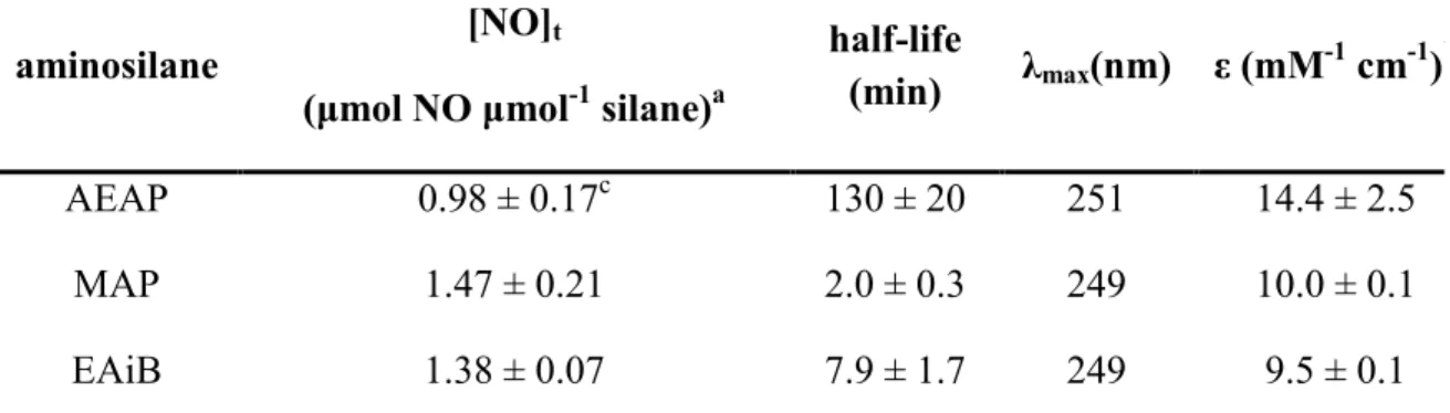
Table 2.1 Nitric oxide release, conversion efficiency, and spectroscopic parameters of N-diazeniumdiolate-modified
silanes ...58 Table 2.2 Nitric oxide release characteristics from
N-diazeniumdiolate modified xerogels measured using
chemiluminescence ...65 Table 2.3 Nitric oxide release totals and conversion efficiency from
pre- and post-diazeniumdiolated xerogels ...67 Table 2.4 Carbon, hydrogen, and nitrogen content of
pre-diazeniumdiolated xerogels as determined via elemental
combustion analysis ...69 Table 2.5 Comparison of NO release totals measured directly using
chemiluminescence with NO release totals predicted by
elemental analysis ...71 Table 2.6 Total amount of silicon liberated from each xerogel system
after 14 d. Xerogels were stored in 10 mM PBS (pH 7.4, 37
o
C) and transferred to new solutions at 4 or 7 d. Cumulative Si concentrations are the integrated totals from each time
point as determined via ICP-OES ...76 Table 2.7 Nitric oxide release totals from 15 mol% AEAP/PT
xerogels when stored in different atmospheric conditions and temperatures for 10 d. The control xerogel was analyzed immediately after drying. Each film was analyzed
in 30 mL PBS (pH 7.4, 37 oC) (n=1) ...78 Table 2.8 Properties of enzyme-based glucose biosensors coated with
pre-diazeniumdiolated 15 mol% AEAP/NO-PTMOS
xerogels after 0, 4 or 7 d immersion in PBS ...83 Table 3.1 Variation in NO-release kinetics as a function of
superhydrophobic coating thickness ...102 Table 4.1 Cumulative NO and silver doses delivered from the dual
NO- and silver-releasing xerogels following 3 h exposure to P. aeruginosa and the resulting log-decrease in viable
adhered bacterial colonies. ...132 Table 4.2 Cumulative NO and silver doses delivered from the NO-
xiv
aureus and the resulting log-decrease in viable adhered
bacterial colonies ...136 Table 4.3 Bacterial surface coverage on NO- and silver-releasing
xerogels following 3 h immersion in 108 cfu mL-1 P.
aeruginosa...139 Table 5.1 Nitric oxide-release characterization of
N-diazeniumdiolate-modified G4-PAMAM dendrimers. All values were determined using chemiluminescent nitric
oxide analysis. ...162 Table 5.2 Nitric oxide-release characterization of
N-diazeniumdiolate-modified dendrimers doped within 75TP470:25TPU polyurethanes (25 wt%) and coated with TP470 barrier layers (30 μL; 40 mg mL-1). Adhesion layers (75TP470:25TPU) were used for ACN and
G4-ACN/SO systems ...169 Table 5.3 Fiber diameters and dendrimer leaching from NO-releasing
G4-PAMAM-doped electrospun TP470 fibers. Dendrimers were doped at a concentration of 5 wt% relative to the TP470 polyurethane. Fiber diameters were determined from SEM and leaching was determined through
RITC-tagged dendrimers measured using fluorometry ...175 Table 5.4 Nitric oxide release properties from G4-PAMAM-doped
electrospun TP470 fibers determined via chemiluminescence. Dendrimers were doped at a
xv
LIST OF FIGURES
Figure 1.1 The stages of biofilm formation that occur when bacteria interact with surfaces. After adhering to a surface, bacteria excrete a protective exopolysaccharide matrix (represented in the graphic as a yellow film), multiply, and disperse into
planktonic bacteria ...4 Figure 1.2 Passive strategies for reducing bacterial adhesion and
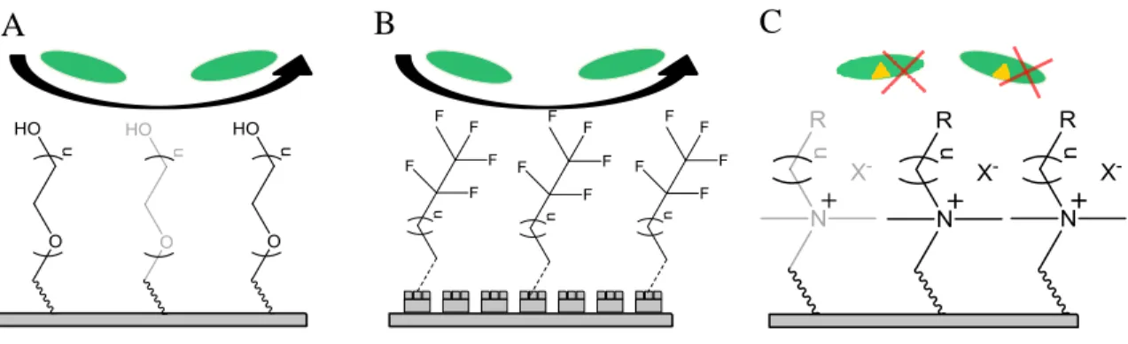
viability on a surface. (A) Poly (ethylene glycol) and (B) superhydrophobic modifications prevent bacterial adhesion, while (C) polycationic quaternary ammonium surfaces kill
adhered bacteria via membrane disruption ...6 Figure 1.3 A water droplet atop a rough surface obeying the (A)
Wenzel or (B) Cassie model...10 Figure 1.4 (A) Acid and (B) base-catalyzed sol-gel hydrolysis and
condensation reactions ...13 Figure 1.5 Minimum bactericidal concentration of NO-releasing
N-diazeniumdiolate formation on a secondary amine following exposure to NO and base. In the presence of a proton, the N-diazeniumdiolate decomposes to yield two
equivalents of NO and the parent amine ...21 Figure 1.6 (A) Poly (amido amine) (PAMAM) dendrimers display 1o
amines, the number of which depends on the dendrimer generation (Generation 0 is shown). (B) Conversion of the 1o to 2o amines allows for N-diazeniumdiolate modification, with subsequent NO-release kinetics
determined by the precursor used to form the 2o amine ...23 Figure 1.7 Nitric oxide-releasing xerogels synthesized via sol-gel
chemical approaches. Amine-modified silanes are incorporated into a silica matrix and then exposed to high pressure NO to convert the 2o amines to
N-diazeniumdiolate NO donors ...24 Figure 1.8 (A) Fixation pins coated with non-NO-releasing xerogels
become infected as foreign microbes migrate into the pin tract and proliferate in tissue. (B) Fixation pins coated with NO-releasing xerogel are killed by NO, and infection
xvi
Figure 2.1 Chemical structures of the N-diazeniumdiolate-modified
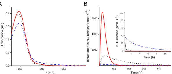
aminosilanes used to fabricate NO-releasing xerogels ...57 Figure 2.2 A) UV-vis spectra and B) NO-release curves of AEAP/NO
(blue dashed line), MAP/NO (red solid line), and EAiB/NO (black dotted line) precursors. Absorption spectra were obtained at a concentration of 50 mM in 1 M NaOH. Nitric
oxide release was measured in PBS (pH 7.4, 10 mM) ...60 Figure 2.3 Nitric oxide release from (A) AEAP/NO, (B) MAP/NO and
(C) EAiB/NO either immediately after synthesis (red line) or vacuum sealed at -20 oC for approximately 6 months
(black line) ...61 Figure 2.4 Total NO release from MAP/NO, EAiB/NO, and
AEAP/NO sols before (black bar; left) and after (red bar; right) the sol-gel reaction. Data is normalized to the “0” reaction time, which was acquired immediately after
addition of the N-diazeniumdiolate NO donor (n=1). ...68 Figure 2.5 The initial 12 h of NO release from 15 mol%
MAP/NO-PTMOS (red), EAiB/NO-MAP/NO-PTMOS (black) and AEAP/NO-PTMOS (blue). During measurement, films submerged in
30 mL PBS (pH 7.4) at 37 oC ...72 Figure 2.6 Silicon leaching from pre-diazeniumdiolated xerogels as a
function of NO donor A) identity and B) concentration. Silicon content was measured at 4, 7, and 14 d from A) 15 mol% AEAP/NO-PTMOS (black square), MAP/NO-PTMOS (red diamond), and EAiB/NO-MAP/NO-PTMOS (blue triangle) and B) 5 (blue triangle), 10 (red diamond) and 15 mol% (black square) AEAP. Error bars represent the standard deviation of the mean from n=3 independent
syntheses. ...75 Figure 2.7 Cytotoxicity of AEAP/PT, MAP/PT, and EAiB/PT leachate
solutions against L929 fibroblast cells. Error bars represent the standard deviation of the mean from n=3 independently
prepared samples ...77 Figure 2.8 Representative calibration curve for glucose biosensors
coated with 15 mol% AEAP/NO-PTMOS xerogels, both real-time (main graph) and as a function of glucose concentration (inset). Error bars represent the standard deviation of the mean from n=3 independently synthesized
sensor membranes ...81 Figure 3.1 Scanning electron micrograph of 30 mol% 17-FTMS
xvii
Figure 3.2 Scanning electron micrographs of superhydrophobic xerogels at A) 300x (scale bar = 100 μm), B) 3,000x (scale bar = 100 μm), and C) 10,000x (scale bar = 5 μm)
magnification ...101 Figure 3.3 Representative integrated NO-release totals from uncoated
and superhydrophobic-coated xerogels as a function of superhydrophobic layer thickness (# layers) and immersion
time. ...103 Figure 3.4 Contact angle stability of superhydrophobic-modified
NO-releasing xerogels after soaking in 37 oC PBS up to 28 d. Error bars represent the standard deviation of the mean
from at least n=7 measurements on n=3 xerogels. ...105 Figure 3.5 Cumulative silicon concentrations from
superhydrophobic-modified NO-releasing xerogels after soaking in 37 oC PBS for up to 28 d measured by ICP-OES. Error bars represent the standard deviation of the mean from n=3 independent
samples ...106 Figure 3.6 Reduction in viable P. aeruginosa adhesion vs. controls for
(NO) NO-releasing xerogels, (SH) superhydrophobic xerogel controls and (NO/SH) NO-releasing superhydrophobic-modified xerogels after (red) 6 h exposure in 108 cfu mL-1 PA and (grey) an additional 12 h in PBS. Error bars represent the standard deviation of the
mean from at least n=3 independent experiments. ...108 Figure 3.7 Relative viability of L929 fibroblasts exposed to (grey)
non-NO-releasing and (blue) NO-releasing superhydrophobic xerogels as a function of superhydrophobic layer thickness. Viability was determined using the MTS assay. Error bars represent the standard deviation of the mean from n=3 measurements of
n=1 samples ...111 Figure 4.1 (A) Cumulative silver release from AG-1 films with 0
(black), 20 (red), 40 (green), or 60 μL (red) μL barrier layers separating the xerogel from the underlying amine layer. The open-boxed line displays silver-release from AG-1 release in the absence of an underlying AHAP/BTMOS film. (B) Silver release from AG-1 (black), AG-2.5 (red), and AG-5 (blue) on 40 μL barrier layers. Silver was quantified using ICP-OES. Error bars represent the standard deviation of the mean from at least n=3
xviii
Figure 4.2 (A) Representative NO-release curves from AG-0/NO (black) and AG-1/NO (red) xerogels obtained using chemiluminescence. (B) Silver release from AG-1, AG-2.5, and AG-5 xerogels following N-diazeniumdiolate NO donor formation measured using ICP-OES. Error bars represent the standard deviation of the mean from at least
n=3 independently prepared samples. ...128 Figure 4.3 (A) Viable adhered colonies on NO- and silver-releasing
xerogels after 3 h exposure to 108 cfu mL-1 P. aeruginosa (B) Viable adhered colonies on AG-0, AG-0/NO, AG-1, and AG-1/NO in the same conditions. The traced box represents theoretical additive killing. Error bars represent the standard deviation of the mean from n=3 independent
experiments ...130 Figure 4.4 (A) Viable adhered colonies on NO- and silver-releasing
xerogels after 3 h exposure to 108 cfu mL-1 S. aureus. (B) Viable adhered colonies on AG-0, AG-0/NO, AG-1, and AG-1/NO following an additional 6 h in bacteria-free PBS (9 h total). The traced box represents theoretical additive killing. Error bars represent the standard deviation of the
mean for at least n=3 experiments. ...134 Figure 4.5 Fluorescent images of P. aeruginosa on (A) 0 (B)
AG-1 (C) AG-0/NO, and (D) AG-AG-1/NO visualized using the
images Syto 9 fluorescent probe. Scale bar = 20 μm ...138 Figure 4.6 Representative confocal micrographs of AG-0/NO, AG-1,
and AG-1/NO xerogels for visualizing intracellular NO uptake (DAF-2 DA) and cell death (PI). Fluorescent images showing DAF-2 DA and PI were converted to grey scale for improved visualization. Full-color images are provided
in the overlays. Scale bar = 10 μm. ...141 Figure 4.7 Cytotoxicity of AG-0, 1, 2.5, and 5 films without (grey bar)
and with (blue bar) NO-release capability against L929 fibroblasts. Cells are normalized to non-NO-releasing AG-0 xerogels. Error bars represent the standard deviation of
the mean from n=3 independent experiments. ...143 Figure 5.1 Structures of the 2o amines formed on (A) PAMAM from
(B) 1,2-expoy-9-decene (ED), (C) styrene oxide (SO), and (D) acrylonitrile (ACN). The PAMAM structure provided
for reference is G0 (4 primary amines) ...155 Figure 5.2 Dendrimer leaching from 10 wt% G4-ED doped
xix
of 100:0 (grey square), 75:25 (black square), 50:50 (red square), 25:75 (grey triangle) and 0:100 (black triangle). Leaching was determined through RITC-tagged dendrimers measured using fluorometry. Error bars represent the standard deviation of n=3 measurements from n=1 film. Error bars represent the standard deviation of n=3
measurements from n=1 film. ...164 Figure 5.3 Real-time nitric oxide release from 75:25 TP470:TPU
polyurethanes containing 10 (light grey), 18 (red), and 25 (black) wt% G4-ED. Measurements were acquired through
chemiluminescent nitric oxide analysis. ...166 Figure 5.4 Cumulative leaching from ACN/SO (grey) and
G4-ACN (blue) with (triangle) and without (square) an adhesion layer. Leaching was determined through RITC-tagged dendrimers and fluorometry. Error bars represent the standard deviation of the mean (n=3 measurements of
n=1 film per data point) ...168 Figure 5.5 Cytotoxicity of L929 fibroblasts following 24 h exposure to
G4-ED, G4-SO, G4-ACN, or G4-ACNSO-doped polyurethanes. Films are either 10 wt% dendrimer; no barrier layer (blue), 10 wt% dendrimer; with barrier layer (white stripe), 20 wt% dendrimer (light grey) or 30 wt% dendrimer (dark grey). Error bars represent the standard deviation of the mean from triplicate measurements of n=1
film. ...171 Figure 5.6 Electrospun TP470 fibers doped with 5 wt% (A) G4-ED,
(B) G4-SO, (C) G4-ACN, or (D) G4-ACN/SO viewed
using SEM. Scale bar = 10 μm. ...174 Figure 6.1 A) Xray photoelectron spectra of 0 (black), 10 (red), and 20
mol% (blue) DDTMS (balance MTMOS) silica composites (B) A water droplet (149o) on a 20 mol% DDTMS (balance
MTMOS) silica composite ...190 Figure 6.2 Aspergillus niger growth on (A) uncoated (B)
MTMOS-coated (C) 30 mol% 17FTMS (balance MTMOS)-MTMOS-coated and (D) superhydrophobic card stock. White areas represent the card stock substrate, and black areas are the spore-laden mycelia of the fungus. Each image displays the
xx
LIST OF SCHEMES
Scheme 2.1 Formation of N-diazeniumdiolates on secondary amines
and pH-dependent decomposition to produce NO ...47 Scheme 2.2 Synthesis of the “pre-diazeniumdiolated” NO-releasing
xerogels. After reacting AEAP with NO to yield AEAP/NO, the N-diazeniumdiolated precursor is reacted with PTMOS, and cast onto an appropriate substrate. Subsequent drying/curing results in the formation of an
N-diazeniumdiolate-modified xerogel film...49 Scheme 3.1 (I) Amine-modified xerogels on glass subtrates are (A)
exposed to 10 atm NO to yield (II) N-diazeniumdiolate-modified xerogels. (B) A fluorinated silica composite is then spraycoated onto the xerogels to yield (III)
superhydrophobic NO-releasing xerogels...93 Scheme 6.1 Proposed synthesis of QA-modified surfaces. (A) An alkyl
halide-functionalized silane and tertiary amine are reacted via nucleophilic aliphatic substitution to yield QA-modified silanes. (B) QA-modified silanes are co-condensed with linker silanes and silica colloids, before (C) application to a substrate to yield superhydrophobic QA-modified
xxi
LIST OF ABBREVIATIONS AND SYMBOLS
o
C degree(s) Celcius
~ approximately
% percentage
[…] concentration
ε molar absorptivity
λ wavelength
17FTMS (heptadecafluoro-1,1,2,2-tetrahydrodecyl)-trimethoxysilane
3D three-dimensional
μL microliter(s)
μM micromolar
ACN acrylonitrile
AEAP N-2-(aminoethyl)-aminopropyltrimethoxysilane AEAP/NO N-diazeniumdiolate-modified N-2-(aminoethyl)-
aminopropyltrimethoxysilane
AFM atomic force microscope
Ag+ silver ion
AgNO3 silver nitrate
AHAP N-(6-aminohexyl) aminopropyltrimethoxysilane
BP bandpass
BTMOS isobutyltrimethoxysilane
cfu colony forming units
d day(s)
DMEM Dulbecco’s modified essential media
xxii e.g. exemplia grata (for example)
et al. and others
EAiB N-ethylaminoisobutyltrimethoxysilane
EAiB/NO N-diazeniumdiolate-modified N-ethylaminoisobutyltrimethoxysilane
ED 1, 2-epoxy-9-decene
ETMOS ethyltrimethoxysilane
FBS fetal bovine serum
FDA United States Food and Drug Administration
G4 generation 4
h hour(s)
H20 water
HCl hydrochloric acid
i.e. id est (in other words)
iNOS inducible nitric oxide synthase
M molar
MΩ Megaohm(s)
MAP N-methylaminopropyltrimethoxysilane
MAP/NO N-diazeniumdiolate-modified N-methylaminopropyltrimethoxysilane
MTMOS methyltrimethoxysilane
mM millimolar
min minute(s)
mg milligram(s)
mL milliliter(s)
mm millimeter(s)
xxiii
MTS 4,5-dimethylthiazol-2-yl)-5-(3-carboxymethoxyphenyl)-2-(4- sulfophenyl)-2H-tetrazolium
NaOMe sodium methoxide
nm nanometer(s)
NO nitric oxide
N2 nitrogen gas
O3 ozone
P. aeruginosa Pseudomonas aeruginosa
PBS phosphate buffered saline
pH -log of proton concentration
PI propidium iodide
PMS phenazine methosulfate
ppb parts per billion
ppm parts per million
PTMOS propyltrimethoxysilane
PMMA poly (methyl methacrylate)
PROLI/NO disodium 1-[2-(Carboxylato)pyrrolidin-1-yl]diazen-1- ium-1,2-diolate (1)
PVC poly (vinyl chloride)
rpm revolutions per minute
s second(s)
S. aureus Staphylococcus aureus S. epidermidis Staphylococcus epidermidis
SSD silver sulfadiazene
xxiv
SO styrene oxide
t time
THF tetrahydrofuran
TP470 tecoplast tp-470-00
TPU tecophilic sg-80a
TSA tryptic soy agar
TSB tryptic soy broth
Chapter 1:
The Development of Antifouling and Biocidal Coatings for Biomedical Implants
The advent of modern biomaterials research began when clinicians serendipitously discovered that certain polymers (e.g., poly(methyl methacrylate) canopies “implanted” into the eyes of fighter jet pilots via machinegun fire) evoked a favorable healing response when compared to other materials.1 As the scientific understanding of molecular biology and materials science has grown, researchers have studied the processes governing the tissue biocompatibility of implants. Given the seemingly endless combination of polymer chemistries, 3D-architectures and mechanical properties that have been developed, it is best to categorize these materials by their function rather than their form. A clear delineation in function appears between two types of biomaterials: 1) implantable devices, where the material in-and-of-itself is the purpose of the implant (e.g., prosthetic joints, catheters, stints, engineered tissue scaffolds, drug-release reservoirs, etc.); and, 2) device coatings, where the biomaterial serves as a companion to an existing implant to improve its utility. This introductory chapter focuses on the latter, specifically coatings that reduce bacterial adhesion or kill bacteria on implanted materials.
1.1 Microbial colonization on medical devices
2
with infection rates ranging from 1—20% depending on the implant type as well as the expertise of the surgical staff performing the procedure.2, 3 At worst, bacterial colonization on an implanted material may lead to life-threatening infections.4 In milder cases, the presence of bacteria on a surface discourages successful integration of the device into native tissue, necessitating device removal.5,6 These episodes are costly; for instance, the expense of arthoplasty infections exceed >$50,000 per case and extend average hospital stays by an average of 11 days.3,7 Mechanisms to reduce the incidence of infection on implanted devices are greatly needed.
The critical density of bacteria required to infect an implant is much lower than the amount required when an implant is absent.2 This discrepancy arises from bacteria’s ability to adhere readily to surfaces. Furthermore, surgical implantation procedures themselves cause inflammation and a localized immuno-incompetent zone, weakening the natural response of the host to microbes introduced during surgery.5 The weakened host response is evident from the vast number of implant infections caused by Staphylococcus epidermidis; in normal tissue, S. epidermidis is virtually avirulent.5,8 Following implantation, localized inflammatory responses produce reactive oxygen intermediates such as superoxide, while production of interferon-γ and interleukin-1 are inhibited.9 As a result, macrophages (immune cells that combat bacteria) become less effective and the activity of other immune cells such as lympochytes, monocytes and neutrophils is depleted.8 Moreover, some immune cells (e.g., leukocytes) are not adept at killing surface-adhered bacteria due to shear stresses at the fluid-solid interface.10
3
that render conventional antibiotic treatments ineffective.11,12 Biofilms are extremely common and implicated in at least 90% of catheter related infections.13 Biofilm bacteria are thus best considered the norm rather than the exception. Biofilms form when bacteria adhere to a surface and secrete a thick polysaccharide matrix. Bacteria within biofilms also occupy a broad range of phenotypes and metabolic states, many of which are less susceptible to antibiotics.12 The environmental heterogeneity within biofilms causes broad variations in nutrient concentrations, oxygen levels and pH that interfere with the action of many antibiotics.12, 14-17 While still not fully understood, cells in biofilms utilize quorum sensing (e.g., cell-to-cell communication) to rapidly change gene expression patterns within the colony.18 This form of communication may control biofilm dispersal, where bacteria within the sessile biofilm are “released” into the local environment in their planktonic form to colonize new surfaces.19 As a result, biofilms on clinical devices cause repeated incidents of acute infection (Figure 1.1).
Complicating matters further, most biofilms are polymicrobial.20 Polymicrobial biofilms are even less susceptible to antibiotics. Harriott et al. examined the efficacy of vancomycin against polymicrobial biofilms containing Staphylococcus aureus and Candida albicans, finding that C. albicans caused S. aureus to become highly resistant to treament.21 Polymicrobial biofilms can also grow faster; for example, Peters et al. found that polymicrobial biofilms of C. albicans and S. aureus grew synergistically.22
4
Figure 1.1 The stages of biofilm formation that occur when bacteria interact with surfaces. After adhering to a surface, bacteria excrete a protective exopolysaccharide matrix (represented in the graphic as a yellow film), multiply, and disperse into planktonic bacteria.
I. Non-specific adhesion
II. Irreversible adhesion
5
microbial adhesion from the start.23 To this end, researchers have developed antimicrobial materials that quell the adhesion of bacteria to surfaces and/or kill bacteria that do adhere. The two major branches of antifouling/antimicrobial materials, passive and active, are discussed in sections 1.3 and 1.4.
1.2 Passive surface modifications for reducing infection
A number of coatings feature topographies or covalently bound chemical moieties that reduce bacterial adhesion or kill bacteria. Coatings of this type (“passive”) rely on mechanisms intrinsic to the surface itself (Figure 1.2) to combat infection. In this section, the strengths and limitations of such surfaces as they relate to clinical applications are discussed.
1.2.1 Non-fouling surfaces
Surfaces that resist fouling (i.e., adherence of proteins and bacteria) represent perhaps the simplest route towards antimicrobial coatings.24 Perhaps the most thoroughly studied antifouling interface utilizes poly(ethylene glycol) (PEG) to resist protein adhesion.25 The PEG macromolecule is easily grafted onto a number of existing surfaces (“PEGylation”), including polyurethanes,26 silicon,27 silica,28 and stainless steel.29 Not limited to grafting, PEG can also comprise 3-dimensional scaffolds such as hydrogels.30
The protein-resistant capabilities of PEGylated materials arise from its hydrophilic nature. When proteins in solution come in contact with these surfaces, the hydrated PEG chains become compressed, forcing steric interactions that are not energetically favorable.31,
32
6
Figure 1.2 Passive strategies for reducing bacterial adhesion and viability on a surface. (A) Poly (ethylene glycol) and (B) superhydrophobic modifications prevent bacterial adhesion, while (C) polycationic quaternary ammonium surfaces kill adhered bacteria via membrane disruption.
7
proteins are forced into the PEGylated layer. As such, it was concluded that steric hindrance alone does not fully account for the antifouling properties of PEG.33 A second, more likely mechanism involves competition between the adhesion of water molecules and the adhesion of larger macromolecules to the PEG chains.33
Despite a proven ability to resist protein adhesion, the ability for PEGylated surfaces to resist bacterial adhesion is less promising. Whitesides and coworkers found that a resistance to protein adhesion does not correlate with a resistance to bacterial adhesion,34 owing to mechanisms of bacterial adhesion that do not rely on proteins. Likewise, PEG surfaces on stainless steel that were highly resistant to protein showed very little resistance in adhesion to Pseudomonas sp. and Listeria monocytogenes.29 These shortcomings were initially attributed to the instability of such coatings in physiological solutions. Roosjen et al. observed hydrolysis of the bond connecting poly(ethylene oxide) to its substrate in saliva and urine. Kingshott et al. also demonstrated that stable, covalent attachment of PEG is critical for any success in resisting bacterial adhesion. By grafting PEG onto a covalently-immobilized branched poly(ethylenimine) macromolecule on stainless steel, the surfaces were able to reduce bacterial adhesion by 2—4 orders of magnitude over a 5-h period.35
8
The tetravalent PEG materials were able to resist S. aureus adhesion and subsequent biofilm formation.36, 37
Even if PEG is stably bound to a substrate, other issues arise with the PEG moiety itself. In oxygenated conditions, or those where transition metal ions are present, the PEG surface may degrade through oxidation.38 PEG hydroxyl groups are also enzymatically converted to aldehydes in vivo.38 As such, coatings less susceptible to degradation may be more appropriate for preventing bacterial adhesion.
1.2.2 Superhydrophobic materials
9
obeying the Wenzel model remain pinned to the interface even at high tilt angles (a “Wenzel state” droplet). In practice, Wenzel state droplets occur when water impregnates the valleys of the roughened surface, giving water droplets direct contact with the material itself rather than air (Figure 1.3). For a surface to be effective in self-cleaning and fouling prevention, the surface properties need to be tailored so that the droplets remain in the Cassie state. This condition is most effectively achieved through hierarchal roughness that exhibits both microscale and nanoscale dimensions.42
10
Figure 1.3 A water droplet atop a rough surface obeying the (A) Wenzel or (B) Cassie model.
11
superhydrophobic plant) such that no plastron remained, and still observed a resistance to P. aeruginosa adhesion. This anti-adhesive effect was attributed to surface architecture, as areas featuring dense nanostructures tended to have the fewest adhered bacteria. Those bacteria that did adhere were mostly located in the boundary regions between nanostructures.44 Spatial variations in adhesive forces were further corroborated via atomic force microscopy, where those areas with high nanostructure densities had less adhesion than areas lacking nanostructures.
When bacteria require specific proteins to adhere to a surface, their adhesion to superhydrophobic substrates may be inhibited through reduced protein adsorption. Stallard and co-workers utilized spectroscopic ellipsometry to quantify fibrinogen adhesion to surfaces with water contact angles between <5 and >150o. Adsorbed fibrinogen was lowest on the fluorinated superhydrophobic substrates. To determine if this reduction in protein binding led to decreased bacterial adhesion, the authors exposed the substrates to a suspension of SH1000, a strain of S. aureus that specifically binds to fibrinogen.47 Compared to fibrinogen-bound controls, bacterial adhesion was reduced by ~99% for superhydrophobic substrates regardless of fibrinogen exposure. These findings indicate that superhydrophobicity reduces bacterial adhesion in the absence of proteins, but also reduces the adhesion of proteins that bind specifically to bacteria.48
12
aqueous reaction conditions and low-temperature drying environments.49 We first fabricated colloidal silica particles (to impart roughness), and then crosslinked those particles within a fluorinated sol-gel film (for low surface energy).49 Utilized in chapters 2, 3, and 4 of this dissertation, the sol-gel method consists of the hydrolysis and condensation of silane precursors to yield a variety of materials depending on the reaction conditions (Figure 1.4). For example, low silane concentrations with basic ammonium hydroxide catalysts favor rapid silane condensation and tend to form particles. In contrast, sol-gel reactions in acidic conditions produce linear chains that further crosslink to form a 3-dimensional network (referred to as a “xerogel” when dried).
13
Figure 1.4 (A) Acid and (B) base-catalyzed sol-gel hydrolysis and condensation reactions.
A
14 1.2.3 Superhydrophobic materials for drug release
15
10 d. However, the cells could be killed (99%) after this period by releasing the entrapped drug with ultrasound. In summary, superhydrophobic fiber mats are an elegant approach for tuning drug release rates; however, doping drugs into fibers may not be feasible for all drug release chemistries. These superhydrophobic fiber mats serve as stand-alone implants (i.e., devices) rather than coatings on existing medical implants. A need still exists for a facile superhydrophobic coating that can be applied in bulk to a wide range of drug-releasing substrates for tuning release of the drug contained within.
1.2.4 Polycationic antimicrobial surfaces
Small molecule quaternary ammonium (QA) compounds have seen widespread use as antiseptics and disinfectants.58 Mechanistically, the cationic charge on QAs promotes association of the molecules with the negatively charged surface of bacteria. Once QAs are surface-associated, the hydrophobic chains pendant from the ammonium cation cause bacterial membrane disruption, ultimately leading to cell death.59 Additional antimicrobial action may be attributed to diffusion and uptake of the molecules into bacterial cells, but membrane-specific mechanisms have inspired the development of coatings with tethered-on antimicrobial QA moieties.58, 60, 61
16
slow release of the compound, the surfaces were repeatedly washed in distilled water and their antimicrobial efficacy was reexamined. The authors found that the coatings largely retained their antimicrobial efficacy after washing, and thus attributed the mechanism of action to the surface-bound QAs.
17
declined by 42%. While the antimicrobial activity could be restored by cleaning off the dead bacteria with surfactants, such a step would be impractical for implanted devices. Furthermore, these surfaces are much less effective at killing bacteria when challenged with high concentrations of bacteria (>1 x 10⁷ cfu mL-1).65 The biocidal action of such coatings is only considered “permanent” insofar as the surface remains clean, or can be regenerated through cleaning. Since even dead bacteria are able to invoke an inflammatory response in vivo,66 mechanisms that reduce bacterial adhesion in addition to killing bacteria are needed.
1.3 Active release strategies for antimicrobial surfaces
The other major branch of antimicrobial materials utilizes the active release of biocidal agents from an implant surface.67 This approach offers several advantages. First, large systemic doses of the biocidal compound in question are avoided because antimicrobial concentrations of the agent are relegated to an area close to the device itself. Concentrations of a released agent in the stagnant layer immediately adjacent to the implant are several orders of magnitude larger than systemic concentrations due to dilution that occurs as the agent diffuses outward.8 Second, active release mechanisms, unlike passive mechanisms, are not typically obstructed by surface fouling or the adhesion of dead cells.67
18
host immune response is severely compromised at the implant site due to localized inflammation. While longer durations may certainly be beneficial, a finite biocidal duration by no means renders the device unusable. A number of biocidal agents have been explored as active release materials, including those that release conventional antibiotics, silver, and nitric oxide. Each is discussed in detail below.
1.3.1 Release of antibiotics
To reduce the need for systemic antibiotic therapy, researchers have incorporated antibiotics into materials to facilitate localized release.69 Since release of the drug is dictated by diffusion, the water uptake by the polymer significantly impacts release kinetics. Risbud et al. reported on amoxicillin-loaded chitosan/polyvinyl pyrrolidone (PVP) hydrogels that released a majority (73%) of their contents within 3 h. In a follow on-study, polyacrylamide-chitosan hydrogels with comparatively lower water uptake were able to slow the release, such that only ~75% of the total payload was released over 3 d.70
19
isolatable viable bacterial colonies in addition to improving bone integration and reducing osteomyelytis.74 After 3 and 4 weeks, the films were no longer able to kill bacteria, suggesting that the vancomycin supply was exhausted.74 Such findings support the benefit of long-term antibiotic release.75 Unfortunately, extremely long-term sustained release of antibiotics may actually serve as a pitfall to successful treatment. Neut and colleagues analyzed a patient whom had been treated with gentamicin-loaded poly(methyl methacrylate) beads 5 years prior.76 The beads still released residual levels of the antibiotic. This sustained, sub-inhibitory release fostered development of a gentamicin-resistant staphylococcal strain of bacteria. The authors concluded that antibiotic-loaded materials must be designed to prevent long-term sustained release, possibly through biodegradability mechanisms.77,78 Similar concerns have been brought to light in other systems that release antibiotics. Antibiotic-loaded bone cements, for instance, were criticized by Hanssen for their ability to encourage drug-resistance.79 Furthermore, multiple antibiotics are required from these materials to successfully inhibit a broad range of microorganisms.79 Active release agents that operate via less specific pathways (i.e., broad-spectrum agents) and overcome bacterial resistance mechanisms may be more suitable candidates for localized antimicrobial release.
1.3.2 Nitric oxide-releasing surfaces
20
that form peroxynitrite (by reaction with the superoxide anion), nitrogen dioxide, and dinitrogen trioxide (by reaction with oxygen).85-87 These species place oxidative and nitrosative stress on bacteria, ultimately disrupting membrane, protein, enzyme, and DNA function.88 Mammalian cells, like those located in the endothelium, are able to combat the effects of oxidative stress (especially via peroxynitrite) by lowering superoxide concentrations through the superoxide dismutase enzyme.89 The wide array of mechanisms by which NO kills makes it difficult for bacteria to develop resistance to the molecule, as multiple simultaneous genetic mutations would be required for a survival advantage.90 Taken together, NO’s low toxicity, wide-spread distribution in the mammalian body, antimicrobial activity, and lack of demonstrated bacterial resistance make it an ideal candidate for active releasing antimicrobial surfaces.
Supplying exogenous NO to the body requires a means of delivery that is stable until the time of treatment and releases at controlled rates during treatment. A number of chemical NO donors exist for this purpose, including organic nitrates and nitrates,91 metal nitrosyls,92 S-nitrosothiols,93 and N-diazeniumdiolates.94, 95 While the clinical use of organic nitrates and metal nitrosyls is well-established (and in fact predates knowledge of NO’s biological activity), the mechanisms by which these donor classes decompose into NO are highly dependent on host-specific factors such as enzyme activity.96-99 At least some of the action of organic nitrates and metal nitrosyls involves the formation of S-nitrosothiol intermediates.100 Thus, N-diazeniumdiolate (Figure 1.5) and S-nitrosothiol NO donors remain the most promising donors for their well-understood NO release triggers, rates, and mechanisms.
21
Figure 1.5 N-diazeniumdiolate formation on a secondary amine following exposure to NO and base. In the presence of a proton, the N-diazeniumdiolate decomposes to yield two equivalents of NO and the parent amine.
2 NO
base
22
established in the late 1980s.103 Formed via the reaction of secondary amines and NO under basic conditions, these compounds exhibit pH-dependent decomposition to two NO equivalents and the parent (i.e., precursor) amine. The decomposition rate (i.e., NO-release rate) is dependent on the structure, with certain chemical groups capable of stabilizing the N-diazeniumdiolate.94 To control the release of NO and target bacteria directly, these functional groups have been incorporated within larger macromolecular scaffolds such as silica particles and dendrimers.104-110 Like small molecules, the NO-release kinetics from such scaffolds are controllable by varying the environment surrounding the N-diazeniumdiolate (Figure 1.6).
23
Figure 1.6 (A) Poly (amido amine) (PAMAM) dendrimers display 1o amines, the number of which depends on the dendrimer generation (Generation 0 is shown). (B) Conversion of the 1o to 2o amines allows for N-diazeniumdiolate modification, with subsequent NO-release kinetics determined by the precursor used to form the 2o amine.
A
24
25
initiate biofilm dispersal.119 The ability for NO-releasing materials to both resist bacterial adhesion and kill adhered bacteria suggest that NO may be a suitable active release agent for reducing infection in medical implants.
Follow-on work demonstrated NO’s ability to combat infection in vivo.120, 121 Nablo et al. implanted NO-releasing xerogels into the subcutaneous tissue of male rats and inoculated the wound site with S. aureus (1 x 10⁶ cfu). Compared to controls, an 82% reduction in infection incidences was observed in rats with NO-releasing implants.120 Building upon this work, Holt et al. coated NO-releasing xerogels onto external fixation pins implanted into the tibia of rats. Because external fixators are percutaneous (i.e., exposed through skin), such devices are prone to infection as bacteria are able to migrate through the pin tract following implantation (Figure 1.8). Following 28 d implantation, significant reductions in bacterial colonies and clinical signs of infection were observed at the NO-releasing pins compared to controls.121 Together, these studies illustrate NO’s ability to reduce infection in implants.
26
Figure 1.8 (A) Fixation pins coated with non-NO-releasing xerogels become infected as foreign microbes migrate into the pin tract and proliferate in tissue. (B) Fixation pins coated with NO-releasing xerogels are killed by NO, and infection incidences are reduced.
NO NO
NO
NO NO B
A
27
acute inflammation and inhibited analyte diffusion by collagen encapsulation) lead to erratic responses. Nitric oxide-release may alleviate some of these pitfalls. Unfortunately, sensor membranes made to release NO from xerogels are mostly impermeable to glucose. Thus while they reduce the FBR, they are not analytically useful. More permeable (to glucose) NO-releasing sensor membranes would enable the development of better performing continuous monitoring glucose sensors.
1.3.3 Antimicrobial silver-releasing materials
Silver’s origins as a broad-spectrum antimicrobial agent date back to ancient times, where Persians, Greeks, Romans and Egyptians used it to preserve water and food.128 Modern-day silver releasing biomaterials hold distinction as one of the few active release systems with demonstrated success in clinical trials.129 Current Food and Drug Administration (FDA) approved devices include wound dressings for burn patients and coatings for urinary and intravascular catheters.130-132 Most often, silver is impregnated into (or on) materials/devices in the form of ionic silver (e.g., silver nitrate,133, 134 silver sulfadiazine,135 etc.), silver metal,136 or silver colloids and nanoparticles.73, 137
28
state are largely indirect, relying on oxidation of the metals to free Ag+. Xiu et al. added convincing support to this theory by testing silver nanoparticles against bacteria in anaerobic conditions (i.e., conditions precluding the formation of Ag+). They found that the toxicity of these materials against bacteria was drastically inhibited,140 and ultimately concluded that differences in antimicrobial activity between different shapes and sizes of silver nanoparticles are intrinsically intertwined with differences in the rate of Ag+ production.
These findings have since guided the development of silver releasing materials. For example, Marini et al. doped sol-gel films with both silver nitrate and silver nanoparticles, finding almost no antimicrobial efficacy from the nanoparticle-doped silica materials despite high antimicrobial activity from silica doped with silver nitrate.133 The authors concluded that diffusion of Ag+ from the tortuous silica network, combined with the slow release of Ag+ from the nanoparticles resulted in a silver release rate too low for antimicrobial activity. Antimicrobial fluxes from silver nanoparticle-doped materials may be achieved if the materials are thin and/or the surrounding framework is open.137, 141 However, direct addition of a silver salt is a simpler approach.
29
relevance.136 Hardes et al. examined the long-term toxicity of an implanted anti-infective silver megaprosthesis in humans over a mean period of 19 months. No damage to liver or kidneys (or any systemic side effects for that matter) was noted. Some bioaccumulation of silver occurred, with blood silver concentrations around 56.4 ppb (compared to basal levels of 2 ppb in untreated patients145) but these concentrations would not be expected to produce systemic toxic effects.146 Trop et al. reported agyria-like symptoms, liver toxicity, and elevated levels of silver in plasma and urine for a burn patient treated with silver sulfadiazine (SSD)-coated polyethylene meshes.147 These conflicting results likely emerge from the varied Ag+ release rates of different materials. The Ag+ release rate may also change for a specific material due to changes in localized pH,128 ionic strength,148 and dissolved oxygen content.149 Nonetheless, clear evidence of dose-dependent toxicity for Ag+ and its associated compounds exists. Given silver’s ability to accumulate systemically, it is best to pursue strategies that minimize overall silver release while maximizing localized antimicrobial activity.
30
31 1.4 Summary of dissertation research
The goal of my research was to design antimicrobial coatings that both resist bacterial adhesion and kill adhered bacteria. Rational approaches were employed to reduce toxicity to healthy cells. Specifically, my research aims included:
1. the development of NO-releasing coatings via facile sol-gel techniques that can easily be applied to functional glucose sensors;
2. the synthesis of superhydrophobic NO-releasing xerogels that combine passive and active antimicrobial strategies, exhibit superior resistance to bacterial adhesion and kill adhered bacteria;
3. the development of coatings that simultaneously release Ag+ and NO for the synergistic killing of adhered bacteria; and
4. utilization of NO-releasing dendrimers as dopants within polyurethane films and fibers to create materials that release low levels of NO over prolonged periods of time.
32
33 REFERENCES
1. Ratner, B.D.; Bryant, S.J., "Biomaterials: Where we have been and where we are going." Annu. Rev. Biomed. Eng. 2004, 6, 41-75.
2. Campoccia, D.; Montanaro, L.; Arciola, C.R., "The significance of infection related to orthopedic devices and issues of antibiotic resistance." Biomaterials 2006, 27, 2331-2339.
3. Katsikogianni, M.; Missirlis, Y.F., "Concise review of mechanisms of bacterial adhesion to biomaterials and of techniques used in estimating bacteria-material interactions." Eur. Cells. Mater. 2004, 8, 37-57.
4. Tiesenhausen, K.; Amann, W.; Koch, G.; Hausegger, K.; Thalhammer, M., "Endovascular stentgraft infection—a life-threatening complication." Vasa 2000, 29, 147-150.
5. Gristina, A.G., "Implant failure and the immuno-incompetent fibro-inflammatory zone." Clin. Orthop. Relat.R. 1994, 106-18.
6. Poelstra, K.A.; Barekzi, N.A.; Rediske, A.M.; Felts, A.G.; Slunt, J.B.; Grainger, D.W., "Prophylactic treatment of gram-positive and gram-negative abdominal implant infections using locally delivered polyclonal antibodies." J. Biomed. Mater. Res. A 2001, 60, 206-215.
7. Berbari, E.F.; Hanssen, A.D.; Duffy, M.C.; Steckelberg, J.M.; Ilstrup, D.M.; Harmsen, W.S.; Osmon, D.R., "Risk factors for prosthetic joint infection: Case-control study." Clin. Infect. Dis. 1998, 27, 1247-1254.
8. Schierholz, J.M.; Beuth, J., "Implant infections: A haven for opportunistic bacteria." J. Hosp. Infect. 2001, 49, 87-93.
9. Kaplan, S.S.; Basford, R.E.; Kormos, R.L.; Hardesty, R.L.; Simmons, R.L.; Mora, E.M.; Cardona, M.; Griffith, B.L., "Biomaterial associated impairment of local neutrophil function." ASAIO J. 1990, 36, M172-174.
10. Sapatnekar, S.; Kao, W.J.; Anderson, J.M., "Leukocyte—biomaterial interactions in the presence of Staphylococcus epidermidis: Flow cytometric evaluation of leukocyte activation." J. Biomed. Mater. Res. 1997, 35, 409-420.
11. Marrie, T.J.; Nelligan, J.; Costerton, J., "A scanning and transmission electron microscopic study of an infected endocardial pacemaker lead." Circulation 1982, 66, 1339-1341.
34
13. Mukherjee, P.K.; Zhou, G.; Munyon, R.; Ghannoum, M.A., "Candida biofilm: A well-designed protected environment." Med. Mycol. 2005, 43, 191-208.
14. Zhang, T.C.; Bishop, P.L., "Evaluation of substrate and pH effects in a nitrifying biofilm." Water Environm. Res. 1996, 1107-1115.
15. Tack, K.J.; Sabath, L., "Increased minimum inhibitory concentrations with anaerobiasis for tobramycin, gentamicin, and amikacin, compared to latamoxef, piperacillin, chloramphenicol, and clindamycin." Chemotherapy 1985, 31, 204-210. 16. Tuomanen, E.; Cozens, R.; Tosch, W.; Zak, O.; Tomasz, A., "The rate of killing of
Escherichia coli by β-lactam antibiotics is strictly proportional to the rate of bacterial growth." J. Gen. Microbiol. 1986, 132, 1297-1304.
17. Mah, T.F.; O'Toole, G.A., "Mechanisms of biofilm resistance to antimicrobial agents." Trends Microbiol. 2001, 9, 34-9.
18. Zimmerli, W., "Infection and musculoskeletal conditions: Prosthetic-joint-associated infections." Best. Prac. Res. Cl. Rh. 2006, 20, 1045-1063.
19. Parsek, M.R.; Greenberg, E., "Sociomicrobiology: The connections between quorum sensing and biofilms." Trends Microbiol. 2005, 13, 27-33.
20. Mermel, L.A.; Farr, B.M.; Sherertz, R.J.; Raad, I.I.; O'Grady, N.; Harris, J.S.; Craven, D.E., "Guidelines for the management of intravascular catheter-related infections." Clin. Infect. Dis. 2001, 32, 1249-1272.
21. Harriott, M.M.; Noverr, M.C., "Candida albicans and Staphylococcus aureus form polymicrobial biofilms: Effects on antimicrobial resistance." Antimicrob. Agents Ch. 2009, 53, 3914-22.
22. Peters, B.M.; Ward, R.M.; Rane, H.S.; Lee, S.A.; Noverr, M.C., "Efficacy of ethanol against Candida albicans and Staphylococcus aureus polymicrobial biofilms." Antimicrob. Agents Ch. 2013, 57, 74-82.
23. Cheng, G.; Zhang, Z.; Chen, S.; Bryers, J.D.; Jiang, S., "Inhibition of bacterial adhesion and biofilm formation on zwitterionic surfaces." Biomaterials 2007, 28, 4192-9.
24. Krishnan, S.; Weinman, C.J.; Ober, C.K., "Advances in polymers for anti-biofouling surfaces." J. Mater. Chem. 2008, 18, 3405-3405.
25. Andrade, J.; Hlady, V.; Jeon, S., "Poly(ethylene oxide) and protein resistance: Principles, problems, and possibilities." Advances in Chemistry 1996, 248, 51-60. 26. Park, K.D.; Kim, Y.S.; Han, D.K.; Kim, Y.H.; Lee, E.H.; Suh, H.; Choi, K.S.,
35
27. Zhang, M.; Desai, T.; Ferrari, M., "Proteins and cells on PEG immobilized silicon surfaces." Biomaterials 1998, 19, 953-960.
28. Alcantar, N.A.; Aydil, E.S.; Israelachvili, J.N., "Polyethylene glycol-coated biocompatible surfaces." J. Biomed. Mater. Res. 2000, 51, 343-51.
29. Wei, J.; Ravn, D.B.; Gram, L.; Kingshott, P., "Stainless steel modified with poly(ethylene glycol) can prevent protein adsorption but not bacterial adhesion." Colloid Surface B. 2003, 32, 275-291.
30. Raeber, G. P.; Lutolf, M. P.; Hubbell, J. A., "Molecularly engineered PEG hydrogels: A novel model system for proteolytically mediated cell migration." Biophys. J. 2005, 89, 1374-88.31.
31. Jeon, S.; Andrade, J., "Protein—surface interactions in the presence of polyethylene oxide: II. Effect of protein siz." J. Colloid. Interface Sci. 1991, 142, 159-166.
32. Jeon, S.; Lee, J.; Andrade, J.; De Gennes, P., "Protein-surface interactions in the presence of polyethylene oxide: I. Simplified theory." J Colloid. Interf. Sci. 1991, 142, 149-158.
33. Sheth, S.R.; Leckband, D., "Measurements of attractive forces between proteins and end-grafted poly(ethylene glycol) chains." Proc. Natl. Acad. Sci USA 1997, 94, 8399-404.
34. Ostuni, E.; Chapman, R.G.; Liang, M.N.; Meluleni, G.; Pier, G.; Ingber, D.E.; Whitesides, G.M., "Self-assembled monolayers that resist the adsorption of cells." Langmuir 2001, 17, 6336-6343.
35. Kingshott, P.; Wei, J.; Bagge-ravn, D.; Gadegaard, N.; Gram, L., "Covalent attachment of poly(ethylene glycol) to surfaces, critical for reducing bacterial adhesion." Langmuir 2003, 209, 6912-6921.
36. Khoo, X.; Hamilton, P.; O'Toole, G.A.; Snyder, B.D.; Kenan, D.J.; Grinstaff, M.W., "Directed Assembly of PEGylated-Peptide Coatings for Infection-Resistant Titanium Metal." J. Am. Chem. Soc. 2009, 131, 10992-10997.
37. Khoo, X.; O'Toole, G.A.; Nair, S.A.; Snyder, B.D.; Kenan, D.J.; Grinstaff, M.W., "Staphylococcus aureus resistance on titanium coated with multivalent PEGylated-peptides." Biomaterials 2010, 31, 9285-92.
38. Ostuni, E.; Chapman, R.G.; Holmlin, R.E.; Takayama, S.; Whitesides, G.M., "A survey of structure-property relationships of surfaces that resist the adsorption of protein." Langmuir 2001, 17, 5605-5620.
36
40. Cassie, A.; Baxter, S., "Wettability of porous surfaces." T. Faraday Soc. 1944, 40, 546-551.
41. Wenzel, R.N., "Resistance of solid surfaces to wetting by water." Ind. Eng. Chem. 1936, 28, 988-994.
42. Bhushan, B.; Koch, K.; Jung, Y.C., "Fabrication and characterization of the hierarchical structure for superhydrophobicity and self-cleaning." Ultramicroscopy 2009, 109, 1029-34.
43. Shirtcliffe, N.J.; McHale, G.; Newton, M.I.; Perry, C.C.; Pyatt, F.B., "Plastron properties of a superhydrophobic surface." Appl. Phys. Lett. 2006, 104106, 1-10. 44. Ma, J.; Sun, Y.; Gleichauf, K.; Lou, J.; Li, Q., "Nanostructure on taro leaves resists
fouling by colloids and bacteria under submerged conditions." Langmuir 2011, 27, 10035-10040.
45. Fadeeva, E.; Truong, V. K.; Stiesch, M.; Chichkov, B. N.; Crawford, R. J.; Wang, J.; Ivanova, E. P., "Bacterial retention on superhydrophobic titanium surfaces fabricated by femtosecond laser ablation." Langmuir 2011, 3012-3019.
46. Poetes, R.; Holtzmann, K.; Franze, K.; Steiner, U., "Metastable underwater superhydrophobicity." Phys. Rev. Lett. 2010, 105, 166104.
47. Corrigan, R.M.; Rigby, D.; Handley, P.; Foster, T.J., "The role of Staphylococcus aureus surface protein SasG in adherence and biofilm formation." Microbiology 2007, 153, 2435-2446.
48. Stallard, C. P.; McDonnell, K. A.; Onayemi, O. D.; O'Gara, J. P.; Dowling, D. P., "Evaluation of protein adsorption on atmospheric plasma deposited coatings exhibiting superhydrophilic to superhydrophobic properties." Biointerphases 2012, 7, 1-12.
49. Privett, B. J.; Youn, J.; Hong, S. a.; Lee, J.; Han, J.; Shin, J. H.; Schoenfisch, M. H., "Antibacterial fluorinated silica colloid superhydrophobic surfaces." Langmuir 2011, 27, 9597-601.
50. Yu, M.; Gu, G.; Meng, W.-D.; Qing, F.-L., "Superhydrophobic cotton fabric coating based on a complex layer of silica nanoparticles and perfluorooctylated quaternary ammonium silane coupling agent." Appl. Surf. Sci. 2007, 253, 3669-3673.
51. Ogihara, H.; Xie, J.; Okagaki, J.; Saji, T., "Simple method for preparing superhydrophobic paper: spray-deposited hydrophobic silica nanoparticle coatings exhibit high water-repellency and transparency." Langmuir 2012, 28, 4605-4608. 52. Banerjee, I.; Pangule, R.C.; Kane, R.S., "Antifouling coatings: Recent developments
37
53. Shateri Khalil-Abad, M.; Yazdanshenas, M. E., "Superhydrophobic antibacterial cotton textiles." J. Colloid Interface Sci. 2010, 351, 293-8
54. Wolinsky, J. B.; Yohe, S. T.; Colson, Y. L.; Grinstaff, M. W., "Functionalized hydrophobic poly(glycerol-co-ε-caprolactone) depots for controlled drug release." Biomacromolecules 2012, 13, 406-411.
55. Yohe, S. T.; Freedman, J. D.; Falde, E. J.; Colson, Y. L.; Grinstaff, M. W., "A Mechanistic Study of Wetting Superhydrophobic Porous 3D Meshes." Adv. Funct. Mater. 2013, 23, 3628-3637.
56. Yohe, S.T.; Colson, Y.L.; Grinstaff, M.W., "Superhydrophobic Materials for Tunable Drug Release: Using Displacement of Air To Control Delivery Rates." J. Am. Chem. Soc. 2012, 134, 2016-2019.
57. Yohe, S. T.; Kopechek, J. a.; Porter, T. M.; Colson, Y. L.; Grinstaff, M. W., "Triggered drug release from superhydrophobic meshes using high-intensity focused ultrasound." Adv. Healthc. Mater. DOI: 10.1002/adhm.201200381, [Available online April 17, 2013]
58. Isquith, A.J.; Abbott, E.A.; Walters, P.A., "Surface-bonded antimicrobial activity of an organosilicon quaternary ammonium chloride." Appl. Microbiol. 1972, 24, 859-63. 59. Denyer, S.P., "Mechanisms of action of antibacterial biocides." Int. Biodeter.
Biodegr. 1995, 36, 227-245.
60. Gottenbos, B.; van der Mei, H.C.; Klatter, F.; Nieuwenhuis, P.; Busscher, H.J., "In vitro and in vivo antimicrobial activity of covalently coupled quaternary ammonium silane coatings on silicone rubber." Biomaterials 2002, 23, 1417-1423.
61. Oosterhof, J. J. H.; Buijssen, K. J. D. A.; Henk, J.; Laan, B. F. A. M. V. D.; Van, H. C.; Mei, D.; Busscher, H. J.; Mei, H. C. V. D., "Effects of quaternary ammonium silane coatings on mixed fungal and bacterial biofilms on tracheoesophageal shunt prostheses." ACS Nano 2006, 12, 7699-7707.
62. Tiller, J.C.; Liao, C.J.; Lewis, K.; Klibanov, a.M., "Designing surfaces that kill bacteria on contact." Proc. Natl. Acad. Sci USA 2001, 98, 5981-5.
63. Tiller, J.C.; Lee, S.B.; Lewis, K.; Klibanov, A.M., "Polymer surfaces derivatized with poly(vinyl-N-hexylpyridinium) kill airborne and waterborne bacteria." Biotechnol. Bioeng. 2002, 79, 465-71.
64. Mukherjee, K.; Rivera, J.J.; Klibanov, A.M., "Practical aspects of hydrophobic polycationic bactericidal "paints"." Appl. Biochem. Biotech. 2008, 151, 61-70.