Distal axotomy enhances retrograde presynaptic
excitability onto injured pyramidal neurons via
trans-synaptic signaling
Tharkika Nagendran
1,2
, Rylan S. Larsen
2,3,8
, Rebecca L. Bigler
4
, Shawn B. Frost
5,6
, Benjamin D. Philpot
2,3,7
,
Randolph J. Nudo
5,6
& Anne Marion Taylor
1,2,7
Injury of CNS nerve tracts remodels circuitry through dendritic spine loss and
hyper-excit-ability, thus in
fl
uencing recovery. Due to the complexity of the CNS, a mechanistic
understanding of injury-induced synaptic remodeling remains unclear. Using micro
fl
uidic
chambers to separate and injure distal axons, we show that axotomy causes retrograde
dendritic spine loss at directly injured pyramidal neurons followed by retrograde presynaptic
hyper-excitability. These remodeling events require activity at the site of injury, axon-to-soma
signaling, and transcription. Similarly, directly injured corticospinal neurons in vivo
also exhibit a speci
fi
c increase in spiking following axon injury. Axotomy-induced
hyper-excitability of cultured neurons coincides with elimination of inhibitory inputs onto injured
neurons, including those formed onto dendritic spines.
Netrin-1
downregulation occurs
following axon injury and exogenous netrin-1 applied after injury normalizes spine density,
presynaptic excitability, and inhibitory inputs at injured neurons. Our
fi
ndings show that
intrinsic signaling within damaged neurons regulates synaptic remodeling and involves
netrin-1 signaling.
DOI: 10.1038/s41467-017-00652-y
OPEN
1UNC/NCSU Joint Department of Biomedical Engineering, UNC-Chapel Hill, Campus box 7575, Chapel Hill, NC 27599-7575, USA.2UNC Neuroscience
Center, UNC-Chapel Hill, Campus box 7250, Chapel Hill, NC 27599-7250, USA.3Department of Cell Biology and Physiology, UNC-Chapel Hill, Campus box
7545, Chapel Hill, NC 27599-7545, USA.4Curriculum in Genetics and Molecular Biology, UNC-Chapel Hill, Chapel Hill, NC 27599, USA.5Landon Center
On Aging, University of Kansas Medical Center, 3901 Rainbow Blvd., Kansas City, KS 66160, USA.6Department of Rehabilitation Medicine, University of
Kansas Medical Center, 3901 Rainbow Blvd., Kansas City, KS 66160, USA.7Carolina Institute for Developmental Disabilities, Campus box 7255, Chapel Hill,
NC 27599-7255, USA.8Present address: Allen Institute for Brain Science, 615 Westlake Avenue North, Seattle, WA 98109, USA. Correspondence and
A
cquired brain injuries, such as occur in stroke and
traumatic brain injury, induce signi
fi
cant synaptic
reorga-nization, even in uninjured cortical regions remote from the
site of damage
1–3. This enhanced neural plasticity supports
formation of new connections and expansion of cortical territories,
well-described in humans using neuroimaging and non-invasive
stimulation techniques
1,2,4,5. However, the cellular mechanisms of
this injury-induced plasticity remain largely unknown.
In healthy brains, long projection neurons with somatodendritic
domains housed in cerebral cortex extend axons into numerous
distant areas of the central nervous system (CNS), including the
spinal cord and the apposing cortical hemisphere. When these
remote areas are injured, long projection axons are damaged and
injury signals propagate retrogradely to somatodendritic domains.
Retrograde injury signal propagation leads to somatic responses such
as chromatolysis and new transcription
6, 7. For example, after
damage to corticospinal axons resulting from spinal cord injury,
dendritic spines in motor cortex undergo time-dependent changes in
morphology including decreased spine density and alterations in
spine length and diameter
8. Loss of local inhibition also occurs at
somatodendritic regions following injury, which is thought to
unmask preexisting excitatory connections and result in enhanced
excitability
2, 9,10. These
fi
ndings suggest that a cascade of events
occurs following distal axonal injury involving retrograde
axon-to-soma signaling and then trans-synaptic signaling from the injured
neuron to uninjured presynaptic neurons causing synaptic changes
and enhanced excitability.
Due to the heterogeneity and complexity of the CNS, intrinsic
neuronal responses to distal axon injury and their contributions
to synaptic remodeling remain unclear. Reduced preparations are
necessary for examining neuron-speci
fi
c responses and provide a
more experimentally tractable model system to identify and
screen drugs to improve neuronal function following injury.
Micro
fl
uidic chambers are useful for compartmentalization of
many types of neurons, including cortical and hippocampal
neurons, and allow axons to be injured and manipulated without
physically disturbing the proximal neurons housed within the
chamber
’
s somatodendritic compartment
11–13. Because brain
injury and disease preferentially affect long projection pyramidal
neurons within the CNS
14,15, we sought to determine the
pro-gression of events that occur intrinsically in these neurons
fol-lowing distal axotomy that lead to synaptic remodeling.
Here we show that axotomized pyramidal neurons undergo
dendritic spine loss followed by a trans-synaptic enhancement in
a
c
Axot. Somatodendritic
compartment
Axonal compartment 900 µm
d
Spine density / 10
µ
m
b
0.0 0.5 1.0 1.5
0.0 0.5 1.0 1.5
0.0 0.5 1.0 1.5
Uninj. cntl
0 h / before 24 h / after
Uninj. cntl
Axotomy
*
**
**
**
**
**
0 h
24 h after
Before
24 h after
Before
48 h after
presynaptic excitability. We
fi
nd that directly injured neurons
preferentially exhibit enhanced excitability, which coincides with
the loss of inhibitory inputs onto injured neurons. Our evidence
suggests that these synaptic remodeling events require retrograde
signaling from the site of axon injury to the nucleus to rapidly
activate transcription.
Netrin-1
is signi
fi
cantly downregulated
following axotomy and the application of exogenous netrin-1
protein several hours after axotomy restores spine density and
normalizes presynaptic excitability, including the fraction of
inhibitory inputs onto injured neurons.
Results
In vitro model to study axon injury of pyramidal neurons
. To
investigate how distal axon injury remodels synapses contacting
injured neurons, we used a micro
fl
uidic approach to
compart-mentalize cultured neurons. Micro
fl
uidic chambers containing
microgroove-embedded barriers ~900
µ
m in length were used
to compartmentalize axons independently from dendrites
and somata of rat central neurons as demonstrated previously
(Supplementary Fig.
1
a)
11, 12, 16. Using this approach we
subjected neurons to distal axotomy ~1 mm away from their
physically undisturbed dendrites and somata
11, 16. We used
hippocampal neurons harvested from embryonic rats to generate a
more consistent, enriched population of pyramidal neurons
(85
–
90% pyramidal) compared with similarly harvested cortical
neurons. Further, hippocampal neurons exhibit morphology
characteristic of maturing pyramidal neurons in vivo
17; the
remaining hippocampal neurons are mostly inhibitory GABAergic
interneurons
18. To identify neurons with axons projecting into the
axonal compartment, we retrogradely labeled neurons by applying
a G-deleted rabies virus expressing
fl
uorescent proteins
(incom-petent for trans-synaptic transfer) to the axonal compartment and
characterized the morphology of the labeled neurons. We found
that 94% (42 of 45) of virally labeled neurons were pyramidal
neurons and the remaining were unclassi
fi
able (Fig.
1
a). When
these neurons were cultured within the micro
fl
uidic chamber and
axotomized within the axonal compartment (Fig.
1
b), there was
no loss in viability post-axotomy (Supplementary Fig.
1
), similar
to in vivo
fi
ndings
19, and injured axons regrew
11,16. Supporting
the use of this approach, we previously found that axotomy
performed within the micro
fl
uidic chambers induced rapid
expression of the immediate early gene
c-fos
11, as reported
in vivo
20. Further, neurons labeled with retrograde tracer, Alexa
568-conjugated cholera toxin, showed a signi
fi
cant decrease in
Nissl staining in the somata re
fl
ective of chromatolysis at 24 h
post-axotomy
21(Supplementary Fig.
1
). Together, this model
recapitulated key features of axotomy in vivo.
Post-axotomy
Spine density / 10
µ
m
Before 24 h after 0 h
24 h after
Before 48 h after
0 h / before 24 h after
*
*
*
*
*
Uninj. cntl
Axotomy
0 10 20 30 40 50
% Spine formation
0 10 20 30 40 50
% Spine elimination
0.0 0.2 0.4 0.6 0.8
0.0 0.2 0.4 0.6 0.8
0.0 0.2 0.4 0.6 0.8
Uninj. cntl
**
**
**
**
**
**
**
**
**
**
**
Uninj. cntl24 h post-axotomy Uninj. cntl24 h post-axotomy Mushroom
Stubby Thin Mushroom
Stubby Thin
Mushroom
Stubby Thin
a
b
c
d
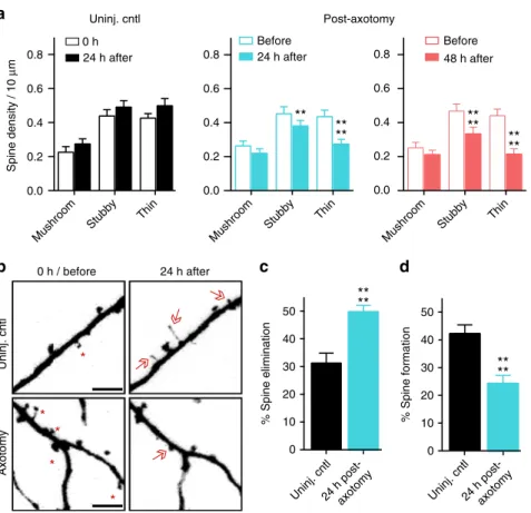
Spine density decreases after distal axon injury
. Decreased spine
density is seen in vivo in models of traumatic brain injury and
spinal cord injury
22,23. To determine whether similar structural
changes occur in cultured pyramidal neurons following distal
axotomy, we quanti
fi
ed spine density within the somatodendritic
compartment of axotomized neurons that were retrogradely
labeled using mCherry rabies virus. Spine density signi
fi
cantly
declined 24 h and 48 h post-axotomy compared to before
axot-omy (Fig.
1
c, d). In contrast, uninjured control neurons showed
increased spine density as expected to occur during normal
maturation (Fig.
1
c, d).
We next analyzed speci
fi
c spine types that were lost. We found
a preferential loss in the density of thin and stubby spines at
both 24 h and 48 h post-axotomy compared to pre-axotomy
(Fig.
2
a). The density of mushroom spines remained stable at
both 24 h and 48 h after axotomy, unlike in the uninjured
control neurons where spine density of all spine types increased.
The reduction in spine density following axotomy suggests
that either dendritic spines were being eliminated or, conversely,
that there was a reduction in new spine formation following
axotomy. Further analysis of our before and after axotomy
images revealed that axotomy caused both a signi
fi
cant
a
c
d
e
# of FM puncta
24 h post-axotomy
FM Fluoresc. (norm.)
Time (s) Stim
48 h post-axotomy 24 h post-axotomy
b
Unresp.
*
48 h post-axotomy
0.2 s 50 ms 20 pA
f
48 h post-axotomy
Time (s) Stim
Uninj cntl. Axotomy
Cut Uncut
g
h
0
Uncut Cut
mEPSC amplitude (pA)
NS
0 2 4 6
mEPSC frequency (Hz)
Uncut Cut
*
Cut-labeled
i
0 50 100 150
0.0 0.2 0.4 0.6 0.8 1.0
Uncut neighbors-unlabeled
Time (s) Stim Uncut-labeled Uninj. cntl
Before stim.
After stim.
Uninj. cntl Uninj. cntl
Uninj. cntl
0 2 4 6
48 h post- axotomy
mEPSC frequency (Hz)
*
Uninj.
cntl
0
48 h post- axotomy
mEPSC amplitude (pA)
NS
Uninj.
cntl
0 200 400 600
*
Resp.
Unresp.
FM Fluoresc. (norm.)
48h post-axotomy 48h post-axotomy
0 25 50 75 100
Axotomy Axotomy
24 h
48 h
Resp.
Proportion of FM puncta (%) Uninj. cntl
Uninj. cntl
*
48 h post- axotomy
0
(S)
NS
***
***
1.0
0.8
0.6
0.4
0.2
0.0
90
0 15 30 45 60 75 0 15 30 45 60 75 90
150
100
50
0
(S)
150
100
50
1.0
0.8
0.6
0.4
0.2
0.0
Uninj.
cntl
30
20
10
30
20
10
0 15 30 45 60 75 90
increase in the percentage of spines eliminated and a signi
fi
cant
reduction in the percentage of new spines formed 24 h
post-axotomy (Fig.
2
b
–
d). Thus, axotomy affected both
elimina-tion and formaelimina-tion of spines to result in lower dendritic spine
density.
Increased synaptic vesicle release rate follows axon injury
. To
further evaluate how synapses are modi
fi
ed following distal
axon injury, we next investigated whether presynaptic release
properties were altered at synapses onto injured neurons. To
address this question, we retrogradely infected neurons using a
modi
fi
ed enhanced green
fl
uorescent protein (eGFP) rabies virus
to label injured neurons and then used FM dyes to optically
measure synaptic vesicle release onto these directly injured
neu-rons (Fig.
3
a). The use of FM dyes provided us with an unbiased
method to label a majority of presynaptic terminals within the
somatodendritic compartment
24. FM puncta highly colocalized
with synapsin1 immunolabeling (93%), which is present at both
inhibitory and excitatory terminals, validating our FM dye
load-ing strategy (Supplementary Fig.
2
). We examined the synaptic
vesicle release rate of FM puncta that colocalized with axotomized
eGFP expressing neurons. At 24 h post-axotomy, there was no
change in synaptic vesicle release rate compared to eGFP
expressing uninjured control samples (Fig.
3
b, c). In contrast, 48
h after axotomy synaptic vesicle release rate was signi
fi
cantly
enhanced (Fig.
3
c). Further, the FM decay time constant,
τ
, which
has been inversely correlated with release probability
25was
sig-ni
fi
cantly reduced at 48 h post-axotomy (control: 124.8 s
±
5.487
vs.
axotomy: 78.65 s
±
3.922;
p
<
0.0001). These results were similar
to those obtained by examining the entire image
fi
eld of FM
puncta closest to the barrier region within the somatodendritic
compartment where a large percentage of axotomized neurons
reside (Supplementary Fig.
3
). The difference in presynaptic
release rate persisted, though modestly, at 4 day post-axotomy in
these cultured neurons (control: 95.14 s
±
1.282 vs. axotomy:
77.19 s
±
1.165;
p
<
0.0001; Supplementary Fig.
3
). Together,
these data suggest a delayed and persistent increase in synaptic
vesicle release rate that occured following dendritic spine loss.
Next, we performed two control experiments to determine (1)
whether cortical cultures, which have more neuron variaiblity
than hippocampal cultures, would behave similarly to
hippo-campal cultures used in our experimental model, and (2) whether
axotomy of axons forming synapses onto postsynaptic targets
would yield similar effects as axotomy of untargeted axons.
First, we performed the FM unloading experiments with cortical
neurons harvested from embryonic rats and found that
these cultures showed similar changes in presynaptic release 48
h post-axotomy (Supplementary Fig.
3
). To address the second
question, we added a small number of target neurons to
the axonal compartment during cell plating. We previously
demonstrated
that
synapses
form
between
two
neuron
populations plated into opposing compartments
12. Axotomy of
this targeted population of neurons resulted in similar changes in
presynaptic release rate as axotomy of untargeted axons
(Supplementary Fig.
3
).
Dendritic spine density is lower following injury, suggesting
fewer synapses, thus, we next wondered whether the balance of
responsive to unresponsive presynaptic terminals might be
altered following axotomy to account for the enhancement in
excitability. We measured the proportion of FM puncta that
unloaded (responsive) or did not unload (unresponsive) in
response to
fi
eld stimulation using extracellular electrodes
24(Fig.
3
d). At 24 h post-axotomy when spine density was
decreased, we observed no change in the fraction of responsive
and unresponsive FM puncta compared to uninjured controls
(Fig.
3
d). However at 48 h post-axotomy, a signi
fi
cantly larger
proportion of puncta were responsive compared to puncta
within uninjured control chambers (Fig.
3
d). Further, at 48 h
post-axotomy we found an overall decrease in the number of
loaded FM puncta (Fig.
3
e). Together, our data suggest that distal
axon injury leads to an overall decrease in synapses, including the
number of presynaptic terminals, but that the smaller number of
presynaptic terminals is more responsive to stimulation.
Enhanced glutamate release at synapses onto injured neurons
.
Our results support that distal axotomy triggers a retrograde
and trans-synaptic cascade of events leading to enhanced
neurotransmitter release rate. To con
fi
rm this, we performed
electrophysiological recordings of
α
-amino-3-hydroxy-5-methyl-4-isoxazolepropionic acid receptor (AMPAR)-mediated
minia-ture excitatory postsynaptic currents (mEPSCs) from axotomized
neurons 48 h post-axotomy and their age-matched uninjured
controls. Biocytin was used to
fi
ll neurons following each
recording to determine whether neurons extended axons into the
axonal compartment and were axotomized. Axotomized neurons
had a signi
fi
cant increase in mEPSC frequency, con
fi
rming our
FM data and supporting an increased rate of presynaptic
glutamate release (Fig.
3
f, g). Membrane properties were
equivalent between axotomized and uninjured control neurons,
demonstrating that the health of these axotomized neurons was
not substantially compromised (Supplementary Table
1
). We
Fig. 3Presynaptic excitability at synapses onto axotomized neurons.aA representative neuron retrogradely labeled with a modified eGFP rabies virus via the axonal compartment. Enlarged region shows FM puncta colocalized with eGFP dendrites and spines (arrows). ImageJ‘fire’color look-up-table for FM puncta shown in the next panel.Scale bar, 20µm.bRepresentative images show FM puncta colocalized with eGFP dendrites (outlined inwhite dashed lines) before and afterfield stimulation in uninjured control, and 24 h and 48 h post-axotomy.Arrowshighlight destaining at spines.Scale bars, 10µm.cFM unloading curves of colocalized puncta 24 h post-axotomy (control,n=185 puncta; axotomy,n=256 puncta) and 48 h post-axotomy (control,n=232 puncta; axotomy,n=322 puncta). Two-way ANOVA, Bonferroni post hoc test. Inset shows FM decay time constant (τ) for puncta withτ<360 s (24 h control,n=151; 24 h axotomy,n=201; 48 h control,n=211; 48 h axotomy,n=304).b,cUnpaired two-tailedt-test. Each condition includes 5–6 chambers/neurons over 3 experiments.dPercent responsive and unresponsive FM puncta per neuronfield (n=8fields/chambers; 4 experiments). Unpaired two-tailedt-test, axotomy vs. control for each time point.eNumber of responsive and unresponsive FM puncta per frame at 48 h post-axotomy (n=11 chambers; 5 experiments). Unpaired two-tailedt-test, for unresponsive puncta.fRepresentative mEPSC traces 48 h post-axotomy.gmEPSC frequency and amplitude at 48 h post-axotomy (control,n=17 neurons; axotomy,n=20 neurons; 4 experiments).Inset: cartoon depicts recordings from either uninjured control neurons (black) or directly injured neurons (red).hAnalysis of mEPSC frequency and amplitude of cut neurons [cut (red),n=10 neurons] compared to neighboring uncut neurons within axotomized chambers [uncut (gray),n=10 neurons].g,hUnpaired two-tailedt-test, Welch’s correction.iFM unloading of neighboring uncut neurons identified by lack of eGFP (uncut neighbors,n=816 puncta), uninjured control neurons (uncut-labeled,n=232), and axotomized labeled neurons (cut-labeled,n=322). Two-way ANOVA, Bonferroni post hoc test; each condition, 5 chambers and 3 experiments. Decay time constant (τ) of FM puncta at 48 h post-axotomy (uncut-labeled,n=211; cut-labeled,n=304; and uncut neighbors unlabeled,n=
observed a trend towards an increase in mEPSC amplitude
following axotomy, however this effect was not signi
fi
cant
(Fig.
3
g).
We next wondered if the increased spontaneous release rate of
glutamate was speci
fi
c to directly injured neurons or more
globally affected neighboring, uncut neurons. To address this, we
quanti
fi
ed
mEPSC
frequency
between
uncut
and
cut
neurons within the same axotomized chamber. In recordings
from directly injured neurons, axotomy speci
fi
cally increased
mEPSC frequency. However, neighboring uncut neurons that did
not extend axons into the axonal compartment, did not have an
increased mEPSC frequency (Fig.
3
h). To further examine the
effects of direct injury to axotomized neurons, we quanti
fi
ed FM
release rate at nearby uninjured neurons that were not infected
with the retrograde eGFP rabies virus. We found that the release
rate was signi
fi
cantly decreased at these locations compared with
synapses on directly axotomized neurons and not signi
fi
cantly
different than at control uninjured neurons labeled with eGFP
rabies virus (Fig.
3
i). These observations con
fi
rmed that axotomy
altered glutamatergic synaptic input onto injured neurons.
Further, directly injured neurons trans-synaptically in
fl
uenced
presynaptic glutamate release without affecting nearby synapses
at uninjured neurons.
SCI induces persistent and enhanced
fi
ring in layer Vb
. To
evaluate the in vivo relevance of our
fi
ndings, we sought to
determine whether distal injury of long projection neurons
in vivo would preferentially induce enhanced excitability in these
injured neurons. To do this, we wanted to use an in vivo model in
which axonal damage occurs far from somata to minimize other
effects of injury (e.g., in
fl
ammation and metabolic changes). We
used a rat SCI model described previously
26in which animals
were subjected to a spinal cord contusion injury at thoracic
level T9-10, and recording electrodes were implanted into the
neurophysiologically identi
fi
ed hindlimb motor cortex in
ketamine-anesthetized animals. Electrode sites on single-shank
microelectrode arrays (Neuronexus, Ann Arbor, MI, USA)
extended through cortical layers V and VI, allowing simultaneous
recording throughout these cortical layers. Effective injury to the
corticospinal neurons innervating hindlimb motor neuron pools
in the spinal cord was con
fi
rmed by stimulating electrode sites
and con
fi
rming loss of evoked hindlimb movement. At each
cortical location, 5 min of neural data was collected for of
fl
ine
analysis. At the end of the procedure, neural spikes were
discriminated using principle component analysis. We examined
fi
ring rates
27within layers Va, Vb, and VI between 4 weeks and
18 weeks post-SCI and compared the data to sham control
animals. We found that the
fi
ring rate within layer Vb was
signi
fi
cantly increased after SCI compared to sham controls
(Fig.
4
). Layer Vb contains the highest density of corticospinal
somata, with estimates of nearly 80% of large pyramidal cells
28.
Also, after spinal cord injury, chromatolytic changes occur
preferentially in layer Vb
29. In layers Va and VI, which have
few (layer Va) or no (layer VI) corticospinal neurons, we found
that
fi
ring rates were not statistically different between SCI
animals and sham controls. Together, these data con
fi
rm a
persistent increase in spontaneous
fi
ring rates in remotely
injured corticospinal neurons, and support the relevance of our
in vitro model system.
Axotomy eliminates inhibitory terminals onto injured
neu-rons
. Loss of inhibition following distal injury contributes to
enhance excitability in vivo, thus we wanted to test whether
axotomy in our culture system results in a similar loss of
inhibitory terminals that release
γ
-aminobutyric acid (GABA).
We performed retrospective immunostaining to determine the
fraction of vGLUT1 or GAD67-positive FM puncta at 48 h
post-axotomy (Fig.
5
a, b). We found that axotomy did not alter the
fraction of glutamatergic terminals, but signi
fi
cantly diminished
the fraction of GAD67-positive puncta within the
somatoden-dritic compartment. Further, we examined the fraction of
vGLUT1 or vGAT puncta colocalized with axotomized neurons
labeled with an eGFP rabies virus (Fig.
5
c). These results
con-fi
rmed the preferential absence of inhibitory terminals following
axotomy while the fraction of vGLUT1 puncta remained
equivalent to uninjured control neurons.
To determine whether inhibitory synapses were functionally
altered following axotomy, we recorded miniature inhibitory
postsynaptic currents (mIPSCs) from axotomized and uninjured
chambers 48 h post-axotomy (Fig.
5
d
–
f). We found that mIPSCs
were more frequent in axotomized cultures compared with
uninjured neurons, suggesting that while there are fewer
inhibitory terminals, the remaining terminals have an increased
rate of spontaneous GABA release. We next asked whether this
change in inhibitory synapse function was restricted to directly
injured neurons. Within the axotomized cultures, we compared
both cut and uncut neurons and found that the mIPSC frequency
was increased in both groups, but was not different between the
directly axotomized neurons and their uncut neighbors. This
suggests that the alteration of inhibitory synaptic transmission
following axotomy affects both directly injured and neighboring,
uninjured neurons.
Although the majority of GABAergic synapses are found on
dendritic shafts or cell bodies, a minor population is also found
on dendritic spines
30, 31(Supplementary Fig.
4
). Inhibitory
synapses formed on dendritic spines allow for
compartmentaliza-tion of dendritic calcium levels involved in regulacompartmentaliza-tion of neuronal
activity
32, 33. To investigate whether dendritic spines receiving
inhibitory inputs (i.e., inhibited spines) are lost following
axotomy, we quanti
fi
ed the number of inhibitory and excitatory
presynaptic terminals onto spines of cultured pyramidal neurons
using retrospective immunostaining for inhibitory (vGAT) and
excitatory (vGLUT1) synapse markers. We found a signi
fi
cant
00.05 0.10 0.15 0.20 0.25
Firing rate (spikes/sec)
Control SCI Control SCI
****
Control SCI 0
0.05 0.15 0.25
0 0.05 0.15 0.25
0.10 0.20
0.10 0.20
NS
NS
Layer Va Layer Vb Layer VI
decrease in the fraction of vGAT-positive spines at 48 h
post-axotomy compared to uninjured control (Fig.
5
g
–
i) with
no signi
fi
cant in
fl
uence on glutamatergic spines. Together, our
data suggest that axotomy caused a preferential loss of inhibitory
terminals onto axotomized neurons, including inhibitory
terminals formed onto dendritic spines, and that increased
spontaneous GABAergic transmission might compensate to some
degree for these lost terminals.
Local activity and transcription regulate remodeling
. Ef
fi
cient
axon regeneration requires signaling from the site of injury to the
nucleus in multiple model systems
6, yet the signaling events
required for synaptic remodeling following distal axotomy
remain unclear. Breach of the axonal membrane following
axon injury causes an in
fl
ux of calcium and sodium ions into the
intra-axonal space, potentially in
fl
uencing signaling to the
nucleus and gene expression. To determine whether local
in
fl
ux of sodium and calcium ions at the time of injury is required
for axotomy-induced spine loss, we performed axotomy within
h
i
c
mIPSC frequency (Hz) mIPSC amplitude (pA) mIPSC frequency (Hz)
Uncut Cut
mIPSC amplitude (pA)
Uncut Cut 0.2 s
20 pA
50 ms
48 h post-axot.
a
g
d
e
f
0 1 2 3 4
*
0 1 2 3
4 NS
0 10 20
30 NS
Axot. Uncut Cut
b
Axot.
FM vGLUT1
Uninj. cntl
FM
Axot.
GAD67
Uninj. cntl
Fraction of
vGLUT1 / FM puncta
0.5 1.0 1.5
0.0
NS
Fraction of
GAD67 / FM puncta
**
0.5 1.0 1.5
0.0
vGLUT1 puncta /
area (
µ
m
2)
vGAT puncta / area (
µ
m
2)
48 h post-axot. 0
10 20
30 NS
Uninj. cntl
24 h post-axot.
48 h post-axot.
GFP
/
vGLUT1
GFP
/
vGAT
GFP
Uninj. cntl
Uninj. cntl
0.0 0.2 0.4 0.6 0.8 1.0
0.00 0.05 0.10 0.15 0.20
vGLUT1+ spine fraction
vGAT+ spine fraction
**
0.0 0.2 0.4 0.6 0.8 1.0
vGLUT1+ spine fraction
0.00 0.05 0.10 0.15 0.20
vGAT+ spine fraction
NS
****
NS 0.00 0.05 0.10 0.15 0.20 0.0 0.1 0.2 0.3 0.4
**
NS
Uninj.
cntl
48 h
post-axot.
Uninj.
cntl
48 h
post-axot. Uninj. cntl
48 h
post-axot. Uninj. cntl
48 h
post-axot.
Uninj.
cntl
48 h
post-axot. Uninj. cntl
48 h
post-axot.
Uninj.
cntl
24h
post-axot. Uninj. cntl
48h
post-axot.
Uninj.
cntl
24h
post-axot. Uninj. cntl
48 h
the axonal compartment in which axons were treated with a
local activity blockade during axotomy. This local activity
blockade solution (ABS) included low-Ca
2+, high-Mg
2+, and
tetrodotoxin citrate (TTX; 0.5 mM CaCl2, 10 mM MgCl2, 1
µ
M
TTX) to prevent in
fl
ux of sodium and reduce calcium in
fl
ux.
This local activity blockade was applied solely to the axonal
compartment for 1 h during axotomy. We labeled neurons
extending axons into the axonal compartment using a retrograde
eGFP rabies virus and quanti
fi
ed spine density before and
24 h following axotomy and compared these measurements
to cultures with vehicle applied to axons during axotomy
(Fig.
6
a, b). Strikingly, we found that local activity blockade at
the injury site prevented axotomy-induced spine loss. These
data suggest that local activity instructs retrograde signaling and
spine loss.
To determine whether injury-induced transcription is required
for these trans-synaptic changes, we treated the somatodendritic
compartment
with
the
reversible
transcriptional
blocker
5,6-dichloro-1-
β
-D-ribofuranosyl-1H-benzimidazole
(DRB)
15 min prior to axon injury and removed the drug 45 min later.
We found that blocking transcription during this brief time was
suf
fi
cient to prevent axotomy-induced spine loss 24 h
post-axotomy compared with similarly treated uninjured control
chambers (Fig.
6
c). Further, DRB treatment at the time of injury
prevented signi
fi
cant changes in the proportion of responsive
FM puncta (Fig.
6
d) and in synaptic vesicle release rate 48 h
post-axotomy (Fig.
6
e). However, action potential blockade with
TTX in the somatodendritic compartment for ~1 h at the time of
injury did not affect injury-induced changes in presynaptic
release or the proportion of responsive puncta 48 h after axotomy
(Fig.
6
f, g). Further, application of Hank
’
s balanced salt solution
(HBS) or dimethyl sulfoxide(DMSO) as respective vehicle
controls to TTX or DRB treatments did not alter
injury-induced increase in presynaptic release. We conclude that both
local activity at the site of injury and a transcriptional response
were critical mediators of the delayed trans-synaptic changes in
presynaptic release properties following distal axon injury.
Differential gene expression at 24 h post-axotomy
. Our data
show that a transcriptional response was required immediately
after axotomy to induce retrograde changes in synaptic vesicle
release onto injured neurons. To identify genes that might
mediate this process within a longer therapeutically relevant time
window, we performed a gene expression study to identify
differentially expressed transcripts within the somatodendritic
compartment at 24 h post-axotomy compared to uninjured
controls (Supplementary Fig.
5
). We found 615 transcripts that
were signi
fi
cantly changed following injury (one-way
between-subject ANOVA,
p
<
0.05) (Fig.
7
a; Supplementary Table
2
).
Con
fi
rming that the transcription response in vitro recapitulated
in vivo
fi
ndings, we found Jun upregulated 1.41 fold in our
micro
fl
uidic cultures 24 h post-axotomy
19.
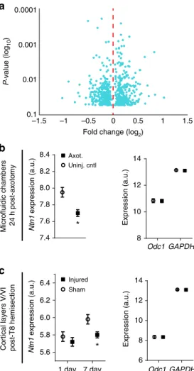
Netrin-1 mRNA downregulated post-axotomy
. Next we sought
to identify potential trans-synaptic mediators that may in
fl
uence
synaptic vesicle release at synapses onto injured neurons. We
focused on differentially expressed transcripts that are known to
localize to cell
–
cell contacts, such as synapses (Supplementary
Table
3
). We identi
fi
ed netrin-1 (
Ntn1
) as signi
fi
cantly
down-regulated 24 h following axotomy, consistent with published
fi
ndings that netrin family proteins are downregulated following
injury in adult rats
34(Fig.
7
b). Netrin-1 is a secreted axon
guidance and synaptogenic cue that is enriched at mature
den-dritic spines
35where it induces clustering of its receptor, deleted
in colorectal carcinoma (DCC), and enhances synapse
matura-tion
36. To con
fi
rm that
Ntn1
expression is downregulated
following nerve injury in vivo, we analyzed microarray data from
a previously published study which examined cortical gene
expression from retrograde labeled layer V cortex of young adult
rats (2 months) subjected to either sham injury or spinal cord
hemisection at thoracic level 8
37(Fig.
7
c). Our analysis of this raw
data con
fi
rmed that spinal cord injury signi
fi
cantly reduced
netrin-1 expression in cortical layer V by 7 days, consistent with
our in vitro
fi
ndings. Together, the signi
fi
cant decrease in
Ntn1
expression both in vitro and in vivo suggests a reliable response
induced by distal axonal damage.
Exogenous netrin-1 normalizes injury-induced changes
. The
downregulation of netrin-1 following injury led us to ask whether
adding exogenous netrin-1 might rescue, to some degree, the
axotomy-induced synaptic changes. To test this we applied
exogenous netrin-1 to the somatodendritic compartment 40 h
after axotomy and evaluated the resulting changes in spine
density, synaptic vesicle responsiveness, and disinhibition. We
performed live imaging of somatodendritic domains before and
after axotomy to measure spine density changes and found that
netrin-1 treatment for 8 h was suf
fi
cient to normalize spine
density to pre-axotomy levels (Fig.
8
a, b). We then used FM dyes
to compare presynaptic release properties between axotomized
and uninjured controls. Exogenous netrin-1 increased the total
number of FM puncta at 48 h post-injury to levels found in
uninjured controls and reduced the percentage of responsive
puncta to levels found in uninjured controls (Fig.
8
c, d). We
next tested whether netrin-1 might rescue axotomy-induced
Fig. 5Absence of inhibitory terminals following distal axotomy and frequency of their spontaneous release events.aRepresentative images offixable FM4-64FX puncta (red) and vGLUT1 (green) co-immunolabeling in uninjured chambers and 48 h following axotomy.White circleshighlight vGLUT1 expression at FM-labeled terminals.Scale bars, 10µm. Fraction of vGLUT1 + FM puncta per neuronfield at 48 h post-axotomy normalized to uninjured controls.n=18 neuronfields; 5 chambers over 3 experiments.bFixable FM4-64FX puncta (red) and GAD67 (green) co-immunolabeling. Quantification of GAD67-positive FM puncta at 48 h post-axotomy normalized to control.n=21 neuronfields; 5 chambers over 3 experiments.cNumber of vGLUT1 and vGAT puncta per neuron area (axotomized or control) 48 h post-axotomy.n=8–9 neurons; 3 chambers per condition over 3 experiments.a–cUnpaired two-tailed t-test. dRepresentative traces of mIPSC recordings 48 h post-axotomy.eQuantification of mIPSC frequency and amplitude 48 h post-axotomy (control,n=9 neurons; axotomy,n=17 neurons). (mIPSC frequency: unpaired two-tailedt-test with Welch’s correction,p=0.05; mIPSC amplitude: unpaired two-tailed
disinhibition. Netrin-1 treatment following axotomy normalized
the density of inhibitory terminals (vGAT labeled) at axotomized
neuron without signi
fi
cantly altering the density of glutamatergic
terminals (vGLUT1 labeled) (Fig.
8
e, f).
Because DCC protein levels parallel netrin-1 expression
changes
38, 39, we next con
fi
rmed that DCC levels were
down-regulated at synapses formed onto the somatodendritic domain of
axotomized neurons (Fig.
8
g, h). Local synaptic DCC
immuno-fl
uorescence at spines of axotomized neurons were decreased at
48 h post-injury. Further, application of exogenous netrin-1
normalized synaptic DCC levels to that similar to uninjured
controls (Fig.
8
g, h).
If downregulation of netrin-1 signaling regulates
axotomy-induced synaptic remodeling, we would expect that blocking
netrin-1 signaling in uninjured neurons would be suf
fi
cient to
cause both reductions in spine density and the density of
inhibitory terminals. Spine density in uninjured cultures treated
with a DCC function blocking antibody for 24 h was signi
fi
cantly
reduced after treatment compared to control chambers treated
with an IgG antibody (Fig.
8
i). Further, we found that blocking
DCC was suf
fi
cient to cause a reduction in the density of
vGAT puncta, but not vGLUT1 puncta, per eGFP-
fi
lled neuron
area (Fig.
8
j). Together, our data suggest that netrin-1 signaling
may play a critical role in regulating synaptic remodeling
following axonal damage, including in modulating inhibition
following injury.
Discussion
While axon regeneration following injury is extensively studied,
much less is known about how proximal neurons within the
mammalian brain are affected following axonal damage
40and
more speci
fi
cally how synapses onto injured neurons are
remodeled. We used a model system to study the cellular
mechanisms of synaptic remodeling following axon injury; this
model recapitulated several hallmarks of neurons subjected to
axonal injury in vivo, including chromatolysis
6, 21, retrograde
spine loss
4,22,23, retrograde hyper-excitability
1–3, and
disinhibi-tion
2, 9, 10. Axotomy-induced transcriptional changes in this
in vitro model are also consistent with in vivo
fi
ndings
7, 20.
Because of the ability to separate neuronal compartments, this
tool facilitates the investigation of axotomy-induced retrograde
signaling intrinsic to neurons and the resulting effects to
inter-neuronal communication.
Our results suggest that retrograde remodeling requires
local signaling at the site of injury mediated by sodium and/or
calcium in
fl
ux to activate a rapid transcriptional response. Both
postsynaptic dendritic spine loss and trans-synaptic changes in
g
d
e
f
FM puncta (%)
FM puncta (%)
0 25 50 75 100
NS Unresp.
Resp.
Time (s)
Uninj.-TTX Axot.-TTX
Stim
Uninj.-DRB Axot.-DRB
Time (s) Stim
0 h / before 24 h after
0.0 0.5 1.0 1.5
****
Spine density / 10
µ
m length
Somatodendritic compartment
a
b
c
Axot. (ABS)
Uninj. (DRB)
Axot. (DRB) NS
0.0 0.5 1.0 1.5
Spine density / 10
µ
m length
Axot. (ABS)
Axot. (veh.)
Before 24 h after
Unresp.
Resp.
*
0 25 50 75 100
FM fluoresc. (norm.) FM fluoresc. (norm.)
**
NS
Somatodendritic compartment
Somatodendritic compartment
Axot. (veh.)
Applied to axons only
Uninj. DRB
Axot. DRB
1.0
0.0 0.8
0.6
0.4
0.2
90 0 30 60
Uninj. TTX
Axot. TTX
1.0
0.0 0.2 0.4 0.6 0.8
0 30 60 90
Fig. 6Influence of local activity and transcription on the initiation of injury-induced synaptic remodeling.aRepresentative images of neurons within microfluidic chambers retrograde labeled with eGFP rabies virus (inverted grayscale) before and 24 h post-axotomy with vehicle or local activity blockade solution (ABS) applied to axons for 1 h during axotomy. Inset shows zoomed in dendritic regions. Image and insetscale bars, 50 and 5µm, respectively. bQuantification of before and after spine density data described ina. Axotomy (vehicle):n=29 dendrites; 5 neurons; 3 chambers over 3 experiments; #spines/TDL: 437/4185µm (before), 274/3868µm (after). Axot. (ABS):n=33 dendrites; 5 neurons; 3 chambers over 3 experiments; #spines/TDL: 426/ 3889µm (before), 446/3700µm (after).cQuantification of spine density changes following application of transcription blocker (DRB) to the somatodendritic compartment for 1 h during axotomy within microfluidic chambers. DRB (uninj.):n=27 dendrites; 6 neurons; 3 chambers over 3 experiments; #spines/TDL: 362/4186µm (0 h), 262/3934µm (24 h after). DRB (axot.):n=28; 6 neurons; 3 chambers over 3 experiments; #spines/TDL: 343/3414µm (before) 283/3248µm (after).b,cUnpaired two-tailedt-test.dPercentage of responsive and unresponsive FM puncta at 48 h post-axotomy.n=6 neuronfields/chambers per condition over 3 experiments. Unpaired two-tailedt-test for % responsive.eFM 5–95 unloading following 1 h application of DRB during axotomy/control. Uninj. DRB:n=1580 puncta; 6 chambers per condition over 3 experiments. Axot. DRB: 2213 puncta; 6 chambers per condition over 3 experiments.fPercent of responsive and unresponsive puncta at 48 h post-axotomy following application of TTX to the somatodendritic compartment for 1 h during injury.n=6 neuronfields/chambers per condition over 3 experiments. Unpaired two-tailed t-test, % responsive.gFM unloading curves following application of TTX. Uninj. TTX:n=1360 puncta; 6 chambers per condition over 3 experiments. Axot. TTX:
presynaptic inputs required immediate transcription. Our data is
consistent with axonal injury signaling in other non-CNS model
systems
6. Localized reversal of a sodium calcium exchanger at the
site of injury may amplify calcium in
fl
ux and contribute to long
range signaling
41. In peripheral neurons calcium waves can
locally propagate to the nucleus to induce a transcriptional
response
42. The localized in
fl
ux of calcium may be a priming
effect for retrograde transport of signaling complexes required to
initiate transcription
6.
Our data showed that axotomy-induced spine loss was
followed by a speci
fi
c loss of inhibitory inputs. Interestingly,
speci
fi
c loss of inhibitory, and not excitatory, terminals suggests
that preserved excitatory inputs may remain available for some
period of time following injury. Because of the spine loss, these
excitatory inputs could form shaft synapses or some may become
orphan presynaptic sites following injury. Large headed dendritic
spines could also receive multiple excitatory inputs, stabilizing
them, and allowing them to
fi
nd new partners over time.
The increased spontaneous release of glutamate at injured
neurons 48 h following axotomy, without an increase in the
number of excitatory terminals, suggests that the maintained
excitatory inputs may contribute to the hyper-excitability
post-injury.
The sequential post- and then pre- synaptic changes following
axotomy suggest a trans-synaptic mechanism. These post- and
then pre-synaptic changes are consistent with the involvement of
synaptic homeostasis where retrograde molecules are released
post-synaptically to in
fl
uence presynaptic release. Further support
for the involvement of trans-synaptic mechanisms comes from
our observation of an increase in spontanenous neurotransmitter
release localized at excitatory synapses onto axotomized neurons,
but not at neighboring excitatory synapses onto uninjured
neurons. The signi
fi
cantly enhanced
fi
ring rate following SCI in
cortical layer Vb, but not layers Va and VI, provides additional
support for this speci
fi
city. Interestingly, we also found an
increase in spontaneous release at inhibitory terminals, although
fewer inhibitory terminals remain following axotomy. The
increase in GABA release rate may serve to compensate for the
axotomy-induced hyper-excitability.
Dendritic release of secreted proteins (e.g., BDNF, NT-3, and
NT-4) and diffusible molecules, such as nitric oxide, can
trans-synaptically regulate neurotransmitter release
43–45. Injury of
motoneuron projections to myocytes caused synaptic remodeling
of inputs to motoneurons which was in
fl
uenced by nitric oxide
synthesis
46. While these previously reported trans-synaptic
signaling pathways were not detectably altered in our
micro-array analysis, we did identify the secreted protein, netrin-1,
as signi
fi
cantly downregulated in our axotomized cultures 24 h
post- axotomy. Downregulation of netrin-1 gene expression was
further con
fi
rmed in vivo through an analysis of independently
acquired microarray data. Netrin-1 is secreted locally from target
cells and signals DCC receptors that are present along axons
36to
in
fl
uence presynaptic release and maturation
47,48. While netrin-1
signaling is historically thought of in a developmental context,
there is increasing evidence of the importance of netrin-1
signaling in the adult CNS. Consistent with our in vitro
fi
ndings, netrin family members are downregulated in vivo
following spinal cord injury in adult rats
34,49and DCC remains
persistently low after 7 months post-injury in adult rats
34.
Netrin-1 has also recently been tested as a potential therapeutic
agent following injury and has been shown to improve recovery
outcomes
50–52.
We found that adding exogenous netrin-1 one and a half
days after axotomy dramatically increased spine density and the
density of inhibitory terminals to levels found in uninjured
controls. The restoration of inhibitory terminals in axotomized
samples treated with netrin-1 is a novel and exciting
fi
nding.
Evidence from
C.elegans
con
fi
rms a link between netrin-1
signaling and stabilization of GABAA
receptors
53. Yet, it
remains unclear how netrin-1 signaling modulates inhibitory
input and will be an important topic for future studies. In
contrast to previous reports, we found that there was no
reduction in the number of vGLUT1 positive terminals
when blocking DCC
36, which could be explained by our shorter
treatment times with DCC function blocking antibody.
Axonal damage within the CNS occurs in numerous
disorders and diseases, but little is known about the overall
impact on cortical circuit function. Importantly, our cell-based
fi
ndings have broader applicability beyond spinal cord injury
to numerous conditions where axonal damage is prevalent,
such as other forms of traumatic brain injury, Alzheimer
’
s
0.00010.001
0.01
0.1
–1.5 –1 –0.5 0 0.5 1 1.5
Fold change (log2)
P
-value (log
10
)
Microfluidic chambers 24 h post-axotomy
Cortical layers V/VI post-T8 hemisection 5.6
5.8 6.0 6.2 6.4
Sham Injured 7.4
7.6 7.8 8.0 8.2 8.4
Uninj. cntl Axot.
8 10 12 14
Ntn1
expression (a.u.)
Ntn1
expression (a.u.)
Expression (a.u.)
Expression (a.u.)
6 8 10 12 14
*
*
Odc1 GAPDH
1 day 7 day Odc1 GAPDH
a
b
c
Fig. 7Netrin-1 gene expression following axotomy in microfluidic cultures and in vivo following spinal cord hemi-transection Microarray analysis was performed on somatodendritic samples of uninjured control and 24 h post-axotomy chambers. Quality control data is presented in
Supplementary Fig.5.aVolcano plot showing differentially expressed RNAs that are significantly changed at 24 h post-axotomy (One-way between-subject ANOVA;n=3 individual chambers each condition; Supplementary Table2).bMicroarray expression levels forNtn1within microfluidic chambers (left) and for housekeeping genesOdc1andGAPDH(right). One-way between-subject ANOVA.cMicroarray expression levels for
disease, and multiple sclerosis. Further, remodeling is enhanced
in embryonic or neonatal neurons, making the use of an
in vitro approach using these neurons, together with in vivo
models, advantageous for identifying pathways instrumental
for neurological recovery
54.
Methods
Hippocampal cultures. Animal procedures were approved by the University of North Carolina at Chapel hill Institutional Animal Care and Use Committee (IACUC). Dissociated hippocampal cultures were prepared from Sprague Dawley rat embryos (E18-E19)11,24. Hippocampal tissue was dissected in ice cold
dissociation media (DM) containing 82 mM Na2SO4, 30 mM K2SO4, 5.8 mM
b
a
c
d
200 400 600
HBS Ntn1 0
# of FM puncta
Unresp.
Resp.
*
0 25 50 75 100
HBS Ntn1
% Responsive FM puncta
*
NS0.0 0.5 1.0 1.5
Spine density / 10
µ
m length
Before
48 h after
**
**
Axot. Axot.+Ntn1
e
f
Axotomy
Before 48 h after
h
Uninj. cntl Axot. Axot.+Ntn1
DCC fluoresc.
per spine ROI (norm.)
0.0 0.5 1.0 1.5
g
Before 24 h after
i
j
IgG ab. DCC ab.
vGAT
vGLUT1
FP
0.0 0.5 1.0 1.5
FP
/
DCC
DCC
FP
Uninj. cntl Axot. Axot. + Ntn1
Axotomy+Ntn1
* *
Uninj. cntl 48 h post-Axot.
Spine
density / 10
µ
m length
NS 0.00
0.10 0.20 0.30
vGLUT1
puncta / area (
µ
m
2)
vGAT
puncta / area (
µ
m
2)
0.00 0.05 0.10 0.15
0.0 0.1 0.2 0.3
0.0 0.1 0.2 0.3
vGAT puncta / area (
µ
m
2)
vGLUT1 puncta /
area (
µ
m
2)
*
NS
**
**
**
**
**
**
NS Uninj. cntl
Axot. Axot.+Ntn1
**
**
NS
*
NS NS
**
**
IgG ab. DCC ab.
MgCl2, 0.25 mM CaCl2, 1 mM HEPES, 20 mM Glucose and 0.001% Phenol red. For
enzymatic digestion, equal volumes of TrypLE Express (Invitrogen) and DM were added to the tissue and incubated at 37 °C for 8 min. Tissue was then rinsed and gently triturated in neuronal culture media consisting of Neurobasal media (Invitrogen) supplemented with 1 × B27 (Invitrogen), 1 × Antibiotic-antimycotic (Invitrogen), and 1 × Glutamax (Invitrogen). Dissociated cells were resuspended in neuronal culture media to yield 12 × 106cells per ml.
Microfluidic chambers. Poly(dimethylsiloxane) (PDMS) (Sylgard 184 Silicon Elastomer, Dow Corning) microfluidic chambers were replica molded against silicon wafers photolithographically patterned with SU-8 negative photoresist (MicroChem)11. Each silicon wafer contained afirst layer of SU-8 to pattern microgrooves 3–4µm tall and 7.5–8µm wide. A second layer of SU-8 generated the 85–100µm high somatodendritic and axonal compartments. All experiments used chambers with 900µm long microgrooves to separate the somatodendritic and axonal compartments11,16,24. Microfluidic chambers were sterilized using 70% ethanol and placed onto sterile German glass coverslips coated with 500–550 kDa Poly-D-Lysine (BD Biosciences). Approximately ~90,000 cells were plated into the somatodendritic compartment and axons extended into the adjacent axonal compartment after 5–7 days of culture. Axotomy was performed between 11 and 15 days in vitro (DIV) byfirst removing media from the axonal compartment and storing for future use. The axonal compartment was then aspirated until completely devoid offluid11,16. Stored culture media was returned immediately to
the axonal compartment for the duration of the culture time. Microfluidic devices with equivalent viable cell populations were randomly chosen for either axotomy or uninjured control groups.
Retrograde labeling. Retrograde labeling was performed using either modified cholera toxin or rabies virus. Cholera Toxin Subunit B Alexa Fluor 488 or 568 (Life technologies, Molecular Probes; 1µg in 200μl of neuronal culture media) was added to the axonal compartment of the microfluidic chamber and incubated for ~ 15 h at 37 °C. After 15 h of incubation, the axonal compartment media was removed, rinsed and replaced using fresh neuronal culture media before per-forming axotomy or imaging.
G-deleted Rabies-mCherry or eGFP virus55(Salk Institute; 1 × 10⁵viral units) in 50μl- conditioned media was added to the axonal compartment of each chamber and incubated for 2 h at 37 °C. Conditioned media was added back to the axonal compartments following two washes with fresh Neurobasal media. Chambers were maintained in 37 °C incubator for ~48 h untilfluorescence expression was visible.
Cell viability assay. Dead cells were labeled using SYTOX Green (Invitrogen) at a
final concentration of 1µM and all cell nuclei were labeled with NucBlue Hoechst Stain (Invitrogen). Cells were incubated with SYTOX/Hoechst solution simulta-neously in 1xPBS for 5 min at 37 °C, washed with PBS, andfixed with 4% paraf-ormaldehyde (PFA) in PBS containing 40 mg/ml sucrose, 1µM MgCl2, and 0.1µM
CaCl2for 15 min at room temperature (RT). Coverslips were then rinsed three
times with PBS and mounted onto the glass slide using Fluoromount G (Southern Biotech). SYTOX positive (Sytox+) cells were manually counted in ImageJ using sum projected z-stack confocal images. Percent cell viability is calculated using [(Hoechst - Sytox+)/Hoechst]×100.
Nissl Staining. Neuronal cultures retrogradely labeled with Cholera Toxin were either axotomized or left uninjured. PDMS chambers were carefully lifted off from
PDL coated coverslips 24 h post-axotomy. Cultures on the coverslips were quickly rinsed twice with PBS,fixed with 4% PFA for 30 min at RT, washed twice in PBS, and incubated in 0.1% Triton X-100/PBS for 10 min at RT. Cultures were incubated for 20 min in NeuroTrace 500/525 Green Fluorescent Nissl Stain (1:100; Invitrogen) and washed for 10 min in 0.1% Triton X-100/PBS. Cell nuclei were stained with DAPI (Sigma-Aldrich), rinsed three times in PBS, and then the coverslip was mounted onto a microscope slide using Fluoromount G.
Immunocytochemistry. PFAfixed neuronal cultures were permeabilized in 0.25% Triton X-100 and blocked in 10% normal goat serum for 15 min each. Coverslips were incubated with anti-MAP2 (1:1000; Millipore # AB5622), anti-beta tubulin III (1:2000; Aves #TUJ), GAD67 (1:2000; Aves labs # GAD), anti-vGLUT1 (1:100; NeuroMab, clone N28/9, cat. #75-066), anti-vGAT (1:1000; Synaptic Systems #131 003), DCC (1:100; Calbiochem #OP45), or anti-synapsin1 (1:500; Calbiochem #574778) primary antibodies in 1% blocking solution for overnight at 4 °C. Coverslips were then incubated with goat anti-rabbit or goat anti-mouse or anti-chicken secondary antibodies conjugated to Alexa-fluorophores (1:1000; Invitrogen) for 1 h at RT. Following PBS washes coverslips were mounted onto glass slides.
RNA isolation. Total RNA from each of 3 axotomized chambers and 3 sham manipulated chambers (6 total samples) was isolated from the somatodendritic compartment of 14 DIV cultures, 24 h after manipulation. RNA was collected from the entire somatodendritic compartment for our gene expression analysis; thus, a fraction of neurons in the axotomized chambers were axotomized and the remaining fraction uninjured or“uncut”. RNA was isolated using an RNAqueous-Micro Kit (Ambion) according to the manufactures instructions including DNase treatment, with modifications specific to accessing the microfluidic compartment16. Briefly, 50µl lysis solution was added to one somatodendritic well and collected from the other somatodendritic well after solutionflowed through the somato-dendritic compartment to this adjacent well. Lysate was added to 50µl of fresh lysis solution and mixed well by careful pipetting. Further RNA purification steps were performed according to the manufacturer’s guidelines. Samples were maintained at
−80 °C until prepared for microarray gene expression.
Microarray analysis. Quantification of RNA integrity and concentration was confirmed with an Agilent TapeStation 2200 at the UNC Lineberger Comprehensive Cancer Center Genomics Core. Microarrays were processed at the UNC School of Medicine Functional Genomics Core using the Affymetrix GeneChip WT Plus Reagent Kit for cRNA amplification, cDNA synthesis, fragmenting and labeling. Samples were hybridized to Rat Gene 2.0 ST Arrays (Affymetrix). Data analysis was performed with Affymetrix Expression Console software and Affymetrix Transcriptome Analysis Console v2.0 software to compare axotomized cultures to uninjured control samples using one-way between-subject ANOVA of Robust Multi-array Average (RMA) normalized intensities. Quality control data is presented in Supplementary Fig.5. Because a fraction of the har-vested cells were uninjured in our axotomized samples, we used modest fold change values for defining our list of significantly changed transcripts (fold change absolute value≥1.1 and ANOVAp-value<0.05). To identify cell–cell adhesion transcripts we searched for the biological process gene ontology category“cell–cell adhesion”. Fold change shown in Fig.7was calculated by dividing the mean log2
intensity value of the uninjured control by the mean log2intensity value of the
axotomized culture samples.