Targeting Ras and Rho Family GTPases for the Treatment of Cancer Through Inhibition of CAAX-Signaled Modifications and the ERK MAPK Pathway.
By Patrick J. Roberts
A dissertation submitted to the faculty of the University of North Carolina at Chapel Hill in partial fulfillment of the requirements for the degree of Doctor of Philosophy in the Department of Pharmacotherapy and Experimental Therapeutics.
Chapel Hill 2007
Approved by
Channing J. Der, Ph.D. (Advisor) Kim L.R. Brouwer, Pharm.D., Ph.D. Adrienne D. Cox, Ph.D.
ABSTRACT
Patrick Roberts: Targeting Ras and Rho Family GTPases for the Treatment of Cancer Through Inhibition of CAAX-Signaled Modifications and the ERK MAPK Pathway.
(Under the direction of Dr. Channing Der)
Ras and Rho family GTPases share many structural and biochemical similarities while regulating distinct cellular functions. One such similarity is the carboxyl-terminal CAAX motif (C = cysteine, A = aliphatic amino acid, and X = terminal amino acid), which signals a series of post-translational modifications important for proper subcellular localization. These modifications are catalyzed by prenyltransferase enzymes, farnesyltransferase (FTase) or geranylgeranyltransferase I (GGTase-I), and subsequently by the endoprotease Ras converting enzyme 1 (Rce1) and by isoprenylcysteine-O-carboxyl methyltransferase (Icmt). Fully processed proteins are thus prenylated, clipped and methylated. Prenylation is known to be absolutely required for correct subcellular localization and biological function of small GTPases. The importance of the post-prenyl processing steps has not been as well validated.
downstream effector pathways, such as the MAPK pathways. We evaluated these two approaches in Ras-mutant non-small cell lung cancer (NSCLC).
And if you feel that you can't go on. And your will's sinkin' low. Just believe and you can't go wrong.
ACKNOWLEDGEMENTS
My graduate training, while difficult at times, has been a truly enjoyable and rewarding journey. I have learned a lot, laughed a lot, and grown both as a scientist and as a person. None of this would have been possible without the love, friendship, encouragement and guidance of my friends, family and mentors, all of whom I will be forever indebted to for their support and generosity. My wife, Kari, has been and will remain my inspiration and my strength. She is my best friend and the love of my life. Without her unrelenting support and confidence in me this journey would not have been possible. My parents, Joe Roberts and Pam Dugan, have loved and nurtured me since the day I was born and are responsible for the man I am today. Their influence and example ultimately led me to pursue graduate training, and for that I am grateful.
Along the way many people have contributed to my growth and development, however none more than my mentor, Dr. Channing Der. His generosity and support has been unimaginable. While I will always strive to emulate Channing’s meticulous work ethic and scientific vision, the one attribute that I would like to take from him is his uncanny ability as a mentor. Like Channing, Dr. Adrienne Cox has been an unbelievable mentor, whose guidance has been critical in the development of my scientific thinking and writing. I am also grateful to the rest of my committee, Dr. Kim Brouwer, Dr. Roy Hawke, and Dr. Howard McLeod, for all of their time and effort.
TABLE OF CONTENTS
List of Tables………...…..x
List of Figures………...xi
Abbreviations………..xiii
Chapter I. Introduction: State of the art approaches to inhibiting Ras, Rho, and the MAPK pathways……….……….…1
A. The Raf-MEK-ERK mitogen-activated protein kinase cascade………....2
B. Ras family small GTPase are key upstream activators of Raf……….14
C. The epidermal growth factor receptor: upstream activation of Ras and the ERK cascade and a component of Ras-mediated autocrine growth loop………..15
D. Inhibitors of the Raf-MEK-ERK cascade………17
E. Inhibitors of EGFR Signaling………..28
F. Evidence of non-classical Rho proteins in cancer………...………39
G. Targeting CAAX-signaled modifications: FTase/GGTase, Rce1, Icmt...…...52
H. Biomarkers: verifying target inhibition and predicting response………...….58
I. Scope of current work………….……….…61
J. References………63
II. Targeting isoprenylcysteine-O-carboxyl methyltransferase for the treatment of non-small cell lung cancer……….82
A. Abstract………....83
C. Materials and ………...88
D. Results………..92
E. Discussion……….……….103
F. References………..108
III. Rho Family GTPases exhibit highly divergent requirements for ispoprenylation, proteolysis and carboxyl methylation to promote membrane association and function……….…111
A. Abstract……...……...………112
B. Introduction………..………..…113
C. Materials and Method…..………..…117
D. Results………..………..121
E. Discussion………..……140
F. References………..149
IV. K-Ras mutations are associated with activation of the Raf-MEK-ERK mitogen-activated protein kinase cascade and disease recurrence: implications for rational clinical trial design for the treatment of Non-Small Cell Lung Cancer……….……….……..154
A. Abstract………...………..…….155
B. Introduction………..…..………156
C. Materials and Methods………...162
D. Results………..………..…166
E. Discussion………..177
F. References………..…183
V. Conclusions and future directions……….…188
growth and tumorgenicity. What is the mechanism of this growth
inhibition? ……….190 C. What is the impact of impaired Icmt activity in animal models:
additional mechanisms of anti-tumor activity or severe toxicity?………….196 D. Rho proteins in cancer: novel targets or sources of toxicity?…..…………..198 E. Defining the roles ERK activation in NSCLC: incorporating Raf
and MEK inhibitors into NSCLC treatment paradigms……….…204
F. Summary...……….…208
LIST OF TABLES Table
1-1. Germline mutations of the Ras-Raf-MEK-ERK cascade in human disorders……….10 1-2. Small molecule MAPK inhibitors………12 1-3. Monoclonal antibody EGFR-directed therapeutic agents………31 1-4. Small molecule tyrosine kinase inhibitor EGFR-directed therapeutic agents……….32 1-5. Properties of monoclonal antibody and small molecule tyrosine kinase
inhibitors of EGFR………...33 3-1. Carboxyl-terminal sequences and membrane targeting elements of human
Ras and Rho family GTPases………126 3-2. Evolutionary conservation of the Rac1 carboxyl-terminal cysteine………..132 3-3. Contribution of prenylation, palmitoylation, Rce1 and Icmt activities to
subcellular membrane association and localization………...148
LIST OF FIGURES Figure
1-1. Oncogene activation of the ERK MAPK cascade……….…..…..5
1-2. Mammalian MAPK cascades……….6
1-3. The Raf-MEK-ERK cascade is a core signaling component of a complex signaling network………...…………..12
1-4. Ras stimulates diverse cytoplasmic signaling networks………..16
1-5. EGFR Monoclonal Antibodies………35
1-6. Human Rho GTPase Family………40
1-7. Post-translational processing of Ras GTPases.………….……….…..………....53
2-1. Impaired plasma membrane association of K-Ras(12V) mutant proteins impaired in CAAX-signaled posttranslational modifications……….……….94
2-2. Reduced transforming activity of K-Ras(12V) mutant proteins impaired in CAAX-signaled posttranslational modifications. ……….………..95
2-3. Transient shRNA suppression of ectopically expressed GFP-Icmt………98
2-4. Icmt shRNA stably-suppresses ICMT carboxyl methylation activity in A549 lung carcinoma cells………..……98
2-5. Suppression of Icmt activity reduces the anchorage-independent growth of A549 cells………99
2-6. A549 tumor formation in athymic nude mice………100
2-7. Cysmethynil inhibits the anchorage-dependent and independent growth of A549 cells………..……102
3-1. The specific isoprenoid modifications of Ras and Rho GTPases are accurately defined using specific FTase and GGTase inhibitors………...122
3-2. Rnd, TC10, TCL, and RhoD are farnesylated in vivo………....125
3-4. Conserved cysteines in Rac1, Rac2, Rac3 and TCL
are not palmitoylated………..………131 3-5. Rce1-mediated –AAX proteolysis is differentially required for
subcellular localization of Rho-family proteins……….………134 3-6. ICMT-mediated methylation is differentially required for subcellular
localization of Rho family proteins………138 3-7. Methylation and palmitoylation act as redundant membrane targeting signals
for TC10 localization……….………139 4-1. ERK activation is not associated with worse overall survival………...169 4-2. Smokers have increased ERK activation………...169 4-3. K-Ras mutation-positive tumors demonstrate increased ERK activation…………..171 4-4. Patients with K-Ras mutations are at increased risk of disease recurrence
LIST OF ABREVIATIONS 17-AAG 17-allylamino-17-demethoxygeldanamycin ADCC Antibody-dependent cell mediated cytotoxicity
BMCC 1-Biotinamido-4-[4′-(maleimidomethyl) Cyclohexanecarboxamido] Butane
CRC Colorectal cancer
EGF Epidermal growth factor
EGFR (Erb1) Epidermal growth factor receptor EMT Epithelial to mesenchymal transition ERK Extracellular signaled-regulated kinase
F Farnesyl
FDA Food and Drug Administration FGF Fibroblast growth factor
FGFR Fibroblast growth factor receptor FTase Farnesyltransferase
FTI Farnesyltransferase inhibitor GAP GTPase-activating protein
GDI Guanine nucleotide dissociation inhibitor GDP Guanosine diphosphate
GEF Guanine nucleotide exchange factor
GG Geranylgeranyl
GGTase Geranylgeranyltransferase
HA Hemagglutinin
Hsp90 Heat-shock protein 90
Her2 (Erb2) Human epidermal growth factor 2 HNSCC Head and neck squamous cell carcinoma
Icmt Isoprenylcysteine-O-carboxyl methyltransferase JNK c-Jun N-term kinase
KSR Kinase suppressor of Ras
LARG Leukemia-associated protein kinase
LF Lethal factor
PBMCs Peripheral blood mononuclear cells mAB Monoclonal antibody
MAPK Mitogen-activated protein kinase
MAPKK MAPK kinase
MAPKKK MAPK kinase kinase MDCK Madin-Darby canine kidney MEFs Mouse embryonic fibroblasts
MEK Mitogen-activated/extracellular signaled-regulated kinase
MEKK MEK kinase
MP1 MEK partner-1
NSCLC Non-small cell lung cancer NF-κB Nuclear factor κB
PAK p21-activated kinase
RCC Renal cell carcinoma Rce1 Ras converting enzyme 1 ROS Reactive oxygen species RTK Receptor tyrosine kinase
SDS/PAGE Sodium dodecyl sulfate polyacrylamide gel electrophoresis SRF Serum response factor
STAT Signal transducers and activators of transcription protein TGF Transforming growth factor
TMA Tissue microarray
TKI Tyrosine kinase inhibitor
VEGF Vascular epidermal growth factor
Chapter 1
Overview of thesis – Mutational activation of the Ras oncogenes is found in 30% of all human cancers. Hyperactivation of Ras by aberrant upstream signaling by receptor tyrosine kinases (RTKs) is also commonly seen in human cancers. In particular, overexpression or mutational activation of the epidermal growth factor receptor (EGFR) is associated with many cancers. Aberrant Ras and EGFR signaling is associated with a high percentage of non-small cell line cancers (NSCLCs). Because of my strong interest in lung cancer therapeutics, I have chosen to focus my research on the identification and validation of approaches to develop anti-Ras inhibitors for lung cancer treatment. Currently, two major directions considered for the development of anti-Ras drugs. First, Ras activation of cytoplasmic signaling networks is critical for Ras-mediated oncogenesis. Therefore, inhibitors of Ras signaling have been considered, with much of the current focus on the Raf-MEK-ERK mitogen-activated protein kinase (MAPK) cascade. Second, inhibitors of the enzymes that catalyze Ras posttranslational modifications that promote Ras association with the plasma membrane have also been pursued. My research studies have concentrated on inhibitors of the ERK MAPK cascade and on validating specific enzymes involved in CAAX-signaled posttranslational modification of Ras and Ras family proteins. In this introduction, I summarize the state-of-the art in current efforts to target these two aspects of Ras function.
A. The Raf-MEK-ERK mitogen-activated protein kinase cascade.
disease, with few successful treatment options. While we have made great strides in unraveling the mysteries of cancer genetics and biology, the important task now facing researchers and clinicians is to translate these discoveries into novel therapeutics that will improve patient outcomes. Since many of the genetic alterations found in cancers involve genes whose products are regulators of signal transduction (Hanahan and Weinberg, 2000), much of the focus in the development of novel therapeutics has involved inhibitors of signal transduction molecules, in particular protein kinases (Arslan et al., 2006; Davies et al., 2006; Sebolt-Leopold and English, 2006). The human kinome is comprised of over 518 protein kinases (http://kinase.com), with disease associations reported for over 150 kinases (http://www.cellsignal.com/reference/kinase_disease.asp). Presently, the Food and Drug Administration (FDA) has approved 10 protein kinase inhibitors and over 100 kinase-targeted agents are currently undergoing clinical evaluation.
Target-based therapies are widely considered to be the future of cancer treatment and much attention has been focused on developing inhibitors of the Raf-MEK-ERK mitogen-activated protein kinase (MAPK) signaling pathway and its upstream activators (Sebolt-Leopold and Herrera, 2004). Evidence that ERK MAPK signaling promotes cell proliferation, cell survival, and metastasis, along with the overwhelming frequency in which this pathway is aberrantly activated in cancer, in particular by upstream activation by the epidermal growth factor receptor (EGFR) and the Ras small GTPases (Figure 1), support current efforts to identify approaches to inhibit this pathway.
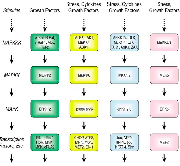
phosphorylation: a MAPK kinase kinase (MAPKKK), a MAPK kinase (MAPKK), and a MAPK (Johnson and Lapadat, 2002). The terminal serine/threonine kinases (MAPKs) are the extracellular signal-regulated kinases (ERK1/2), the c-Jun amino terminal kinases (JNK12/3; also called SAPKs), p38 kinases (p38α/β/γ/δ) and ERK5. Generally, the ERK pathway is activated by growth factor-stimulated cell surface receptors, whereas the JNK, p38 and ERK5 pathways are activated by stress and growth factors.
pathways to mediate oncogenesis, thus providing additional support for the development of inhibitors of these pathways as novel anticancer agents.
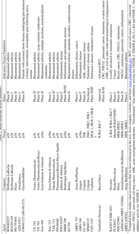
Quite an extensive array of potent and specific inhibitors of p38 are being evaluated in phase I and II clinical trials (Dominguez et al., 2005; Hynes and Leftheri, 2005; O'Neill, 2006) (Table 1). They have been developed primarily for the treatment of chronic inflammatory diseases (e.g., rheumatoid arthritis, Crohn’s disease), although some trials are also evaluating possible applications in cancer. One consequence of p38 inhibitors is blockage of p38-induced transcriptional expression of genes that encode pro-inflammatory cytokines. While Ras has been shown to cause activation of p38, for example by activation of the Rac and Cdc42 Rho family GTPases, whether p38 inhibitors will be useful for blocking Ras-mediated oncogenesis is uncertain. For example, our laboratory showed previously that inhibition of p38 enhanced, rather than blocked, Ras transformation (Pruitt et al., 2002). Therefore, p38 inhibitors are not currently considered for anti-Ras therapy
been considered for the treatment of respiratory diseases (Table 1). Currently, there is limited evidence for a therapeutic value in blocking JNK to block Ras-mediated oncogenesis. Like p38, JNK has been shown to be activated by Ras and there is evidence that JNK activation is necessary for Ras transformation (Westwick et al., 1994). However, the critical role of JNK activation in Ras-mediated growth transformation is unresolved, and consequently, the validation of JNK as an important target for anti-Ras development remains to be done.
The MAPKKK component of the ERK cascade is comprised of the Raf serine/threonine kinases (c-Raf-1, A-Raf and B-Raf) (Schreck and Rapp, 2006; Wellbrock et al., 2004). Raf kinases phosphorylate and activate the MEK1 and MEK2 dual specificity protein kinases. Although Raf has been reported to phosphorylate other proteins, to date, the only validated physiologically relevant substrates remain the two closely related MEK1 and MEK2 proteins. MEK1/2 (MAPKK) then phosphorylates and activates the ERK1 and ERK2 MAPKs. Activated ERKs phosphorylate and regulate the activities of an ever-growing roster of substrates that are estimated to comprise over 160 proteins (Yoon and Seger, 2006). The majority of ERK substrates are nuclear proteins, but others are found in the cytoplasm and other organelles. Activated ERKs can translocate to the nucleus, where they phosphorylate and regulate various transcription factors, such as Ets family transcription factors (e.g., Elk-1), ultimately leading to changes in gene expression (Schulze et al., 2004; Zuber et al., 2000).
potent retrovirus oncogenes (Schreck and Rapp, 2006). Subsequently, laboratory-generated constitutively activated mutants of Raf and MEK were shown to potently transform rodent fibroblasts and other cell types. Furthermore, studies using genetic or pharmacologic approaches have shown that MEK and ERK are required for the transforming activities of Ras and other oncogenes. More recently, mutationally activated B-Raf has been identified in a variety of human cancers (Davies et al., 2002) and the finding that mutationally activated Ras and Raf occur in a non-overlapping occurrence in melanomas, colorectal carcinomas (CRC), papillary thyroid carcinomas, serous ovarian carcinomas, and lung cancers suggests that Ras function is facilitated primarily by activation of Raf (Mercer and Pritchard, 2003; Rajagopalan et al., 2002; Sieben et al., 2004; Singer et al., 2003; Vos et al., 2003). Interfering RNA suppression of mutant B-Raf demonstrated the importance of continued B-B-Raf activity for the transformed and tumorigenic growth of melanomas (Hingorani et al., 2003; Hoeflich et al., 2006; Sharma et al., 2005; Sumimoto et al., 2006).
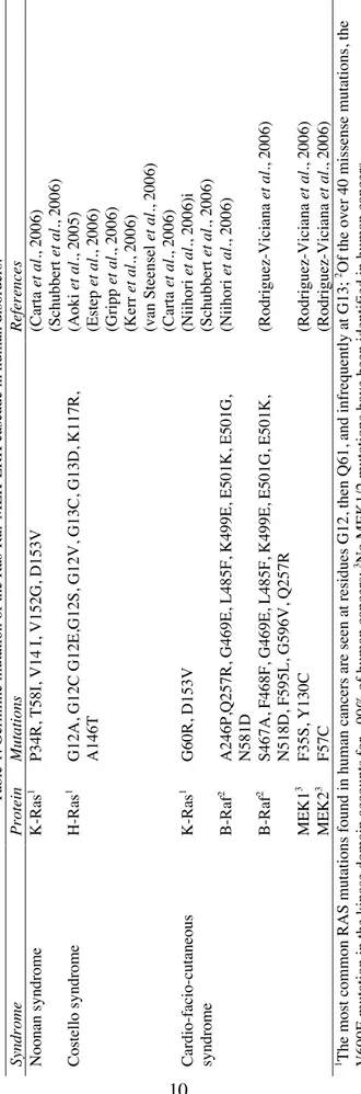
Table 1. Germline mutation of the Ras-Raf-MEK-ERK cascade in human disorders. Protein Mutations References K-Ras 1
P34R, T58I, V14 I, V152G, D153V
(Carta et al. , 2006) (Schubbert et al. , 2006) H-Ras 1
G12A, G12C G12E,G12S, G12V, G13C, G13D, K117R, A146T
(Aoki et al. , 2005) (Estep et al. , 2006) (Gripp et al. , 2006) (Kerr et al. , 2006) (van Steensel et al. , 2006) (Carta et al. , 2006) K-Ras 1 G60R, D153V (Niihori et al. , 2006)i (Schubbert et al. , 2006) B-Raf 2
A246P,Q257R, G469E, L485F, K499E, E501K, E501G, N581D
(Niihori
et al.
, 2006)
B-Raf
2
S467A, F468F, G469E, L485F, K499E, E501G, E501K, N518D, F595L, G596V, Q257R
(Rodriguez-Viciana et al. , 2006) MEK1 3 F35S, Y130C (Rodriguez-Viciana et al. , 2006) MEK2 3 F57C (Rodriguez-Viciana et al. , 2006)
2 Of the over 40 missense mutations, the
spectrum seen in these syndromes differs from those seen in cancer, and generally leads to weakly activated proteins. This reflects the likelihood that the more potent activating mutations found in cancer cannot be tolerated during development. Taken together, these observations provided strong support for the therapeutic value of blocking ERK signaling in cancer and developmental disorders.
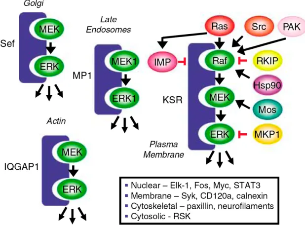
Although the Raf-MEK-ERK cascade is typically drawn as a simple linear, unidirectional cascade of protein kinases, the more appropriate depiction of this cascade is that it is a key core element of a complex signaling network, with many other interactions (Figure 3) (Kolch, 2005). This complexity is best represented at the level of Raf, where multiple signals converge to regulate Raf activation. As described below, the Ras small GTPases are major activators of Raf kinases. However, Raf activation is complex, with Ras facilitating the plasma membrane association of normally cytosolic Raf, where additional signaling activities, including phosphorylation (e.g., by PAK serine/threonine and Src family tyrosine kinases) and dephosphorylation (by protein phosphatase 2A) events are required to fully activate the kinase function. Raf function is also regulated by interaction with other proteins, including 14-3-3 proteins and heat shock protein 90 (Hsp90). Hence, inhibitors of Raf and MEK are not likely to cause similar consequences or to show equivalent clinical responses. These regulatory events also suggest other approaches for antagonizing Raf-MEK-ERK signaling.
Table 2. Small molecule MAPK Inhibitors
1.
Company
Target
Status
Indication /Target Population
Hoffmann-LaRoche p38 Phase II Rheumatoid arthritis Hoffmann-LaRoche p38 Phase I Rheumatoid arthritis Pfizer p38 Phase II Rheumatoid arthritis AstraZeneca p38 Phase I Rheumatoid arthritis GlaxoSmithKline p38 Phase II
Patients with coronary heart disease undergoing percutaneous coronary interventions; neuropathic pain following nerve trauma
Vertex Pharmaceuticals p38 Phase II 2 Rheumatoid arthritis Vertex Pharmaceuticals/Kissei p38 Phase II
Rheumatoid arthritis, acute coronary syndromes
Scios/Johnson & Johnson
p38
α
Phase II
Rheumatoid arthritis, multiple myeloma, myelodysplastic syndromes
Scios/Johnson & Johnson
p38 α Phase I Rheumatoid arthritis Takeda Pharmaceuticals p38 Phase II 2 Rheumatoid arthritis Pharmacopeia/Bristol-Myers Squibb p38 Phase I Rheumatoid arthritis
Johnson & Johnson
p38 Phase I 2 Inflammatory disease Boehringer Ingelheim p38 α Phase IIb/III 2
Inflammatory and autoimmune diseases
Kémia
p38
Phase II
Rheumatoid arthritis, metabolic disorders, cardiovascular disease
Array BioPharma
p38
Phase I
Inflammatory disease, cancer
Amgen p38 α Phase I 2 Inflammatory disease Celgene JNK Phase II
AML, inflammatory disease
Celgene
JNK1, JNK2, JNK3
Preclinical
Parkinson’s disease, asthma
Cephalon
MLK-1, MLK-2, MLK- 3
Phase II/III
2
Parkinson’s disease, Alzheimer's disease
Bayer/Onyx B-Raf, Raf-1 3 Approved Advanced RCC Phase II-III
CRC, NSCLC, pancreatic cancer, melanoma, ovarian cancer, AML, as well as other solid and hematological malignancies
Novartis
A-Raf, B-Raf, c-Raf-1
4
Phase I
Locally advanced or metastatic melanoma
Roche/Plexxikon Mutant B-Raf(V600E) Phase I Cancer Pfizer MEK,MEK2 Phase II
Breast cancer, CRC, NSCLC, melanoma
AstraZeneca/Array BioPharma
MEK,MEK2
Phase II
NSCLC, melanoma, pancreatic cancer; inflammation
Array BioPharma
MEK, MEK2
Phase I
Rheumatoid arthritis
www.clinicaltrials.gov
; RCC, renal cell cancer, CRC, colorectal
2Discontinued; 3Also inhibitory activity for VEGFR-2,
PDGFR-β
, Flt-3, c-Kit and FGFR-1;
Figure 3. The Raf-MEK-ERK cascade is a core signaling componenet of a complex
signaling network. The ERK MAPK cascade is regulated by a complex array of
KSR has been one of the best studied, and KSR function has been shown to be critical for
Ras activation of Raf. Mouse embryo fibroblasts deficient in KSR1 function showed
impaired sensitivity to Ras transformation (Kortum and Lewis, 2004). Furthermore,
anti-sense suppression of KSR1 expression was found to impair the growth of Ras mutation
positive human tumor cells, supporting KSR as a possible therapeutic target for blocking
Ras (Xing et al., 2003).
B. Ras family small GTPases are key upstream activators of Raf
Ras proteins (H-, K- and N-Ras) function as a GDP/GTP-regulated switch.
GDP/GTP cycling is regulated by guanine nucleotide exchange factors (RasGEFs; e.g.,
Sos) that promote formation of active Ras-GTP, whereas GTPase activating proteins
(GAPs; e.g., NF1 neurofibromin) stimulate GTP hydrolysis and formation of inactive
Ras-GDP (Mitin et al., 2005). In normal quiescent cells, Ras is GDP-bound and inactive.
Extracellular stimuli (e.g., EGF) cause transient formation of the active, GTP-bound form
of Ras. Activated Ras-GTP binds to a spectrum of downstream effector targets, of which
the Raf kinases are the best characterized. Ras is mutationally activated in 30% of all
cancers, with pancreas (90%), colon (50%), thyroid (50%), lung (30%) and melanoma
(25%) having the highest prevalence (Malumbres and Barbacid, 2003). The mutant ras
genes in human cancers encode mutated proteins that harbor single amino acid
substitutions primarily at residues G12 or Q61. Mutant Ras proteins are GAP-insensitive,
rendering the proteins constitutively GTP-bound and activated, leading to
stimulus-independent, persistent activation of downstream effectors, in particular, of the
Although there is considerable experimental evidence that the Raf-MEK-ERK
cascade is a critical mediator of Ras-induced oncogenesis, recent studies have also clearly
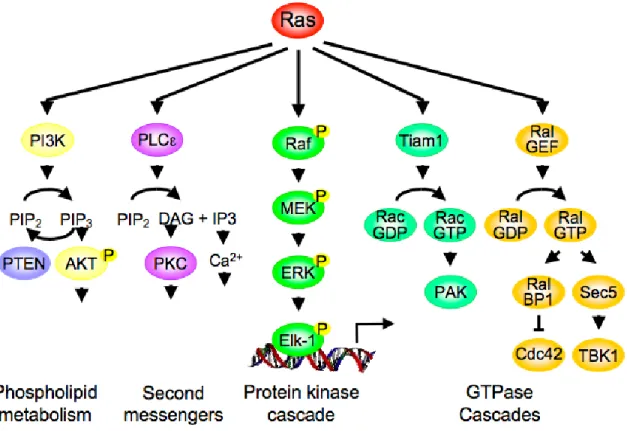
demonstrated that Ras utilizes additional effectors to promote tumorigenesis (Repasky et
al., 2004) (Figure 4). Currently, at least four other effector classes have demonstrated
roles in Ras transformation: the p110 catalytic subunits (p110α, β, γ and δ) of class I
phosphatidylinositol 3-kinases (PI3K), the Tiam1 Rac small GTPase-specific GEF, the
Ral small GTPase-specific GEFs (RalGDS, Rgl, Rgl2 and Rgl3), and phospholipase C
epsilon. The evidence for their involvement and roles in Ras-mediated oncogenesis has
been summarized in recent reviews (Repasky et al., 2004; Shaw and Cantley, 2006). The
existence of non-Raf mechanisms of Ras-mediated oncogenesis prompts the question of
whether blocking the Raf-MEK-ERK pathway alone will be sufficient to effectively
block oncogenic Ras function, or will concurrent inhibition of multiple effector pathways
be required? Alternatively, since this protein kinase pathway is central to many signaling
networks beyond Ras, will blocking the Raf-MEK-ERK pathway be too deleterious and
result in significant normal cell toxicity? Instead, will blocking a subset of downstream
functions of the ERK pathway be a more effective approach? My studies in Chapter 4
have focused on defining the importance of the Raf-MEK-ERK pathway in NSCLC, a
disease in which Ras mutations are frequent events.
C. The epidermal growth factor receptor (EGFR): upstream activation of Ras
and the ERK cascade and a component of a Ras-mediated autocrine growth loop.
In addition to activating mutations of the Ras oncogene, the ERK MAPK
For example, aberrant overexpression or mutational activation of receptor tyrosine
kinases (RTKs; e.g., EGFR and HER2) can cause hyperactivation of Ras leading to
upregulated MAPK signaling (Hynes and Lane, 2005; Lynch et al., 2004; Stephens et al.,
2004). In particular, the EGFR is overexpressed or mutationally activated in many human
cancers (Grandis and Sok, 2004). The regularity with which this signaling cascade is
activated suggests that it may be critical in oncogenesis and makes it an appealing
pathway for drug development. Another linkage between Ras and the EGFR receptor is
mediated by the upregulation of expression of EGFR ligands by Ras signaling (Figure 1).
One important gene target of Ras activation involves transcriptional activation of the
gene for transforming growth factor alpha (TGFα), a ligand for the EGFR. Increased
TGFα gene expression and secretion of TGFα in turn causes persistent stimulation of the
EGFR. The upregulated expression and secretion of TGFα and other EGFR ligands (e.g.,
heparin-binding EGF (HB-EGF), amphiregulin) has been observed in a wide variety of
Ras- or Raf-transformed cell types (Gangarosa et al., 1997; McCarthy et al., 1995;
Schulze et al., 2001). The importance of this autocrine signaling loop for Ras
transformation has been demonstrated by the ability of inhibitors of EGFR to block
oncogenic Ras transformation. Additionally, the majority of Raf-induced changes in gene
expression were found to be dependent on EGFR function (Schulze et al., 2004). Hence,
EGFR can function both upstream, as well as downstream, of Ras and the ERK MAPK
cascade. In addition to activating Ras, EGFR utilizes Rho signaling pathways through its
binding and activation of RhoGEFs, such as Vav2 (Pandey et al., 2000; Schiller, 2006).
Currently, inhibitors of the kinase function of Raf and MEK represent the most
studied and advanced approaches for blocking ERK signaling, with several inhibitors
under evaluation in clinical trials and additional inhibitors in preclinical analyses (Table
1). To date, no inhibitors of ERK1 or ERK2 have been described.
Raf inhibitors
The three Raf kinases exhibit the same substrate specificity, with MEK1 and
MEK2 the only known substrates. However, these highly related isoforms do exhibit
differences in regulation and biological function (Schreck and Rapp, 2006; Wellbrock et
al., 2004). Furthermore, functions independent of MEK activation have been described
although these activities remain poorly characterized. Several structurally distinct classes
of compounds have been developed as potential Raf kinase inhibitors (Smith et al.,
2006)). In addition to small molecule inhibitors of Raf kinase, other anti-Raf efforts
include the development of antisense inhibitors of Raf expression, particularly ISIS-5132,
a 20-base phosphorothioate DNA oligonucleotide that inhibits c-Raf-1 protein
expression. ISIS-5132 showed anti-tumor activity in preclinical xenograft models and
early clinical trials (Cripps et al., 2002; Monia et al., 1996; Oza et al., 2003; Tolcher et
al., 2002). However, no patient response or anti-tumor efficacy was seen in phase II
clinical trials (Cripps et al., 2002; Oza et al., 2003; Tolcher et al., 2002) and it has been
withdrawn from further clinical evaluation. A related approach, using
liposome-encapsulated antisense c-raf-1 oligonucleotide (Gokhale et al., 2002), showed anti-tumor
activity in xenograft analyses (Mewani et al., 2004). Liposome entrapment serves to
completed initial Phase I clinical trials where it was evaluated as monotherapy and in
combination with radiation or chemotherapy (Dritschilo et al., 2006; Rudin et al., 2004).
Further clinical development of LErafAON is ongoing.
Small molecule inhibitors of Ras-Raf interaction (MCP1 and derivatives) have
been described and shown to have anti-tumor activity in cell culture studies
(Kato-Stankiewicz et al., 2002). MCP1 was identified as an inhibitor of Ras interaction with
Raf in a yeast two-hybrid based screen. The exact mechanism by which MCP1 functions
is currently unclear and this information will be important for further clinical
development of this novel class of Raf inhibitors. Currently, preclinical evaluation of
MCP continues in human tumor xenograft mouse models in combination with other
chemotherapeutic agents (Skobeleva, 2007).
Hsp90 functions as a chaperone that is required for the stability and function of
Raf and other oncogene proteins, including HER2 and the Met RTK, as well as steroid
hormone receptors that include the androgen and estrogen receptors (Neckers, 2006). The
anti-tumor activity of the antibiotic benzoquinone ansamycin geldanamycin is due to
binding to and promoting Hsp90 degradation, and hence, indirectly inhibiting the
function of Raf and other client proteins by promoting their proteosomal degradation.
Interestingly, a recent study found that Hsp90 is required for wild type c-Raf-1 and
A-Raf, but not B-A-Raf, stability (Grbovic et al., 2006). However, mutant B-Raf showed
dependency on Hsp90 for function. Treatment of melanoma cells with the geldanamycin
analog 17-allylamino-17-demethoxygeldanamycin (17-AAG) caused degradation of
mutant B-Raf and inhibition of ERK, and showed anti-tumor activity. Another study also
al., 2005). 17-AAG is currently in phase II clinical trials (Sharp and Workman, 2006).
Although 17-AAG has shown promising activities in preclinical and clinical trials, the
further clinical development may be limited by problems with solubility, stability, and
hepatotoxicity. Hsp90 inhibitors are relatively unique anti-tumor agents in that they
simultaneously inhibit multiple, functionally diverse, signaling components that promote
oncogenesis. Hence, the precise biomarker for defining and monitoring anti-tumor
response may be complicated.
To date, the most successful anti-Raf inhibitor has been sorafenib (tosylate salt of
BAY 43-9006; Nexavar®), an orally available compound that received FDA approval in
2005 for the use in advanced renal cell carcinoma (RCC). Sorafenib is a bi-aryl urea
compound (Smith et al., 2006) that was originally developed as an inhibitor of Raf-1
(Lyons et al., 2001). Subsequent analysis revealed that sorafenib was a potent inhibitor of
both wild type and mutant (e.g., V600E, the most frequent mutation found in human
cancers) B-Raf kinases in vitro. Crystallographic analyses of sorafenib complexed with
the kinase domain of B-Raf showed that the inhibitor bound to the ATP-binding pocket
and prevented kinase activation, thus preventing substrate binding and phosphorylation
(Wan et al., 2004).
Shortly after clinical analyses begin, it was revealed that sorafenib also showed
very potent activity for other protein kinases in vitro and in vivo, in particular, for the
pro-angiogenic RTKs such as VEGFR-2, VEGFR-3, PDGFR-β, Flt-3, c-Kit and FGFR-1
(Wilhelm et al., 2004). While sorafenib demonstrated multi-kinase inhibition, it was also
found to maintain some specificity, as it did not inhibit other protein kinases such as
In cell culture and mouse models representing a wide range of tumor cell types,
sorafenib exhibited broad anti-tumor activity and was associated with reduced MEK and
ERK activation, supporting the possibility that its anti-tumor activity involves, in part,
inhibition of Raf (Wilhelm et al., 2004). However, despite its association with reduced
ERK activation, sorafenib anti-tumor activity must also be a consequence of its ability to
inhibit angiogenesis-related kinases as well as other non-Raf kinases. This possibility is
supported by the potent anti-tumor activity that was independent of Ras or B-Raf
mutation status that was seen with sorafenib in xenograft studies.
Phase I clinical trials established sorafenib as a safe and well-tolerated oral agent
with skin rash and diarrhea as the most common adverse effects (Awada et al., 2005)
(Moore et al., 2005a). Results from phase I clinical trials suggested clinical activity in
several patients with RCC, resulting in subsequent clinical trials focused on RCC.
Ultimately this effort led to a large phase III clinical trial that enrolled more than 900
patients with advanced RCC who previously failed prior systemic therapy. The primary
endpoint of this study was improved survival. This trial met its surrogate endpoint of
significantly longer progression-free survival in the sorafenib arm compared to the
placebo arm of the study. In addition, sorafenib treatment doubled the disease
progression-free survival from 12 to 24 months when compared to the placebo-control
arm (Escudier et al., 2005); Nexavar website has updated survival data]. In addition to
RCC, several single agent and combination clinical studies are ongoing in hepatocelluar
carcinoma, non-small cell lung cancer (NSCLC), prostate cancer, breast cancer, ovarian
cancer, pancreatic cancer, melanoma and hematological malignancies (Hahn and Stadler,
The results of clinical trial analyses of sorafenib have not provided sufficient
information to conclude that inhibition of Raf provides a clinical value. Since B-Raf
mutations are not seen in RCC, and since sorafenib is not a potent Raf inhibitor, the
anti-tumor activity seen may be attributed to the anti-angiogenic, rather than anti-Raf, activity
of sorafenib. Although preclinical cell culture and mouse model analyses showed that
continued expression of mutant B-Raf is critical for melanoma growth and
tumorigenicity, phase II clinical trial analyses with sorafenib observed little or no
antitumor activity when evaluated as monotherapy for advanced melanomas (Eisen et al.,
2006). Some clinical efficacy had been seen for melanomas when sorafenib was used in
combination, with clinical trials ongoing for advanced melanomas with sorafenib
combination with bevacizumab or carboplatin and paclitaxel. However, a recently
completed phase III trial administering sorafenib or placebo tablets in combination with
carboplatin and paclitaxel in patients with advanced melanoma found no difference in the
primary endpoint of improving progression-free survival.
RAF265 (formerly CHIR-265) is another orally bioavailable Raf inhibitor
currently being investigated in phase I clinical trials in locally advanced or metastatic
melanoma (http://www.clinicaltrials.gov/ct/show/NCT00304525). RAF265 inhibits all
three Raf isoforms as well as mutated B-Raf. Like sorafenib, RAF265 may also have
anti-angiogenic activity through inhibition of VEGFR2. PLX4032 is a potent and
selective inhibitor of mutant B-Raf that is currently in phase I clinical evaluation. Other
Raf inhibitors are also in preclinical evaluation and should be entering clinical evaluation
The clinical success of sorafenib and another approved multi-kinase inhibitor
(sunitinib) has prompted a debate regarding the advantages and disadvantages of highly
specific versus broad specificity inhibitors (Sebolt-Leopold and English, 2006). Since
cancer is a multi-step process, requiring multiple alterations, it is expected that effective
cancer treatment requires concurrent activities that target different defects (Hanahan and
Weinberg, 2000). The greater success of combination chemotherapy is consistent with
this premise. The ability to optimize the pharmacokinetics and pharmacodynamic
properties of a single agent with multiple activities is also a great advantage for the
successful clinical development of a drug. In the development of sorafenib, the intention
was the identification of a Raf inhibitor, with the activity against other protein kinases
fortuitous and unplanned. Hence, this has complicated a full understanding of the
mechanism of action of sorafenib and the importance of its anti-Raf activities for its
clinical efficacy. Therefore, whether blocking Raf will be a clinically effective approach
will require future clinical evaluation of more specific and potent Raf kinase inhibitors.
The clinical success of highly selective protein kinase inhibitors, in particular mAb-based
drugs (e.g., trastuzumab, bevacizumab), demonstrates that there is clinical value for both
highly selective and multi-targeted inhibitors.
MEK inhibitors
MEK1 and MEK2 are closely related dual-specificity kinases, capable of
phosphorylating both serine/threonine and tyrosine residues of their substrates ERK1 and
ERK2. They are the only known catalytic substrates of Raf kinases. The fact that ERK is
commonly activated in both tumor cell lines and patient tumors, has fueled strong interest
in developing pharmacological inhibitors of MEK as a means to block ERK activation
(Hoshino et al., 1999).
In contrast to sorafenib, small molecule inhibitors of MEK1/2 are highly specific
protein kinase inhibitors. While the first two MEK inhibitors, PD98059 and U0126, were
highly specific (Davies et al., 2000) they lacked the pharmaceutical properties needed to
be successful clinical candidates. Nonetheless, these compounds have been invaluable
academic research tools for dissecting the MEK-ERK pathway and have provided
enormous insight into the importance of ERK MAPK signaling in cancer (Cox and Der,
2002b; Sebolt-Leopold and Herrera, 2004).
The first MEK inhibitor to enter clinical trials was CI-1040 (PD184352), an orally
active, highly potent and selective inhibitor of MEK1 and MEK2 (Sebolt-Leopold et al.,
1999). Preclinical evaluation found that CI-1040 inhibited the growth of human colon
cancer cells and human melanoma cells in athymic nude mice (Collisson et al., 2003;
Sebolt-Leopold et al., 1999). Subsequent phase I and II clinical trials reported the most
common toxicities were mild skin rash, diarrhea, and fatigue (Lorusso et al., 2005)
(Rinehart et al., 2004). During the phase I trial, a partial response was seen in one patient
with pancreatic cancer and 25% of patients with a variety of tumors had stable disease for
greater than three months (Lorusso et al., 2005). Tumor tissues from treated patients
showed significant reduction in activated phosphorylated ERK, indicating that the target
was inhibited. These encouraging results prompted a phase II study in patients with
advanced NSCLC, breast cancer, CRC, and pancreatic cancer. Unfortunately the results
properties (Rinehart et al., 2004). However, when considered together with the
significant body of positive preclinical data as well as early indications from the phase I
trial, it is still believed that MEK is a valid therapeutic target for the treatment of cancer.
Thus, two second-generation MEK1/2-specific inhibitors (PD325901 and ADZ6244)
believed to have superior pharmacological and biopharmaceutical properties have been
developed and are currently in clinical trials (Table 1).
In contrast to the majority of protein kinase inhibitors, MEK inhibitors are
non-ATP competitive inhibitors, which may account for their highly selective properties.
Structural studies with an analog of CI-1040 in complex with MEK1 or MEK2 showed
inhibitor binding did not perturb ATP binding, and instead, bound to a unique inhibitor
binding pocket adjacent to the ATP binding site (Ohren et al., 2004). Inhibitor binding
locked MEK in a catalytically-inactive conformation. This recognition of MEK
sequences that are not shared with other protein kinases, and their association with an
inactivate conformation, account for MEK inhibitor target selectively.
PD0325901 is a derivative of CI-1040 where several slight modifications to the
chemical structure have resulted in more than a 50-fold increase in potency against
MEK1/2, improved bioavailability, and longer duration of target suppression compared to
CI-1040 (Sebolt-Leopold and Herrera, 2004). Anti-tumor activity for PD0325901 was
demonstrated for a variety of tumor xenografts and this inhibitor is now being evaluated
in phase I/II clinical trials with a focus on tumors expected to have activated ERK MAPK
signaling (Solit et al., 2006; Thompson and Lyons, 2005).
AZD6244 (ARRY-142886) is an orally bioavailable benzimidazole derivative
2004; Yeh et al., 2004). Like other MEK inhibitors, AZD6244 is ATP-non-competitive.
Preclinical evaluation of AZD6244 showed anti-tumor activity in several human
xenograft models including colon, pancreas, breast, NSCLC and melanoma (Lee et al.,
2004). Additionally, AZD6244 anti-tumor activity was found to correlate with
suppression of ERK activation, which further validates that its mechanism of action is
MEK-dependent. Results from preclinical analysis have been extremely promising and
thus AZD6244 has moved into clinical development. Recently, initial results of a first in
human dose-ranging study to assess the pharmacokinetics, pharmacodynamics, and
toxicities of AZD6244 in patients with advanced solid tumors concluded that AZD6244
is well tolerated, and the most common treatment-related adverse events were rash,
diarrhea, nausea, fatigue, peripheral edema, and vomiting. The best clinical response seen
in the 57 patients was stable disease (19 patients at the end of cycle two; 9 of whom
achieved stable disease for 5 months or greater). The pharmacokinetic and
pharmacodynamic analyses showed good systemic exposure, which correlated with high
levels of ERK inhibition in peripheral blood mononuclear cells (PBMCs)
(http://www.arraybiopharma.com). AZD6244 is now being evaluated in multiple phase II
trials in a variety of solid tumors.
Rosen and colleagues recently reported that B-Raf mutant tumors are exquisitely
sensitive to MEK inhibition (Solit et al., 2006). In this study the authors used genetic and
pharmacological (CI-1040 and PD0325901) approaches to evaluate MEK-dependence in
a variety of tumor types and found that human tumor cell lines possessing mutant B-Raf
were much more sensitive to MEK inhibition than cells with wild-type B-Raf or mutant
had completely abrogated tumor growth whereas xenografts of Ras mutation positive
tumor cells were only partially inhibited, perhaps reflecting the fact that Ras also utilizes
non-Raf effector pathways to promote oncogenesis. Whether Ras or B-Raf mutation
status will accurately identify the patients who will respond to MEK inhibitor treatment,
and whether suppression of ERK activity is an accurate measure of drug inhibition of
MEK, are issues that remain to be determined.
In addition to small molecule inhibitors of MEK kinase activation, bacterial toxins
have been identified that inhibit MEK function by unique biochemical mechanisms.
Anthrax lethal factor (LF) is a protease and a component of Bacillus anthracis exotoxin,
the Gram-positive bacterium responsible for the disease anthrax. LF, together with a
second exotoxin component (protective antigen), comprise lethal toxin (LeTx). LeTx
inactivates multiple MAPKKs, including MEK1 and MEK2 by proteolytic cleavage and
inactivation of kinase function (Bodart et al., 2002). LeTx was shown to block the
transformed and tumorigenic growth of Ras-transformed rodent fibroblasts (Duesbery et
al., 2001). LeTx also showed preferential inhibition of growth of melanoma cell lines that
harbored mutated B-Raf and elevated ERK activity (Abi-Habib et al., 2005). Yersinia
outer protein J (YopJ) is another bacterial toxin that inhibits MEK function. The bacterial
pathogen Yersinia pestis, the causative agent for the plague (Black Death), uses a type III
secretion system to inject YopJ and other virulence factors into target cells. YopJ was
shown to bind directly to MEK1, MEK2 and other MAPKKs and to block their
phosphorylation and activation (Orth et al., 1999). YopJ functions as an acetyltransferase,
using acetyl-coenzyme A (CoA) to modify the critical serine and threonine residues in
activation (Mukherjee et al., 2006). YopJ also causes acetylation of a threonine residue in
the activationloop of IκB kinase β, preventing its phosphorylation and inactivation of
IκB, thus preventing nuclear translocation and activation of the NF-κB transcription
factor. Since inhibition of NF-κB also has an anti-tumor consequences (Karin, 2006),
YopJ can inhibit concurrently at least two important pathways that promote oncogenesis.
The unique biochemical activities of these bacterial proteins may identify novel
approaches for blocking MEK-ERK signaling.
E. Inhibitors of EGFR signaling
Another approach that has been considered for anti-Ras therapy involves targeting
the EGFR. The rationale for targeting this RTK for blocking Ras is based on two issues.
First, a common consequence of mutated Ras function is upregulated expression of the
gene for transforming growth factor alpha (TGFα) and other EGFR ligands, resulting in
increased protein expression and secretion. This results in the induction of an autocrine
growth factor loop where Ras mutation positive tumor cells secrete growth factors that
stimulate a receptor expressed on the same cell, leading to uncontrolled cellular
proliferation(Gangarosa et al., 1997; McCarthy et al., 1995; Schulze et al., 2001).
Inhibition of TGFα stimulation of the EGFR impairs Ras transformation, demonstrating
the critical role of this autocrine loop in Ras-mediated growth transformation. Second,
as described above, a key consequence of EGFR signaling is activation of Ras and ERK
MAPK pathway (Schulze et al., 2004). Therefore, targeting EGFR signaling may be an
The EGFR family consists of four transmembrane receptors: EGFR
(HER1/ErbB-1), HER2 (ErbB-2/Neu), HER3 (ErbB-3) and HER4 (ErbB-4) (Mendelsohn and Baselga,
2006; Scaltriti and Baselga, 2006). Multiple ligands bind to and activate EGFR, HER3
and HER4. Ligands for EGFR include EGF, TGF_, HB-EGF, betacellulin and epiregulin.
Ligand binding results in the formation of receptor homodimers and heterdimers leading
to subsequent receptor activation and ultimately the activation of downstream signal
transduction pathways (Wiley, 2003; Yarden, 2001).
The best-characterized cytoplasmic signaling pathway activated by the EGFR is
the ERK MAPK pathway, and ERK activation has been utilized as a biomarker for EGFR
inhibitor action. Additional EGFR-activated pathways include the phosphatidylinositol
3-kinase (PI3K) and AKT serine/threonine 3-kinase, signal transducer and activator of
transcription (STAT), as well as protein kinase C and phospholipase D pathways (Citri et
al., 2003). EGFR activation of these pathways results in enhanced proliferation,
angiogenesis, invasion and metastasis, as well as inhibition of apoptosis (Jimeno and
Hidalgo 2006). Aberrant activation of the EGFR signaling commonly occurs in cancer
(NSCLC, CRC, breast cancer, gastric cancer, and others) and multiple mechanisms
describing its activation have been reported including EGFR overexpression, EGFR gene
amplification, acquisition of activating mutations and overexpression of EGFR ligands
(Baselga and Arteaga, 2005).
Two strategies to inhibit EGFR signaling have been successfully developed and
include monoclonal antibodies (mAbs) directed against the extracellular domain of EGFR
(Table 3) and small molecule tyrosine kinase inhibitors (TKIs) of the intracellular
receptor homo- and heterodimerization resulting in receptor internalization and inhibition
of EGFR signaling pathways. Additionally, the clinical activity of some EGFR mAbs
may also be attributed to their ability to stimulate an immune response. Alternatively,
EGFR receptor tyrosine kinase inhibitors (TKIs) bind to the intracellular tyrosine kinase
domain preventing ATP-binding and receptor activation, thus blocking EGFR activation
of ERK and other signaling pathways. Mab and small molecule inhibitors of EGFR
possess shared and distinct features that distinguish their mechanistic and clinical
activities (Table 5). Hence, it is believed that their use in combination may act
synergistically and improve anti-EGFR therapy.
As with other signal transduction inhibitors, EGFR inhibitors lack the severe
myelosuppressive toxicities seen with conventional cytotoxic drugs. Toxicities are most
evident in tissues that are dependent on EGFR function, in particular the skin (Lacouture,
2006). This includes a papulopustular rash that affects the face and upper trunk,
abnormalities in hair growth, and dry and itchy skin. There is incomplete evidence that
the strength of skin rash may be a good indication of drug activity and possibly favorable
patient response and survival (Perez-Soler and Saltz, 2005).
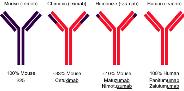
Several EGFR mAbs have been developed that recognize the extracellular domain
of the EGFR (Figure 5). However, since they recognize distinct sequences and vary in
binding affinities and composition (Figure 5), they also vary in their biological activities.
The first FDA-approved EGFR mAb was cetuximab, a chimeric monoclonal IgG1
antibody initially approved for use in combination with irinotecan for the treatment of
EGFR-detectable metastatic CRC refractory to irinotecan, as well as monotherapy for the
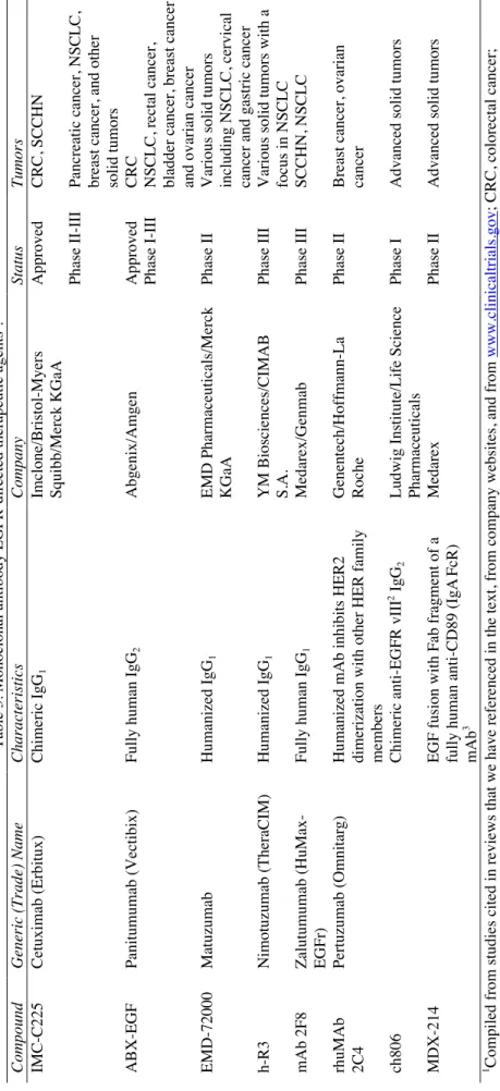
Table 3. Monoclonal antibody EGFR-directed therapeutic agents 1 .
Generic (Trade) Name
Characteristics Company Status Tumors Cetuximab (Erbitux) Chimeric IgG 1
Imclone/Bristol-Myers Squibb/Merck KGaA
Approved
CRC, SCCHN
Phase II-III
Pancreatic cancer, NSCLC, breast cancer, and other solid tumors
Panitumumab (Vectibix)
Fully human IgG
2
Abgenix/Amgen
Approved
CRC
Phase I-III
NSCLC, rectal cancer, bladder cancer, breast cancer and ovarian cancer
Matuzumab
Humanized IgG
1
EMD Pharmaceuticals/Merck KGaA
Phase II
Various solid tumors including NSCLC, cervical cancer and gastric cancer
Nimotuzumab (TheraCIM)
Humanized IgG
1
YM Biosciences/CIMAB S.A.
Phase III
Various solid tumors with a focus in NSCLC
Zalutumumab (HuMax- EGFr)
Fully human IgG
1
Medarex/Genmab
Phase III
SCCHN, NSCLC
Pertuzumab (Omnitarg)
Humanized mAb inhibits HER2 dimerization with other HER family members Genentech/Hoffmann-La Roche
Phase II
Breast cancer, ovarian cancer
Chimeric anti-EGFR vIII
2 IgG
2
Ludwig Institute/Life Science Pharmaceuticals
Phase I
Advanced solid tumors
EGF fusion with Fab fragment of a fully human anti-CD89 (IgA
FcR)
mAb
3
Medarex
Phase II
Advanced solid tumors
www.clinicaltrials.gov
; CRC, colorectal cancer;
2 Generated against the deletion mutant of EGFR lacking exons 2-7
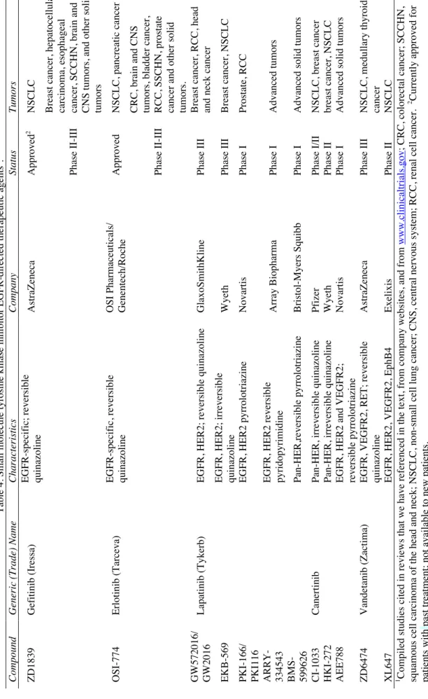
Table 4. Small molecule tyrosine kinase inhibitor EGFR-directed therapeutic agents 1 .
Generic (Trade) Name
Characteristics
Company
Status
Tumors
Gefitinib (Iressa)
EGFR-specific; reversible quinazoline
AstraZeneca
Approved
2
NSCLC
Phase II-III
Breast cancer, hepatocellular carcinoma, esophageal cancer, SCCHN, brain and CNS tumors, and other solid tumors
Erlotinib (Tarceva)
EGFR-specific, reversible quinazoline OSI Pharmaceuticals/ Genentech/Roche
Approved
NSCLC, pancreatic cancer
Phase II-III
CRC, brain and CNS tumors, bladder cancer, RCC, SSCHN, prostate cancer and other solid tumors.
Lapatinib (Tykerb)
EGFR, HER2; reversible quinazoline
GlaxoSmithKline
Phase III
Breast cancer, RCC, head and neck cancer
EGFR, HER2; irreversible quinazoline
Wyeth
Phase III
Breast cancer, NSCLC
EGFR, HER2 pyrrolotriazine
Novartis
Phase I
Prostate, RCC
EGFR, HER2 reversible pyridopyrimidine
Array Biopharma Phase I Advanced tumors Pan-HER,reversible pyrrolotriazine Bristol-Myers Squibb Phase I
Advanced solid tumors
Canertinib
Pan-HER, irreversible quinazoline
Pfizer
Phase I/II
NSCLC, breast cancer
Pan-HER, irreversible quinazoline
Wyeth
Phase II
breast cancer, NSCLC
EGFR, HER2 and VEGFR2; reversible pyrrolotriazine
Novartis
Phase I
Advanced solid tumors
Vandetanib (Zactima)
EGFR, VEGFR2, RET; reversible quinazoline
AstraZeneca
Phase III
NSCLC, medullary thyroid cancer
EGFR, HER2, VEGFR2, EphB4
Exelixis
Phase II
NSCLC
www.clinicaltrials.gov
; CRC, colorectal cancer; SCCHN,
2 Currently approved for
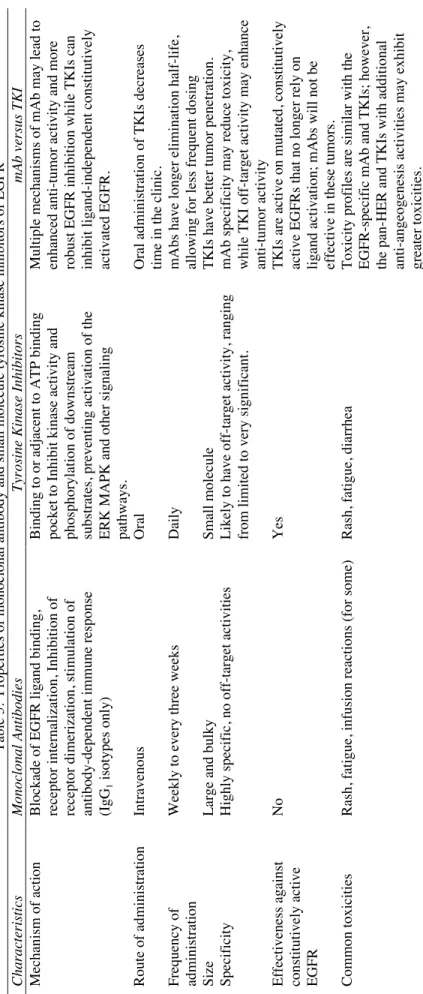
Table 5. Properties of monoclonal antibody and small molecule tyrosine kinase inhibitors of EGFR
Monoclonal Antibodies
Tyrosine Kinase Inhibitors
mAb versus TKI
Blockade of EGFR ligand binding, receptor internalization, Inhibition of receptor dimerization, stimulation of antibody-dependent immune response (IgG
1
isotypes only)
Binding to or adjacent to ATP binding pocket to Inhibit kinase activity and phosphorylation of downstream substrates, preventing activation of the ERK MAPK and other signaling pathways. Multiple mechanisms of mAb may lead to enhanced anti-tumor activity and more robust EGFR inhibition while TKIs can inhibit ligand-independent constitutively activated EGFR.
Intravenous
Oral
Oral administration of TKIs decreases time in the clinic.
Weekly to every three weeks
Daily
mAbs have longer elimination half-life, allowing for less frequent dosing
Large and bulky
Small molecule
TKIs have better tumor penetration.
Highly specific, no off-target activities
Likely to have off-target activity, ranging from limited to very significant. mAb specificity may reduce toxicity, while TKI off-target activity may enhance anti-tumor activity
No
Yes
TKIs are active on mutated, constitutively active EGFRs that no longer rely on ligand activation; mAbs will not be effective in these tumors.
Rash, fatigue, infusion reactions (for some)
Rash, fatigue, diarrhea
(Cunningham et al., 2004). More recently, cetuximab has received additional approval for use in combination with radiation to treat inoperable squamous cell cancer of the head and neck (Bonner et al., 2006). Finally, although EGFR expression was an initial basis for patient selection, patient response has not correlated with the degree of overexpression. Thus, a reliable biomarker for defining patient response remains elusive.
While cetuximab has proven to be an effective treatment in the aforementioned indications and continues to be investigated for use in other tumor types, one of its major drawbacks is the associated risk of anaphylactic reactions during cetuximab infusion (Bonner et al., 2006; Cunningham et al., 2004). This has led to the development of two humanized EGFR (matuzumab and nimotuzumab) and two fully human (panitum umab and zalutumumab) mAbs.
Matuzumab is a humanized IgG1 anti-EGFR mAb. As a humanized mAb matuzumab, like panitumumab, is expected to have reduced infusion-related anaphylactic reactions. Furthermore, as an IgG1 antibody matuzumab induced potent ADCC against tumor cells in vivo, which distinguishes matuzumab from the recently approved panitumumab (Bier et al., 1998). While results from several phase II trials are expected within the next year, initial phase I trials have shown matuzumab is well-tolerated, with rash and diarrhea the most common toxicities. In these trials, evaluation of both tumor tissue and skin biopsies determined that matuzumab inhibited phosphorylation of EGFR, ERK, and AKT (Doi et al., 2005; Salazar et al., 2004; Tabernero et al., 2003; Vanhoefer et al., 2004). In phase I trials, activity has been evaluated in colorectal, cervical, and esophageal cancers and in squamous cell cancer of the head and neck. Current therapeutic targets in phase II trials include cervical and gastric cancers, and NSCLC.
data suggest that pertuzumab may have greater clinical benefit when used in combination with other therapies (Friess et al., 2005). Thus, current phase II trials of pertuzumab are focused on combination therapy, including a study of pertuzumab in combination with erlotinib in patients with locally advanced or metastatic NSCLC.
Two EGFR small molecule kinase inhibitors have received approval for use in NSCLC (Table 4). The first was gefitinib, which received accelerated approval as third-line monotherapy in NSCLC based on its 12% response rate and 43% rate of tumor control. However, gefitinib was subsequently evaluated in a large phase III trial comparing gefitinib to placebo in NSCLC patients who had failed previous treatment regimens, and results from this trial showed no improvement in survival compared to placebo resulting in a FDA-mandated labeling change. Currently, gefitinib is approved only for the treatment of cancer patients who have previously received and benefited from treatment. Ongoing clinical trials are evaluating the use of gefitinib for the treatment of other cancers.
As opposed to gefitinib, erlotinib has demonstrated improved survival in a randomized phase III placebo-controlled trial. Although the overall response rate was only 9% in the erlotinib arm, erlotinib prolonged overall survival (6.7 versus 4.7 months) and increased one-year survival (31% versus 22%). These results led to FDA approval in 2004. More recently, erlotinib has also gained FDA approval for use in combination with gemcitabine for the first-line treatment of patients with locally advanced, unresectable or metastatic pancreatic cancer (Moore et al., 2005b).
particular, laptinib (Tykerb) is a dual EGFR and HER2 tyrosine kinase inhibitor that has been extensively evaluated in multiple solid tumors including breast, NSCLC, CRC, head and neck cancer, hepatocellular carcinoma and billary carcinoma (Nelson and Dolder, 2006). Initial evaluation of this compound revealed diarrhea and skin rash as the most common toxicities (Nelson and Dolder, 2006). While the clinical activity of this agent has been evaluated in multiple tumor types, the main focus has been in the treatment of breast cancer, a disease known to rely on both HER-2 and EGFR signaling. Numerous phase I and II clinical trials have been reported, including several looking at the use of lapatinib in refractory metastatic breast cancer and in first line treatment of advanced breast cancer (Moy and Goss, 2006). An international, multi-center, randomized, open label phase III trial in patients with documented HER2 overexpressing refractory advanced or metastatic breast cancer treated with lapatinib in combination with capecitabine versus capecitabine alone was stopped after the interim analysis. Of the 321 evaluable patients, 161 were treated in the combination arm and 160 in the monotherapy arm. Median time to progression in the combination arm was 8.5 months, compared with 4.5 months in the capecitabine alone arm (Tykerb at ASCO 2006, http://www.gsk.com/investors/presentations_webcasts.html). While these data need to mature so that an overall survival advantage can be determined, this compound is likely to become part of the standard of care in breast cancer and possibly other tumor types as well. The results of this phase III trial are so promising that it has been made available through an Expanded Access Protocol of lapatinib combined with capecitabine in metastatic breast cancer in a non-randomized, open label, uncontrolled trial.
EGFR/HER2 inhibitors in clinical trials (Table 4). Furthermore, canertinib (CI-1033), BMS-599626 and HKI-272 are pan-HER inhibitors, while several EGFR inhibitors also inhibit RTKs involved in tumor angiogenesis. While targeting multiple protein kinases may enhance anti-tumor activity, it may also result in greater normal cell toxicity.
F. Rho family small GTPases: key downstream components of Ras signaling
RhoGAP. Current evidence suggests that activation of the RalGEF-Ral pathway causes inactivation of RalBP1, leading to activation of the Rac and Cdc42 Rho GTPases (Cantor et al., 1995). Another link between Ras and Rho GTPases is demonstrated by the
observations that the Rnd3/RhoE Rho GTPase is transcriptionally upregulated by Ras activation of the Raf-MEK-ERK cascade (Hansen et al., 2000; Shields et al., 2007). Ectopic expression of Rnd3 proteins cause cell rounding, disruption of actin stress fibers, reduced cell adhesion and increased cell migration. These functions of Rnd3 are mediated, in part, by inhibition of RhoA function. Since these cellular alterations are associated with Ras transformation, it is speculated that Rnd3 upregulation may contribute to these Ras-induced cellular changes. The importance of Rho GTPases in Ras transformation is supported by studies showing that genetic or biochemical inhibition of RhoA, Rac1 or Cdc42 inhibits Ras-mediated growth transformation of rodent fibroblasts(Khosravi-Far et al., 1995; Qiu et al., 1997; Qiu et al., 1995a; Qiu et al., 1995b; Roux et al., 1997). Finally, in addition to the functional signaling links that connect Ras with Rho GTPases, Ras and Rho GTPases undergo a similar set of posttranslational modifications by lipids that promote their membrane association. Hence, the anti-Ras approaches that are designed to block Ras membrane association may also block Rho GTPase membrane association and function.
main classes of regulatory proteins. Guanine nucleotide exchange factors promote the formation of the active GTP-bound form (Schmidt and Hall, 2002) and GTPase-activating proteins catalyze the intrinsic GTPase activity and promote the formation of inactive GTP-bound Rho (Bernards and Settleman, 2004). Distinctly, Rho family members are also regulated by a third class of proteins, the Rho guanosine nucleotide dissociation inhibitors (RhoGDIs). This class of proteins regulates Rho localization and effector activation by sequestering Rho in the cytoplasm (Gosser et al Nature 1997; Hoffman et al, Cell 2000). To accomplish this level of regulation, RhoGDIs complex with Rho GTPases by providing a hydrophobic pocket to mask the c-terminal isoprenylated cysteine critical for membrane targeting.
Active, GTP-bound Rho GTPases bind preferentially to downstream effectors, stimulating diverse cytoplasmic signaling cascades that control actin reorganization and regulate cell polarity, cell motility, cell shape, cell adhesion, and membrane trafficking. (Etienne-Manneville and Hall, 2002). As such, it is thought that Rho proteins contribute to cancer progression by influencing the cell’s ability to migrate and thus invade and metastasize. In addition to these alterations in cellular function, aberrant activation of Rho proteins have also been shown to contribute to other cancer phenotypes such as cell growth, proliferation, survival, and angiogenesis (Ridley, 2004). Therefore, defining pharmacologic approaches for inhibition of Rho GTPase function represents an important direction for target-based anti-cancer drug discovery, specifically inhibitors of post-translational CAAX processing.
proper subcellular localization and biological function. Recognition of the C-terminal CAAX tetrapeptide motif (C = cysteine, A = aliphatic, X = any amino acid) initiates the same series of post-translational modifications as has been well described for Ras proteins and ultimately leads to proper membrane targeting. In brief, this includes the addition of a farnesyl or geranylgeranyl isoprenoid lipid to the cysteine residue of the CAAX sequence, the subsequent cleavage of the AAX peptide from the carboxyl-terminus and finally the addition of a methyl group to the prenylated cysteine residue (Sebti and Der, 2003; Winter-Vann and Casey, 2005). These steps are described more completely in section G below. Where studied, mutation of the cysteine residue of the CAAX motif, which prevents all three modifications, renders Rho GTPases cytosolic and inactive, suggesting that pharmacological inhibition of such modifications would also interfere with Rho GTPase function.
![Figure 1. Oncogene activation of the ERK MAPK cascade. Mutationally activated B- B-Raf, Ras and mutationally activated [by missense mutations in the cytoplasmic kinase domain in NSCLC or by extracellular domain truncations (e.g., VIII) in glioblastomas]](https://thumb-us.123doks.com/thumbv2/123dok_us/8247313.2185442/20.918.204.748.118.531/activation-mutationally-activated-mutationally-cytoplasmic-extracellular-truncations-glioblastomas.webp)