Thyroid Stem Cells: Concept and Clinical Implications
Nani ML
1,2, Mahkamova K
1, Aspinall S
1,3, Meeson A
11
Institute of Genetic Medicine, Faculty of Medical Sciences, Newcastle University, Newcastle
Upon-Tyne, NE1 3BZ, United Kingdom.
2
Department of Surgery, Faculty of Medicine, Universiti Kebangsaan Malaysia Medical Centre, Jalan
Yaacob Latif, Bandar Tun Razak, 56000 Cheras, Kuala Lumpur, Malaysia.
3
Northumbria Healthcare NHS Foundation Trust, North Tyneside General Hospital, Rake Lane, North
Shields, NE29 8NH, United Kingdom.
Abstract
Thyroid pathology is the commonest endocrine surgical problem encountered. However, the study of thyroid stem cells is relatively new in the field of stem cell research. Since the identification of thyroid stem cells in 1992, research interest in this area has been increasing mainly based on furthering our knowledge of the biology of these important cells that are thought to be responsible for tumourigenesis and propagation of cancers. This article reviews the current science and biology of thyroid stem cells and summarizes their potential role in the general management of thyroid disorders.
Keywords: Cancer stem cells, clinical implications, stem cells, thyroid
Correspondence:
Nani Harlina Md Latar, Institute of Genetic Medicine, Faculty of Medical Sciences, Newcastle University, Newcastle Upon-Tyne, NE1 3BZ, United Kingdom. Tel: +44-0191-241-8856 Fax: +44 (0)191 241 8666 Email: [email protected]
Date of submission: 30 Sept, 2016 Date of acceptance: 24 Oct, 2016
I
ntroductionThyroid nodular disease, either symptomatic or incidental, is the most common endocrine surgical disorder encountered. Nodules are not a single disease entity but represent a wide range of pathological processes ranging from the normal physiological process to lethal malignancy (1). The main pathophysiological process that drives the formation of these nodules is attributed to chronic stimulation of thyrocytes via the thyrotropin hormone receptor (TSHR) and its downstream signaling cascade. In one population based study, it was noted that the incidence of thyroid cancer increased with higher TSH and interestingly, high level of TSH is associated with more advanced differentiated cancers (2). Thyroid nodular disease is also one of the predisposing factors for development of thyroid cancer, although this relationship is still not clearly understood.
The worldwide incidence of thyroid cancer has progressively risen over the past three decades by an average of 4.5% each year from 2002 to 2012 in the USA (3,4). While this can partly be attributed to the ease of access to thyroid sonography that has resulted in more sensitive detection, more accurate diagnostics work up and increasing patient awareness are other contributing factors. With this steady upward trend, detection of smaller cancers (i.e. micropapillary carcinoma) becomes an increasingly common scenario (5) and this may result in thyroid cancer becoming the third most common cancer in the United States by 2030 (6,7). Despite the increasing reported incidence, mortality due to thyroid cancers has been stable at 0.3 and 0.6 per 100,000 population in men and women, respectively in high-resourced countries (7).
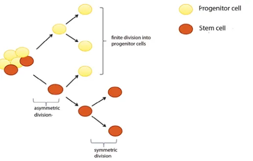
Figure 1: Schematic diagram of the classical self-renewal and differentiation properties of stem cells (SC). Symmetric division occurs when SC divides into two similar progenitor cells or SC, whilst asymmetric division occurs when SC divides into one identical SC and another daughter cell that becomes differentiated cell.
cancers with/without metastasis and anaplastic, poorly or undifferentiated thyroid cancers. In anaplastic cancers, the 5-year survival is often very poor at <5%. It is thus, reasonable to focus future research on these tumours in order to improve survival. In radio-resistant cancers, the tumour cells are thought to have changed their phenotype to a less differentiated phenotype that makes them less susceptible to I131 treatment (8). The remaining modes of treatment with chemotherapeutic drugs and external beam radiation are often poorly tolerated and rarely curative (9,10). One possible reason for the development of these resistant tumours could be due to the presence of cells that have the capability to self-divide, metastasize and resist chemotherapeutic interventions. These properties mimic closely those of stem cells (11,12).
Stem Cells
Stem cells are fundamental cells from which all our cells arise or ‘stem’ from. These unique undifferentiated cells have the properties to divide and thus renew themselves for very long period of time in the body and in vitro (13). They are broadly classified into embryonic stem cells (ESC) and somatic/adult stem cells based on their differentiation potential (potency). The pluripotent ESC can differentiate into any of the three germ layers i.e. endoderm (gastrointestinal tract and its derivatives e.g. lung, liver, pancreas and thyroid), mesoderm (muscle, bone, blood and cardiovascular system) and ectoderm (skin and nervous system). However, this ability is limited in adult stem cells. While there is no doubt that the therapeutic potential of ESC seems limitless, the ethical implications that surround their use have restricted their application in clinical practice.
Stem cells are able to divide giving rise to similar progenitor cells (symmetric division) or asymmetrically. Asymmetric division allows for two different daughter cells to be produced, one to replenish the stem cell pool and the other to become a differentiated cell, so the stem cell daughter would be capable of generating more stem cells (Fig. 1). This clonogenic capacity contributes to the presence of cell heterogeneity within tissues and ensures homeostatic control within tissues is maintained (13). In order to retain and safe guard this special feature, stem cells need to reside in a specific microenvironment called the niche (14).
There are a number of transcriptional regulatory circuits which are responsible for maintaining pluripotency and self-renewal capability in stem cells
(15). Homeodomain transcription factors are
evolutionary conserved and play important roles in cell fate (16). Oct4/POU5F1 and NANOG are essential regulators of early development and their expression provides evidence of embryonic stem cell identity (17,18). OCT4 acts in concert with another transcription factor SOX2. Their combination is thought to be fundamental in developmental control of gene expression involved in pluripotency.
It was reported that OCT4 controls pluripotency in a quantitative manner, that is when expressed at high levels in embryonic stem cells OCT4 drives these cells to endoderm and mesoderm lineages (19). Among these widely studied core transcriptional factors, OCT4 has been found to be indispensable in the maintenance of pluripotency (20).
and cancer development and progression. The use of adult SC in regenerative treatment in clinical practice e.g. limbal transplantation and cardiac regeneration has shown promising results (21,22). However, their impact in solid cancer management is still under investigation due to challenges in identifying clinically relevant biomarkers to specifically target cancer SC (23).
Normal Thyroid SC
Over the past decade, a number of investigators have reported on the identification of putative human thyroid stem/progenitor cells. The presence of thyroid SC populations was first postulated by Dumont et al. (24), who found that it was possible to grow thyroid tissue in recipient animals using only a minimum number of cells. This idea was further elaborated and reinforced by Thomas et al. (25) and Fierabracci et al. (26) who identified precursor cells of endodermal origin and isolated a specific subset of cell populations from goitrous nodules that have the ability for clonal expansion respectively. These cells detected by flow cytometry comprise of less than 1.5% of human thyroid tissues and lack the expression of thyroid specific markers such as thyroglobulin (Tg).
Human thyrocytes have a slow turnover rate (estimated at 8.5 to 14.4 years) (27) with limited regeneration capacity. Furthermore, thyroid SC niches have not been demonstrated in thyroid tissues unlike the brain (28) and intestines (29). Due to these facts, other than the mature follicular cells, the source of thyroid SC has been postulated to derive from embryological remnants (30), microchimerism (31) and SC from bone marrow (32).
There was a forward leap in the study of thyroid stem cells a few years ago, when Antonica et al. successfully demonstrated the ability of murine ESC to differentiate into a functional thyrocytic lineage in vitro. These cells were able to support hormone deficiency when transplanted under the renal capsules in athyroid mice (33).
Cancer Stem Cells (CSC)
The cancer stem cells (CSC) theory was first introduced in the study of haematological malignancies (34) where a small population of cells was found to constitute a reservoir of self-sustaining cells with exclusive ability to self-renew, maintain the tumour and also produce heterogenous non-tumourigenic cancer cell types that constituted the bulk of the tumour (35). Due to these properties, SC are able to multiply uncontrollably, metastasize and
resist chemotherapeutic interventions resulting in treatment resistance.
The advancement of CSC theory in solid malignancy however lagged behind until 5 years later when Ignatova et al. identified clonogenic, stem-like cells in human cortical gliomas, providing the first evidence of CSC in solid cancers (36). Other solid tumours with established SC properties were later identified in breast (37), prostate (38), colon (39)and pancreas (40). For thyroid cancers, a small percentage of CSC has been successfully isolated from both tissues and cell lines (41-44).
Takano has put forward the notion of fetal cell carcinogenesis, which suggests that thyroid cancer cells arise from abnormal development of fetal thyroid cells possibly secondary to genetic mutations (45). This is based on gene expression profiling data seen in anaplastic thyroid cells, where onco-fetal fibronectin was found to be highly expressed in conjunction with lack of expression of differentiated thyroid markers
(46). Another aspect involved in thyroid
carcinogenesis relates to the CSC theory. Thyroid CSC was first isolated by Mitsutake et al. who postulated that the presence of a small subset of cells in the mature thyroid is responsible for cancer initiation and maintenance (42). At this point, it is not clear whether the origin of this subset of cells is normal cells that have undergone transformation, or existing stem cells that have become activated (47).
The CSC model has evolved from a static to dynamic hierarchical one in which the new progenies acquire the ability of self-renewal through de-differentiation, as well as reversal of differentiated cells (48). In addition to this, the theory of clonal evolution, which emerged recently, demonstrated the acquisition of self-renewal capabilities by non-CSC populations within a tumour (49,50). This interconverison ability is likely to be acquired through oncogenic transformation (49). These recent advances, identified through extensive research on leukaemic SC, further establishes the importance of developing strategies that target both CSC and their progenies as both contribute to the development of therapy resistant cancers (48).
Epithelial-Mesenchymal Transition (EMT) and CSC
metastasis (52,53). Thus, during this reversible process carcinoma cells acquire mesenchymal gene expression patterns and properties(47).
Numerous data has documented the presence of EMT in malignant cells in vitro, however its significance in human cancer tissues has remained a matter of debate. This is due to the difficulty in distinguishing stromal cells and other tumour associated fibroblasts from individual mesenchymal cells derived from epithelial tumour cells by EMT (52). However, Prall et al. have now demonstrated the existence of aggregated colorectal cancer cells detaching from the main tumour mass into adjacent stroma which poses EMT characteristics (54). This supports the concept that cancer cells with EMT properties are present at the invasive fronts of human tumours, which is the immediate interphase for tumour and stromal signalling from which metastasis occurs (52).
Clinical Implications
Current chemotherapeutic agents generally target actively dividing cells, leading to reduction in tumour bulk at certain time points during treatment but fail to eradicate the relatively dormant CSC (55). Following treatment, SC normally remain inactive for some time; however the change in the tumour microenvironment along with acquired genetic transformations that occur after initial treatment may later trigger the development of a more aggressive SC phenotype (56). In well differentiated thyroid cancers, often tumour cells demonstrate sensitivity towards initial radioactive iodine treatment. In those that recur presumably, the previously radio avid cells, acquire changes that reduce their sensitivity towards later treatment. As tumours are composed of heterogenous groups of cells, with SC thought to be responsible for recurrence, it is reasonable to assume that we should change our perspective of how we manage malignant tumours in the future. Designing agents that would be able to target both cancer and CSC will hopefully result in more effective treatment. Identifying the pathways involved in this respect may open avenues for the development of more targeted therapy in cancer management.
The development of resistance in cancer cells towards chemotherapeutic agents may partly be attributed to the presence of cells that are able to pump out these drugs. This efflux property, which is the most widely studied aspect of multidrug resistance (MDR) in cancer cells, is due to the overexpression of the ATP-binding cassette (ABC) transporters that reside in the transmembrane region of the cells. This feature has been exploited to isolate a CSC population with a
distinct phenotype known as side population (SP) cells. This subpopulation of cell was first identified by Goodell et al. (57). Using the SP assay, SC have been successfully isolated from benign and malignant thyroid tissues and cells lines (42,58). There have been approaches involving nanoparticle administration aimed at inhibiting the activity of these transporters as means of overcoming drug resistance in cancer cells (59).
The presence of distant metastases denotes stage IV disease with lowest survival rate. Evidence is now accumulating that relates EMT and prognosis. Expression of transcription factors involved in EMT such as Snail and Zeb2 are poor prognostic markers in hepatocellular (60) and ovarian carcinoma (61) respectively. Activation of EMT itself has been linked to poor prognosis in colorectal carcinoma (62). In addition to this, EMT markers have also been linked to tumour recurrence, which generally indicates poor prognosis in metastatic breast cancer patients (63). Celina and Buehler et al. have also found a correlation between the presence ZEB1, SNAIL and TWIST transcription factors and high grade thyroid cancers (64). Defining and targeting the appropriate pathways involved in EMT will potentially broaden treatment options for cancers.
Stem Cells in Benign Tumours
The role of normal SC in the initiation and development of benign tumours has been gaining attention. In the pathogenesis of benign prostatic hyperplasia, in addition to the complex interaction of growth stimulation, apoptosis and cell proliferation, SC have been shown to play a major role (65). Understanding the behaviour of SC in normal and benign diseases, may allow us to identify a suitable focused treatment in order to minimise harm to surrounding normal tissues.
Stem Cells in Regenerative Medicine for Hypothyroidism
administration of lifelong thyroxine, with a potential impact on both quality of life and healthcare costs.
Conclusion
In conclusion, knowledge of the presence of SC residing in thyroid tissues and cell lines has sparked new ideas and avenues in search for a cure for thyroid diseases, specifically anaplastic and recurrent cancers. Further studies are needed to determine if targeting thyroid CSC will provide a platform on which to build new more effective options for the treatment of this group of thyroid cancers that currently have poor prognostic outcomes.
References
1. Schlumberger, M, Pacini F. Thyroid Tumours. Paris: Editions Nucleon, 1999, pp-13.
2. Haymart MR, Repplinger DJ, Leverson GE, et al. Higher serum thyroid stimulating hormone level in thyroid nodule patients is associated with greater risks of differentiated thyroid cancer and advanced tumor stage. J Clin Endocrinol Metab J Clin Endocrinol Metab. 2008; 93(3): 809-14.
3. Pellegriti G, Frasca F, Regalbuto C, Squatrito S, Vigneri R. Worldwide increasing incidence of thyroid cancer: update on epidemiology and risk factors. J Cancer Epidemiol 2013; 2013: 965212.
4. SEER Cancer Statistics Factsheets: Thyroid Cancer. National Cancer Institute. Bethesda, MD, 2012.
5. Chen AY, Jemal A, Ward EM. Increasing incidence of differentiated thyroid cancer in the United States, 1988-2005. Cancer 2009; 115(16): 3801-7.
6. Rahib L, Smith BD, Aizenberg R, Rosenzweig AB, Fleshman JM, Matrisian LM. Projecting cancer incidence and deaths to 2030: the unexpected burden of thyroid, liver, and pancreas cancers in the United States. Cancer Res 2014; 74(11): 2913-21.
7. Ferlay J, Soerjomataram I, Dikshit R, et al. Cancer incidence and mortality worldwide: sources, methods and major patterns in GLOBOCAN 2012. Int J Cancer 2015; 136(5): E359-86.
8. Li F., Zhou K., Gao L., et al. Radiation induces the generation of cancer stem cells: A novel
mechanism for cancer radioresistance (Review). Oncology Letters 2016; 12(5): 3059-65.
9. Sherman SI. Cytotoxic chemotherapy for
differentiated thyroid carcinoma. Clin Oncol (R Coll Radiol) 2010; 22(6): 464-8.
10. Lin JD, Tsang NM, Huang MJ, Weng HF. Results of external beam radiotherapy in patients with well differentiated thyroid carcinoma. Jpn J Clin Oncol 1997; 27(4): 244-7.
11. Davies TF, Latif R, Minsky NC, Ma R. Clinical review: The emerging cell biology of thyroid stem cells. J Clin Endocrinol Metab 2011; 96(9): 2692-702.
12. Reya T, Morrison SJ, Clarke MF, Weissman IL. Stem cells, cancer, and cancer stem cells. Nature. 2001; 414(6859): 105-11.
13. Maenhaut C, Dumont JE, Roger PP, van Staveren WC. Cancer stem cells: a reality, a myth, a fuzzy
concept or a misnomer? An analysis.
Carcinogenesis 2010; 31(2): 149-58.
14. Morrison SJ, Spradling AC. Stem Cells and Niches: Mechanisms that promote stem cell maintenance throughout life. Cell 2008; 132(4): 598-611.
15. Boyer LA, Lee TI, Cole MF, et al. Core transcriptional regulatory circuitry in human embryonic stem cells. Cell 2005; 122(6): 947-56.
16. Hombría JC, Lovegrove B. Beyond homeosis--HOX function in morphogenesis and organogenesis. Differentiation 2003; 71(8): 461-76.
17. Chambers I, Colby D, Robertson M, et al. Functional expression cloning of Nanog, a pluripotency sustaining factor in embryonic stem cells. Cell 2003; 113(5): 643-55.
18. Hay DC, Sutherland L, Clark J, Burdon T. Oct-4 knockdown induces similar patterns of endoderm and trophoblast differentiation markers in human and mouse embryonic stem cells. Stem Cells. 2004; 22(2): 225-35.
20. Pan GJ, Chang ZY, Schöler HR, Pei D. Stem cell pluripotency and transcription factor Oct4. Cell Res 2002; 12(5-6): 321-9.
21. Health Quality Ontario. Limbal stem cell transplantation: an evidence-based analysis. Ont Health Technol Assess Ser 2008; 8(7): 1-58.
22. Sun R, Li X, Liu M, Zeng Y, Chen S, Zhang P. Advances in stem cell therapy for cardiovascular disease (Review). Int J Mol Med 2016; 38(1): 23-9.
23. Wicha MS, Liu S, Dontu G. Cancer stem cells: an old idea--a paradigm shift. Cancer Res 2006; 66(4): 1883-90; discussion 1895-6.
24. Dumont JE, Lamy F, Roger P, Maenhaut C. Physiological and pathological regulation of thyroid cell proliferation and differentiation by thyrotropin and other factors. Physiol Rev 1992; 72(3): 667-97.
25. Thomas T, Nowka K, Lan L, Derwahl M. Expression of endoderm stem cell markers: evidence for the presence of adult stem cells in human thyroid glands. Thyroid 2006; 16(6): 537-44.
26. Fierabracci A, Puglisi MA, Giuliani L, Mattarocci S, Gallinella-Muzi M. Identification of an adult stem/progenitor cell-like population in the human thyroid. J Endocrinol 2008; 198(3): 471-87.
27. Coclet J, Foureau F, Ketelbant P, Galand P, Dumont JE. Cell population kinetics in dog and human adult thyroid. Clin Endocrinol (Oxf) 1989; 31(6): 655-65.
28. Decimo I, Bifari F, Krampera M, Fumagalli G. Neural stem cell niches in health and diseases. Curr Pharm Des 2012; 18(13): 1755-83.
29. Potten CS. Stem cells in gastrointestinal epithelium: numbers, characteristics and death. Philos Trans R Soc Lond B Biol Sci 1998; 353(1370): 821-30.
30. Reis-Filho JS, Preto A, Soares P, Ricardo S, Cameselle-Teijeiro J, Sobrinho-Simões M. p63 expression in solid cell nests of the thyroid: further evidence for a stem cell origin. Mod Pathol 2003; 16(1): 43-8.
31. Klintschar M, Schwaiger P, Mannweiler S, Regauer S, Kleiber M. Evidence of fetal
microchimerism in Hashimoto's thyroiditis. J Clin Endocrinol Metab 2001; 86(6): 2494-8.
32. Sherwood RI, Christensen JL, Weissman IL, Wagers AJ. Determinants of skeletal muscle contributions from circulating cells, bone marrow cells, and hematopoietic stem cells. Stem Cells 2004; 22(7): 1292-304.
33. Antonica F1, Kasprzyk DF, Opitz R, et al. Generation of functional thyroid from embryonic stem cells. Nature 2012; 491(7422): 66-71.
34. Bonnet D, Dick JE. Human acute myeloid leukemia is organized as a hierarchy that originates from a primitive hematopoietic cell. Nat Med 1997; 3(7): 730-7.
35. Clarke MF, Dick JE, Dirks PB, et al. Cancer stem cells--perspectives on current status and future directions: AACR Workshop on cancer stem cells. Cancer Res 2006; 66(19): 9339-44.
36. Ignatova TN, Kukekov VG, Laywell ED, Suslov ON, Vrionis FD, Steindler DA. Human cortical glial tumors contain neural stem-like cells expressing astroglial and neuronal markers in vitro. Glia 2002; 39(3): 193-206.
37. Al-Hajj M, Wicha MS, Benito-Hernandez A, Morrison SJ, Clarke MF. Prospective identification of tumorigenic breast cancer cells. Proc Natl Acad Sci U S A 2003; 100(7): 3983-8.
38. Collins AT, Berry PA, Hyde C, Stower MJ, Maitland NJ. Prospective identification of tumorigenic prostate cancer stem cells. Cancer Res 2005; 65(23): 10946-51.
39. O'Brien CA, Pollett A, Gallinger S, Dick JE. A human colon cancer cell capable of initiating tumour growth in immunodeficient mice. Nature 2007; 445(7123): 106-10.
40. Niess H1, Camaj P, Renner A, et al. Side population cells of pancreatic cancer show characteristics of cancer stem cells responsible for resistance and metastasis. Target Oncol 2015; 10(2): 215-27.
42. Mitsutake N, Iwao A, Nagai K, et al. Characterization of side population in thyroid cancer cell lines: Cancer stem-like cells are
enriched partly but not exclusively.
Endocrinology 2007; 148(4): 1797-803.
43. Hoshi N, Kusakabe T, Taylor BJ, Kimura S. Side population cells in the mouse thyroid exhibit stem/progenitor cell-like characteristics. Endocrinology 2007; 148(9): 4251-8.
44. Ke CC, Liu RS, Yang AH, et al. CD133-expressing thyroid cancer cells are undifferentiated, radioresistant and survive radioiodide therapy. Eur J Nucl Med Mol Imaging 2013; 40(1): 61-71.
45. Takano T. Fetal cell carcinogenesis of the thyroid: a modified theory based on recent evidence. Endocr J 2014; 61(4): 311-20.
46. Takano T. Fetal cell carcinogenesis of the thyroid: a hypothesis for better understanding of gene expression profile and genomic alternation in thyroid carcinoma. Endocr J 2004; 51(6): 509-15.
47. Zane M, Scavo E, Catalano V, et al. Normal vs cancer thyroid stem cells: the road to transformation. Oncogene 2016; 35(7): 805-15.
48. O'Connor ML, Xiang D, Shigdar S, et al. Cancer stem cells: A contentious hypothesis now moving forward. Cancer Lett 2014; 344(2): 180-7.
49. Chaffer CL, Brueckmann I, Scheel C, et al. Normal and neoplastic nonstem cells can spontaneously convert to a stem-like state. Proc Natl Acad Sci U S A 2011; 108(19): 7950-5.
50. Iliopoulos D, Hirsch HA, Wang G, Struhl K. Inducible formation of breast cancer stem cells and their dynamic equilibrium with non-stem cancer cells via IL6 secretion. Proc Natl Acad Sci U S A 2011; 108(4): 1397-402.
51. Lamouille S, Xu J, Derynck R1. Molecular mechanisms of epithelial-mesenchymal transition. Nat Rev Mol Cell Biol 2014; 15(3): 178-96.
52. Thiery JP, Acloque H, Huang RY, Nieto MA. Epithelial-mesenchymal transitions in development and disease. Cell 2009; 139(5): 871-90.
53. Książkiewicz M, Markiewicz A, Zaczek AJ. Epithelial-mesenchymal transition: a hallmark in
metastasis formation linking circulating tumor cells and cancer stem cells. Pathobiology 2012; 79(4): 195-208.
54. Prall F. Tumour budding in colorectal carcinoma. Histopathology 2007; 50(1): 151-62.
55. Colak S, Medema JP. Cancer stem cells--important players in tumor therapy resistance. FEBS J 2014; 281(21): 4779-91.
56. Lagasse E. Cancer stem cells with genetic instability: the best vehicle with the best engine for cancer. Gene Ther 2008; 15(2): 136-42.
57. Goodell MA, Brose K, Paradis G, Conner AS, Mulligan RC. Isolation and functional properties of murine hematopoietic stem cells that are replicating in vivo. J Exp Med 1996; 183(4): 1797-806.
58. Lan L, Cui D, Nowka K, Derwahl M. Stem cells derived from goiters in adults form spheres in response to intense growth stimulation and require thyrotropin for differentiation into thyrocytes. J Clin Endocrinol Metab 2007; 92(9): 3681-8.
59. Xue X, Liang XJ. Overcoming drug efflux-based multidrug resistance in cancer with nanotechnology. Chin J Cancer 2012; 31(2): 100-9.
60. Miyoshi A, Kitajima Y, Kido S, et al. Snail accelerates cancer invasion by upregulating MMP expression and is associated with poor prognosis of hepatocellular carcinoma. Br J Cancer 2005; 92(2): 252-8.
61. Elloul S, Elstrand MB, Nesland JM, et al. Snail, Slug, and Smad-interacting protein 1 as novel parameters of disease aggressiveness in metastatic ovarian and breast carcinoma. Cancer 2005; 103(8): 1631-43.
62. Spaderna S, Schmalhofer O, Hlubek F, et al. A transient, EMT-linked loss of basement membranes indicates metastasis and poor survival in colorectal cancer. Gastroenterology 2006; 131(3): 830-40.
63. Blanco MJ, Moreno-Bueno G, Sarrio D, et al. Correlation of Snail expression with histological grade and lymph node status in breast carcinomas.
Oncogene 2002; 21(20): 3241-6.
markers in thyroid carcinoma progression. Endocr Pathol 2013; 24(4): 206-12.