NANOBIO BIONANO
www.journal.hibiscuspublisher.comMorphological Structure of
Kappaphycus alvarezii
Under Scanning Electron Microscope After
Degradation in Acidic Solution
Helmi Wasoh
1,*, Faiqah Abd-Rahim
1, Mohd Izuan E
ff
endi Halmi
2, Siti Aqlima Ahmad
2, Mohd
Yunus Abd. Shukor
2, Mohd Termizi Yusof
3, Arbakariya Ari
ff
1,4, Rizal Kapri
1,41. Department of Bioprocess Technology, Faculty of Biotechnology and Biomolecular Sciences, University Putra Malaysia UPM Serdang, Selangor, Malaysia. 2. Department of Biochemistry, Faculty of Biotechnology and Biomolecular Sciences, University Putra Malaysia UPM Serdang, Selangor, Malaysia. 3. Department of Microbiology, Faculty of Biotechnology and Biomolecular Sciences, University Putra Malaysia UPM Serdang, Selangor, Malaysia. 4. Bioprocessing and Biomanufacturing Research Centre, Faculty Biotechnology and Biomolecular Sciences, Universiti Putra Malaysia, UPM Serdang, Selangor, Malaysia. 5. School of Chemical Sciences and Food Technology, Faculty of Science and Technology, Universiti Kebangsaan Malaysia, UKM Bangi, Selangor, Malaysia. *Corresponding author: Dr. Helmi Wasoh Email: [email protected]
eaweeds are labeled as distinctive macroalgae that are macroscopic plant life of maritime benthoses [1]. They are unique having midrib of the leaf-like lamina attaches towards the stalk. The majority of them are attached straight to the holdfast and a few are free floating. The body of the seaweed, or thallus, is strap-like and forks at frequent intervals, a growth from called dichotomous branching (branching into two) hardly ever observed in 'higher' plants. Each and every developing branch tip includes a region of dividing epidermal cells
in which growth takes place, developing an apical meristem, that is located within the invagination on the tip. This meristem is governed through the large apical cell that rests in the center of the meristem. The apical cell occurs in the base of the terminal, hair-like filament. Beneath the epidermis is the meristoderm, consisting of a few cell layers. The epidermis and meristoderm are photosynthetic. Beneath the meristoderm is an outer cortex of tissue. Beneath the cortex is the medulla which fills the interior of the frond.
History
Received: Dec 22, 2014 Revised: Dec 25, 2014 Accepted: Dec 29, 2014
Keyword
Kappaphycus alvarezii Gracilaria salicornia Seaweed
microfibers
Scanning Electron Mic-roscope
Seaweeds are an increasingly popular macroalgae with intensive cultivation being carried carried out in East Malaysia especially from the species Kappaphycus alvareziiand Gracilaria salicornia. These species have unique saccharides that warrant further exploration. K. alvarezii is rich in carrageenan and sulphated sugars. Other important polysaccharides are agars, xylans, floridean starch and water-soluble sulphated galactan which can be used in many biotechnology applications. Despite this only a few studies have been carried out to understand their hydrolysis behavior into the nanosize level morphological structure using Scanning Electron Microscope (SEM). Seeaweeds are also rich in oxygen believed to be in the form of hydroxyl bond making them harder to be broken down. Generally, chemical method is used to hydrolyse seaweed polysaccharides into their respected monosaccharides. This study shows that the morphologies of K. alverazii exhibits smooth surface with salt crystalloid deposition cover-ing the area. The study samples also show some reticulated and blocky image. The shrink fibrils could be seen clearly due to the dried sample used before treatment. The fractured surface after heating treatment at high temperature shows a removal of surface impurities. The treatment also resulted in the leaching out of the salt crystalloid deposition layer and more internal structure was exposed. Another observation revealed the existence of pores on the surface. Finally the surfaces of K. alverazii contain less microstructures and the microfibrils structure become broken as the broken microfibers can be clearly seen on the fiber surfaces. This may benefit to increase total surface area for further hydrolysis process.
S
Communication
The most important property of the seaweeds is the variety of saccharides in their storage cells. K. alverazii (red algae) is rich in polysaccharides making it an important source for biotechnological application and innovations. Kumar et al (2008) reported that most red algae contain agars, carrageenans, xylans, floridean starch (amylopectin-like glucan) and water-soluble sulphated galactan located in the intercellular spaces [2]. Borines, and McHenry (2011) reported that the growth rate of algae is enormously high and can potentially be used as raw materials [3]. However they are very hard to be hydrolyzed due to their unique functional group on their surface area. Regarding to this property, the higher the surface area of the seaweed, the harder its potential to be hydrolyzed. Yang et al (2008) reported that oxygen was a major compound on the surfaces of the leaf and stem of the raw sargassum and were believed to be related to the presence hydroxyl bond [4].
In order to convert the intermediate sugars into other compounds, first they need to be extracted from seaweeds. Generally, chemical and enzymatic methods are used to hydrolyse the polysaccharides into monosaccharides. Chemical hydrolysis is widely used in the industry due to its efficiency and low cost. Several studies were done on seaweed hydrolysis such as on Gelidum amansii [5], K. alverazii [6] and Gelidum salicornia [7]. Meinita et al (2011) recently reported that high yield of reducing sugar had been extracted from K. alvarezii (galactose) through acid hydrolysis. However, very few articles were carried out on the SEM micrograph study of seaweed especially in order to understand their morphology during hydrolysis.
Materials and Methods
Seaweed material
In this study the red seaweed (K. alverazii) was obtained from East Malaysia, Sabah. First the seaweed was cleaned to remove any deposition material using tap water followed by drying at 70 ºC (in the oven) until constant weight was achieved. The seaweed was grounded in a grinder (Hsiangtai, Model no: CW-1, Volts: AC 320 V, Fuse: 10 A, Taiwan) until 0.2 mm in size. Chemicals such as dinitrosalicylic acid, natrium acetate, citric acid, hydrochloric acid and sulphuric acid obtained from Sigma Aldrich, Malaysia, Sdn. Bhd. which were of technical grade quality. Glucose and galactose (synthetic) were obtained from MERCK Sdn. Bhd., Malaysia. The technical-grade carrageenan which consists mainly of κ-carrageenan was obtained from Sigma Aldrich, Malaysia, Sdn. Bhd..
Acid hydrolysis treatment
The hydrolysis of K. alverazii was carried out in 250 mL shake flasks using an autoclave with 100 mL working volume. The prepared samples were autoclaved at a specific time and temperature while the concentration of acid (H2SO4 or HCl) and K. alvarezii used were from the works of Abd-Rahim et al., (2014) [8]. Once the acid hydrolysis was completed, the residues were separated from liquid by filtration using muslin cloth and centrifuged at 12,000 x g for 5 min (Eppendorf Mini Spin, Model No: 36914) and viewed under SEM.
Scanning electron microscopy
The untreated and treated seaweed samples were coated with platinum and examined by scanning electron microscopy as conducted by Boston et al., (2014) at magnifications ranging from 300 to 4500 X (Field Emission Scanning Electron Microscope; S-3400 N, Tokyo, Japan).
Open Access
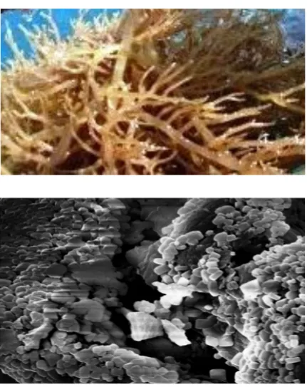
Figure 1. Scanning electron micrographs of (a) raw Kappaphycus alverazii under naked eye, (b) raw Kappaphycus alverazii under scanning electron microscope
!
Communication Communication
Open Access
Result and Discussions
Untreated Seaweed (Raw Seaweed)
The morphologies of the surface of K. alverazii was determined by SEM analysis and shown in Figure 1. The untreated K. alverazii (Figure 1a) exhibited a smooth surface with some impurities that appears to be salt crystalloid deposition covering the area. Obviously the samples of raw seaweed resulted in a reticulated, blocky morphology, closely similar to the seaweed sample observed previously by Boston et al (2014) [9]. Yang et al (2008) observed similar structure on the raw Sargassum sp. and noticed that surface prot uberance and microstructures can be seen which may be due to calcium and other salt crystalloid deposition [4]. Raize et al (2004) also reported a shrinking and sticking of layers were seen in the cell well matrix of Sargassum biomass morphological surfaces [10]. Yang reported that, calcium is a major metal ion on the surfaces of the leaf (2.80 wt. %) and stem (1.16 wt.%) of the sargassum sp. Besides this, several ionic element could also be found such as silicon (1.00 wt.%), sulphur (1.43 wt.%) and kalium (1.09 wt.%) on the surface of leaf and silicon (0.79 wt.
%), sulphur (1.52 wt.%) and kalium (1.05 wt.%) on the surface of stem. The metal may be in the forms of CaCO3, CaO or Ca(OH)2. This may favor the microstructure to be further developed if the metal ions are adsorbed on the raw sargassum.
Under the SEM (1000x), the surface morphology of raw sample seaweed (Ulva spp. and P. palmate) were appeared not to be flattened. Raw Ulva spp. has fold-like structures in a random arrangement on the surface (Murphy et al., 2009). Raw P. palmata contained fold structures similar to those observed in Ulva spp. These structures were less pronounced giving the surface a rough appearance [11]. For magnification at 350 x, the surface of the material was dense and planar without any crevices. The bulge fibrils could be seen clearly on the surface since the K. alvarezii is a typical type of seaweed with huge amount of hydrocolloid substance compounds. Previously Harada and Harada (1998) reported that κ-carrageenan showing 8-nm widths, with the gelling k-and ι-carrageenan showed microfibrils approximating 8-nm and 5-nm widths, respectively, whereas the nongelling λ-carrageenan shows a width of approximately 1.5 nm. The bulging surface may provide a system for advance
!
!
Communication Communication
network during gelling process in food system and farmaceutical product applications [12]. It was reported that the major cell wall and matrix carbohydrate in the commercially farmed K. alverazii is carrageenan [13]. They also showed the differential distribution of and -carrageenan in the cell walls of K. alverazii, with epidermal cells rich in i-carrageenan and cortical and medullary cells rich in carrageenan. The fact that -carrageenan is present is not surprising since the majority of protoplasts originated from the epidermis and outer cortex and, as pointed out by Zablackis et al (1991), the epidermis of K. alverazii is rich in -carrageenan. There appears to be either a deficiency in the assembly of a normal cell wall in these protoplasts.
Treated Seaweed (Chemical Treatment)
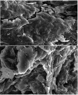
The morphologies of the fractured surface of K. alverazii after heating treatment in acidic solution was determined by SEM analysis as shown in Figure 2. After the fiber undergoes this process in autoclave at high temperature some physical changes such as a rougher fiber surface can be observed indicating fiber breakage occurred during treatment. Changes in the fiber surface occur due to the removal of surface impurities and wax compounds. The treatment also resulted in the leaching out of the salt crystalloid deposition layer and more internal structure was exposed. Another observation revealed is the existence of pores on the surface as presented in (Figure 2a). The micrographs show that treatment of fiber with acid was capable to increase hydrolysis activity. This may benefit to increase total surface area for further hydrolysis process. The -OH group of K. alverazii may react with the acid at high temperature resulted in the broken covalent bonding in its microfibrils structure during autoclave to form hydrolysis products, which causes the transformation of surfaces morphology. This indicates that the fiber undergo a hard breakage process during heating treatment. Finally the surfaces of K. alverazii contain less microstructures and the microfibrils structure become broken and shrunk as the broken microfibers can be clearly seen on the fiber surfaces (Figure 2b). This observation is closely similar to those degradation process described by other researchers [14-16].
surface may provide a system for advance network during gelling process in food system and pharmaceutical product applications. It was reported that the major cell wall and matrix carbohydrate in the commercially farmed K. alverazii is carrageenan [13]. They also showed the differential distribution of κ- and ι-carrageenan in the cell walls of K. alverazii, with epidermal cells rich in ι-carrageenan and cortical and medullary cells rich in κ-carrageenan. The fact that ι-carrageenan is present is not surprising since the majority of protoplasts originated from the epidermis and outer cortex and the epidermis of K. alverazii is rich in ι-carrageenan. There appears to be
either a deficiency in the assembly of a normal cell wall in these protoplasts,
Conclusion
Seaweeds, especially K. alverazii has unique saccharides in their storage cells awaiting for potential exploration such as sulphated sugars making them a potential source for innovation. The morphology structure during hydrolysis was carried out using Scanning Electron Microscope (SEM). The seaweed is very hard to be broken down may be resulted from hydroxyl and covalent bond in their galactant structure. Raw K. alverazii under SEM exhibits some surface protuberance and microstructures which may be due to calcium and other salt crystalloid deposition. The morphology structures after heating treatment in autoclave at high temperature shows some physical changes such as a rougher fiber surface indicating fiber breakage occurred after the leaching out of the salt crystalloid deposition layer until more internal structure was exposed. The -OH group of K. alverazii may react with the acid at high temperature resulted in the broken covalent bonding in its microfibrils structure during autoclave, which causes the morphology transformation. This indicates that the fiber undergo a hard breakage process during heating treatment. Th is s t udy shows changes in the morphological structure of K. alverazii after acidic treatments which can be further optimized in seaweed hydrolysis methods for fut ure biotechnological applications. Open Access References Vision (x) Species Morphology Structure
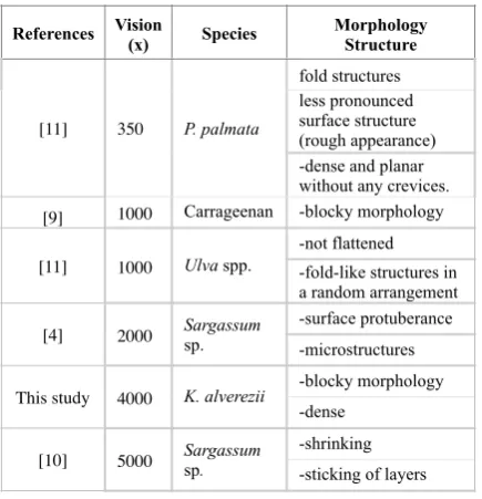
[11] 350 P. palmata
fold structures less pronounced surface structure (rough appearance) -dense and planar without any crevices. [9] 1000 Carrageenan -blocky morphology
[11] 1000 Ulva spp.
-not flattened -fold-like structures in a random arrangement
[4] 2000 Sargassum sp.
-surface protuberance -microstructures
This study 4000 K. alverezii
-blocky morphology -dense
[10] 5000 Sargassum sp.
-shrinking -sticking of layers
Communication
References
1. Darcy-Vrillon B. Nutritional aspects of the enveloping use of marine macro algae for the human industry. Int J Food Sci Nutr. 1993; 44: 23-35.
2. Kumar KS, Ganesan K, Subba Rao PV. Antioxidant potential of solvent extracts of Kappaphycus alvarezii (Doty) Doty – An edible seaweed. Food Chem. 2008; 107: 289-295
3. Borines MG, de Leon RL, McHenry MP. Bioethanol production from farming non-food macroalgae in Pacific island nations: Chemical constituents, bioethanol yields, and prospective species in the Philippines. Renew Sust Energ Rev. 2011; 15: 4432-4435.
4. Yang L, Paul Chen J. Biosorption of hexavalent chromium onto raw and chemically modified Sargassum sp. Bioresource Technol. 2008; 99: 297-307.
5. Jang SS, Shirai Y, Uchida M, Wakisaka M. Production of mono sugar from acid hydrolysis of seaweed. Afr J Biotechnol. 2012; 11: 1953-1963.
6. Meinita MDN, Kang JY, Jeong GT, Koo HM, Park SM, Hong YK. Bioethanol production from the acid hydrolysate of the carrageenophyte Kappaphycus alvarezii (cottonii). J Appl Phycol. 2011; 24: 857-862.
7. Wang X, Liu X, Wang G. Two-stage hydrolysis of invasive algal feedstock for ethanol fermentation. J Integr Plant Biol. 2011; 53: 246-252.
8. Abd-Rahim F, Wasoh H, Zakaria MR, Ariff A, Kapri R, Ramli N, Siew-Ling L. Production of high yield sugars from Kappaphycus alvarezii using combined methods of chemical and enzymatic hydrolysis. Food Hydrocolloid. 2014; 42(2): 309-315.
9. Boston R, Awaya K, Nakayama T, Ogasawara W, Hall SR. Formation of superconducting yttrium barium copper oxide
using sulphur-containing templates. Roy Soc Chem Adv. 2014; 4: 26824-26828.
10. Raize O, Argaman Y, Yannai S. Mechanisms of biosorption of different heavy metals by brown marine macroalgae. Biotechnol Bioeng. 2004; 87(4): 451-458.
11. Murphya V, Tofail SAM, Hughes H, McLoughlin P. A novel study of hexavalent chromium detoxification by selected seaweed species using SEM-EDX and XPS analysis. Chem Eng J. 2009; 148: 425-433
12. Harada T, Harada A. 1998. Gel formation and ultrastructure in food polysaccharides. In Polysaccharide Association Structures in Food, ed. Walter H, pp. 37-56. New York: Marcel Dekker, Inc.
13. Zablackis E, Vreeland V, Kloareg B. Isolation of protoplasts from Kappaphycus alvarezii van tambalang (rhodophyta) andsecretion of ι-carrageenan fragments by cultured cells. J Exp Bot. 1993; 44(266): 1515-1522.
14. Ibrahim NA, Hadithon KA, Abdan K. Effect of fibre treatment on mechanical properties of kenaf-Ecoflex composites. J Reinf Plast Comp. 2010; 29: 2192-2198. 15. Nik Mahmud NA, Baharuddin AS, Bahrin EK, Sulaiman A,
Naim MZ, Zakaria R, Hassan MA, Nishida H, Shirai Y. Enzymatic saccharification of oil palm mesocarp fiber (OPMF) treated with superheated steam. Bioresources. 2013; 8: 1320-1331.
16. Ahamad Nordin NIA, Ariffin H, Andou Y, Hassan MA, Shirai Y, Nishida H, Wan Yunus WMZ, Karuppuchamy S, Ibrahim NA. Modification of oil palm mesocarp fiber characteristic using superheated steam treatment. Molecules. 2013; 18: 9132-9146.
17. Mouritsen, G. 2013. Seaweeds are marine algae. In Seaweeds, Edible, Available and Sustainable, ed. G., Mouritsen, pp. 1-61. London: The University of Chicago Press, Ltd.