University of Pennsylvania
ScholarlyCommons
Publicly Accessible Penn Dissertations
2013
Forkhead Transcription Factors Foxp1 and Foxp4
Regulate T Cell Development and Function
Karla Rose Wiehagen
University of Pennsylvania, [email protected]
Follow this and additional works at:
http://repository.upenn.edu/edissertations
Part of the
Allergy and Immunology Commons
,
Immunology and Infectious Disease Commons
,
and the
Medical Immunology Commons
This paper is posted at ScholarlyCommons.http://repository.upenn.edu/edissertations/943 For more information, please [email protected].
Recommended Citation
Wiehagen, Karla Rose, "Forkhead Transcription Factors Foxp1 and Foxp4 Regulate T Cell Development and Function" (2013).
Forkhead Transcription Factors Foxp1 and Foxp4 Regulate T Cell
Development and Function
Abstract
Transcription factors regulate T cell fates at every stage of development and differentiation. Members of the FoxP family of Forkhead transcription factors are essential for normal T lineage development; Foxp3 is required for regulatory T cell generation and function, and Foxp1 is necessary for the development of naïve T cells. FoxP family member Foxp4 is highly homologous to Foxp1 and has been shown to dimerize with other FoxP proteins.
In this thesis, we report the first studies of Foxp4 in T lymphocytes. Using a CD4Cre-mediated conditional knockout approach we evaluated the roles for Foxp4 regulation in the T lineage. T cell development and homeostasis are normal in the absence of Foxp4. Despite effective control of infection withToxoplasma gondii
or acute Lymphocytic choriomeningitis virusin vivo, cytokine production during antigen-specific rechallenge is reduced in the absence of Foxp4. We conclude that Foxp4 is dispensable for T cell development, but necessary for normal memory T cell recall responses to antigen in acutely or chronically infected mice. Next we determined whether FoxP family members compensate for one another in Foxp1- or
Foxp4-knockout models. We utilized a similar CD4Cre approach to delete both Foxp1 and Foxp4 in T cells. Foxp1/4-deficient T cells exhibit abnormal thymic development and T cell receptor signaling. Loss of Foxp1/ 4 results in significantly reduced T cell numbers, and altered T cell effector function, reminiscent of
Foxp1-deficient T cells.
Lastly, we examined the functions of Foxp1/4 in Foxp3+ regulatory T cells (Tregs). Tregs are critical for prevention of autoimmunity and controlling immune responses during infection. While conditional deletion of either Foxp1 or Foxp4 in T cells has little effect on Tregs, combined deletion results in abnormal Treg generation. Foxp1/4-deficient Tregs exhibited significant defects in both development and homeostasis. Under competitive conditions, double-deficient Tregs are at a significant developmental disadvantage relative to wild-type competitors. Furthermore, Foxp1/4-deficient Tregs exhibit impaired cytokine-induced STAT5 phosphorylation and reduced expression of Foxp3, suggesting Foxp1/4 is required for normal Treg
generation.
Together, these findings demonstrate that the FoxP family regulates multiple facets of T cell development and function, and actively contributes to the maintenance of immunological tolerance.
Degree Type Dissertation
Degree Name
Doctor of Philosophy (PhD)
First Advisor
Jonathan S. Maltzman
Keywords
Foxp1, Foxp3, Foxp4, Regulation, T cells, Transcription
Subject Categories
FORKHEAD TRANSCRIPTION FACTORS FOXP1 AND FOXP4 REGULATE T CELL
DEVELOPMENT AND FUNCTION
Karla Rose Wiehagen
A DISSERTATION
in
Immunology
Presented to the Faculties of the University of Pennsylvania
in
Partial Fulfillment of the Requirements for the
Degree of Doctor of Philosophy
2013
Supervisor of Dissertation
__________________________
Jonathan S. Maltzman
Assistant Professor of Medicine
Graduate Group Chairperson
__________________________
Avinash Bhandoola, MBBS, Ph.D.
Professor of Pathology and Laboratory Medicine
Dissertation Committee
Terri M. Laufer, MD, Ph.D. (Chair), Associate Professor of Medicine
Christopher A. Hunter, Ph.D., Professor of Pathology
Edward E. Morrisey, Ph.D., Professor of Medicine
FORKHEAD TRANSCRIPTION FACTORS FOXP1 AND FOXP4 REGULATE T CELL
DEVELOPMENT AND FUNCTION
COPYRIGHT
2013
Karla Rose Wiehagen
This work is licensed under the Creative Commons Attribution- NonCommercial-ShareAlike 3.0 License
To view a copy of this license, visit
iii To my parents, Paul and Anne Wiehagen, for their incredible love and support
and impressive work ethic, and to my sisters, Katherine Wiehagen Leonard, Theresa Samways, and Joyce Wiehagen,
ACKNOWLEDGMENTS
I would like to offer my most sincere gratitude to my thesis mentor, Dr. Jonathan S. Maltzman, from whom I have learned so much. Jon has been a tireless proponent of my thesis work and of my development as a scientist. He has taught me to be rigorous when designing models and experiments, and to read widely and often. His quick and critical thinking about models have helped tremendously in navigating the complex networks of FoxP transcriptional regulation. It has been my pleasure to be mentored by one of the best scientific minds I’ve met, and to aspire to be as well-rounded a scientist.
The work included in this thesis would not have been possible without the technical expertise and assistance of past and present Maltzman lab members: Michelle Schmidt, Eleni Argyropoulou, Evann Corbo-Rodgers, and Elizabeth S. Staub. Special thanks go to Liz Staub for her excellent support in genotyping and maintaining our extensive mouse colony. Her optimism, energy, and humor made her the best colleague and friend I could have asked for. In addition to technical help, her critical thinking and helpful discussions over lunch and lab meetings have contributed to this thesis work.
I would like to thank my thesis committee members for their guidance and advice throughout these studies. Special thanks also go to Dr. Ed Morrisey and Dr. Shanru Li, for generously providing the Foxp4FLOX mice that initiated my thesis project.
To the entire Immunology Graduate Group community, I thank you for the critical feedback and thoughtful insights offered in Research in Progress seminars, numerous poster sessions, and Immunology Graduate Group Retreats. Suggestions and questions generated from those forums have been instrumental to the work in this thesis. Additionally, I would like to thank the personnel of the Hunter, Bhandoola, Koretzky, Kambayashi, Laufer, Morrisey, Caton, Pear, Wells, and Wherry labs for sharing reagents, offering technical advice, and for helpful conversations that directly shaped the studies described in this thesis. Within the UPenn community, I would like to also thank the Flow Cytometry and Sorting Core personnel and the animal facility care and support teams, without whom this work would not have been completed.
I’d also like to thank Drs. Scott Lieberman, Martha Jordan, Andrew Wells, Paige Porrett, and Michael Cancro for critical reading of manuscripts prepared from this work. Many thanks go to Drs. Betsy Taylor, Helen Simkins, and Natasha Girgis for critical reading of this thesis, and helpful suggestions for arranging this diverse set of data into a palatable layout.
This degree would not be awarded without the generous support and mentorship of Dr. Habib Zaghouani, who first hired me in his lab at the University of Missouri - Columbia. At 17, I had never used a pipette or loaded a centrifuge, but Habib offered me a fantastic opportunity to learn and love Immunology research. I am eternally grateful for that serendipitous job offer.
The encouragement and mentorship from Dr. Jenni Punt has been invaluable to me as well. Her enthusiasm and positivity are infectious, and her thoughtful questions and feedback have helped fuel my work with the FoxP transcription factors. I regard Jenni as one of my favorite scientists and all-around human beings. I’m grateful for the opportunities I have had to work with her.
ABSTRACT
FORKHEAD TRANSCRIPTION FACTORS FOXP1 AND FOXP4 REGULATE T CELL
DEVELOPMENT AND FUNCTION
Karla R. Wiehagen
Jonathan S. Maltzman
Transcription factors regulate T cell fates at every stage of development and
differentiation. Members of the FoxP family of Forkhead transcription factors are
essential for normal T lineage development; Foxp3 is required for regulatory T cell
generation and function, and Foxp1 is necessary for the development of naïve T cells.
FoxP family member Foxp4 is highly homologous to Foxp1 and has been shown to
dimerize with other FoxP proteins.
In this thesis, we report the first studies of Foxp4 in T lymphocytes. Using a
CD4Cre-mediated conditional knockout approach we evaluated the roles for Foxp4
regulation in the T lineage. T cell development and homeostasis are normal in the
absence of Foxp4. Despite effective control of infection with Toxoplasma gondii or acute
Lymphocytic choriomeningitis virus in vivo, cytokine production during antigen-specific
rechallenge is reduced in the absence of Foxp4. We conclude that Foxp4 is dispensable
for T cell development, but necessary for normal memory T cell recall responses to
antigen in acutely or chronically infected mice.
Next we determined whether FoxP family members compensate for one another
both Foxp1 and Foxp4 in T cells. Foxp1/4-deficient T cells exhibit abnormal thymic
development and T cell receptor signaling. Loss of Foxp1/4 results in significantly
reduced T cell numbers, and altered T cell effector function, reminiscent of Foxp1cKO T
cells.
Lastly, we examined the functions of Foxp1/4 in Foxp3+ regulatory T cells
(Tregs). Tregs are critical for prevention of autoimmunity and controlling immune
responses during infection. While conditional deletion of either Foxp1 or Foxp4 in T cells
has little effect on Tregs, combined deletion results in abnormal Treg generation.
Foxp1/4-deficient Tregs exhibited significant defects in both development and
homeostasis. Under competitive conditions, double-deficient Tregs are at a significant
developmental disadvantage relative to wild-type competitors. Furthermore,
Foxp1/4-deficient Tregs exhibit impaired cytokine-induced STAT5 phosphorylation and reduced
expression of Foxp3, suggesting Foxp1/4 is required for normal Treg generation.
Together, these findings demonstrate that the FoxP family regulates multiple
facets of T cell development and function, and actively contributes to the maintenance of
TABLE OF CONTENTS
ABSTRACT ... V
TABLE OF CONTENTS ... VII
LIST OF TABLES ... XI
LIST OF FIGURES ... XII
CHAPTER I
:
INTRODUCTION ... 1
A brief introduction to the adaptive immune system ... 1
Forkhead transcription factors govern cell fate decisions ... 4
The FoxP subfamily of transcription factors ... 6
FoxP family transcriptional regulation in mammalian development ... 8
FoxP transcription factors in the adaptive immune system ... 11
FoxP protein Interactions in T cells ... 13
Structure of Thesis ... 15
CHAPTER II: FOXP4 REGULATION IS DISPENSABLE FOR T CELL
DEVELOPMENT, BUT ESSENTIAL FOR MEMORY RECALL RESPONSES. . 18
Introduction ... 18
Results ... 21
Foxp4 can be efficiently deleted in thymocytes ... 24
Foxp4 is not required for T cell development and peripheral homeostasis ... 26
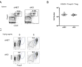
Normal Regulatory T cell generation in Foxp4 cKO CD4 T cells. ... 30
Foxp4 is not necessary for T cell proliferation or activation. ... 31
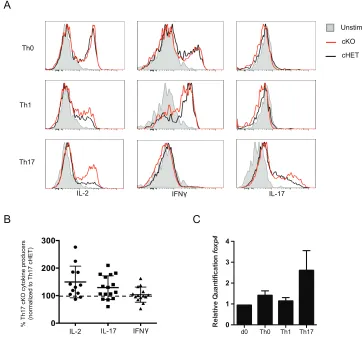
Foxp4 does not regulate T helper cell commitment ... 34
Foxp4-deficient T cells control chronic infection ... 37
Foxp4 is not required for acute viral clearance. ... 43
Discussion ... 47
CHAPTER III:
FOXP1 AND FOXP4 DIRECT T CELL FATES AND
FUNCTIONS. ... 52
Introduction ... 52
Results ... 54
Generation of Foxp1/Foxp4 conditional double knockout mice. ... 54
Foxp1 and Foxp4 in thymic development. ... 57
FoxP members maintain peripheral CD4 and CD8 T cell number ... 62
CD44 expression is regulated by Foxp1 but not Foxp4. ... 65
cDKO CD4 and CD8 T cells acquire an effector memory phenotype. ... 66
Loss of Foxp1/4 results in systemic T lymphopenia. ... 67
Cell-intrinsic regulation of T cell development by FoxP proteins. ... 69
Cell-intrinsic regulation of T cell central and effector memory phenotype ... 73
Foxp1 regulates T cell effector functions. ... 75
FoxP transcription factors regulate IL-7R expression and cytokine signaling. ... 83
cDKO T cells exhibit increased proximal STAT5 signals but poor survival. ... 85
Discussion ... 89
CHAPTER IV: THE FOXP FAMILY REPRESSES CD4 EXPRESSION IN T
LYMPHOCYTES. ... 91
Results ... 93
cDKO CD4 and CD8 T cells express increased levels of CD4. ... 93
CD4 is intrinsically regulated by FoxP family members. ... 96
CD4 is a transcriptional target of FoxP family regulation. ... 98
Discussion ... 100
CHAPTER V:
FOXP1 AND FOXP4 REGULATE FOXP3+ TREG CELL
DEVELOPMENT. ... 102
Introduction ... 102
Results ... 106
Foxp1 and Foxp4 are expressed in regulatory T cells. ... 106
Foxp1 and Foxp4 are required for normal Treg generation. ... 106
cDKO Tregs phenotypically resemble memory Tregs ... 112
cDKO Tregs exhibit loss of suppressive function in vitro. ... 115
FoxP members regulate adaptive Treg development. ... 116
Defective cDKO Treg development is cell intrinsic. ... 119
cDKO thymocytes signal abnormally through the T cell receptor. ... 123
STAT5 signals are dysregulated in cDKO Tregs. ... 125
Impaired cytokine signaling and Foxp3 induction is an intrinsic defect in cDKO thymocytes. ... 128
Discussion ... 131
CHAPTER VI:
DISCUSSION ... 134
Introduction ... 134
I. Foxp4 regulation in effector and memory T cells ... 135
Future Directions: Foxp4 regulation in lymphocyte development ... 138
II. Foxp1 and Foxp4 in the regulation of lymphocyte development. ... 140
cDKO thymocytes exhibit altered T cell receptor signaling. ... 140
Foxp1 and Foxp4 regulation of IL-7 receptor signaling and survival. ... 144
Future Directions: Foxp1 and Foxp4 regulation of immune responses in vivo. ... 145
III. Foxp1 and Foxp4 regulation of Foxp3+ Treg generation ... 146
Future Directions: Rescuing cDKO Treg development ... 147
Future Directions: Investigation of Foxp1/4 regulation of Treg maintenance ... 149
Future Directions: Runx:FoxP family interactions ... 150
The relationship between Foxp1 and Foxp4 in T cells ... 154
CHAPTER VII:
MATERIALS AND METHODS ... 158
CHAPTER VIII:
BIBLIOGRAPHY ... 167
LIST OF TABLES
Table 3.1. Generation of cDKO mice and littermate controls. 54
Table 3.2. Summary of cDKO CD4 and CD8 T cell cytokine production in vitro. 83
LIST OF FIGURES
Chapter II.
Figure 2.1. Foxp4 is expressed in lymphoid tissues and T cell subsets 23
Figure 2.2. Generation of Foxp4 cKO mice 25
Figure 2.3. Deletion of Foxp4 at the DP stage does not alter thymocyte development 27
Figure 2.4. Foxp4 is not required for the generation of peripheral CD4 and CD8
T cells 29
Figure 2.5. Foxp3+ Regulatory T cell development does not require Foxp4
expression 31
Figure 2.6. T cell activation induces normal proliferation and effector T cell
differentiation in the absence of Foxp4 33
Figure 2.7. Foxp4 is not required for .T helper cell differentiation. 36
Figure 2.8. Toxoplasma gondii infection does not lead to wasting or lymphopenia in
Foxp4 cKO mice 39
Figure 2.9. Foxp4 deletion alters recall responses to Toxoplasma gondii in the
spleen and brain. 42
Figure 2.10. Antigen specific Foxp4 cKO T cells persist after antigen clearance 45
Figure 2.11. Foxp4 deficient CD4 T cells exhibit reduced memory recall responses
following LCMV infection. 47
Chapter III.
Figure 3.1. Generation of cDKO mice 57
Figure 3.2. Reduced thymic cellularity in Foxp1cKO and cDKO mice 58
Figure 3.3. Foxp1/4 deletion alters CD5 expression and DP cellularity but not SP
polyclonality 60
Figure 3.4. Normal numbers of SP thymocytes develop in cDKO mice 62
Figure 3.5. Reduced cDKO CD4 and CD8 T cells contribute to significant T
Figure 3.6. cDKO SP thymocytes and splenic T cells express T cell signaling
proteins and markers of activation. 67
Figure 3.7. Reduced cDKO CD4 and CD8 T cells were detected in multiple tissues 69
Figure 3.8. Schematic of generation of competitive bone marrow chimeras. 71
Figure 3.9. Competitive development of cDKO DP and SP thymocytes in mixed
bone marrow chimeras. 72
Figure 3.10. Foxp1cKO- or cDKO-derived CD4 and CD8 T cells have a developmental
disadvantage in peripheral lymphoid tissues. 74
Figure 3.11. Expression of effector memory cell surface markers are intrinsically
regulated by Foxp1 and Foxp4. 76
Figure 3.12. Foxp1, but not Foxp4, regulates IL-2 and IFNγ production by CD4
T cells. 79
Figure 3.13. Altered CD4 T cell cytokine production reflects cell-intrinsic
Foxp1-dependent regulation of IFNγ and IL-2. 81
Figure 3.14. Decreased frequencies of cDKO CD8 T cells produce IFNγ. 82
Figure 3.15. Competitive chimeras reveal comparable frequencies of cDKO CD8 T cells
produce IFNγ. 83
Figure 3.16. cDKO CD4 T cells exhibit increased STAT5 phosphorylation but decreased
survival 89
Chapter IV.
Figure 4.1. cDKO CD4 and CD8 T cells express increased levels of CD4
co-receptor 96
Figure 4.2. Increased CD4 expression is cell-intrinsically regulated by FoxP proteins. 98
Figure 4.3. Total CD4 protein level and cd4 transcript is increased in cDKO CD4
T cells 100
Chapter V.
Figure 5.1. Conditional deletion of Foxp1 and Foxp4 alters thymic Foxp3+ tTreg
frequencies. 108
Figure 5.2. Foxp1/4 deletion results in significantly reduced numbers of
peripheral Tregs. 110
Figure 5.3. Frequencies of cDKO Tregs are significantly increased in spleen
Figure 5.4 cDKO Tregs express an altered cell surface phenotype. 115
Figure 5.5. cDKO Tregs exhibit reduced suppressive ability in vitro. 117
Figure 5.6. Inducible Treg differentiation requires Foxp1 expression. 119
Figure 5.7. Foxp1 or Foxp4 expression is necessary for normal tTreg generation. 121
Figure 5.8. CD45.2+ cDKO Tregs are nearly absent in peripheral lymphoid tissues
of competitive chimera mice. 123
Figure 5.9. cDKO CD4SP T cells display increased signaling to anti-CD3
and anti-CD28 stimulation in vitro. 125
Figure 5.10. cDKO tTregs express increased cytokine receptors but
exhibit impaired survival. 128
Figure 5.11. cDKO thymocytes have intrinsic cytokine signaling defects. 131
Chapter VI.
Figure 6.1. Model of TCR signaling defects in cDKO T cells. 144
Figure 6.2. Schematic of Runx1-regulated gene targets in the CD4 T lineage. 152
Figure 6.3. Models of Foxp1/4 and Runx regulation of selected gene targets
1
CHAPTER I
Introduction
A brief introduction to the adaptive immune system
The purpose of the immune system is to protect the host from infection with
pathogens, including bacteria, viruses, fungi, and parasites. There are two branches of
the immune system: innate and adaptive. The innate immune system exists in different
forms in all multicellular organisms, whereas the adaptive immune system evolved in
vertebrates in the animal kingdom. While innate immune mechanisms provide initial
protection to the host, adaptive mechanisms mount specific responses to control and
clear foreign substances and pathogens.
The primary cellular components of the adaptive branch of the immune system
are a type of white blood cell called lymphocytes, which are further divided into T cells
and B cells. Compared to cells of the innate system, T and B lymphocytes are uniquely
suited to initiate specialized soluble and cellular responses and to conduct a systemic
response upon exposure to a pathogen. For instance, activated lymphocytes undergo
clonal expansion in which rapid cellular division produces an exponential increase in
Because of the potent functions of lymphocytes, activation of these cells is tightly
regulated from the early stages of development. During lymphocyte maturation, somatic
rearrangement of antigen receptor genes causes each cell to express a unique antigen
receptor complex on the cell surface. In the T lineage, this antigen receptor is called the
T cell receptor (TCR). A successfully rearranged TCR complex licenses a single
specificity to each cell. A large percentage of developing lymphocytes are selectively
culled, to ensure that each antigen receptor rearrangement signals appropriately and
that clones with self-reactive antigen receptors are eliminated. Similar processes occur
in B cells to restrict the survival of potentially autoreactive lymphocytes.
During development, lymphocytes are further committed to distinct subsets that
best suit their antigen receptor specificity. Within the T lineage, cells are divided into two
main T cell subsets. CD4+ TCR+ lymphocytes (CD4 T cells) respond to immunologic
challenges originating outside host cells, such as bacterial infections and allergenic
antigens. CD8+ TCR+ cells (CD8 T cells) are cytotoxic lymphocytes that clear
intracelullar pathogens, like viruses, by killing infected host cells. Signals specific for
each kind of immunologic insult activate the correct cell type and induce effector cell
differentiation, proliferation, and expression of gene programs encoding lymphocyte
functions.
Mature lymphocytes circulate through blood and lymphatic vessels to patrol host
tissues for pathogens. To ensure inflammatory lymphocyte reactions occur only when
warranted, layers of transcriptional regulation prevent non-specific initiation of effector
programs. Naïve, or antigen-inexperienced, lymphocytes remain functionally silent until
ligation, which triggers a cascade of intracellular signaling and initiation of an adaptive
immune response.
Successful clearance of infection requires expansion of lymphocytes expressing
antigen receptor complexes specific for epitopes unique to the pathogen. Of the millions
of lymphocytes circulating in the body, only a fraction will be specific to any one
pathogen. Upon activation, these pathogen-specific T cells and B cells expand
exponentially to produce significant numbers of identical daughter cells, equally reactive
to pathogenic epitopes. B cell clones become activated and produce soluble antibodies
against the pathogen, which aid in detection and killing of extracellular pathogens. T
cells orchestrate responses by secreting cytokines that recruit other immune cells to
infected tissues. CD8 T cells can also participate by mounting cytotoxic responses
against cells harboring intracellular pathogens. This process of clonal expansion and
activation affords the host access to an army of effector T or B cells to control the spread
of bacterial, fungal, viral or parasitic pathogens at the time of infection, while maintaining
fewer lymphocytes at steady-state.
Following pathogen clearance, there is no need for the large numbers of
daughter effector cells; further, prolonged maintenance of these cells can harm the host.
Effector cells undergo apoptosis during the contraction phase, which occurs after an
infection is controlled. However, a population of pathogen-specific lymphocytes is
retained and persists in the host to guard against repeated infection. These cells allow
the host to mount more rapid and larger immune responses upon re-exposure to a
specific pathogen. A cardinal feature of adaptive immunity is the provision of this
immunological memory, which benefits the longevity of the host by preventing chronic
The protective benefits of adaptive immunity described above are offset by the
potential damage caused by dysregulated immune reactions. At every point in the life
cycle of lymphocytes, both environmental and intracellular signals direct cell fate
decisions to maintain a diverse repertoire of quiescent antigen-specific T and B cells in a
healthy host. Unprovoked expansion and activation of T or B cell clones commonly
results in non-specific immune responses and damage to host tissues. Therefore, the
selective activation of pathogen-specific cells is tightly regulated. Lymphocyte specificity,
controlled activation, and restrictions on self-reactive lymphocyte development are
examples of regulatory mechanisms in the adaptive immune system. These precautions
provide protection for vertebrate organisms against foreign pathogens, while minimizing
the risk of uncontrolled cytotoxic and inflammatory responses.
Forkhead transcription factors govern cell fate decisions
In every nucleated cell in the body, cell fate decisions are regulated by both
extrinsic and intrinsic mechanisms. Environmental cues such as soluble growth factors
support cell metabolism, migration, and survival. Intrinsic processes include cell cycle
checkpoints and manipulation of genomic DNA to express proteins necessary for
proliferation and differentiation. Successful execution of gene expression requires both
temporal and contextual specificity. In eukaryotic cells, regulatory mechanisms have
evolved to correctly interpret extrinsic and intrinsic signals to prevent unproductive gene
expression. A fundamental source of regulation is a class of proteins called transcription
factors that translate extracellular signals and control the expression or silencing of gene
One family of transcription factors is the forkhead family. Members of this family
are expressed in all eukaryotic cells except plant cells (Jonsson and Peng, 2005). All
forkhead box (Fox) proteins possess a highly conserved sequence of 80-100 amino
acids, originally identified in Drosophila in 1990 (Kaufmann and Knochel, 1996). X-ray
crystallography and nuclear magnetic resonance (NMR) studies of the forkhead domain
determined this sequence forms three alpha helices and three beta sheets flanked with
two loops or wings, giving the three-dimensional structure a butterfly-like appearance
(Hannenhalli and Kaestner, 2009). This winged-helix structure facilitates binding of Fox
proteins to the major groove of DNA sequences containing consensus binding sites
(Myatt and Lam, 2007). Fox proteins recognize sites with the core sequence 5’
(A/G)(C/T)(A/C)AA(C/T)A 3’ (Carlsson and Mahlapuu, 2002; Myatt and Lam, 2007).
More than 100 genes in the human genome encode forkhead proteins that are
organized into 27 subfamilies based on phylogenetic hierarchy and sequence homology
(Hannenhalli and Kaestner, 2009; Uhlenhaut and Treier, 2011). In humans, expression
of forkhead proteins has been implicated in the generation of most tissues in the body.
Forkhead family members are required to orchestrate organogenesis throughout
embryonic development, and mutations in forkhead proteins can result in embryonic
lethality. Defective Fox regulation in children and adults is associated with pathological
disorders and increased incidence of cancer (Myatt and Lam, 2007).
Forkhead proteins are required for normal hematopoiesis and the maintenance of
both innate and adaptive immunity. In both myeloid and lymphocyte blood cell lineages,
expression of forkhead proteins is essential for normal development and function (Coffer
and Burgering, 2004; Myatt and Lam, 2007). Fox transcription factors directly regulate
proteins), and differentiation of multiple blood cell lineages (Foxj1) (Coffer and
Burgering, 2004). Therefore, forkhead proteins play a crucial role in maintaining adaptive
immunity by dictating cell fate decisions in the hematopoietic lineage.
The FoxP subfamily of transcription factors
The FoxP subfamily is comprised of four proteins, Foxp1-Foxp4, which generally
act as transcriptional repressors (Lopes et al., 2006; Teufel et al., 2003). FoxP family
proteins share common characteristics including a conserved leucine zipper domain, a
zinc finger motif, a long glutamine repeat, and the forkhead box DNA-binding domain
(Teufel et al., 2003; Wang et al., 2003). The sequences of FoxP member genes foxp1,
foxp2 and foxp4 are more closely related, while foxp3 is least like its family members (Lu
et al., 2002; Uhlenhaut and Treier, 2011). While FoxP proteins recognize the nucleotide
sequence unique to forkhead DNA binding, sequence variation among the family
members is thought to license each protein to recognize independent targets and
assume distinct regulatory roles. For example, Foxp3 binds the nucleotide sequence 5’
(G/a)TAAACA 3’, while Foxp1 binds the sequence 5’ AA(C/t)A(C/t)AAATA 3’ (Koh et al.,
2009; Wang et al., 2003). Each contains the core sequence recognized by forkhead
transcription factors, but also illustrates variable specificity in gene targets of the FoxP
family. Furthermore, the flanking sequences around the forkhead domain in FoxP
proteins are all unique, which may affect how these proteins bind DNA targets and other
proteins.
The FoxP proteins are unique from all other forkhead subfamilies because of the
way these proteins dimerize to bind DNA. The leucine zipper domain is required for
FoxP family members can also heterodimerize with one another via the leucine zipper
motif. Mutation of the highly conserved leucine residues within the leucine zipper domain
disrupts protein-protein interactions and inhibits transcriptional regulation by FoxP
dimers (Li et al., 2004a; Liu et al., 2012; Zhou et al., 2009). Heterodimerization provides
for regulation of specific combinations of gene sequences, depending on which proteins
participate in the FoxP complexes. Ectopic overexpression studies have demonstrated
that Foxp1, Foxp2, and Foxp4 can homo- and heterodimerize, and that Foxp3 can form
heterodimers with Foxp1. FoxP family heterodimerization may offer another level of DNA
site recognition, elegantly controlled by the expression or availability of different FoxP
partners.
Schematic 1. The FoxP family. Representation of the features and relative length of
the four members of the FoxP family of transcription factors.
More complexity of FoxP protein regulation is provided by interactions with other
remodeling. Conformational changes in FoxP proteins as they bind partners may also
expose different sites involved in protein-protein interactions. Competition for FoxP
protein binding sites by potential partners and the stability of multi-protein complexes
may affect transcription factor function. Therefore, the conserved FoxP family
characteristics support complex protein interactions, and highly specific regulation of
gene programs.
FoxP family transcriptional regulation in mammalian development
Each member of the FoxP family regulates diverse tissue-specific cell fate
decisions throughout development (Carlsson and Mahlapuu, 2002; Kaufmann and
Knochel, 1996; Shu et al., 2001). Although Foxp1, Foxp2, and Foxp4 are detected in
adult tissues, expression of these transcription factors is highest during embryonic
development of neural, intestinal, pulmonary, and cardiovascular tissues. Foxp3
expression is detected during development in the embryonic neuronal tissues, but is
otherwise limited to immune cells, which will be discussed later.
The function and regulation of FoxP proteins have been characterized through
germline deletion, overexpression, and gene mutation. Initial studies demonstrated that
germline deletion of Foxp1, Foxp2, or Foxp4 blocks development, resulting in embryonic
lethality (French et al., 2007; Li et al., 2004a; Wang et al., 2004). However, more recent
studies describe the effects of lineage-specific FoxP family deletion, which bypass
blocks in embryonic development. This growing body of literature illustrates the
versatility of FoxP transcription factor functions, including regulation of cellular
Studies of cardiopulmonary development offer excellent examples of the ways
FoxP proteins regulate cell fate decisions. Foxp1 expression is highest in developing
lung tissue and is required for the normal development of the distal pulmonary
epithelium. Foxp4 is co-expressed with Foxp1—together, these proteins repress
abnormal cell differentiation of pulmonary epithelium throughout development. Deletion
of either Foxp1 or Foxp4 does not alter differentiation of epithelial cells, but Foxp1/Foxp4
double deficiency has profound effects on secretory cell development in the pulmonary
airway that cause neonatal morbidity. Co-regulation of pulmonary development by Foxp1
and Foxp4 highlights the ability of these homologous Fox proteins to compensate for
one another to preserve cell fate potential. In contrast, Foxp2 is also required for normal
lung development, but regulates the development of proximal-distal epithelium
independently of Foxp1 and Foxp4. The expression patterns and regulatory functions of
Foxp1/Foxp2/Foxp4 neatly demonstrate the roles FoxP members can play
independently and in combination to direct cell fate decisions.
In addition to directing tissue differentiation, FoxP family proteins regulate cell
survival and proliferation. The FoxP family is essential for normal cardiac development,
both in the formation of vasculature and cardiomyocyte development. Foxp1 and Foxp2
are implicated in cardio vasculogenesis. Germline deletion of Foxp1 causes defects in
heart valve and ventricle formation, resulting in death by day 16.5 of embryogenesis
(E16.5) (Wang et al., 2004). This defect is further characterized by dysregulated
myocardium maturation, which is dependent on Foxp1 regulation. Foxp1-/-
cardiomyocytes exhibit hypertrophy, increased proliferation, and failure to differentiate
normally (Bai and Kerppola, 2011). These findings suggest Foxp1 is required for
Like Foxp1 and Foxp2, Foxp4 is expressed in and required for development of
cardiopulmonary tissues. Foxp4 expression in the developing embryo programs
localization of cardiac tissues and proper heart development. Foxp4-deficient mice
exhibit embryonic lethal cardia bifida (Li et al., 2004b). Foxp4-deficient precardiac
tissues fail to migrate toward the midline of the fetus, resulting in two symmetrical hearts,
bilateral to the midline (Li et al., 2004b). Defects in the developing foregut of embryonic
Foxp4-/- mice also suggest Foxp4 not only directs localization, but also directs the
survival of critical tissues during embryogenesis.
Defects observed in cardiopulmonary development demonstrate transcriptional
regulation by Foxp1 and Foxp2 represses cellular proliferation. Dysregulation of cellular
proliferation is a hallmark of cancer, and FoxP family members have been implicated in
tumor repression. Oncogenic cell growth occurs with loss of Foxp1 and Foxp2
transcriptional regulation in multiple tissues (Myatt and Lam, 2007). FoxP family member
Foxp3 also protects against tumor development (Heinze et al., 2011; Li et al., 2011;
Wang et al., 2010). Mutations that perturb Foxp3 regulation lead to cancer development
in the breast, brain and intestine (Hannenhalli and Kaestner, 2009; Myatt and Lam,
2007; Triulzi et al., 2013).
Together, these studies demonstrate how FoxP family proteins regulate multiple
kinds of cell fate decisions across a variety of tissues. FoxP-mediated regulation of
target genes affects development, survival, and differentiation. FoxP proteins direct
FoxP transcription factors in the adaptive immune system
All members of the FoxP family are expressed in blood cells, except Foxp2.
Previous studies in lymphocytes demonstrate how Foxp1 and Foxp3 regulate cell fate
decisions and impact adaptive immunity. In B cells, Foxp1 initiates B lymphopoiesis, and
controls antigen receptor gene recombination later in development (Hu et al., 2006).
Expression of the enzyme RAG (recombination activating gene) is required for normal B
cell receptor rearrangement and B cell selection (Hu et al., 2006). Deletion of Foxp1
impairs RAG expression and blocks B cell development. Humoral immunity may also
rely on expression of Foxp4. Preliminary data from our work suggest Foxp4 deficiency
causes abnormal B cell development. In contrast, loss of FoxP family proteins does not
block T cell development. FoxP members do not regulate somatic rearrangement of
TCR genes, but instead regulate cell fate decisions during differentiation and
commitment to T cell subsets.
Of the FoxP proteins, the function of Foxp3 transcriptional regulation is best
defined. Expression of Foxp3 in T lymphocytes is critical for the maintenance of
self-tolerance (Brunkow et al., 2001; Hori et al., 2003; Ramsdell and Ziegler, 2003). Foxp3
was first identified in mouse models of autoimmunity, and later associated with IPEX
(Immunodysregulation polyendocrinopathy enteropathy X-linked) syndrome (Bennett et
al., 2001; Gambineri et al., 2003). Patients with IPEX inherit gene mutations in Foxp3
that cause dysregulation of immune cells, manifesting in symptoms of severe
autoimmunity (Bennett et al., 2001; Gambineri et al., 2003).
The mechanism of Foxp3-dependent tolerance was traced to a subset of CD4+ T
cells, called regulatory T cells, or Tregs (Hori et al., 2003; Rudensky, 2011; Sakaguchi et
responses, and to return balance to immune cell homeostasis following infection (Khattri
et al., 2001). Foxp3 is regarded as a master regulator of Treg commitment among the
scores of transcription factors expressed in T cells, since it is necessary to initiate
Treg-associated gene programs. In both animal models and humans, Tregs normally develop
during thymic selection, but conversion of conventional CD4 T cells during immune
responses also occurs upon Foxp3 induction (Chen et al., 2003; Fu et al., 2004; Zheng
et al., 2004). In addition to Foxp3 expression, Tregs are identified by cell surface
expression of the high affinity IL-2 receptor alpha chain (CD25), GITR (a member of the
tumor necrosis factor receptor family) (Cuzzocrea et al., 2005; Gavin and Rudensky,
2003). Several groups have determined Foxp3 regulates transcription of these cell
surface proteins and other gene programs required for tolerance, although the precise
mechanisms of Treg suppression are not well understood.
Initial studies in Foxp1-deficient T cells determined Foxp3+ Treg differentiation
does not require Foxp1 expression (Feng et al., 2010). Instead, Foxp1 is necessary for
normal development of conventional T cells (Feng et al., 2010; Feng et al., 2011).
Deletion of Foxp1 in the T lineage demonstrated that Foxp1 normally represses
activation-induced gene programs in both CD4 and CD8 T cells. Foxp1-deficient T cells
acquired an activated phenotype, including upregulation of the cell surface marker CD44
(Feng et al., 2010). Furthermore, T cells lacking Foxp1 expression exhibited effector
functions, including increased proliferation and cytokine production. Experiments in
which Foxp1 was temporally deleted after normal T cell development demonstrated that
loss of Foxp1 immediately resulted in abnormal activation (Feng et al., 2010). Therefore,
sustained Foxp1 expression is required to repress effector T cell fate and to maintain the
As observed in other cell types, Foxp1 also regulates survival and proliferation of
T lymphocytes. The survival of both B and T lymphocytes is impaired without Foxp1
expression despite increased expression of the interleukin-7 receptor (IL-7R) (Feng et
al., 2011). Foxp1 acts as a transcriptional repressor of the il7r gene, which encodes the
alpha chain of the Interleukin-7 receptor (IL-7R) (Feng et al., 2010; Hu et al., 2006).
Foxp1 and forkhead factor Foxo1 compete for a forkhead binding site in the il7r
promoter. In the absence of Foxp1, T cells express increased levels of IL-7R, and CD8 T
cells proliferate abnormally to IL-7 stimulation (Feng et al., 2011). However, despite
increased sensitivity to anti-apoptotic IL-7 signals, survival is impaired. Loss of Foxp1
causes T lymphopenia, which becomes more severe with age. Therefore, Foxp1
regulation maintains the quality and quantity of the T lymphocyte repertoire.
Preliminary work has demonstrated Foxp4 expression in lymphoid tissues (Teufel
et al., 2003). However, the function of Foxp4 in lymphocytes had not been described.
Like all FoxP family members, Foxp4 forms homodimers, and can heterodimerize with
Foxp1 and Foxp2 in non-lymphoid cells (Li et al., 2004a). Examples of Foxp4 regulation
in other cell types indicate this transcription factor also directs cell differentiation and
survival. Yet it was unclear what processes Foxp4 regulates in adaptive immune cells,
and whether Foxp4 expression is required to the same degree as its FoxP family
members for normal T cell development.
FoxP protein Interactions in T cells
FoxP transcription factors regulate gene expression by participating in
multi-protein complexes. Multiple domains, including the conserved FoxP forkhead domain,
functional alterations in gene transcription. Foxp3 binding partners include other
transcription factors: Foxp1, NFAT, NFκB, Runx family members, retinoic acid-related
orphan receptor (ROR). FoxP proteins also interact with chromatin remodeling proteins
such as histone deacetylases (HDACs), which are proteins capable of altering DNA
accessibility (Bettelli et al., 2005; Du et al., 2008; Li et al., 2006; Li et al., 2007; Ono et
al., 2007; Wu et al., 2006). Direct interaction between Foxp4 and HDACs has also been
reported in transfected fibroblasts, but not yet in lymphocytes (Chokas et al., 2010). The
ability of FoxP proteins to work with a variety of partners ostensibly increases the variety
of potential gene targets.
Interactions with different binding partners rely on conserved motifs within the
FoxP protein sequence. Models of Foxp3 dimerization indicate this protein changes
conformation as it binds another Foxp3 protein, other partners, or target genes. For
example, homodimerization of Foxp3 exposes a number of hydrophobic amino acid
residues encoded in sequences flanking the leucine zipper domain (Zhou et al., 2009).
Normally, these residues would provide better stability for dimerization if aligned along
the axis of protein-protein interaction. Instead these residues are interspersed around
and throughout the leucine zipper domain. Such unusual placement suggests these
sequences provide a scaffold for other proteins to bind to Foxp3 dimers. Furthermore,
Foxp3 binds different target sequences along the major or minor groove of DNA, which
also changes the availability of motifs (Myatt and Lam, 2007; Song et al., 2012; Zhou et
al., 2009). FoxP platforms may be fundamental for the recruitment of transcriptional
machinery or for binding of chromatin remodeling proteins. Sequence variability around
conserved motifs in each FoxP member may shape protein-protein interactions, or
Structure of Thesis
This thesis begins by specifically addressing how Foxp4 regulates T cell
development and function. Preliminary evidence of Foxp4 expression in lymphoid
tissues suggested this FoxP protein plays a role in adaptive immunity. Requirements for
related proteins Foxp1 and Foxp3 for normal immune regulation, as well as the high
degree of homology with the Foxp1 gene sequence, suggested Foxp4 could participate
in similar regulation of lymphocyte development. These findings raised several
hypotheses that provoked investigation of Foxp4 regulation in lymphocytes.
Studies described in Chapter II of this thesis assessed the effects of Foxp4
deletion on the development and function of the T lineage. Unlike its FoxP relatives,
deletion of Foxp4 does not affect T cell development. Foxp4 does not appear to
cooperatively regulate Treg and conventional T cell gene programs with Foxp1 or Foxp3.
Instead, our data demonstrate Foxp4 regulates memory T cell recall responses and may
be important for maintaining long-term immunity.
Although our work suggested Foxp1 and Foxp4 have independent functions in T
cells, we had not formally ruled out the possibility that Foxp1 also compensates for loss
of Foxp4, and can assume the roles normally filled by Foxp4. In Chapter III, we
expanded our studies to examine how loss of both Foxp1 and Foxp4 affected T cell
development. Combined deletion of FoxP proteins provides the first characterization of
Foxp1- and Foxp4- deficient T cells. Double-deficient T cells were compared to T cells in
which Foxp1 or Foxp4 were deleted. Without Foxp1/4, many Foxp1-associated defects
Deletion of both Foxp1 and Foxp4 altered the expression of the CD4 co-receptor
on both CD4 and CD8 T cells. In Chapter IV, we examine the defect in CD4 expression
in depth, and demonstrate cell-intrinsic dysregulation of this critical co-receptor at the
transcriptional level. Furthermore, the expression patterns of CD4 were significantly
dysregulated in double-deficient T cells, but not in Foxp1- or Foxp4-deficient
conventional T cells or in the Treg subset of CD4 T cells that express Foxp3. These data
suggest FoxP members can compensate for one another in select gene programs, and
expression of at least one FoxP protein is sufficient to regulate levels of CD4.
Chapter V addresses how Foxp1, Foxp3, and Foxp4 regulate peripheral
tolerance. Deletion of Foxp1 and Foxp4 revealed a critical role for FoxP family members
in Foxp3+ Treg development. Defects appeared in double-deficient Treg differentiation,
but not in Foxp1 or Foxp4 knockout T cells, suggesting expression of Foxp3 with at least
one FoxP family member was sufficient for normal generation of regulatory T cells.
Signaling pathways downstream of cytokine and TCR stimulation are dysregulated in
Foxp1/4-deficient cells, resulting in abnormal Treg development, survival, and function.
Together, this work expands the model of FoxP family transcriptional regulation
in T lymphocytes to include Foxp4-specific functions. Unlike Foxp1 and Foxp3, Foxp4
regulation is limited to memory T cell responses. With few exceptions, our studies
indicate Foxp4 otherwise serves as a redundant regulator of Treg development under
steady-state conditions. Combined deletion of Foxp1 and Foxp4 causes a unique set of
developmental and survival defects, which are exacerbated under competitive
conditions. Studies of double knockout T cells demonstrate some cases in which Foxp1
and Foxp4 may compensate for one another to support T cell survival and generation of
implications for Foxp4 regulation of adaptive immune cell effector and memory
CHAPTER II.
Foxp4 regulation is dispensable for T cell development, but
essential for memory recall responses.
Introduction
In the immune system, members of the FoxP family of transcription factors
regulate gene programs important for normal development and function. FoxP family
member Foxp4 regulates developmental programs in heart and lung tissues, and shares
a high degree of homology with Foxp1 (Li et al., 2004b). Preliminary work has
demonstrated expression of Foxp4 in lymphoid tissues (Teufel et al., 2003). However,
the function and role of Foxp4 in T cells has not been described.
FoxP family members regulate gene expression by forming homo- and
heterodimers (Li et al., 2007). The FoxP protein Foxp3 also interacts with other
transcription factors through participation in multi-protein complexes. Multiple domains,
including the conserved DNA-binding forkhead domain, mediate interactions between
Foxp3 and a variety of partners (Bettelli et al., 2005; Li et al., 2006; Ono et al., 2007; Wu
et al., 2006). Studies in transfected fibroblasts indicate Foxp4 forms homo- and
heterodimers. In non-lymphoid cells, Foxp4 has been shown to heterodimerize with
Foxp1 and Foxp2 (Li et al., 2004a). Foxp4 protein-protein interactions with members of
demonstrated. Direct interaction of Foxp4 with histone deacetylases (HDAC) has also
been reported in transfected fibroblasts, but not in lymphocytes (Chokas et al., 2010).
This is the first study of Foxp4 expression and function in T lymphocytes. Foxp4
is expressed in immature thymocytes and in mature T lymphocyte subsets. In contrast to
previously reported studies of FoxP family members, we find Foxp4-deficient thymocytes
undergo normal development to become naïve, quiescent peripheral T cells. Foxp4
expression has no effect on development of Foxp3+ T regulatory cells, induction of
Foxp3 in naïve CD4 T cells in vitro, or Treg function. T cell-specific deletion of Foxp4
does not immediately suggest functional disruption of gene programs required for
adaptive immunity.
To further investigate the role of Foxp4 in T cell responses, mice lacking Foxp4
expression in the T lineage were challenged in vivo and assessed for normal adaptive
immune responses. In this study, we utilized two model pathogens: Toxoplasma gondii
and lymphocytic choriomeningitis virus (LCMV).
Toxoplasma gondii is an obligate intracellular protozoan parasite that can infect
both humans and mice through contaminated food or water (Dubey, 2008; Dupont et al.,
2012). In immunocompetent hosts, innate and adaptive responses control infection and
pathogen replication, but fail to clear the parasites from the body completely. (Dubey,
2008; Johnson, 1992; Weiss and Dubey, 2009) Immune pathology declines during
transition to chronic toxoplasmosis when immune responses contain T. gondii to tissue
cysts in muscle and neural tissues. However, the parasite remains in the host in the form
of dormant bradyzoites (Dupont et al., 2012; Johnson, 1992).
Previous studies have highlighted the importance of regulating T cells responses
disease in the brain and increased immune pathology (Araujo, 1992). Regulation by T
helper Th1 (Th1) and Tregs are important to orchestrate immune cell responses (Hall et
al., 2012; Stumhofer et al., 2006). Loss of either pro-inflammatory or suppressive signals
causes mice to succumb to infection, indicating that a balance of lymphocyte function is
required to maintain effective immune control over parasite replication.
LCMV is a rodent-borne arenavirus, originally isolated after an outbreak in St.
Louis, Missouri more than 80 years ago . Models of classic antiviral immune responses
and immunological memory have emerged from extensive studies of LCMV infection.
Additionally, an impressive array of reagents has been developed to examine both acute
and chronic LCMV-specific responses, enabling researchers to observe and study
lymphocyte activation, expansion, contraction and memory T cell development. In this
study, we utilized the Armstrong strain of LCMV, which is cleared 7-8 days post
infection; a population LCMV-specific memory cells capable of mounting recall
responses develop and reach stasis approximately 30 days after infection.
Infection of Foxp4-deficient mice demonstrated Foxp4 was not required to control
infection with either T. gondii or LCMV. However, the capacity to produce effector
cytokines was reduced in peripheral T cells generated by either pathogen. Rechallenge
with cognate antigen revealed reduced memory recall responses in the absence of
Foxp4 regulation. Therefore, Foxp4 appears to be unnecessary for T cell development,
Results
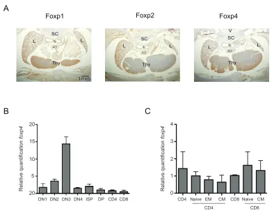
Foxp4 is expressed in T lymphocytes
foxp4 transcript expression was detected by northern blot in multiple tissues,
including the spleen (Teufel et al., 2003). We extended these studies to assess Foxp4
expression in developing thymi. In collaboration with Drs. Ed Morrisey and Shanru Li,
protein was detected by immunohistochemistry in thymic tissues of fetal E18.5 day-old
mice. Immunostaining of transverse sections with Foxp4 polysera revealed Foxp4
protein expression in the thymus, particularly in cortical regions (Figure 2.1A, right
panel). Foxp4 expression in the thymus was compared to other FoxP family members.
Staining with Foxp1-specific polysera of embryonic thymi appeared a darker brown
color, denoting Foxp1 protein expression (Figure 2.1A, left panel). In contrast, Foxp2
staining of the thymi exhibited reduced expression, which correlates with previous
reports that Foxp2 is not expressed in the hematopoietic lineage (Figure 2.1A center
panel) (Shu et al., 2001). These data are consistent with published findings, and
demonstrate Foxp4 protein expression in the thymus.
Next, we examined Foxp4 expression in developing T lymphocyte populations
using highly purified T lymphocyte subsets isolated from wild-type thymi (Figure 2.1B).
Steady-state Foxp4 mRNA levels were assessed using a TaqMan-based real-time
polymerase chain reaction (PCR) PCR assay. Thymocytes were sorted from 8-week-old
C57Bl/6 wild-type mice using flow cytometry, based on expression of CD4 and CD8.
Lineage markers were used to exclude CD11c, CD11b, and F4/80 expressing non-T
cells. CD4-CD8- double negative (DN) thymocytes were further separated based on
CD25 and c-Kit expression. In all thymocytes populations, Foxp4 mRNA was expressed
-(DN3) subset. Following β-selection, we observed that Foxp4 expression declined but
remained constitutively expressed in both CD4 and CD8 single positive T cell subsets.
Previously, studies have demonstrated Foxp4 expression in peripheral lymphoid
tissues. However, these studies did not determine the specific cell types expressing
Foxp4. Real-time PCR was used to determine relative Foxp4 levels in various sorted T
cell populations. Foxp4 mRNA was expressed in both peripheral mature CD4 and CD8 T
cells (Figure 2.1C). Based on CD44 and CD62L expression, CD4 and CD8 T cells can
be further divided into CD44loCD62Lhi naïve, CD44hiCD62Llo effector memory (EM), and
CD44hiCD62Lhi central memory (CM) populations. While Foxp4 was expressed in sorted
sub-populations, no statistically significant differences in Foxp4 levels were detected in
naive, EM, or CM T cells of both the CD4 and CD8 T lineage. Therefore, Foxp4
expression is dynamically regulated throughout thymic development but decreased in
Figure 2.1. Foxp4 is expressed in lymphoid tissues and T cell subsets. A) Expression of Foxp1, Foxp2 and Foxp4 in wild-type embryonic thymi by immunostaining with FoxP member antisera. 10µm cryosections of E18.5 mice were mounted and fixed with formaldehyde and H&E stained. Frames are magnified 40X. Representative of two independent experiments. L=lung, SC=spinal cord, AO=aortic arch, Tr=trachea, Thy=thymus. B) Thymocytes from C57BL/6 mice were stained with a lineage cocktail, CD4, CD8, CD44 and CD25, and HSA. Stained cells were collected using flow cytometric cell sorting. Steady state mRNA levels of Foxp4 and ß-actin were determined by TaqMan PCR. Foxp4 levels were normalized to ß-actin. Double positive thymocytes were given a relative value of 1. All isolated populations were lineage negative. DN1: CD4-CD8-CD25-CD44+; DN2: CD4-CD8-CD25+CD44+; DN3:CD4-CD8-CD25+CD44-; DN4: CD4-CD8-CD25-CD44-; ISP: CD4-CD8+HSA+CD5-; DP: CD4+CD8+; CD4: CD4+CD8-; CD8: CD4-CD8+HSA-CD5+. C) C57BL/6 splenocytes were stained with CD4, CD8, CD44, CD62L and CD25 and collected by flow cytometry. Relative levels of Foxp4 were determined as in (A). Total CD8+ were given a normalized value of 1. Naive: CD44loCD62Lhi, EM: CD44hiCD62Llo; CM: CD44hiCD62Lhi. Error bars represent standard deviation among triplicate wells from TacMan assay. Data is representative of two independent experiments.
DN1 DN2 DN3 DN4 ISP DP CD4CD8 20
15
10
5
20
4
3
2
1
0
CD4 Naive EM CM CD8 Naive CM
CD4 CD8
Relative quantification
foxp4
Relative quantification
foxp4
B C
Foxp1 Foxp2 Foxp4
mm
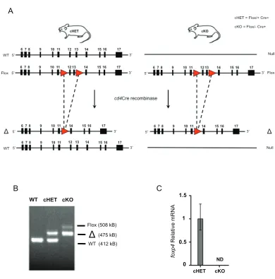
Foxp4 can be efficiently deleted in thymocytes
To investigate the role of Foxp4 in T lymphocyte development and function, we
sought to generate Foxp4-deficient thymocytes. Germline deletion of Foxp4 results in
embryonic lethality at day E12.5 due to cardiac defects (Li et al., 2004b), necessitating a
conditional deletion approach. To study the effect of Foxp4 deletion specifically in the T
lineage, we used mice with LoxP/LoxP sites flanking exons 12-13 of foxp4 that encode
the forkhead domain (Figure 2.1A). Foxp4FLOX mice were then crossed with CD4Cre mice
in which Cre recombinase expression is under transcriptional control of the cd4 promoter
(Lee et al., 2001). Under cd4 promoter regulation, Cre is first expressed at the transition
from the double negative to double positive stage in the thymus. Gene deletion is
observed in DP, mature SP thymocytes, and peripheral T cells. To increase likelihood of
generating Foxp4 null cells, we used mice carrying one germline null allele and one
floxed allele (Foxp4Flox/null CD4Cre+, henceforth referred to as cKO). Throughout this
study, cKO animals were compared to conditional heterozygous (Foxp4Flox/+CD4Cre+,
henceforth referred to as cHET) littermates that expressed one wild-type allele and one
floxed allele. Thus, cHET T cells express one allele of Foxp4, while cKO mice lack both
genomic Foxp4 alleles following Cre-mediated deletion.
To confirm efficient deletion of Foxp4, we first isolated total thymocytes (>95%
DP and SP cells) and evaluated deletion in genomic DNA using a PCR-based assay.
Deletion of Foxp4 was verified by detection of a wild-type Foxp4 PCR product at 412 kb,
a Foxp4FLOX product including the inserted flanking LoxP sites at 508 kb, or the deleted
Foxp4Δ product at 475kb in total thymocytes and purified peripheral CD4 T cells from
splenic CD4 T cells the level of Foxp4 message was undetectable, as determined by
real-time PCR (Figure 2.2B). Taken together, CD4Cre-mediated deletion results in
Foxp4 deficient thymocytes and peripheral T cells, and allows for further investigation of
the role of Foxp4 in the T lineage.
Figure 2.2. Generation of Foxp4 cKO mice. A) Schematic of conditional deletion of Foxp4 in cHET and cKO mice. LoxP sites (red triangles) were cloned into genomic DNA flanking exons 12 and 13 sequences, to generate the Foxp4FLOX mutant allele. cHET mice express one Foxp4FLOX allele and one wild-type Foxp4 allele. cKO mice were bred
to express one Foxp4FLOX allele and a Foxp4 mutant allele. Cre recombinase under the
WT (412 kB) (475 kB) Flox (508 kB)
WT cHET cKO
B C
1.5
1
0.5
0
cHET cKO
foxp4
Relative mRN
A
cd4 promoter results in excised DNA between LoxP sites. B) DNA isolated from cHET and cKO thymocytes was amplified using primers to detect wild-type, floxed, and deleted allelic sequences. Band sizes and identities are indicated. Representative of ten experiments. C) RNA isolated from WT (C57BL/6) and cKO thymocytes was assessed for Foxp4 by real time PCR. ND=not detectable. Representative of three experiments.
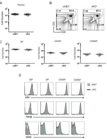
Foxp4 is not required for T cell development and peripheral homeostasis
To determine if thymocyte development was normal in the absence of Foxp4, the
phenotype and cellularity of thymic populations from cHET and cKO animals were
compared. From initial observation, the size and cellularity of Foxp4 cKO thymi was
comparable to cHET and Cre control thymi (Figure 2.3A, and data not shown).
Throughout these studies, there were no changes observed between cHET and Cre
control mice, suggesting loss of one allele of Foxp4 had no impact on T cell
development. The relative frequency of DP, and SP populations, and absolute numbers
were similar between both cohorts of mice (Figure 2.3B, C). ISP, DP and SP populations
were stained for phenotypic expression of markers of development (Figure 2.3D). Loss
of Foxp4 resulted in comparable expression of CD5 or the TCRβ chain between cHET
and cKO mice. CD44 levels on Foxp4 cKO cells were also evaluated, as Foxp1
conditionally deficient CD4SP and CD8SP T cells acquire a CD44hi phenotype. In Foxp4
cKO mice, however, CD4SP and CD8SP cells maintain low levels of CD44 expression,
providing the first evidence that Foxp1 and Foxp4 have non-overlapping roles in
thymocyte development. Furthermore, these data suggest that deletion of Foxp4 during
Figure 2.3. Deletion of Foxp4 at the DP stage does not alter thymocyte development. A) Total cellularity was assessed in both cHET and cKO thymi from four-week-old littermates. Each point represents a single mouse. Mean and standard deviation are indicated. B) Thymocytes were stained for CD4 and CD8 and analyzed by polychromatic flow. Contour plots shown are previously gated on live, singlet-gated cells, negative for myeloid lineage markers (CD11b, CD11C, CD19, B220). Gated frequencies are indicated. Representative of 12 cHET and 12 cKO mice. C) Absolute numbers of thymocyte populations were determined. Each point represents an individual mouse. D) Thymocytes were stained with CD4, CD8, CD5 and HSA. Cells are gated on
A Thymus cHET CD4 CD8 cKO 7.77 2.96 4.25 83.8 7.18 2.44 4.2 85.3 B C 108 106 104 102
cHET cKO
Cell Number
CD8SP CD4SP
cHET cKO
Cell Number Cell Number
D
CD5
DP CD8SP
ISP CD4SP
cHET
cKO
CD44
0102 103 104 105 0102 103 104 105 0102 103 104 105 0102 103 104 105 0102 103 104 105 0102 103 104 105 0102 103 104 105 0102 103 104 105
0102 103 104 105 0102 103 104 105 0102 103 104 105 0102 103 104 105 TCR
cHET cKO cHET cKO
populations as indicated at the top of each column and assessed for CD5, TCRβ, and CD44 expression. Solid histograms are from cHET and bold lines are from cKO mice. Representative of 12 cHET and 12 cKO mice.
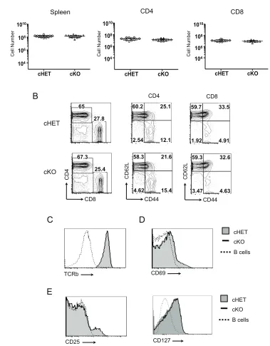
Because thymocytes appear to develop normally, we were able to assess the
effect of Foxp4 deficiency in peripheral T cell populations. Splenic cellularity in cKO mice
was similar to that seen in cHET mice (Figure 2.4A, left panel). Similarly, the frequency
and absolute number of cKO CD4 and CD8 T cells was comparable to controls (Figure
2.4A, center and right panels, Figure 2.4B). As observed in developing Foxp4 cKO
thymocytes, the majority of Foxp4 cKO splenic T cells remain phenotypically naïve,
expressing low levels of CD44 and high levels of CD62L (Figure 2.4B). Similar
frequencies of CD44hi endogenous memory T cells were present in Foxp4 cHET and
cKO mice. This is in contrast to Foxp1 cKO T cells, which are uniformly CD44hi. We
assessed other markers of activation for any indication of aberrant T cell development,
but we found comparable expression of CD69 and the TCRβ chain on splenic CD4 and
CD8 T cells (Figure 2.4C, D). Similarly, Foxp4 deletion does not affect levels of the IL-2
or IL-7 receptor on either CD4 or CD8 T cells (Figure 2.4E), suggesting Foxp4 does not
regulate mechanisms of T cell commitment or homeostasis. Foxp4 cKO mice were
observed for more than 12 months, and did not develop any sign of abnormal immune
activation or overt pathology (data not shown). Thus, Foxp4 deficiency does not result in
abnormal activation or lead to overt autoimmune pathology, allowing for interpretation of