University of Pennsylvania
ScholarlyCommons
Publicly Accessible Penn Dissertations
Fall 12-22-2010
Development as a new Paradigm for Improved
Tendon Healing: A Characterization of Neonatal
Tendon Development, Healing During
Development and An investigation into Differential
Parameters During Accelerated Healing
Heather L. Ansorge
University of Pennsylvania, [email protected]
Follow this and additional works at:
http://repository.upenn.edu/edissertations
Part of the
Biomedical Engineering and Bioengineering Commons
, and the
Developmental
Biology Commons
Recommended Citation
Ansorge, Heather L., "Development as a new Paradigm for Improved Tendon Healing: A Characterization of Neonatal Tendon Development, Healing During Development and An investigation into Differential Parameters During Accelerated Healing" (2010).
Development as a new Paradigm for Improved Tendon Healing: A
Characterization of Neonatal Tendon Development, Healing During
Development and An investigation into Differential Parameters During
Accelerated Healing
Abstract
During neonatal development, tendons undergo a well orchestrated process whereby extensive structural and compositional changes occur in synchrony to produce a normal tissue. Conversely, during the repair response to injury, structural and compositional changes occur, but in this case, a mechanically inferior tendon is produced. An injured tendon that is mechanically inferior has compromised function and reruptures after treatment are commonly observed clinically. As a result, the process of development has been postulated as a potential paradigm through which improved adult tissue healing may occur. First, the role of CD44 in healing was examined through a patellar tendon injury in a CD44 knockout mouse. A beneficial environment post injury was shown to lead to improved function in the CD44 knockout tendons. Second, type XIV collagen was examined in tendons during development and maturity through Col14a1-/-, Col14+/- and Col14+/+ flexor digitorum longus tendons. A lack of type XIV collagen during development was associated with reduced mechanical parameters but had no effect in mature tendons. Third, compositional, structural and mechanical properties were quantitatively characterized during multiple stages of neonatal Achilles tendon development in a mouse. Mechanical parameters, collagen content, fibril diameter average and standard deviation all increased with age. Biglycan expression decreased with age while decorin and angular deviation did not change. Fourth, compositional, structural and mechanical parameters were quantified after injury during two different stages of development, early and late. An accelerated healing process was demonstrated during early development over late development. Lastly, regression analysis was used to determine which compositional and structural parameters predicted mechanical parameters during early and late development and healing. Applying these differentially regulated parameters to new treatments may help improve healing in adult tendons. Importantly, this dissertation was conducted in a mouse model which is allows for future mechanistic studies due to the availability of genetically modified mice and commercially available assays. Over all, this dissertation introduces development as a new paradigm through which to study improved function during healing and potential new therapeutic treatments.
Degree Type
Dissertation
Degree Name
Doctor of Philosophy (PhD)
Graduate Group
Bioengineering
First Advisor
Keywords
Tendon Development Healing Neonatal Injury Mechanics Composition Structure Mouse Model
Subject Categories
DEVELOPMENT AS A NEW PARADIGM FOR IMPROVED TENDON HEALING: A CHARACTERIZATION OF NEONATAL TENDON DEVELOPMENT, HEALING DURING DEVELOPMENT AND AN INVESTIGATION INTO DIFFERENTIAL PARAMETERS DURING
ACCELERATED HEALING
Heather Lynn Ansorge
A Dissertation in Bioengineering
Presented to the Faculties of the University of Pennsylvania
In Partial Fulfillment of the Requirements for the Degree of Doctor of Philosophy
2010
______________________________ Supervisor of Dissertation Dr. Louis J. Soslowsky
______________________________ Graduate Group Chairperson Dr. Susan S. Margulies
Dissertation Committee Dr. Beth A. Winkelstein (chair) Dr. Pedro K. Beredjiklian Dr. David E. Birk
DEVELOPMENT AS A NEW PARADIGM FOR IMPROVED TENDON HEALING: A CHARACTERIZATION OF NEONATAL TENDON DEVELOPMENT, HEALING DURING DEVELOPMENT AND AN INVESTIGATION INTO DIFFERENTIAL PARAMETERS DURING
ACCELERATED HEALING
COPYRIGHT 2010
iii
ACKNOWLEDGEMENTS
I sincerely thank my committee members for their guidance and support through out my
entire graduate career. First and foremost, my thesis advisor, Dr. Soslowsky, for being a great
mentor both in and out of the laboratory. Thank you for guiding me through my education,
teaching me how to think and being understanding. Dr. Winkelstein for your insightful
suggestions on my research and your mentorship over the past 6 years. Dr. Beredjiklian for all
your clinical knowledge and for never saying never when I approached you with an “almost”
impossibly small surgical procedure. Dr. Mauck for all your help when I was doing cell culture
and for being my biology expert in McKay. Dr. Birk, for your enormous help with the TEM in my
experiments, for your amazing support throughout numerous collaborative projects and for letting
me spend many, many hours in your laboratory in Florida.
Many of my projects have been collaboration with other laboratories. In particular, I’d like
to thank the Birk lab for all of their tremendous effort, time and company. Sheila Adams for
receiving, processing, cutting, staining and imaging too many TEM samples to count. In addition,
for your hospitality while I was visiting the Birk lab in Florida. Dr. Mienaltowski for all your efforts
trying to correlate mRNA and protein expression. Dr. Sun for all your guidance on proteoglycan
extractions and mRNA analysis. I’d also like to thank Dr. Nicoll for letting me rotate in your
laboratory for more than a semester while working on the cell/scaffold project and for your
continued mentorship. Dr. Hallie Brink for showing me the ropes in Dr. Nicoll’s lab, working side
by side and for the friendship that grew during that time and still continues. I would like to thank
Dr. Jawad for your guidance and insight on the regression analysis.
I’d also like to thank all the members of the Soslowsky laboratory (past and present), Dr.
Michele Favata, Dr. Stephanie Perry, Dr. Nelly Andarawis-Puri, Dr. Spencer Lake, Dr. Cathy Peltz,
LeAnn Dourte, Kris Miller, Katie Reuther, Spence Szczesny, David Beason, Joseph Sarver, Lena
Edelstein, and Michael Dishowitz. Also all the residents, each of whom has contributed to this
Zgonis, and in particular Dr. Jason Hsu for doing all the neonatal surgeries. I’d like to particularly
thank David Beason, you are the best resource on the 4th floor of McKay and you helped me out tremendously on too many projects. Dr. Elliot for all your guidance and support, and for basically
being a 6th committee member. Dr. Dodge and Paul Matuszewski for your help with the ELISAs and proteoglycan extraction. Lena Edelstein, Nick Trasolini, and Willis Zhu for your enormous
help with the animal husbandry, and collecting and analyzing data for this defense. Barb Gibson
and Fiachra Malone for answering all my silly questions and helping me ship out many packages.
Bob Caron for all your help with histology. Josef Kaplan for helping me try to accomplish an
impossible western blot. I’d like to thank all the members of McKay Laboratory, I really enjoyed
my time at McKay and that’s mostly due to the people I’ve met. Lastly, I’d like to thank all of my
friends in and out of UPenn but particularly those in the BE department and PADA. Your love
and friendship are what got me through these years and I am so lucky to have you!
Last but not least I would like to thank my entire family! You all mean the world to me
and knowing that you were there every step of the way was what kept me moving forward. Mom,
you instilled in me a love for biology at a young age and a thirst to always want to know more.
Dad, our nature walks taught me how to be adventurous in all avenues of life and to keep looking
under the next rock if the current one was empty. Katie and Lisa, you’re both much more than
sisters to me and your friendship has kept me sane many nights. Aunt Sandy, Uncle Joe, Aunt
Pat, and Uncle Keith, you are the most amazing aunts and uncles and I’m still floored by your
support. And all my grandparents, I appreciate your love so much and since you’ve asked so
v
ABSTRACT
DEVELOPMENT AS A NEW PARADIGM FOR IMPROVED TENDON HEALING: A
CHARACTERIZATION OF NEONATAL TENDON DEVELOPMENT, HEALING DURING
DEVELOPMENT AND AN INVESTIGATION INTO DIFFERENTIAL PARAMETERS DURING
ACCELERATED HEALING
Heather L. Ansorge
Louis J. Soslowsky, Ph.D.
During neonatal development, tendons undergo a well orchestrated process whereby extensive
structural and compositional changes occur in synchrony to produce a normal tissue. Conversely,
during the repair response to injury, structural and compositional changes occur, but in this case,
a mechanically inferior tendon is produced. An injured tendon that is mechanically inferior has
compromised function and reruptures after treatment are commonly observed clinically. As a
result, the process of development has been postulated as a potential paradigm through which
improved adult tissue healing may occur. First, the role of CD44 in healing was examined
through a patellar tendon injury in a CD44 knockout mouse. A beneficial environment post injury
was shown to lead to improved function in the CD44 knockout tendons. Second, type XIV
collagen was examined in tendons during development and maturity through Col14a1-/-, Col14+/-
and Col14+/+ flexor digitorum longus tendons. A lack of type XIV collagen during development
was associated with reduced mechanical parameters but had no effect in mature tendons. Third,
compositional, structural and mechanical properties were quantitatively characterized during
multiple stages of neonatal Achilles tendon development in a mouse. Mechanical parameters,
collagen content, fibril diameter average and standard deviation all increased with age. Biglycan
expression decreased with age while decorin and angular deviation did not change. Fourth,
compositional, structural and mechanical parameters were quantified after injury during two
different stages of development, early and late. An accelerated healing process was
used to determine which compositional and structural parameters predicted mechanical
parameters during early and late development and healing. Applying these differentially regulated
parameters to new treatments may help improve healing in adult tendons. Importantly, this
dissertation was conducted in a mouse model which is allows for future mechanistic studies due
to the availability of genetically modified mice and commercially available assays. Over all, this
dissertation introduces development as a new paradigm through which to study improved function
vii
TABLE OF CONTENTS
CHAPTER 1: INTRODUCTION AND BACKGROUND………..………...…....………….1
Significance………..……….….1
Prevalence of tendon injuries in the US………..………..………..1
Scar formation……….………1
Healing parameters throughout development……….….………..……..………..3
Background……….………3
Tendon composition and structure...……….………...………...3
Tendon mechanics………..………4
Reparative Tendon Healing…...………6
Current Strategies for Tendon Repair………7
Regenerative Tendon Healing………...……….10
Structure-Function Relationships………..………...13
Tendon Development………..……….14
Specific Aims and Hypotheses………..………17
Chapter Overviews……….………….20
References……….……..22
CHAPTER 2: CD44 DEFICIENCY IMPROVES HEALING TENDON MECHANICS AND INCREASES MATRIX AND CYTOKINE EXPRESSION IN A MOUSE PATELLAR TENDON INJURY MODEL………..…………...………...……….29
Introduction………..……….………….29
Significance………..…………..………...……….29
CD44 and Hyaluronic Acid……….………29
Objective and Hypotheses ……….………..……….31
Methods……….……….……31
Specimens...……….……….31
Histological Analyses…...………..……33
Gene Expression……….…33
Statistical Analyses………..….35
Results………..…35
Biomechanical Analyses………..………35
Histological Analyses…...………..…36
Gene Expression……….…36
Discussion………....38
Interpretation of Results………..………38
Limitations…...……….…42
Summary………42
References………...44
CHAPTER 3: TEMPORAL AND TISSUE SPECIFIC DYFUNCTION IN THE ABSENCE OF TYPEXIV COLLAGEN IN A MOUSE MODEL.………...…………48
Introduction………..……….………….…48
Tendon Development and Fibril Associated Macromolecules ………...……..48
Type XIV Collagen………….……….………48
Type XIV Collagen in Neonatal Tendon Development……...……….50
Type XIV Collagen Knockout Mouse………..……...……….51
Objective and Hypotheses………...………...……….56
Methods……….……….……56
Mechanical Analysis of Skin.….………..56
Mechanical Analysis of Neonatal Developmental FLD Tendon ………56
Mechanical Analysis of Mature FDL Tendon………..……58
Statistical Analyses………..….58
Results………..…58
ix
Discussion………....59
Interpretation of Results………..………60
Limitations…...……….63
Summary………64
References………...65
CHAPTER 4: MECHANICAL, COMPOSITIONAL AND STRUCTURAL PROPERTIES OF THE NEONATAL MOUSE ACHILLES TENDON...………...……….69
Introduction………..……….……….…69
Significance……….………...…….69
Tendon Development….……….………69
Objective and Hypothesis……….……...………71
Methods……….……….……71
Animals……….….………..71
Mechanical Analysis ……….………72
Hydroxyproline Analysis………..………..……74
Gene Expression……….…75
Histological Analyses…...………..…75
Transmission Electron Microscopy………..…76
Fibril Diameter Distribution………..…76
mRNA Comparison to Protein Expression………..…77
Statistical Analyses………..….77
Results………..…77
Animals……….….………..77
Mechanical Properties……….………79
Collagen Content………..………..……80
Proteoglycan mRNA Expression.……….…80
Comparison of mRNA Expression to Protein Expression………..…81
Fibril Diameter Parameters………..…81
Discussion………....82
Interpretation of Results………..………82
Limitations…...……….…88
Summary………90
References………...91
CHAPTER 5: RECPITULATION OF THE ACHILLES TENDON MECHANAICAL STRENGTH DURIG NEONATAL DEVELOPMENT: A STUDY OF DIFFERENTIAL HEALING DURING TWO STAGES OF DEVELOPMENT IN A MOUSE MODEL………...………….95
Introduction………..……….………….…95
Significance……….………...…….95
Regenerative Healing….……….………96
Tendon Development and Healing……….………96
Objective and Hypothesis……….……...………97
Methods……….……….……98
Animals……….….………..98
Surgical Model..…….……….………98
Mechanical Analysis ……….………99
Hydroxyproline Analysis………..………..…101
Gene Expression……….102
Histological Analysis…...………..103
Transmission Electron Microscopy………..…104
Fibril Diameter Distribution……….105
Statistical Analyses………..105
Results………..106
Pilot Studies……….106
xi
Pilot to Determine “Ruptured” Parameters……….………..107
Animals……….….……….108
Mechanical Properties……….………109
Collagen Content………..………..……110
Proteoglycan mRNA Expression.……….111
Histological Parameters………...………..111
Fibril Diameter Parameters………..112
Discussion………..113
Interpretation of Results………..………113
Limitations…...………121
Summary………126
References………..127
CHAPTER 6: INVESTIGATING STRUCTURE-FUNCTION RELATIONSHIPS IN NEONATAL ACHILLES TENDON DEVELOPMENT AND HEALING IN A MOUSE MODEL USING MULTIPLE REGRESSION..……….………...………132
Introduction………..…..………132
Significance……….………...………132
Correlating Compositional and Structural Parameters to Mechanics………...…….132
Multiple Regression Models ……….………134
Methods……….……….…135
Data Collections………..….………135
Statistical Models………136
General Linear Model..……….………..……137
Model Refinement…..……….………..…138
Final Multiple Regression Models………..….140
Results………..141
Correlations Between the Independent Parameters………..………141
Whole Model Analysis – Backward Stepwise Linear Regression Model………...142
Grouped Analysis – Backward Stepwise Linear Regression Model………...143
Discussion………...143
Interpretation of Results………..………143
Limitations…...………149
Summary………150
References………...152
CHAPTER 7: SUMMARY, LIMITATIONS AND FUTURE DIRECTIONS.………...………155
Overall Conclusions………...155
Background and Significance……….………...…155
CD44 Deficiency Improves Healing Tendon Mechanics and Increases Matrix and Cytokine Expression in a Mouse Patellar Tendon Injury Model………156
Temporal and Tissue Specific Dysfunction in the Absence of Type XIV Collagen in a Mouse Model………...157
Mechanical, Compositional and Structural Properties of the Neonatal Mouse Achilles Tendon………...157
Recapitulation of the Achilles Tendon Mechanical Strength during Neonatal Development: A Study of Differential Healing During Two Stages of Development in a Mouse Model………158
Investigating Structure-Function Relationships in Achilles Tendon Neonatal Development and Healing during Neonatal Development in a Mouse Model using Multiple Regression Models………159
Limitations and Future Directions………...160
CD44 in Adult Healing………160
The Role of Type XIV Collagen in Development………163
Neonatal Tendon Development and Developmental Healing ………166
xiii
References……..………175
LIST OF FIGURES
Figure 1.1: Example of a normal tendon with aligned fibers stained with hemotoxylin and eosin..3
Figure 1.2: Hierarchical structure of tendons from collagen molecule through the fascicle. Inset demonstrates a hypothesized interaction of proteoglycans with the collagen fibrils………3
Figure 1.3: Achilles tendon shown connecting the calcaneous to the gastrocnemius muscle…….4
Figure 1.4: Typical stress vs strain curve from a constant ramp to failure tensile test of tendon. The toe, linear, yield and failure regions are indicated………5
Figure 1.5: Representative schematic for a tendon during a stress-relaxation test………6
Figure 1.6:Maximum stress after injury with and without IL-10 vector……….9
Figure 1.7: H&E stained tendon: 1) adult normal, 2) adult wounded showing significant fiber disruption, 3) fetal normal, 4) fetal wounded with completely reconstituted collagen
architecture………11
Figure 1.8:CD44 increased markedly in the wounded adult tendons but only slightly or not at all in the fetal (black indicates wound, brown represents immunostaining). (A) Adult, original
magnification 100; (B) 1 week fetal, original magnification 200 ………12
Figure 1.9: Linear regression plots for strength versus collagen cross sectional area fraction. Solid line is data from adult mice only (R2=0.61), dashed line includes data from Robinson et al of 3wk old mice (R2=0.69) ……….…13
Figure 1.10: Tendon fibrillogenesis. (A) Molecular assembly of type I collagen generates the fibril intermediate. (B) In the linear growth step, the intermediates in (A) grow by end-to-end growth to
generate longer fibrils. (C) In the lateral growth step, there is a lateral association and growth of
xv
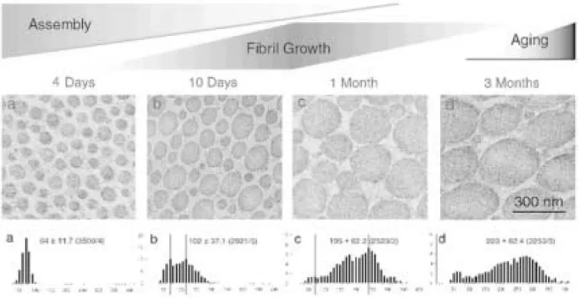
Figure 1.11: Collagen fibril structure during development in mouse tendons. Transmission electron micrographs of transverse sections from mouse flexor tendons and the diameter
distributions at each developmental stage from normal mice (a-d). Bar 300nm ………15
Figure 1.12: Mechanical changes during chick development from prehatchling day 43 to 2 day old hatchling. These stress strain curves demonstrate an increase in mechanical strength during
development………..16
Figu re 2. 1: Sc hem atic of t he c ellul ar rec eptor CD44 ( P hot o: htt p:/ / www. c anc er -therapy.org/CT2A/html/25.%20Coradini%20et%20al,%20201-21%20copy.html)...29
Figure 2.2: Photos that depict the partial transection created in the mouse patellar tendon. A plastic coated backing is placed underneath the tendon to provide support (Figure 2.1.1) and a
circular biopsy punch is used to create a full thickness partial transection (Figure 2.1.2) The
backing is removed, leaving a distinct and reproducible injury (Figure 2.1.3 and 2.1.4)…………31
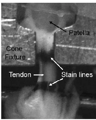
Figure 2.3: Example of a tendon in the custom designed cone fixture and mounted in the Instron………32
Figure 2.4: Cross-sectional area compared across genotype. Cross-sectional area was significantly reduced in knockouts (KO) compared to wild types (WT) at both 3 and 6 weeks post
injury………36
Figure 2.5: Maximum stress and strain energy density were significantly increased in knockouts (KO) compared to wild-type (WT) at both 3 and 6 weeks post injury. Similarly, knockout animals
demonstrated a trend toward greater failure strain compared to wild-type. No significant changes
were seen across genotype in modulus. *:p < 0.05; #:p < 0.1……...37
Figure 2.6: Appreciable differences were seen in angular deviation in the knockout (KO) tendons across time. While the wild-type (WT) tendons remained disorganized, the KO tendons
Figure 2.7: Representative histological images for each genotype and time point. Injury is clear in both genotype at 3 and 6 weeks post injury; however, in the knockout tendons the fibers are
more aligned……….………39
Figure 2.8: Gene expression in a patellar tendon injury in wildtype (WT) and CD44 knockout (KO) represented as relative quantity fold changes (2-∆∆CT) with error bars representing (2--∆∆CT-s), where s is
the standard deviation of the DDCT value. All postinjury time points are normalized to uninjured
tendons (day 0), which is represented by a value of 1………40
Figure 3.1: Model of type XIV collagen in fibril assembly. (a): Type XIV collagen has multiple functional domains. These domains interact with different components of the extracellular matrix
(ECM). (b): Type XIV is localized periodically to the surface of the striated collagen fibrils. (c):
Fibrillogenesis can be divided into distinct phases beginning with the formation of short, immature
fibril segments, then a period of linear growth, followed by a rapid lateral growth characterized by
an increase in fibril diameter and length (Young, 2000). ………49
Figure 3.2: Type XIV collagen expression in normal neonatal developing mouse tendons. Type XIV collagen mRNA expression (between P4-P90) was analyzed by real time quantitative
RT-PCR (a) and semi-quantitative RT-RT-PCR (b). (c) Semi-quantitative immuno-blotting analysis. All
results demonstrate a clear decrease in type XIV collagen in neonatal development……….50
Figure 3.3: Type XIV Collagen expression patterns in mouse tendon neonatal development. Localization of type XIV collagen or type I collagen by immunofluorescence microscopy with
nuclear localization using DAPI (blue). There was a dramatic decrease in reactivity against type
XIV collagen between P10 and P30……….52
Figure 3.4: Absence of type XIV collagen in Col14a1 null mouse skin. (a). Immunofluorecence staining of Col14a1 knockout mouse skin using anti-type XIV collagen antibody was completely
xvii
Western blotting of protein samples extracted from mouse skin showed complete absence of type
XIV collagen in Col14a1 null mice and reduced synthesis in heterozygous mice………53
Figure 3.5: Altered collagen fibril formation in Col14a1-/- tendons. Transmission electron micrographs of FDL tendon at P4 and P60 from (a) Col14a1+/+ and (b) Col14a1-/- mice. In the
Col14a1-/- tendon, there are fewer smaller fibrils and more fibrils of larger diameter as compared
to the wild type control. Bar = 100 nm. (c)Histograms representing fibril diameter measurements
of tendons at P4 and P60………54
Figure 3.6: Proteoglycan expression in the FDLs of Col14a1-/- tendon. (a) No significant difference was found in expression at either P4 or P30 for type XII collagen, biglycan, decorin,
lumican, or fibromodulin. ………...55
Figure 3.7: Dumbbell stamped skin sample. Skin samples were dumbbell stamped for mechanical testing. Stain lines were placed at 10mm for grip to grip gauge length placement and
5mm apart for optical strain measures………56
Figure 3.8: Custom designed fixture for P7 FDL mechanical testing. The tendon (as shown with bold arrow) is secured in grips. The holding fixture, which is attached to the clamps through pins,
ensures that the tendon is in a relaxed position until the fixture is secured in the Instron………57
Figure 3.9: Skin mechanics in P60 wildtype and null mice. No significant differences were found in the cross-sectional area of the P60 skin. The maximum stress was significantly decreased in
the skin of the col14 a1 null mice compared to that of wild-type. Additionally, the modulus
showed a trend toward lower modulus when compared to wild type mice. *:p<0.05; #: p<0.1…58
Figure 3.10: FDL cross-sectional area at P7 and P60. No significant differences were observed in cross-sectional area at P7 or P60 between wildtype, heterozygotes or null FDLs at either stage
of development………59
tendons. In addition, FDLs from the developing deficient mice showed significantly decreased
maximum load and a trend towards decreased maximum stress and stiffness when compared to
the heterozygote mice. FDLs from heterozygote mice also had significantly decreased stiffness
when compared to wild type mice. (b): In contrast, the mature tendons demonstrated no
significant differences between the control, deficient or heterozygote tendons. *:p<0.05; #:
p<0.1………..60
Figure 3.12: Type XIV collagen expression after tendon injury. The panel on the left is a normal P90 tendon and the one on the right is a tendon 1 week post injury, both were stained with
anti-col14a1 antibody. No type XIV is present in the uninjured tendon whereas protein expression is
v i si bl e one we ek po st i nj ur y … … …… … … … …… … … …… … … … …… … … … 62
Figure 4.1:A: Custom designed boat with grips and an AT tendon. B: Custom designed grip-holder, indicated by the arrow, with a tendon loaded between the grips, indicated with the circle.
The grip-holder ensured that the tendon remained unloaded during manipulation and mounting
prior to testing……….….73
Figure 4.2: Example of the testing protocol for neonatal AT. Protocol includes a preload, precycle, stress relaxation and constant ramp to failure. ……….73
Figure 4.3: Growth curve of the body weights of C57BL/6 mice during neonatal development…78
Figure 4.4: Typical 4 days old (A) and 28 days old (B) AT immediately after dissection. Obvious
size increases occur over the period of time investigated in this study………79
Figure 4.5: Increase in cross-sectional area throughout neonatal development; increases
occurred at 21 and 28 days old. -: p<0.05………81
Figure 4.6: Representative load vs displacement curves from the constant ramp to failure for
xix
Figure 4.7: Linear stiffness and modulus significantly increased with age in a neonatal
developing AT. – : p< 0.05……….83
Figure 4.8: Relative expression of biglycan and decorin relative to GAPDH. Biglycan decreased
significantly throughout development while no significant differences were seen in decorin mRNA
expression. 1-way ANOVA was performed on dCT values, while 2-Ct is represented graphically. – : p< 0.05………84
Figure 4.9: Western blot and mRNA expression for 7, 14 and 28 days old pooled AT. Both sets
of data were normalized to 7 days old for easy comparison. ………84
Figure 4.10: Representative examples of H&E stained sections of the AT 4, 7, 10, 14, 21 and 28days old. Fibers were well aligned throughout development………86
Figure 4.11: Frequency of fibril diameters demonstrated increased fibril diameters and fibril
diameter distributions throughout mouse AT neonatal development………87
Figure 4.12: Representative micrographs for each age throughout neonatal development. Fibril
diameters were circular throughout neonatal development and an obvious increase in fibril
diameter size occurred throughout development………..………88
Figure 5.1: Unilateral injuries were performed on neonatal mice. A rubber backing was used for support as the biopsy punch created a partial width, full thickness injury in the Achilles tendo…99
Figure 5.2: Dumbbell stamping device. The stamp is affixed to the upper fixture and the tendon rests on the platform. Linear bearings maintained constant vertical translation of the stamping
device to ensure appropriate placement of the stamp in the neonatal tendon………100
Figure 5.3: Representative sample of a developmental Achilles tendon prepared for mechanical testing, with the ends glued between sandpaper and the stain lines in the midsubstance….101
Figure 5.5: A pilot study was conducted in 8 days old AT to ensure the tendon fibers were being cut. The top panels clearly show the mode of injury with a rubber backing placed under the
tendon and a 0.3mm biopsy punch utilized to create the injury. The panel at right clearly shows
cut fibers when the AT is viewed under polarized light……….………..106
Figure 5.6: An example of a neonatal limb with a normal AT injury (left) and a fully ruptured AT (right). The rough, “sunken in” appearance of the area where the AT should be is indicative of a
fully ruptured AT. ……….……….107
Figure 5.7: Comparison of body weights between normal development and injury during development demonstrating no detrimental effects of the surgical method on developmental
growth……….108
Figure 5.8: Representative samples of AT after gross dissection for each age and time point post injury. One unit = 1 mm………108
Figure 5.9: The cross-sectional area was increased during early development but decreased by 10 days post injury. During late developmental injury, little change was observed at 3 days injury
and an increase in cross-sectional area was seen at 10 days post injury. The horizontal line at 1
indicates no change from normal development………109
Figure 5.10: Differential responses between early and late development were observed in mechanical parameters. Specifically, a return to normal mechanics was observed in early
developmental injury (7 days old), not mechanics are still decreased in late developmental injury
(21 days old). The horizontal line at 1 indicates no change from normal development…………110
Figure 5.11: Collagen content showed no significant changes across any group. The horizontal line at 1 indicates no change from normal development………. 111
xxi
Figure 5.13: Representative histological images from each experimental group at 20x magnification. A marked increase in disorganization, rounded cell shape and cell number from 3
days to 10 days post injury in AT injured at 21 days can be seen………113
Figure 5.14: Median and interquartile range of histological parameters. AT injured at 21 days old show marked increase in disorganization, rounded cell shape and cell number whereas AT
injured at 7 days do not. Inj = Injured; Un = Uninjured………114
Figure 5.15: Representative micrographs for each experimental group. Fibril diameters were circular throughout developmental healing……….115
Figure 5.16: Histogram of uninjured and injured AT during neonatal development. A shift towards small diameter fibrils and decreased spread can be seen in AT injured at 10 days post
LIST OF TABLES
Table 1.1: Example of several key growth factors and cytokines involved in tendon healing……8
Table 2.1: Brief description of the growth factors and matrix components examined……….34
Table 2.2: Q-PCR Primers. Forward and reverse primers and the base pair length of the amplicon (BP) used in Q-PCR………35
Table 4.1: Comparison of elastic parameters in pilot study in 7 days old neonatal tendons……74
Table 4.2: Mean +/- standard deviation and the significance for each mechanical parameter…78
Table 4.3: Mechanical, compositional and structural parameters throughout neonatal
development (mean ± stdev)………...79
Table 4.4: Average and standard deviation of fibril diameters……….88
Table 5.1: Grades and key for each non-paremeteric parameter measured by 3 blinded grader...103
xxiii
LIST OF EQUATIONS
Equation 6.1: General Linear Model………137
Chapter 1: Introduction
1.1 Significance
1.1.1 Prevalence of Tendon Injuries in the US
Tendon injuries affect a large number of individuals and account for enormous associated
costs [1, 2]. These injuries account for 45% of the almost 33 million musculoskeletal injuries that
occur in the United States each year[3]. The work place is a large source of these injuries. In
1996, approximately 44% of all injuries and illness that resulted in days away from work were
sprains, strains and tears [4]. However, soft tissue injuries are also extremely common outside
the work place. Between the years 1991 and 1998, the number of sports related injuries in
people 35-54 years of age increased 33% [5]. It was estimated that from the approximately 1
million sports related injuries in 1998, the cost to the nation was more than $18.7 billion[5]. The
number of tendon injuries will continue to increase in our aging and active population[6]. Tendon
injuries that are especially common, requiring surgical repair, include the shoulder's rotator cuff
tendons (51,000 per year), the Achilles tendon (44,000 per year), and the patellar tendon (42,000
per year) [7] [8]. Tendon injuries are of particular concern because they heal slowly and rarely
regain normal function. Surgery [9], rehabilitation [10], drugs [11] and tendon grafts [7] have all
been employed to improve healing. However, despite surgical advances, adhesion formation,
reruptures and decreased function are still common problems.
1.1.2 Scar Formation
Adult tendons heal through a reparative process [12, 13] and undergo a set of
coordinated responses that include inflammation, extracellular matrix production and remodeling
of the tissue [14] . During these processes, cellular and extracellular matrix components are
quickly deposited by fibroblasts to fill the defect and restore function; however, the quality and the
structure of the scar tissue are both altered from native uninjured tendon. While in some cases
the tendon through the sheath. Reduced quality of tissue results in tendon reruptures and is of
prominent concern. Therefore, it is imperative that new strategies are developed to quickly return
tendon to normal function post-injury.
1.1.3 Healing Parameters throughout Development
Unlike adult reparative healing, fetal tissue is well known to heal through a regenerative
process [15]. While first demonstrated in skin, fetal healing also occurs in tendon [16, 17]. In
general, fetal wound healing occurs at a fast rate and without scar formation [18]. The
mechanism behind fetal healing is still unknown, but it is believed that the regenerative response
is a property of the tissue and not the environment [17]. Some candidates with differential
expression or roles during fetal healing versus adult healing include the presence or absence of
cytokines, relative amounts and organization of extracellular matrix components and mechanical
stresses on the tissue. While fetal healing is an excellent model for scarless healing, it is possible
extensive differences, including load and inflammation, between fetal and adult healing may have
prevented major breakthroughs in improving adult healing. It has been hypothesized that healing
parameters change from the fetal stage, through development and into adulthood. For example,
neonatal developmental tendons may heal through scar formation but regain normal structure
and function faster than adult tendons. Few studies have been able to test this hypothesis due to
the small size and fragile nature of developing tendons. One study, on the healing of subfailure
ligament injury in immature and mature rat, showed that a 21 day old rat regained 95% of its
strength by 7 days post injury whereas an 8 month old rat only regained 81% at 14 weeks [19].
Understanding which parameters contribute most to improved healing will allow the identification
of key factors to examine when developing new therapies for adult healing. The hope of this
dissertation is to develop new therapeutic strategies to improve the outcome of tendon injuries
through basic science investigations into the mechanisms involved in development and injury at
Figure 1.1: Example of a normal tendon with aligned fibers stained with hemotoxylin and eosin.
Figure 1.2: Hierarchical
structure of tendons from collagen
molecule through the fascicle. Inset demonstrates a hypothesized interaction of proteoglycans with the collagen fibrils (Derwin, 1998). 1.2 Background
1.2.1 Tendon Composition and Structure
Tendon is a highly organized soft
connective tissue composed mainly of parallel
collagen fiber bundles surrounded by extracellular
matrix and tendon fibroblasts (Figure 1.1). The
fiber bundle’s main constituent is type I collagen
whereas the matrix that surrounds the fibers is
composed mainly of water, minor collagens,
proteoglycans, and glycosaminoglycans (GAGs).
Water represents 60-80% of the wet weight of tendon while type I collagen represents
approximately 65-80% of the dry weight. Proteoglycans, such as decorin and biglycan, are
approximately 1-5% of the dry weight [20, 21]. Minor collagens, such as type XII and type XIV
collagens, are present in varying, yet small, amounts throughout tendon development and
Figure 1.3: Achilles tendon shown connecting the calcaneous to the gastrocnemius muscle. (Photo: http://www.eorthopod.com)
Achilles Tendon
Calcaneous Gastocnemius Muscle
Achilles Tendon
Calcaneous Gastocnemius Muscle
The development of tendon’s hierarchical structure is a complex process consisting of
fibrillogenesis and higher order organization. Fibrillogenesis alone consists of three distinct
phases [25-27] and each step is independently regulated by specific fibril-associated
macromolecules including proteoglycans and minor collagens. First, immature type I collagen
molecules assemble extracellularly to form immature fibril intermediates. Next, during linear fibril
growth, pre-formed fibril intermediates assemble end-to-end to form longer fibril consistent with
mature, mechanically functional fibrils. Lastly, in lateral fibril growth, the fibrils associate laterally
to generate larger diameter fibrils. As tendons mature, this lateral association of fibrils creates a
wide range of fibril diameter sizes [2, 27-29]. For example, a fibril range from 40 to 400nm is
seen in a three month old mouse[27]. The fully formed fibrils are then organized into fibers which
are primarily parallel and aligned with the long axis of the tendon (Figure 1.2). These fibers,
along with the spindle-shaped tendon fibroblasts, are organized into fascicles. Lastly, sheaths
called endotenon bind fascicles together to form tendon which are in
turn surrounded by epitenon. The formation of this
hierarchy is a highly regulated, multistep process on
which the mechanical integrity and function of tendon
is dependent.
1.2.2 Tendon Mechanics
Tendon is primarily a uniaxial tissue which
transfers load from muscle to bone and stabilizes the
joint (Figure 1.3). Whereas the primary function of
collagen in tendon is to withstand tensile strength,
the non-collagenous components are thought to
serve mainly as resistance to compressive forces.
Tendons have the highest tensile strength of any connective tissue in the body, a characteristic
that is likely due to the high collagen content of the tendon fibers and their densely packed,
Toe
Region
Linear
Region
Yield and
Failure
σ
ε
Toe
Region
Linear
Region
Yield and
Failure
Ramp to Failure
Toe
Region
Linear
Region
Yield and
Failure
σ
ε
Toe
Region
Linear
Region
Yield and
Failure
Toe
Region
Linear
Region
Yield and
Failure
σ
ε
Toe
Region
Linear
Region
Yield and
Failure
Ramp to Failure
Figure 1.4: Typical stress vs strain curve from a constant ramp to failure tensile test of tendon. The toe, linear, yield and failure regions are indicated.
forces during various activities revealed loading in the Achilles tendon as high as 9 kN during
running in humans, which is up to 12.5 times body weight [30].
In vivo, tendons most often experience uniaxial tensile forces. Mechanically testing a
tendon ex vivo in similar conditions produces a nonlinear curve. Stress-strain curves can be
calculated from load-displacement uniaxial tensile
testing and the cross-sectional area of the tendon
(Figure 1.4). There are three distinct regions of
the tendon stress-strain curve produced during
tensile testing [31]. Initially the curve is
characterized by a low modulus (toe) region at low
strains. The toe region is thought to be a result of
the gradual recruitment and uncrimping of fibers
as the tendon is elongated. During this period
there is extensive deformation with little increase
in load. After the toe region, the stress-strain curve is characterized by a high modulus (linear)
region during which the tendon demonstrates linear elastic behavior. It is believed that at these
strains all of the fibers are engaged and bear load. The yield point, where the modulus gradually
decreases, is thought to be the point where collagen fibers begin to break, slide past each other
and eventually fail.
Structural properties, which do not account for differences in size and structure, can be
obtained from the load-displacement curve. Toe region properties are calculated from a bilinear
fit where the inflection point is identified. Subsequently the toe region stiffness is calculated as
the slope of the curve up to the inflection point and the load and deformation can be determined
at the inflection point. Parameters obtained from the linear region include the maximum load (the
highest load reached) and stiffness (the slope of the curve in the linear region). These structural
parameters describe the behavior of the tendon as a whole. On the other hand, material
Figure 1.5: Representative schematic for a tendon during a stress-relaxation test. modulus, can be calculated in a similar fashion as the structural properties but from the
stress-strain curve.
Tendons possess time and history
dependant properties that can be observed
through stress relaxation (Figure 1.5) and
creep mechanical tests. Stress relaxation
tests involve stretching the tendon to a
constant length (strain) and allowing the stress
to vary over time. Subsequently, percent
relaxation can be calculated from peak stress,
the (highest stress after initial strain) and equilibrium stress (the stress reached after relaxation
has occurred). A creep test involves subjecting tissue to a constant force and allowing the length
to vary with time. Viscoelastic parameters are dependant on time and rate of loading of the
tendon. Proteoglycans and water, along with the inherent viscoelasticity of the collagen itself, are
thought to be primarily responsible for viscoelastic behavior [32, 33]. Structural, material and
viscoelastic parameters characterize the functional behavior of tendons; however, one instance
when all these parameters are markedly changed from normal mature tendon values is after
tendon injury.
1.2.3 Reparative Tendon Healing
Adult tendons heal through a reparative process [12, 13] and undergo a set of
coordinated responses that include inflammation, extracellular matrix production and remodeling
of the tissue [14] . During the inflammatory phase, accumulating leukocytes perform extensive
phagocytosis of necrotic tissue and release pro-inflammatory cytokines [34]. Subsequent to
fibroblast proliferation, increased extracellular matrix production then alters the composition of the
tendon from its uninjured state. For example, biglycan levels increase and decorin levels
decrease [35, 36], the cellular receptor CD44 is highly expressed[16] and type XIV collagen is
upregulated [37]. At this stage, not only is the composition altered but also the structure. For
Stress Relaxation
t
σ
Stress Relaxation
Stress Relaxation
t
instance, the fibril diameter size distribution is narrowed and consists mainly of small diameter
fibrils [38-40]. Extracellular matrix is also laid down in a disorganized fashion, altering the parallel
alignment of the fibers[41]. During remodeling, the final stage, collagen fibrils begin to coalesce
laterally and fibers reorganize along the long axis of the tendon. However, even after remodeling,
the overall result of reparative healing is a fibrotic scar with structure, composition and
mechanical properties that are inferior to normal tendon.
Tendon properties improve over time after injury, but they rarely return to normal levels
[42, 43]. For example, the repair tissue in the patellar tendon 1.5 months post-injury only
achieved 20% of normal maximum stress and at 6 months was still only 22% of normal [43].
Similarly, 21 months after a central third defect the patellar tendon only regained 34% of the
uninjured ultimate stress [44]. Numerous studies have tried to improve the reparative healing
response through the application of cytokines and growth factors, mechanical stimulation or
deprivation and gene therapy to name a few. While some studies demonstrate improved
mechanical properties of one treatment versus another, even in the best cases, mechanical
properties remain only a fraction of native tissue. Therefore, it is imperative that new strategies
are developed that promote an efficient return to normal function post-injury.
1.2.4 Current Strategies for Tendon Repair
As previously stated, tendon injuries are still a major area of scientific study due the
inability of most tendons to fully heal on their own. Clinical management of tendon injury currently
follows two basic routes: conservative (rehabilitation, rest and pain relief) or surgical. However,
treatment choices between and within each of these disciplines can be complicated. For
example, in the biceps tendon, nonoperative treatments (rest, activity modification, ice,
non-steroidal anti-inflammatories, immobilization) are employed for older patients while younger
patients receive acute repair[45]. Many factors affect outcome including the specific tendon
injured (flexor, intrasynovial, extrasynovial, other), age of the patient and the type of injury
Cytokine Function
bFGF
Stimulates angiogensis, cellular migration and
proliferatoin
BMP-12 Stimulates cellular differentiation
IL1
β
Intiaties multiple pro-inflammatory cascades
IL-10
Anti-inflammatory
PDGFB
Influences extracellular matrix remodeling and
DNA synthesis
TGF
β
1
Plays a role in the initial inflammatory response
and throught to increase fibrosis
TGF
β
3
Involved in inflammation and is increased in
scarless healing
VEGF
Plays a role in angiogensis
Table 1.1: Example of several key growth factors and cytokines involved in tendon healing.
used to repair rotator cuff tears. However, despite a greater understanding of tendon healing and
advances in surgical treatment, rotator cuff failure after surgery remains common[47]. Reruptures
have been correlated with tear size, tear chronicity, and patient satisfaction[48]. Tendon grafts
are an alternative to tissue repair and are commonly used; however, they can insight
inflammatory reactions and can also mechanically fail [49]. It is clear that despite recent surgical
and rehabilitative advances extensive improvement in the treatment of injured tendon is still
necessary.
New methods to help advance tendon healing are being explored to improve upon
current clinical outcomes. Specific cytokines play a role in wound healing include transforming
growth factor-beta, platelet-derived growth factor, basic fibroblast growth factor and interleukin-1
beta [50] (Table 1.1). While many in vivo animal studies observed promising results for cytokine
application[14], there are still many complications to be overcome before wide clinical application.
For instance, not all cytokines have a monotonic effect. One study found that bFGF promoted the
formation of repair tissue; however, while a high dose was not detrimental, it delayed later
maturation of the tissue [51]. Even when appropriate doses are determined in animal models it is
difficult to scale these doses for clinical application. Animal studies have also shown that growth
factors work well in combination, but again, the appropriate ratios and concentrations are difficult
Figure 1.6:Maximum stress after injury in tendons injected with either saline, empty vector or IL-10 vector. The asterisk (*) indicates a significant difference (p<0.05) and the pound sign (#) denotes a trend
(p<0..1) when comparing between treatment groups within a postinjury time point. (RIcchetti, 2008).
orthopaedic tissues but can be accompanied by unforeseen complications.. Therefore, while
promising, the application of cytokines and growth factors still pose many clinical concerns.
One solution to the issues posed by cytokine application is the use of autologous growth
factors, particularly those obtained through platelet-rich plasma (PRP). PRP is being widely used
clinically for both surgical and non-surgical treatments in sports orthopaedics. Platelets contain
many biologically active factors, including proteins responsible for haemostasis, synthesis of new
connective tissue and revascularisation [52]. It is thought that PRP can stimulate a physiological
release of growth factors to jumpstart healing in chronic injuries, or speed up an acute injury
repair process [53-55]. However, few studies have shown a direct effect from PRP on soft tissue
healing and very few randomized controlled trials have been performed. Even fewer trials are
adequately powered, use appropriate outcome measures or have decent follow-ups[56]. In
general, the results of treating tendon injuries with PRP are unknown.
An alternative method for the delivery of specific growth factors or cytokines is gene
therapy. Genes can be transfected into cells ex vivo or in vivo through retro-viruses, and thereby
increase the expression of a protein or suppress protein synthesis in targeted cells. With this
technology, it might be possible to deliver multiple genes of specific cytokines to cells, and to
et al demonstrated successful gene transfer of IL-10 into healing adult murine patellar tendon
using a lentiviral vector[58] and showed improved mechanics relative to controls (Figure 1.6).
While viruses can be amplified easily, they can also elicit an immune response. The use of viral
vectors also places constraints upon the size of the gene being transferred, and can require that
the target cells be actively dividing which posses a complication with tendon fibroblasts. While
gene therapy is a promising new area, there are still many complicated issues that need to be
unraveled before it is widely used.
Tendon grafts, in conjunction with cell based therapies, are also being used to improve
tendon healing. Autologous cells are removed, expanded in vitro, possibly treated and placed
back in vivo with a carrier device (graft). This consists of removing autologous cells, expanding
them in culture, possibly treating them in vitro, and placing them back in vivo with a carrier device.
Potential cell sources include mesenchymal stem cells (MSCs) or cells from the native tissue,
tendon fibroblasts. Numerous in vitro studies have been done to improve cell/graft constructs
prior to implantation. Some of the methods include application of growth factors and mechanical
stimulation[59, 60]. These methods have been shown to stimulate extracellular matrix
production, cell proliferation and improved mechanical properties. In vivo studies showed that
implantation of MSCs into injured rabbit Achilles tendons significantly improved their structural
properties [61, 62]. Despite these advances cell based therapies have their limitations.
Specifically, using autologous cells requires two surgeries with the possibility of donor site
morbidity. In addition, cell/graft constructs have yet to recapitulate the mechanical strength of
native tissue and could possibly rupture after implantation.
One can easily see from the previous paragraphs that while great advances have been
made in the field of tendon healing, there is still significant room for improvement. By gaining a
greater understanding of the natural development of tendons and their mechanical strength,
these processes may be recapitulated during healing.
Extensive experimental evidence suggests that early and mid-gestation fetal tissue
responds to injury in a fundamentally different way than adult tissue. While first observed in skin,
regenerative healing also occurs in fetal tendon [16] (Figure 1.7). Identifying key factors in fetal
healing has been previously used as a strategy with which to improve adult healing. One main
difference between fetal and adult healing is that fetal healing occurs with almost no inflammatory
response and anti-inflammatory cytokines are elevated. Some authors believe that this differential
response plays a key role in scarless healing; therefore, numerous studies were conducted that
decreased inflammation in adult tissue. For example, hyaluronic acid (HA) is a
glycosaminoglycan that is abundant in tendons and is associated with embryogenesis, and
healing [63-65]. It is believed that high molecular weight HA is anti-inflammatory while low
molecular weight in pro-inflammatory. During adult tendon healing, CD44 (a cellular receptor for
Figure 1.8: CD44 expression in adult and fetal healing. CD44 increased markedly in the wounded adult tendons but only slightly or not at all in the fetal (black indicates wound, brown represents immunostaining). (A) Adult, original magnification 100; (B) 1 week fetal, original magnification 200 (Beredjiklian 2003).
regenerative fetal healing, CD44 expression is down regulated [17] (Figure 1.8) and HA levels
surpass those of healing adult tendons [15, 66]. Studies attempting to recreate regenerative
wound environments have demonstrated that increased levels of HA are indeed beneficial,
possibly through an anti-inflammatory pathway [67, 68]. Therefore, since HA is degraded
primarily through CD44, strategies to decrease CD44 expression or activation may reduce scar
formation
The regenerative response of fetal tendon offers a model for the study of scarless healing
mechanisms. Current investigations however have yet to produce profound effects on adult
healing. However, it is interesting to note that while fetal wounds looked healed histologically,
they still had significantly decreased mechanical parameters post injury [16]. This could be one
reason why current investigations using knowledge gained from fetal studies have yet to produce
profound effects on adult healing. Additional limitations of the fetal healing model system are the
numerous and drastic differences between fetal and adult healing. Specifically, fetal healing
occurs in a relatively unloaded environment which could be the cause of reduced mechanics
despite histological healing. A lack of inflammatory response in fetal healing also presents an
issue when trying to apply fetal healing mechanisms to adult healing. Therefore, while fetal
Figure 1.9: Linear regression plots for strength versus collagen cross sectional area fraction. Solid line is data from adult mice only (R2=0.61), dashed line includes data from Robinson et al of 3wk old mice (R2=0.69) (Goh, 2008).
improved function and mechanical strength. A new model system that employs improved healing
but also incorporates improved function needs to be explored.
1.2.7 Structure-Function Relationships
It has long been hypothesized that the structure and composition of tendons are a result
of the specific function tendons perform. Previous studies have provided insight into the complex
interplay of a tendon’s constituents which is critical to formalizing structure-function relationships
[29, 69-71]. Importantly, some studies have performed statistical correlations between structure
or composition and mechanics. For example, age-related declines in tissue strength have been
positively correlated with a
decrease in fibril diameter
distribution and size. Similarly,
collagen fibril cross-sectional area
fraction explained more than half
of the changes in mechanical
properties of mature to aged
tendons through linear regression
[72] (Figure 1.9). Interestingly,
the addition of data from younger
tendons to this study improved the
regression model, thereby supporting the need for further structure-function analyses of younger,
developing tendons.
While many studies have demonstrated that fibril diameter size is correlated with tendon
mechanics in normal tendons, similar relationships have not been shown in healing tendons. A
study in the healing MCL of rabbits demonstrated that while the injured ligament increased in
mechanical strength to 2/3 the control tendon at 40 weeks, the mean fibril diameter remained
Figure 1.10: Tendon fibrillogenesis. (A) Molecular assembly of type I collagen generates the fibril intermediate. (B) In the linear growth step, the intermediates in (A) grow by end-to-end growth to generate longer fibrils. (C) In the lateral growth step, there is a lateral association and growth of the developing fibrils (Zhang, 2005).
however, it was noted that there was an increased proportion of very small fibrils along with large
fibrils, creating an increased spread in fibril diameter size [42]. This indicates that fibril diameter
spread may be a more appropriate collagen fibril parameter to correlate with mechanics than
mean fibril diameter size.
These studies begin to distinguish the relationships between structure, composition and
mechanics; however several limitations should be noted. First, few studies used quantitative
correlations. Second, few studies have systematically quantified tissue structure, composition
and mechanical properties simultaneously. Further investigation into these relationships,
specifically during development and healing could identify key factors necessary for the
production of functional tendon.
1.2.8 Tendon Development
Tendon development is initiated
in utero and continues through neonatal
development until maturity. Numerous
studies have investigated the
composition and structure of tendon
throughout growth and have found them
intimately connected. The development
of tendon’s hierarchical structure
through fibrillogenesis is a multistep
process with at least three phases
[25-27] (Figure 1.10). First, collagen
molecules assemble extracellularly to
form immature fibril intermediates.
Next, during linear fibril growth, fibril
Figure 1.11: Collagen fibril structure during development in mouse tendons. Transmission electron micrographs of transverse sections from mouse flexor tendons and the diameter distributions at each developmental stage from normal mice (a-d). Bar 300nm (Ezura, 2000). Lastly, in lateral fi bril growth, the fibrils associate laterally to generate larger diameter fibrils. Each step of fibrillogenesis is independently regulated by specific fibril-associated
macromolecules, including minor collagens, type XII and XIV, and the proteoglycans, biglycan
and decorin. Type XIV collagen is believed to play a role in linear growth of the fibrils. Biglycan
is believed to promote fibril diameter growth, where as decorin is believed to control lateral fusion
of the fibrils and increase fibril stability. During early development, biglycan and type XIV
collagen are highly expressed while decorin is relatively low; however, as growth progresses
biglycan and type XIV collagen are down regulated and decorin is upregulated [2, 74-76]. Due to
their role in fibrillogenesis, it has been hypothesized that their temporal expression during
development plays a crucial role in the mechanical formation of tendons. In support of this, in
vivo studies of decorin and biglycan deficient mice showed unregulated lateral growth, structurally
abnormal tendon fibrillogenesis and altered mechanics [77, 78]. Therefore, it has been
hypothesized that type XIV collagen, biglycan and decorin not only play a role in the structure of
tendons but also in the function.
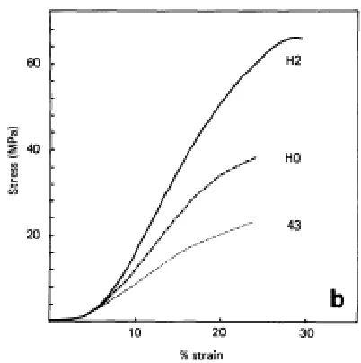
Figure 1.12: Mechanical changes during chick development from prehatchling day 43 to 2 day old hatchling. These stress strain curves demonstrate an increase in mechanical strength during development (McBride, 1988).
diameter sizes [28, 71, 79, 80] (Figure 1.11). Early in development, the distribution is narrow,
consisting of mainly small diameter fibrils. As growth progresses, the distribution increases as
fibrils coalesce to form larger diameters while some remain unaltered. Often this change begins
at the onset of locomotion; therefore, it has been hypothesized that the large diameter fibrils have
higher tensile stress than small diameter fibrils [29, 71]. The small size and fragile nature of
neonatal tendons has made it difficult to obtain mechanical data to test this hypothesis during
development when the largest changes in fibril diameter are occuring. However, one study
showed a rapid increase in ligament mechanical strength throughout development but the
youngest age studied was only 1.5 months old in a r at [81]. An additional study done in chick tendons examined composition, structure and mechanics from post-fertilization through hatchling
[82] (Figure #). While this model does provide mechanical data of tendon development, the chick
is not the ideal model system due to lack of genetically modified animals and available assays.
An ideal model system would allow for the detailed study of the mechanisms behind structural,
compositional and mechanical changes during
development.
As previously mentioned tendons
undergo drastic compositional and structural
changes during both development and healing.
Many of these changes parallel each other. In
both development and healing collagen content
increases and the fibril diameters increase.
Minor matrix molecules, like Collagen XIV and
biglycan, are present during development and
healing but are very sparse in mature
tendon[83]. However, there are also
differences in these two events. In particular,