R E V I E W
Virus
–
Receptor Interactions: Structural Insights
For Oncolytic Virus Development
This article was published in the following Dove Press journal: Oncolytic Virotherapy
Nadishka Jayawardena
1Laura N Burga
1John T Poirier
2Mihnea Bostina
1,31Department of Microbiology and
Immunology, University of Otago, Dunedin, New Zealand;2Perlmutter
Cancer Center, NYU Langone Health, New York, NY, USA;3Otago Micro and
Nano Imaging, University of Otago, Dunedin, New Zealand
Abstract:
Recent advancements in oncolytic virotherapy commend a special attention to
developing new strategies for targeting cancer cells with oncolytic viruses (OVs).
Modi
fi
cations of the viral envelope or coat proteins serve as a logical mean of repurposing
viruses for cancer treatment. In this review, we discuss how detailed structural knowledge of
the interactions between OVs and their natural receptors provide valuable insights into tumor
speci
fi
city of some viruses and re-targeting of alternate receptors for broad tumor tropism or
improved tumor selectivity.
Keywords:
oncolytic viruses, virus
–
receptor interaction, virus entry
Introduction
Oncolytic virotherapy is a dynamic
fi
eld of cancer treatment with over 70 clinical
trials registered to date.
1The majority of oncolytic viruses (OVs) are used in their
native, replication-competent form to cause a direct oncolysis of tumors. For instance,
coxsackievirus, parvovirus, Newcastle disease virus, measles virus, vaccinia virus and
Seneca Valley virus have been used in clinical trials in their native forms.
2–6On the
other hand, human pathogenic viruses such as herpes simplex virus-1, poliovirus and
adenovirus have been genetically modi
fi
ed to limit their replication to tumor sites and
to reduce their virulence in normal tissues.
7–9In addition to the direct oncolysis, OVs
can kill cancer cells via several indirect mechanisms: the activation of immunologic
pathways and antiangiogenesis.
10,11En route to reaching cancer cells, OVs must
overcome a range of complex physical and chemical barriers to
fi
nally interact with
speci
fi
c cellular receptors.
12Perhaps the most exhaustive obstacle in systemic
deliv-ery of OVs is the neutralization of viruses by pre-existing antibodies or triggered
anti-viral immune response.
13One way to bypass the host immunity is to
mask/manip-ulate viral surface proteins to avoid recognition by neutralizing antibodies.
14,15However, eliminating antibody recognition does not guarantee a successful infection
of tumors with OVs as the cellular uptake will ultimately be dependent on virus
binding to the cellular receptors. Expression of virus cellular receptors in cancers
varies depending on tumor type as well as among different patients with the same
type of cancer.
16In such cases, OVs need to be modi
fi
ed to re-target the cancer via
alternative receptors. Thus, the manipulation of OV surface proteins to either
cir-cumvent anti-viral immune response or to exploit different receptors requires in-depth
knowledge of how they interact with their cellular receptors at a structural level. In
this review, we discuss the interactions between clinically evaluated OVs and their
cellular receptors and how they have been modi
fi
ed to target cancers.
Correspondence: Mihnea Bostina Department of Microbiology and Immunology, University of Otago, Dunedin, New Zealand
Tel +64 22 44 5583
Email [email protected]
Oncolytic Virotherapy
Dove
press
open access to scientific and medical research
Open Access Full Text Article
Oncolytic Virotherapy downloaded from https://www.dovepress.com/ by 118.70.13.36 on 25-Aug-2020
Oncolytic Viruses And Cancer
Tropism
Herpes Simplex Virus
Herpes simplex
virus 1 and 2 (HSV-1 and HSV-2) belong to
the family of
Herpesviridae
, genus
Simplexvirus
.
17HSV
virion has a complex architecture characterized by a
dsDNA genome, an icosahedral capsid (nucleocapsid), an
amorphous layer of protein (tegument) and an envelope
(
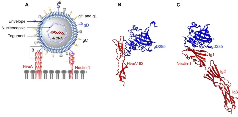
Figure 1A
).
18Both HSV-1 and -2 are genetically stable
and considered to be the most serious human pathogens in
their family. HSV-1 was shown to be associated with
ence-phalitis and orofacial herpes infections, whereas HSV-2
mostly causes genital infections.
19The remarkable
patho-genicity of HSV is attributed to its ability to establish latent
infections in sensory neurons, thus providing a logical
rea-son to manipulate these strains for therapeutic applications.
T-VEC is a genetically modi
fi
ed strain of HSV-1 and
represents a major breakthrough in immunotherapy being
the
fi
rst and only US FDA approved oncolytic virus to
date.
20–23T-VEC is presently used as intralesional
injec-tions to treat non-resectable melanoma with many ongoing
Phase I/II clinical trials showing the possibility of using
the virus in conjunction with other treatments such as
immune checkpoint inhibitors.
24,25Furthermore, two
other strains of HSV-1, G207 (Infected cell protein (ICP)
34.5 and ribonucleotide reductase mutated) and NV1020
(ICP34.5 mutated) have completed Phase I/II clinical trials
in malignant brain tumors and in colorectal cancer liver
metastasis, showing partial clinical responses and
stabili-zation of metastasis, respectively.
26,27While modi
fi
cations to the T-VEC genome are aimed at
reducing the neurotoxicity of the wild-type strain and
stimulat-ing a strong immune response in tumor site, expression levels
of cellular receptors and their interactions with HSV still play a
vital role in virus entry into tumor cells. HSV utilizes four viral
glycoproteins, gB, gD, gH and gL (
Figure 1A
), expressed on
the outer envelope to establish interactions with various cell
surface receptors and to facilitate cell entry.
28,29In order to
initiate HSV cell entry, at least three different classes of cell
surface receptors should interact with the respective
glycoproteins.
29,30Current molecular and structural biology
literature identi
fi
es three steps in penetrating host cells: 1) gB
attachment to heparan sulfate proteoglycans (HSPG)
244, 2) gD
binding to nectin-1,
31,32herpes virus entry mediator
(HVEM),
33or 3-O-sulfated heparan sulfate, and 3) gB binding
to paired Ig-like type 2
α
(PILR
α
),
34nonmuscle myosin IIA
(NMHC-IIA) or myelin-associated glycoprotein (MAG) and
initiation of envelope fusion with plasma membrane.
29Upon
the envelope fusion with host-cell membrane, HSV
nucleocap-sid is translocated to the nuclear pore through which viral DNA
Figure 1Structures of enveloped, DNA oncolytic viruses in complex with their cellular receptors. (A) Schematic diagram of herpes simplex virus-1 (HSV-1). (B) HSV-1 utilizes its surface exposed glycoprotein D ectodomain to bind host cellular receptor herpes virus entry mediator A (HveA) ectodomain (PDB: 1JMA). (C) Glycoprotein D of HSV also interacts with thefirst Ig domain of nectin 1 at 1:1 stoichiometry. Nectin-1 binding site on gD differs from HveA binding site, as evident from the crystal structures arranged in the same orientations (PDB: 3SKU).
Dovepress
Oncolytic Virotherapy downloaded from https://www.dovepress.com/ by 118.70.13.36 on 25-Aug-2020
is released into the nucleus.
35Evidence from various clinical
studies points toward a direct relationship between the
expres-sion of HSV receptors in tumors, cancer progresexpres-sion and
prognosis. For instance, herpesvirus entry mediator (HVEM),
a member of the tumor necrosis factor (TNF) superfamily, has
been shown to play a role in activating inhibitor signaling in
T-cells upon binding to BTLA ligand (B-lymphocyte and
T-lymphocyte attenuator).
36Increased expression of HVEM
has been reported in hepatocellular carcinoma,
37gastric
cancer
38and melanoma.
36Structural evidence for interactions
between HVEM and HSV gD protein arises from a crystal
structure of gD ectodomain truncated at residues 285 (gD285)
bound to the ectodomain of HVEM (
Figure 1B
).
33In both
HSV-1 and -2, gD is structurally unique in comparison to other
members of the family due to diverging N-terminal hairpins.
39The interface between gD285 and HVEM is comprised of
interactions between short, N-terminal hairpin (1
–
37) that
extends towards the V-like immunoglobin core of gD to
estab-lish interactions with two cysteine-repeat-domains (CRDs) of
HVEM. C-termini of gD and HVEM are arranged in opposite
directions, presumably anchored to viral and cellular
mem-branes, respectively. The observation that only a small segment
of gD protein is involved in HVEM binding suggests the
possibility of manipulating gD protein to redirect HSV to a
different receptor, such as nectin-1 or 3-O-sulfated heparan
sulfate, depending on their expression levels in cancers.
14Nectin-1 is another cell surface receptor that binds gD of
HSV.
32Nectin-1 belongs to the family of nectin or
nectin-like receptors that play an important role in cell adhesion.
40Results from various in vitro and clinical studies have
identi-fi
ed increased expression of nectin-1 and nectin-2 in cancers
such as breast cancer,
41highly migratory and invasive
carcinoma,
42squamous cell carcinoma
43and colorectal
cancer.
44In such instances, nectin-1 serves as an excellent
predictor of HSV oncolytic sensitivity. Interactions between
gD and nectin-1 have been characterized by crystallization of
truncated forms of the gD ectodomain (gD285, truncated
resi-dues 1
–
285) complexed with nectin-1 (
Figure 1C
).
31The
crystal structure identi
fi
es both N- and C-termini and a residue
located in Ig core interacting with the
fi
rst Ig domain of
nectin-1 at nectin-1:nectin-1 stoichiometry, resembling an interaction pattern similar
to nectin-1 homodimers. Interestingly, interactions in
gD285-nectin 1 interface are similar to those observed in gD285-nectin-1
homodimer interface and distant from gD285-HVEM
inter-face due to the absence of N-terminal hairpin. From a
physiological point of view, gD binding to nectin-1 can
abolish nectin-1 dimerization, eventually affecting cell
–
cell
adhesion.
31Therefore, modi
fi
ed HSV strains could have an
additional mechanism of hampering tumor progression apart
from triggering anti-tumor immunity.
Because of the wide expression of nectin-1 in human
cells,
45targeting nectin-1 expressing tumors with HSV-1
could be problematic in the case of systemic
immunother-apy. Such off-target effects can be minimized either by
developing HSV mutants capable of escaping nectin-1
while still retaining its ability to bind HVEM, or by
identifying potential bi-soluble adapters for targeting
cog-nate tumor receptors.
46First evidence for latter strategy
comes from targeting of epidermal growth factor receptor
(EGFR) expressing cells with a HSV variant modi
fi
ed with
P-V528LH adapter consisting of gD ectodomain binding
region of nectin-1 fused to an EGFR-speci
fi
c monoclonal
antibody.
46Vaccinia Virus
Vaccinia virus (VV) is a large, enveloped dsDNA virus
(~191
kbp)
from the
genus
Orthopoxvirus
of
the
Poxviridae
family.
47The natural host and origin of VV are
not known.
48Characteristic to VV is its replication strategy
which takes place in cytoplasmic viral factories of infected
cells.
49The genome of VV encodes more than 200 proteins,
of which approximately 20 are envelope proteins.
50During
the life cycle of VV, three distinct particle types are
pro-duced; (1) intracellular mature virions (IMV), (2) wrapped
virions (WV) and (3) extracellular enveloped virions
(EEV).
50Mature virions (MV) are stable under virus
pur-i
fi
cation conditions, remaining the most extensively studied
form of the virus. By contrast to other dsDNA viruses, IMV
has a complex, asymmetric structure that consists of a
nucleoprotein core surrounded by a single lipoprotein
membrane.
51Since its use in eradicating smallpox,
52VV has played
a seminal role as recombinant vectors in gene therapy.
53Both wild-type and recombinant strains of VV have been
of particular interest in oncovirotherapy.
54As an oncolytic
agent, VV has several advantages such as the ability to
incorporate a large amount of foreign DNA, fast and
ef
fi
cient replication and safety.
55Moreover, VV displayed
natural cancer tropism, selectively targeting tumors after
systemic administration.
54Clinical trials on VV thus far
have employed a potent, yet safe form of VV (JX-594),
which encodes granulocyte-macrophage
colony-stimulat-ing factor as an immunomodulator.
55,56,57Vaccinia virus MVs entry into host cells is either mediated
by fusion of MV membrane with the plasma membrane at
neutral pH or through receptor-mediated endocytosis under
Dovepress
Oncolytic Virotherapy downloaded from https://www.dovepress.com/ by 118.70.13.36 on 25-Aug-2020
acidic conditions.
58,59Nonetheless, no receptors have been
unequivocally
identi
fi
ed.
Glycosaminoglycans
(GAGs),
highly polyanionic compounds present on the surface of
stro-mal tumor cells, have been suggested as putative receptors
facilitating VV entry.
59,60VV membrane proteins A27L and
H3L are essential for fusion of viral membrane with cell
membrane.
61,62Positively charged amino-terminal of A27L
can also act as a site for binding of heparan sulfate (HS).
63The
involvement of additional GAGs such as chondroitin sulfate
(CS) in binding the VV surface protein D8L has been shown,
but subsequent studies eliminated the essentiality of these
receptors.
59,64,65To date, an exact mechanism behind
VV-induced oncolysis is unknown. Whether the anti-tumor ef
fi
-cacy is receptor-mediated or attributed to tumor vasculature
66or whether overexpression of ribonucleotide reductase is
essential for viral replication
67still remains an open question.
Rhabdoviruses
Members of
Rhabdoviridae
family are enveloped,
nega-tive-sense single-stranded (ss) RNA viruses with a 11
–
15
kb linear genome encoding
fi
ve proteins: glycoprotein (G),
matrix protein (M), phosphoprotein (P), polymerase (L)
and nucleoprotein (NP).
68Rhabdoviruses (RhVs) virions
are about 180 nm long and 75 nm wide and have a rod- or
bullet-shaped geometry. The G proteindecorating the
envelope is involved in receptor binding, whereas NPs
are associated with RNA (NP-RNA). Together with L
and P, NP-RNA complex forms a ribonucleoprotein
parti-cle, which makes contact with M proteins beneath the
envelope (
Figure 2A
).
RhVs have a broad and diverse host speci
fi
city with
Lyssavirus
and
Vesiculovirus
genera, infecting animals and
the remaining RhVs infecting plants.
69RhVs present
sev-eral advantages that recommend them for development as
oncolytic agents. RhV infections are relatively rare,
there-fore there is no pre-existing immunity. Additionally, they
do not show genetic reassortment, integration in the host
genome or malignant transformation due to cytoplasmic
replication and have a relative ease of large-scale virus
production in a broad range of cell lines. Several RhVs
have been investigated for their oncolytic properties.
70,71Vesicular stomatitis virus (VSV) is a vesiculovirus that
infects cattle, horses, pigs, and other mammals. VSV
infections are usually asymptomatic in human and
non-lethal in animals, with mild
fl
u-like symptoms.
70VSV
exhibits a robust infectivity and broad tropism to tumors,
attributed to the defective interferon (IFN) responses in
tumor cells.
72Entry of VSV into tumor cells is initiated by
the
interactions
between
its
coat
protein
VSV-G
(
Figure 2A
) and highly ubiquitous cellular receptor,
low-density lipoprotein receptor (LDLR).
73LDLR is a
trans-membrane receptor whose functions include cell-signaling,
endocytosis and traf
fi
cking of cellular proteins. The most
abundantly expressed form of LDLR in solid tumors is
LDLR1, shown to be linked to low patient survival rate.
74The ligand binding domain of LDLR is comprised of
cysteine-rich repeats conserved among other members of
LDLR family,
75therefore presenting alternative entry
points for VSV. Crystal structures of VSV-G in complex
with
two
different
cysteine-rich
domains,
CRD2
(
Figure 2B
) and CRD3 (
Figure 2C
) of LDLR demonstrate
that both binding sites on VSV-G are identical.
76VSV-G-LDLR complex is internalized into host cells through a
clathrin-mediated endocytosis.
77,78In the case of
recombi-nant VSV
Δ
M51 encoding reovirus fusion-associated small
transmembrane (FAST) protein, this mechanism extends
from the virus-cell fusion to cell-cell fusion.
79The process
repeats, expanding to un-infected cells and could lead to
large multinucleated giant cells (syncytia).
80In another
study, the use of VSV-G substituted with lymphocytic
choriomeningitis virus glycoprotein (LCMV-GP) has
shown minimal neural toxicity and potent anti-tumor
effect in mice brain tumor models.
81LMCV-GP may
bind differentially glycosylated
α
-dystroglycan (
α
DG) in
brain tumors with high-af
fi
nity
82,83despite the lower
expression levels of
α
DG in human glioblastoma.
84Maraba virus is another vesiculovirus which binds
LDLR and has the capacity to infect a broad range of
human cancers.
85,86In order to speci
fi
cally target cancer
cells and to enhance replication ef
fi
cacy, two mutations
were introduced: L123W and Q242R in M and G proteins,
respectively.
87,88Maraba virus strain MG1 expressing
human melanoma-associated antigen-A3 (MAGE-A3)
and an adenoviral vector (Ad) expressing the same antigen
have been developed as an oncolytic vaccine strategy with
high immune priming ef
fi
ciency.
89Newcastle Disease Virus
Newcastle disease virus (NDV) is a ssRNA virus in the
genus
Avulavirus
of
Paramyxoviridae
family.
90The
envel-oped NDV capsid harbours a non-segmented
negative-sense ssRNA that codes for six proteins (
Figure 2D
).
Nucleoprotein (NP), phosphoprotein (P) and RNA
depen-dent RNA polymerase (RdRP) bind the RNA genome to
form the nucleocapsid.
90Other NDV proteins include
matrix protein (M), which forms the inner layer of virus
Dovepress
Oncolytic Virotherapy downloaded from https://www.dovepress.com/ by 118.70.13.36 on 25-Aug-2020
envelope, hemagglutinin-neuraminidase (HN) and fusion
protein (FP), involved in receptor binding and entry,
respectively.
91Numerous in vitro studies have shown that NDV is
non-pathogenic to humans and elicits anti-tumor effects
without any genetic modi
fi
cations or limitations in
Figure 2Structures of enveloped, RNA oncolytic viruses in complex with their cellular receptors. (A) Schematic diagram of vesicular stomatitis virus. (BandC) Vesicular stomatitis virus (VSV) surface glycoproteins (VSV-G) identify and interact with cysteine-rich domains (CRD) on low-density lipoprotein receptors (LDLR) expressed in cancer cells. Different CRDs interact with VSV-G at identical locations as evident from crystal structures arranged in the same orientation (PDB: 5OLY and 5OY9). (D) Schematic diagram of Newcastle disease virus (NDV). (E) Newcastle disease virus (NDV) surface protein hemagglutinin-neuraminidase (HN) exploits cell surface sialic acid (SA) as the cellular receptors. Two SA binding sites exist on HN dimers, SA1, and SA2 (PDB: 1USR). (F) Schematic diagram of Measles virus (MV). (GandH) measles virus (MV) H binds CD46 short consensus repeats (SCR) 1, SCR2, SCR1-2 interface (PDB: 3INB) and domain 1 of nectin-4 (PDB: 4GJT).
Dovepress
Oncolytic Virotherapy downloaded from https://www.dovepress.com/ by 118.70.13.36 on 25-Aug-2020
delivery methods.
92–94MTH68/H, PV701 and NDV-HUJ
are three attenuated strains of NDV with highly ef
fi
cient
intratumoral replication, tumor cell lysis and
immunosti-mulation, currently in Phase I/II clinical trials.
4,95In
addi-tion, NDV-HUJ strain is able to bypass the effect of an
anti-apoptotic protein Livin.
4NDV binds tumor cells via interactions between HN and
cell surface sialic acids (SA) receptors.
96SA is a derivative
of neuraminic acid overexpressed in multiple cancers
145–147and was shown to be associated with the metastasis of breast
cancer.
97HN has a dual function: to recognize the cell
sur-face receptors, and subsequently to promote the fusion
activ-ity of the F protein and to cleave off the sialic acid from
progeny virus particles.
98HN is composed of a long stalk
connected to a globular head that consists of a six-bladed
β
-sheet propeller.
99Two sites have been identi
fi
ed on HN
dimers that form interactions with sialic acid (
Figure 2E
).
The
fi
rst binding site is involved in mediating neuraminidase
activity, receptor binding and promoting the fusion activity of
F protein.
100The second binding site is located at the
mem-brane-HN distal region interface, which aids in tethering the
virus in close proximity to the host membrane during
fusion.
101,102Measles Virus
Measles virus (MV) is an enveloped, spherical-shaped,
negative-sense ssRNA virus (
Figure 2F
) from the genus
Morbillivirus
of the
Paramyxoviridae
family.
103Due to its
highly contagious nature, MV remains a major human
health concern worldwide, causing approximately 150,000
deaths annually.
104Similar to RhVs, the non-segmented
RNA genome (15
–
16 kb in size) of MV encodes
fi
ve
structural proteins: glycoprotein (G), matrix protein (M),
phosphoprotein (P), large protein (L) and nucleoprotein
(NP).
103,105On the MV envelope there are two types of
glycoproteins
characteristic
to
paramyxoviruses:
1)
hemagglutinin
106and 2) fusion protein,
107responsible for
cell receptor attachment and fusion, respectively.
Live-attenuated MV vaccine strains can be used as
onco-lytic agents to target different receptors overexpressed in
tumors.
3Measles virus hemagglutinin (H) binds CD46,
108signaling lymphocyte activation molecule (SLAM)
109or
nectin-4 in epithelial cells.
110Overexpression of CD46 and
nectin-4 receptors has been identi
fi
ed as a strategy to
prefer-entially target cancers with MV.
111,112In addition, SLAM
expressed on activated B and T-lymphocytes, monocytes,
and dendritic cells has been reported to be the main entry
port for wild-type MV.
113CD46 structure is comprised of a C-terminal domain,
a transmembrane domain, a short region with unknown
function and four modules of short consensus repeats
(SCR) 1-4 at the N terminus.
143,114The crystal structure
of dimeric H-CD46 identi
fi
es interactions between
MV-H and SCR1, SCR1-SCR2 interface and SCR2 of CD46
(
Figure 2G
). CD46 SCR1-2 is pivotal to capsid binding
in adenoviruses,
115discussed later in this review.
However, the binding sites of MV-H for CD46 and
SLAM overlap,
116supporting the need to develop
strains that can preferentially bind CD46. Two amino
acid substitutions, N481Y and S546G in MV-H protein,
have been shown to arm MV strains to ef
fi
ciently use
CD46 as an entry receptor in CD46+ cells.
117In another
study, structural characterization of MV-H-nectin-4
com-plex revealed that the amino-terminal of nectin-4 binds
β
4-
β
5 groove of MV-H (
Figure 2H
).
110This study
iden-ti
fi
ed a hydrophobic pocket located in the groove
sug-gested to be involved in binding all three receptors for
MV, with different residues involved for different
receptors.
110Similar to RhVs, MV exerts its oncolytic activity by a
sequence of virus-cell fusion through H protein, cell-cell
fusion through F protein and subsequent apoptosis.
118The
Edmonston vaccine strain of MV (MV-Edm) has been
modi
fi
ed for non-invasive imaging of MV activity in
tumors by introducing either sodium iodide symporter
(NIS), the
β
subunit of human chorionic gonadotropin
(
β
hCG) or human carcinoembryonic antigen (CEA) into
the MV genome. The MV-CEA strain has been tested in
Phase I clinical trials in patients with platinum-resistant
ovarian cancer, with evidence pointing towards the
recruit-ment of tumor effector T-cells to establish an
anti-tumor immunity.
119,120Selective tumor tropism of MV
was further validated in a Phase I clinical trial in myeloma,
where systemically administered MV-NIS showed
replica-tion within tumors.
121Alternative to live-attenuated
vac-cines, recombinant, replication-competent MV could be
developed
to
re-target
different
surface
receptors
expressed on tumor cells. This requires the mutation of
SLAM and CD46 binding sites, thus resulting in a double
ablated chimeric H protein to prevent the binding of MV
to normal cells expressing SLAM and CD46.
122Mutations
at Y481 and R533 on MV-H and subsequent incorporation
of single-chain antibodies directed against cognate
recep-tors such as EGFR has shown to elicit oncolysis of
EGFR-positive tumor models in mice.
123,124Dovepress
Oncolytic Virotherapy downloaded from https://www.dovepress.com/ by 118.70.13.36 on 25-Aug-2020
Adenovirus
Human adenoviruses (HAdVs) belong to the family of
Adenoviridae
, genus
Mastadenovirus
and are divided into
seven different species from HAdV-A to -G.
125They are
non-enveloped viruses with an icosahedral capsid
protect-ing a dsDNA genome of 26
–
46 kbp (
Figure 3A
). The
icosahedral capsid is comprised of 252 capsomeres,
con-sisting of 240 hexon trimers and 60 penton bases
(PB).
38,126Attached to PB plates are trimers of
fi
ber
mole-cules, which utilize the conserved N-terminal (residues
1
–
20) to bind the PB and the C-terminal knob to bind
cellular receptors. Collectively, hexon trimers, PBs, and
fi
ber molecules are known as the major capsid proteins. In
addition, 240 copies of the minor protein IX, and several
copies of the minor proteins IIIa, VI and VII are located on
the capsid exterior and interior, respectively. All the HAdV
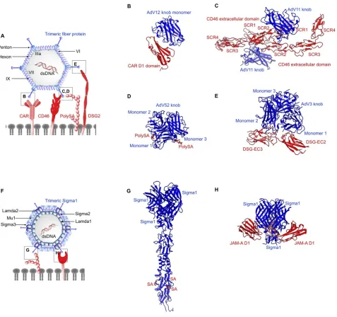
Figure 3Structures of non-enveloped, DNA oncolytic viruses in complex with their cellular receptors. (A) Schematic diagram of human adenovirus (AdV). (B) Coxsackievirus-adenovirus receptor (CAR) extracellular D1 domain interacts with a monomer of AdV12 knob (PDB: 1P69). (C) AdV 11 exploits CD46 as the primary receptor. AdV knob monomer interacts with short consensus repeat (SCR) 1 and SCR1-2 interface. Another knob monomer binds the base of SCR2 (PDB: 3O8E). (D) AdV52 utilizes its shortfibers to bind polysialic acid (polySA) and monomer 1 and 3 of the knob trimers interact with two polySA (PDB: 6G47). (E) Desmoglein 2 (DSG2) acts as the receptor for AdV3 with two distinct receptor:knob ratios of 1:1 and 1:2 observed. DSG2 EC2 and EC3 interact with monomer 1 and 2 of AdV3 knob, respectively (PDB: 6QNT). (F) Schematic diagram of human Reovirus (RV). (G) RV exploits cell surface sialic (SA) acid as its attachment receptor to tether the virus in close proximity to host membrane to interact with an entry receptor, junction adhesion molecule-A (JAM-A). SA binds to the stalk of the trimeric sigma protein (PDB: 3S6X), whereas(H)JAM-A D1 domain interacts with the head of the trimeric sigma protein (PDB: 3EOY).
Dovepress
Oncolytic Virotherapy downloaded from https://www.dovepress.com/ by 118.70.13.36 on 25-Aug-2020
strains can cause gastrointestinal infections, with some
subtypes being reported to cause respiratory, urinary tract
infections and keratoconjunctivitis.
127HAdV is also
responsible for viral-induced tumors in mice, with subtype
A showing the highest oncogenicity, subtype B being
weakly oncogenic,
128,129while C, E and F are known to
be non-oncogenic.
127Adenoviruses are one of the most extensively studied viral
vectors due to ease of genome manipulation. In addition,
HAdVs provide several distinct advantages such as inherently
potent lytic activity and feasibility of manufacturing high viral
titers.
130Numerous HAdV strains have been genetically
engi-neered (ONYX-015 and DNX-2401) to reduce infection in
normal tissues and to selectively target tumors.
131,132HAdV entry into host cells is a two-step mechanism,
which involves the initial attachment of the viral
fi
bers to
cell surface receptors, followed by interactions with other
capsid proteins and internalization receptors.
133Upon
virus internalization by endocytosis, the capsid escapes
into the cytosol through lysis of endosomal membrane
and is subsequently traf
fi
cked to the nuclear envelope
along microtubules, where the viral genome enters the
host nucleus via nuclear pores.
134Most of the HAdVs
and other AdV subtypes except HAdV B use the
coxsack-ievirus and adenovirus receptor (CAR) for cellular
attachment.
135CAR is a type I transmembrane
glycopro-tein that belongs to the immunoglobulin (Ig) superfamily.
It contains a cytoplasmic C-terminal, a hydrophobic
trans-membrane domain and two extracellular Ig-like domains,
D1 and D2.
136CAR D1 domain alone is suf
fi
cient to
establish
interactions
with
HAdV
fi
ber
knob
(
Figure 3B
).
137,138However, the variable expression of
CAR in cancers is a signi
fi
cant challenge for HAdV
oncovirotherapy.
139Low expression of CAR was reported
for gastric, colon, and prostate cancer cell lines under
hypoxic conditions.
140In addition, CAR expression is
downregulated in cancer cells treated with chemotherapy
or radiation, which poses an issue when using HAdVs in
combination therapies.
141The majority of subtype B HAdVs and some of subtype D
(AdV37) have been shown to exploit CD46, a type I
trans-membrane protein overexpressed in tumors, as the attachment
receptor.
142,143Crystallographic studies showed that AdV11
trimeric
fi
bers form a compact knob that interacts with the
SCR1-2 regions of three CD46 molecules (
Figure 3C
).
115Complementary or not to CD46, sialic acid has been shown
to interact with the top region of AdV37 knob trimer. This has
been further con
fi
rmed by structural studies on AdV52, which
utilizes its long
fi
bers to bind CAR and short
fi
bers to bind
polysialic acid (
Figure 3D
).
144,145,146,147CD80 and CD86 are
another two members from the Ig superfamily that play a key
role in subtype B AdV3 entry. Both CD80 and CD86 are
expressed in dendritic cells, thus targeting these receptors by
AdV can elicit a strong immune response via T-cell
activation.
148,149,150Recent studies have identi
fi
ed desmoglein 2 (DSG2) as
a new receptor for HAdV3, HAdV7, HAdV11 and
HAdV14 strains of subtype B.
151DSG2 is a type 1
trans-membrane glycoprotein present in epithelial cells that
plays an essential role in cell-cell adhesion.
152,153The
extracellular domain of DSG2 is comprised of four
cad-herin domains, EC1-EC4, with EC2 and EC3 accounting
for the region that binds trimeric
fi
ber knob of HAdV3.
154DSG2 is overexpressed in a range of epithelial cancers,
acting as a marker for targeting such cancers with
AdV.
155,156,157,158A cryo-EM study showed that DSG2
EC2-EC3 fragment binds the top of the trimeric HAdV3
in 2:1 or 1:1 stoichiometry (
Figure 3E
).
159EC2 and EC3
establish interactions with the loop regions of monomers 1
and 2, respectively, while the third HAdV monomer of the
knob is not engaged. Furthermore, mutagenesis
experi-ments identi
fi
ed D261 as an essential knob residue
required for DSG2 binding.
159Endocytosis of the HAdV-CAR complex is mediated by
the interactions between internalization receptors, integrins
and
fi
ve-fold capsid vertices.
160Structural information is
available on entry receptors
α
v
β
3 integrins bound to
adeno-virus, which shows the requirement of Arg-Gly-Asp (RGD)
moiety on the penton base to interact simultaneously with
several integrins in different orientations to facilitate integrin
clustering and subsequent viral entry into host cells via
endocytosis.
161,162However, mutation of RGD sequence
was associated with only a reduced viral infection but not
complete abolishment.
163Though no plausible mechanisms
have been proposed for an integrin-independent entry
path-way of AdV, there is evidence for compensation for loss of
penton-integrin interactions through recruitment of
fi
ber
receptors.
163,164In the absence of suf
fi
cient levels of CAR
for a successful infection, re-targeting of integrin receptors
by incorporation of an RGD moiety in the
fi
ber knob of
AdV5 has shown to be ef
fi
cient in promoting infection of
ovarian tumor cells.
165Reovirus
Reoviridae
is a family of non-enveloped, dsDNA viruses
with an icosahedral capsid structure (~85 nm in diameter)
Dovepress
Oncolytic Virotherapy downloaded from https://www.dovepress.com/ by 118.70.13.36 on 25-Aug-2020
composed of a large outer layer, and a smaller inner layer
(
Figure 3F
). Reovirus (RV) dsDNA is structured into 12
segments, categorized into three size-dependent groups:
large, medium and small.
166,167The outer shell of the
capsid and at the vertices of the virion are formed by
heterodimers of µ1 and
σ
3 proteins, while pentamers of
λ
2 protein form a channel connecting to trimers of
attach-ment protein
σ
1.
168RV requires interactions with junctional adhesion
molecule-A (JAM-A)
169and cell surface monosaccharides
such as sialic acid
170to penetrate the host cell. JAM-A
expression has been proposed to be linked to tumor cell
proliferation and progression, whereas in some cases an
inverse relationship was observed.
171The
fi
rst step of the
RV binding to host cells involves a low-af
fi
nity interaction
of the lower part (stalk) of
σ
1 protein with cell surface
sialic acid (
Figure 3G
). This process facilitates the
anchor-ing of RV capsid in close proximity to host-cell membrane
in order to initiate interactions with a secondary receptor.
High-af
fi
nity interactions between JAM-A D1 domain and
the head domain of
σ
1 protein (
σ
1H)
169serve as the
second step in RV host-cell attachment (
Figure 3H
).
Reolysin, a wild-type, non-pathogenic, serotype 3 RV,
has been widely investigated in preclinical and clinical
settings.
172Phase I and II clinical trials of advanced
solid tumors and recurrent gliomas,
173–175and
combina-tion therapy with paclitaxel/carboplatin or docetaxel
176,177showed Reolysin to be safe and effective.
Parvovirus
Human parvovirus (HPV) is a single-stranded DNA virus in
the
Parvoviridae
family, associated with a wide variety of
diseases in humans.
178The genome of HPV is packaged
inside an icosahedral capsid of ~280 Å in diameter. The
capsid is composed of 60 structural subunits, in which
major capsid protein VP2 is the primary protein (~95%)
while the minor capsid protein VP1 is less abundant
(~5%).
179Capsid proteins have an eight-stranded,
antipar-allel
β
-barrel
“
jelly roll
”
fold. Engineered and wild-type
strains of HPV demonstrate a tumor-selective replication
with excellent safety pro
fi
les. Oncolytic activity of HPV is
attributed to the direct oncolysis as well as induced
anti-tumor immunity.
180HPV B19 strain binds the erythrocyte P19 antigen
expressed in erythroid progenitor cells.
181However, entry
into host cells requires the involvement of
α
5
β
1 integrin
co-receptor,
182known to be essential for tumor progression in
certain cancers.
183Modi
fi
cations of I367S and H373R in the
dimple region of the capsid in rat parvovirus strain H-1PV
have engineered the virus to re-target integrin receptors
expressed in cancers.
184In another study, transferrin receptor
1 (TFR1) has been identi
fi
ed as the cellular receptor for
canine parvovirus (CPV).
185TFR1 is a membrane
glycopro-tein linked to many diseases including cancers.
185However,
TFR1 expression is variable across different cancers.
186The
structure of CPV-TFR1 complex demonstrates an example
for a receptor saturating only a few of the 60 equivalent
binding sites on the capsid, resulting in an asymmetric
interaction.
185Coxsackievirus
Coxsackievirus (CV) is a non-enveloped, positive-sense
ssRNA virus (~7.4 kb) from the family of
Picornaviridae
,
genus
Enterovirus
(
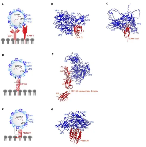
Figure 4A
). CV is a major human
patho-gen causing a number of diseases including myocarditis and
meningoencephalitis.
187CV serotypes are categorized into
two groups; (1) coxsackievirus A (CVA) and coxsackievirus
B (CVB).
188CV RNA genomes code for four structural
proteins VP1
–
VP4 that form an icosahedral capsid, and
seven non-structural proteins.
189,190Characteristic to
enter-oviruses is the presence of four types of particles in their life
cycle: mature virion, procapsid devoid of RNA, an expanded
A-particle and an empty particle after RNA exit.
191Receptor binding in enteroviruses takes place in the
“
canyon
”
, a depression located at 5-fold axis of the capsid.
The binding of the receptor displaces a fatty acid molecule
called the
“
pocket factor
”
located in a hydrophobic pocket
within VP1, below the canyon base. Loss of the pocket
factor induces a series of conformational changes in capsid
architecture, leading to capsid expansion and
externaliza-tion of VP1 N-terminus as well as VP4 for membrane
anchoring and subsequent RNA transfer.
192This
mechan-ism holds true for most of the enteroviruses and has been
well characterized for poliovirus (PV),
193,194enterovirus
71 (EV71)
195and CV
196.
CVs utilize three different receptors for cellular entry.
CAR acts as both attachment and entry receptor for
CVB3.
196Cryo-EM reconstruction of CVB3 bound to
full length human CAR has shown that the N-terminal
region of CAR D1 domain contains the binding sites for
CVB3 (
Figure 4B
).
196In the CVB3-CAR interface, A and
G
β
strands of D1 domain form contacts with the north and
south rims of CVB3 canyon. All the external CVB3 capsid
proteins are involved in CAR binding with majority of the
interactions localized to VP1. Of note is the moderately
conserved nature of these receptor binding residues across
Dovepress
Oncolytic Virotherapy downloaded from https://www.dovepress.com/ by 118.70.13.36 on 25-Aug-2020
six different CVB serotypes.
197VP2 residue N165 has
been suggested to be critical in stabilizing the electrostatic
interactions between the capsid and CAR.
198The distal
end of CAR D1 domain is a shared site for CVB3 and
adenovirus (as previously discussed) and their binding
sites overlap on C
β
strand and FG loop.
196Additionally,
the involvement of decay-accelerating factor (DAF) as an
attachment receptor in the CVB3-RD strain has been
demonstrated by another cryo-EM study,
199with one
DAF molecule linking two adjacent protomers on the
capsid exterior. The northern end of the VP2 puff (residues
129
–
180) in one protomer is linked to the south end of the
Figure 4Structures of non-enveloped, RNA oncolytic viruses in complex with their cellular receptors. (A) Schematic diagram of coxsackievirus (CV). (B) D1 domain of coxsackievirus-adenovirus receptor (CAR) acts as the binding site for coxsackievirus B (CVB) capsid proteins VP1-VP3 (PDB: 1JEW). (C) Coxsackievirus A variant 24 (CVA24v) capsid proteins VP1 and VP2 interact with the D1 domain of intracellular adhesion molecule-1 (ICAM-1) (PDB: 6EIT). (D) Schematic diagram of poliovirus (PV). (E) Poliovirus utilizes CD155 on the cell surface as its cellular receptor. Similar to ICAM1 and CAR, CD155 D1 domain binds PV capsid proteins VP1 and VP2 from one protomer and VP3 from the adjacent protomer (PDB: 3J8F). (F) Schematic diagram of Seneca Valley Virus (SVV). (G) Anthrax toxin receptor 1 binds to surface-exposed loops of VP1–VP3 on SVV capsid (PDB: 6CX1).
Dovepress
Oncolytic Virotherapy downloaded from https://www.dovepress.com/ by 118.70.13.36 on 25-Aug-2020
puff of the adjacent protomer, and the bulk of the
interac-tions are condensed between short consensus repeat (SCR)
2 and the north end of the puff. Unlike CAR D1 domain,
DAF does not enter the canyon and thus, does not induce
the conformational changes required for genome delivery
into the host cell.
200On the other hand, CVA binds both DAF and intercellular
adhesion molecule-1 (ICAM-1).
201,202DAF binding does not
induce conformational changes and primarily acts as an
attach-ment receptor,
203whereas ICAM-1 acts as an attachment/entry
receptor for CVA. Overexpression of DAF and ICAM-1 has
been reported in multiple cancers.
204,205,206,207,208ICAM-1 is a
transmembrane immunoglobin with three structural
compo-nents: extracellular N-terminus, transmembrane domain, and
cytoplasmic C-terminus.
209Structural insights into
CVA-ICAM-1
stem
from
several
cryo-EM
investigations
(
Figure 4C
).
201,202Similar to other enteroviruses, the canyon
of CVA24v binds ICAM-1 D1 domain at the quasi 3-fold axis
of the capsid.
202The CVA24v-ICAM-1 interface is comprised
of interactions established between the FG loop of ICAM-1 D1
domain and VP1. Finally, C and D
β
strands of ICAM-1 D1
domain interact with the VP1 GH loop, whereas the DE loop of
D1 forms additional contact with VP2 in CVA24v. This study
also provides insights into adapting CVA strains for a sialic
acid binding as a secondary receptor by mutating residue 250
of VP2 to tyrosine. Sialic acid metabolism has been shown to
be upregulated in metastatic cancers and acts as a receptor for
other oncolytic viruses discussed here such as adenovirus,
Newcastle disease virus and reovirus.
The therapeutic potential of CVA21 or CAVATAK has
been investigated in various preclinical melanoma studies
as monotherapy
210or in combination with doxorubicin.
211Furthermore, CAVATAK has completed a Phase I clinical
trial in patients with advanced melanoma with promising
safety and anti-tumor activity recorded.
22In vivo studies
of non-small-cell lung cancer xenograft models treated
with CVB3 demonstrated abscopal effect of this therapy,
suggesting an enhancement of antitumor immunity.
212Poliovirus
Poliovirus (PV), a member of
Enterovirus
genus, family
Picornaviridae
, is the main causative agent of paralytic
poliomyelitis.
213Three different PV serotypes can be
differ-entiated according to their antigenic properties.
214Similar
to CV, PV possesses a negative-sense ssRNA genome of 7.5
kb coding for seven non-structural proteins and four
struc-tural proteins, which constitute the icosahedral capsid
(
Figure 4D
) as previously described for CV.
215,216Poliovirus entry into host cells is initiated by the
inter-actions between poliovirus receptor CD155, and capsid
canyon.
217–220PV undergoes the same conformational
changes characteristic to other enteroviruses. A-particle
formation,
194,221exit of RNA genome at a location on
2-fold axis
193and empty particle formation
222have been
extensively characterized. CD155 is an onco-immunologic
protein overexpressed in human cancers with a role in
tumor cell invasion and migration.
223CD155 is a type I
immunoglobulin-like transmembrane protein that contains
three ectodomains D1
–
D3.
218,224CD155 expression is
upregulated in carcinomas
225–227and less abundantly
expressed in normal tissues with the exception of liver
development or regeneration.
228In the PV-CD155
com-plex (
Figure 4E
), the D1 domain of CD155 binds VP1, and
VP2 of one protomer and VP3 of adjacent protomer at the
capsid quasi 3-fold axis.
219In the PV capsid, C-terminal,
GH, EF, BC loops, C
β
strand of VP1, B
β
strand, GH loop
of VP3 and EF loop of VP2 occupy the CD155 binding
site.
Poliovirus infection is rapid and remarkably ef
fi
cient,
releasing as high as 10,000 mature virions per infected cell
at 6 hrs post-infection.
229Even though a rapid replication
warrants the applicability of PV in oncovirotherapy, a
counter mechanism must be in place to minimize the
neurotoxicity associated with wild-type PV. The
neuro-attenuated variant of PV, PVSRIPO has completed a
Phase I dose-
fi
nding clinical study in patients with grade
IV malignant glioma with no neurotoxicity reported.
230Seneca Valley Virus
Seneca Valley Virus is the only member of
Senecavirus
genus of the
Picornaviridae
family. The overall structure of
SVV has an icosahedral symmetry and is comprised of a
non-enveloped protein capsid harboring a positive-sense ssRNA
genome of approximately 7.3 kb (
Figure 4F
).
231Similar to
CV and PV, the SVV genome encodes seven non-structural
proteins and four structural proteins. To date, the SVV strains
have been classi
fi
ed into 3 clades,
232–234with the prototype
SVV-001 being the sole member of clade I.
SVV cell entry is dependent on its cellular receptor:
anthrax toxin receptor 1 (ANTXR1), also known as tumor
endothelial marker 8 (TEM8).
235ANTXR1 is a type I
transmembrane protein overexpressed in many types of
cancers, but weakly expressed in healthy tissues.
236The
role of ANTXR1 is unknown beyond its function as a
toxin and virus receptor; indeed, ANTXR1 knockout
mice exhibit no major phenotypic abnormalities.
237Dovepress
Oncolytic Virotherapy downloaded from https://www.dovepress.com/ by 118.70.13.36 on 25-Aug-2020
However, ANTXR1 blockade has been shown to decrease
tumor angiogenesis and to potentiate an anti-tumor effect
towards certain cancers.
238Our group identi
fi
ed the
sur-face exposed loops on SVV-001 capsid exterior, BC loop,
loop II of VP1, the
“
puff
”
loop of VP2, and the
“
knob
”
loop of VP3 which form the binding site for the
extracel-lular domain of ANTXR1 (
Figure 4G
).
239Furthermore, we
showed that SVV binding site on ANTXR1 is
non-con-served in its paralogous receptor, ANTXR2, which is
expressed in normal cells, thereby providing a structural
basis for tumor speci
fi
city of SVV.
240SVV empty capsid
binds ANTXR1, suggesting it may have potential as a
vaccine or as virus-like particles for the development of
tumor-targeted delivery of drugs.
240As suggested from both functional and structural
stu-dies, the tumor tropism of SVV-001 is attributed to
recep-tor-mediated internalization of the virus, a phenomenon
common to other oncolytic picornaviruses. However, a
successful SVV-001 infection may also require an
addi-tional innate immune defect.
235SVV-001 in its native form
provides several advantages for oncovirotherapy: the
native virus is genetically stable and non-toxic to healthy
tissues, it is safe and it homes to tumors when
adminis-tered systemically and pre-existing immunity for SVV is
rare.
241Several preclinical, Phase I/II clinical studies have
demonstrated the anti-tumor potential, intratumoral
repli-cation and safety of SVV in treating solid tumors with
neuroendocrine features.
242,243Conclusion
Oncolytic viruses (OVs) either have an inherent ability to
successfully replicate in cancer cells or they have been
modi
fi
ed to exploit de-regulated signaling pathways in
tumors. Nevertheless, the attachment of OVs to speci
fi
c
receptors found in cancers plays a pivotal role in OV
tumor cell entry, subsequent viral replication and cell
lysis. However, the expression of these receptors varies
in different cancers and also among individual patients.
Furthermore, the presence of natural receptors of OVs in
normal cells may pose a potential challenge when the virus
is pathogenic in nature. Therefore, understanding the
structural details concerning how OVs interact with their
receptors can inform the development of more ef
fi
cient-targeted therapies to exploit cognate receptors and to
reduce off-target cytotoxicity. Additionally,
oncovirother-apy is constantly facing the challenge of overcoming
anti-viral immunity in cancer patients. In this case, the
knowl-edge of OV-receptor interactions is necessary to modify
the viral capsid or envelope proteins in order to bypass the
immune response without impairing the ability to bind
their cellular receptors.
Disclosure
Dr John T Poirier reports personal fees from Perceiver
Pharmaceuticals LLC, outside the submitted work. In
addition, Dr Poirier has a patent WO2017096201A1
licensed to Perceiver Pharmaceuticals, LLC. The authors
report no other con
fl
icts of interest in this work.
References
1. Peters C, Grandi P, Nigim F. Updates on oncolytic virus immu-notherapy for cancers. Mol Ther Oncolytics. 2019;12:259–262.
doi:10.1016/j.omto.2019.01.008
2. Geletneky K, Hajda J, Angelova AL, et al. Oncolytic H-1 parvo-virus shows safety and signs of immunogenic activity in afirst phase I/Iia glioblastoma trial.Mol Ther.2017;25(12):2620–2634. doi:10.1016/j.ymthe.2017.08.016
3. Aref S, Bailey K, Fielding A. Measles to the rescue: a review of oncolytic measles virus.Viruses.2016;8(10):294.
4. Lazar I, Yaacov B, Shiloach T, et al. The oncolytic activity of newcastle disease virus NDV-HUJ on chemoresistant primary mel-anoma cells is dependent on the proapoptotic activity of the inhi-bitor of apoptosis protein livin.J Virol.2010;84(1):639.
5. Hiley CT, Yuan M, Lemoine NR, Wang Y. Lister strain vaccinia virus, a potential therapeutic vector targeting hypoxic tumours.
Gene Ther.2009;17:281.
6. McCarthy C, Jayawardena N, Burga LN, Bostina M. Developing picornaviruses for cancer therapy.Cancers.2019;11:5.
7. Conry RM, Westbrook B, McKee S, Norwood TG. Talimogene laherparepvec: first in class oncolytic virotherapy. Hum Vaccin Immunother.2018;14(4):839–846.
8. Holl EK, Brown MC, Boczkowski D, et al. Recombinant oncolytic poliovirus, PVSRIPO, has potent cytotoxic and innate infl amma-tory effects, mediating therapy in human breast and prostate cancer xenograft models.Oncotarget.2016;7(48):79828–79841. 9. Nemunaitis J, Ganly I, Khuri F, Arseneau J, Kuhn J, McCarty T.
Selective replication and oncolysis in p53 mutant tumors with ONYX-015, an E1B-55kD gene-deleted adenovirus, in patients with advanced head and neck cancer: a phase II trial.Cancer Res.
2000;60:6359–66.
10. Kaufman HL, Kohlhapp FJ, Zloza A. Oncolytic viruses: a new class of immunotherapy drugs. Nat Rev Drug Discov. 2015; 14:642. doi:10.1038/nrd4663
11. Breitbach CJ, De Silva NS, Falls TJ, et al. Targeting tumor vascu-lature with an oncolytic virus. Mol Ther. 2011;19(5):886–894. doi:10.1038/mt.2011.26
12. Vähä-Koskela M, Hinkkanen A. Tumor restrictions to oncolytic virus.
Biomedicines.2014;2(2):163–194. doi:10.3390/biomedicines2020163 13. Filley AC, Dey M. Immune system, friend or foe of oncolytic
virother-apy?Front Oncol.2017;7:106. doi:10.3389/fonc.2017.00106 14. Anderson DB, Laquerre S, Ghosh K, et al. Pseudotyping of
glycopro-tein D-deficient herpes simplex virus type 1 with vesicular stomatitis virus glycoprotein g enables mutant virus attachment and entry.J Virol.
2000;74(16):7698. doi:10.1128/JVI.74.16.7698-7698.2000
15. Ilett EJ, Bárcena M, Errington-Mais F, et al. Internalization of oncolytic reovirus by human dendritic cell carriers protects the virus from neutralization. Clin Cancer Res. 2011;17(9):2767. doi:10.1158/1078-0432.CCR-10-3266
Dovepress
Oncolytic Virotherapy downloaded from https://www.dovepress.com/ by 118.70.13.36 on 25-Aug-2020
16. Boonstra MC, de Geus SWL, Prevoo HAJM, et al. Selecting targets for tumor imaging: an overview of cancer-associated membrane proteins. Biomark Cancer. 2016;8:119–133. doi:10.4137/BIC. S38542
17. Pellet PE and Roizman B. The family herpesviridae: a brief intro-duction. In: Knipe DM and Howley PM, editors.In Fields Virology. Philadelphia [PA]: Lippincott Williams & Wilkins; 2007:2479– 2499
18. Yuan S, Wang J, Zhu D, et al. Cryo-EM structure of a herpesvirus capsid at 3.1 Å.Science.2018;360(6384):eaao7283. doi:10.1126/ science.aao7283
19. Whitley RJ, Roizman B. Herpes simplex virus infections.Lancet.
2001;357(9267):1513–1518. doi:10.1016/S0140-6736(00)04638-9 20. Greig SL. Talimogene laherparepvec:first global approval.Drugs.
2016;76(1):147–154. doi:10.1007/s40265-015-0522-7
21. Andtbacka RHI, Collichio FA, Amatruda T, et al. Final planned overall survival (OS) from OPTiM, a randomized Phase III trial of talimogene laherparepvec (T-VEC) versus GM-CSF for the treat-ment of unresected stage IIIB/C/IV melanoma (NCT00769704).J Immunother Cancer.2014;2(3):P263.
22. Andtbacka RHI, Curti B, Hallmeyer S, et al. Phase II CALM extension study: enhanced immune-cell infiltration within the tumour micro-environment of patients with advanced melanoma following intralesional delivery of Coxsackievirus A21. Eur J Cancer.2015;51:S677–S677.
23. Andtbacka RHI, Kaufman HL, Collichio F, et al. Talimogene laherparepvec improves durable response rate in patients with advanced melanoma.J Clin Oncol.2015;33(25):2780–2788. 24. O’Donoghue C, Doepker MP, Zager JS. Talimogene laherparepvec:
overview, combination therapy and current practices. Melanoma Manage.2016;3(4):267–272.
25. Senior M. Checkpoint inhibitors go viral. Nat Biotechnol.
2019;37:12.
26. Markert JM, Razdan SN, Kuo H-C, et al. A phase 1 trial of oncolytic HSV-1, G207, given in combination with radiation for recurrent GBM demonstrates safety and radiographic responses.
Mol Ther.2014;22(5):1048–1055.
27. Geevarghese SK, Geller DA, de Haan HA, et al. Phase I/II study of oncolytic herpes simplex virus NV1020 in patients with extensively pretreated refractory colorectal cancer metastatic to the liver.Hum Gene Ther.2010;21(9):1119–1128.
28. Shukla D, Spear PG. Herpesviruses and heparan sulfate: an inti-mate relationship in aid of viral entry. J Clin Invest. 2001;108 (4):503–510.
29. Agelidis AM, Shukla D. Cell entry mechanisms of HSV: what we have learned in recent years.Future Virol.2015;10(10):1145– 1154.
30. Koelle DM, Corey L. Recent progress in herpes simplex virus immunobiology and vaccine research. Clin Microbiol Rev.
2003;16(1):96.
31. Di Giovine P, Settembre EC, Bhargava AK, et al. Structure of herpes simplex virus glycoprotein D bound to the human receptor nectin-1.PLoS Pathog.2011;7(9):e1002277.
32. Zhang N, Yan J, Lu G, et al. Binding of herpes simplex virus glycoprotein D to nectin-1 exploits host cell adhesion. Nat Commun.2011;2:577.
33. Carfı́ A, Willis SH, Whitbeck JC, et al. Herpes simplex virus glycoprotein d bound to the human receptor HveA. Mol Cell.
2001;8(1):169–179.
34. Kuroki K, Wang J, Ose T, et al. Structural basis for simultaneous recognition of an O-glycan and its attached peptide of mucin family by immune receptor PILRα.Proc Natl Acad Sci.2014;111(24):8877. 35. Pasdeloup D, Blondel D, Isidro AL, Rixon FJ. Herpesvirus capsid
association with the nuclear pore complex and viral DNA release involve the nucleoporin CAN/Nup214 and the capsid protein pUL25.J Virol.2009;83(13):6610–6623.
36. Malissen N, Macagno N, Granjeaud S, et al. HVEM: A novel cosignaling molecule of major interest in melanoma. J Clin Oncol.2017;35(15_suppl):e14591–e14591.
37. Hokuto D, Sho M, Yamato I, et al. Clinical impact of herpesvirus entry mediator expression in human hepatocellular carcinoma.Eur J Cancer.2015;51(2):157–165.
38. Lan X, Li S, Gao H, et al. Increased BTLA and HVEM in gastric cancer are associated with progression and poor prognosis.Onco Targets Ther.2017;10:919–926.
39. Spear PG, Eisenberg RJ, Cohen GH. Three classes of cell surface receptors for alphaherpesvirus entry.Virology.2000;275(1):1–8. 40. Ogita H, Takai Y. Nectins and nectin-like molecules: roles in cell
adhesion, polarization, movement, and proliferation.IUBMB Life.
2006;58(5-6):334–343.
41. Martin TA, Lane J, Harrison GM, Jiang WG. The expression of the Nectin complex in human breast cancer and the role of Nectin-3 in the control of tight junctions during metastasis.PLoS One.2013;8 (12):e82696–e82696.
42. Yu Z, Chan M-K, O-Charoenrat P, et al. Enhanced nectin-1 expres-sion and herpes oncolytic sensitivity in highly migratory and inva-sive carcinoma.Clin Cancer Res.2005;11(13):4889.
43. Yu Z, Adusumilli PS, Eisenberg DP, et al. Nectin-1 expression by squamous cell carcinoma is a predictor of herpes oncolytic sensi-tivity.Mol Ther.2007;15(1):103–113.
44. Tampakis A, Tampaki EC, Nonni A, et al. Nectin-1 expression in colorectal cancer: is there a group of patients with high risk for early disease recurrence?Oncology.2019;96(6):318–325. 45. Cocchi F, Menotti L, Mirandola P, Lopez M, Campadelli-Fiume G.
The ectodomain of a novel member of the immunoglobulin sub-family related to the poliovirus receptor has the attributes of a bona
fide receptor for herpes simplex virus types 1 and 2 in human cells.
J Virol.1998;72(12):9992–10002.
46. Nakano K, Asano R, Tsumoto K, et al. Herpes simplex virus targeting to the EGF receptor by a gD-specific soluble bridging molecule.Mol Ther.2005;11(4):617–626.
47. Smith GL, Murphy BJ, Law M. Vaccinia virus motility.Annu Rev Microbiol.2003;57(1):323–342.
48. Wilkinson L. Jenner’s smallpox vaccine. The riddle of vaccinia virus and its origin.Med Hist.1982;26(1):94–95.
49. Joklik WK, Becker Y. The replication and coating of vaccinia DNA.J Mol Biol.1964;10(3):452–474.
50. Chung C-S, Chen C-H, Ho M-Y, Huang C-Y, Liao C-L, Chang W. Vaccinia virus proteome: identification of proteins in vaccinia virus intracellular mature virion particles.J Virol.2006;80(5):2127. 51. Ichihashi Y, Oie M. Neutralizing epitope on penetration protein of
vaccinia virus.Virology.1996;220(2):491–494.
52. Strassburg MA. The global eradication of smallpox.Am J Infect Control.1982;10(2):53–59.
53. Guo ZS, Bartlett DL. Vaccinia as a vector for gene delivery.Expert Opin Biol Ther.2004;4(6):901–917.
54. Haddad D. Genetically engineered vaccinia viruses as agents for cancer treatment, imaging, and transgene delivery.Front Oncol.2017;7:96. 55. Thorne SH, Hwang TH, Kirn DH. Vaccinia virus and oncolytic
virotherapy of cancer.Curr Opin Mol Ther.2005;7(4):359–365. 56. Breitbach CJ, Burke J, Jonker D, et al. Intravenous delivery of a
multi-mechanistic cancer-targeted oncolytic poxvirus in humans.
Nature.2011;477:99.
57. Heo J, Reid T, Ruo L, et al. Randomized dose-finding clinical trial of oncolytic immunotherapeutic vaccinia JX-594 in liver cancer.
Nat Med.2013;19:329.
58. Townsley AC, Weisberg AS, Wagenaar TR, Moss B. Vaccinia virus entry into cells via a low-pH-dependent endosomal pathway. J Virol.2006;80(18):8899.
59. Carter GC, Law M, Hollinshead M, Smith GL. Entry of the vacci-nia virus intracellular mature virion and its interactions with gly-cosaminoglycans.J Gen Virol.2005;86(5):1279–1290.
Dovepress
Oncolytic Virotherapy downloaded from https://www.dovepress.com/ by 118.70.13.36 on 25-Aug-2020