R E S E A R C H A R T I C L E
Open Access
Polyurethane membrane with porous surface for
controlled drug release in drug eluting stent
Eun Ha Seo and Kun Na
*Abstract
Background:Membrane covered drug eluting stents (DES) were prepared to prevent tumor ingrowth and to control drug release. Polyurethane (PU) is commonly used for DES coating material because of high tensile strength. The release of paclitaxel (PTX) may increase from porous PU membrane.
Results:Polyethylene glycol (PEG) was incorporated into PU membranes to form porous structure and control the release of hydrophobic anti-cancer drug such as PTX. The bare metal stents were coated with PEG incorporated PU and then, PEG was washed out to form porous structure. The crystallization of PTX was inhibited in porous PU membranes and the release of PTX from porous PU membranes was approximately 8.6% more extended over 19 days.
Conclusions:The enhanced release of PTX from porous PU membranes may increase the patency for the DES covering materials.
Keywords:Drug eluting stent, Paclitaxel, Polyurethane, Polyethylene glycol, Porous structure, Controlled release
Background
Most cancers of extrahepatic bile ducts cause biliary ob-struction [1]. The insertion of a bare metal stent is a widely used technique for patients with this malignancy, because this technique prolongs survival, shortens hos-pital stay, and improves quality of life [2]. However, these stents also have disadvantages of occlusion over time because of tumor ingrowth or overgrowth [3], and mucosal hyperplasia as a consequence of chronic irrita-tion. Moreover, bare metal stents merely promote biliary drainage and have no antitumor effect [4].
Alternatively the local drug delivery system via a stent that is covered with an antitumor-drug-releasing mem-brane makes it possible to treat a target tissue without adverse systemic effects [5]. The bare metal stent that is covered with paclitaxel (PTX) incorporated membrane that has an antineoplastic effect has been developed [6]. Previously, a polyurethane (PU) membrane was prepared for potential applications to stent-based drug delivery and the local treatment of malignant tumors around non-vascular stents [7]. The PU membrane generally has a high tensile strength that is physically useful as a
covering material for gastrointestinal stents that should be compressed inside an introducer tube with a mini-mum volume during the delivery to the obstructed lumen [8]. Based on the upper reasons, the PU mem-brane was developed and applied using a dip coating method [9] as part of a PTX-loaded controlled-release membrane for drug-eluting non-vascular stents.
However, the release of PTX was inversely proportional to the PTX loading. This type of the smaller drug release rate with the higher drug loading was reported by S. G. Kang group [10] investigated the percentage of PTX re-leased from PU membrane decreased with the increase in PTX loading. They reported this inverse-relationship of cumulative release % with drug loading is expected, since the amount of drug released from the PU membranes was virtually independent of the drug loading.
Polyethylene glycol (PEG) are commonly incorporated as a pore forming agent to enhance the release of hydro-phobic drugs [11]. PEG was incorporated in PU and washed out from PU membranes to form porous ture [12,13]. The increased surface area of porous struc-ture can facilitate a hydrophobic drug release rate even though higher drug loading [14]. Therefore, we assumed that the porous PU membrane using PEG has possibility * Correspondence:[email protected]
Department of Biotechnology, The Catholic University of Korea, 43 Jibong-ro, Wonmi-gu, Bucheon-si, Gyeonggi-do 420-743, South Korea
to enhance the release of PTX from drug eluting stents (DES).
In this study, the influence of porous structure in the PTX incorporated PU membrane was investigated. Also, the surface morphology and pore size were determined by SEM and drug release behavior was confirmed.
Methods Materials
Polyurethane (PU, Pellethane 2363-80AE, Lubrizol) and bare metal stents were supplied by Teawoong medical co. Ltd. (Kimpo-si, South Korea). Polyethylene glycol (PEG, average Mn 2,050),tert-butyl methyl ether (tBME), tween 20 was purchased from Sigma Aldrich (St.Louis, MO, USA) and tetrahydrofuran (THF) was purchased from Junsei chemical (Tokyo, Japan). Paclitaxel (PTX) was pur-chased from Samyang biopharmaceuticals (Seoul, South Korea). All of the other chemicals and solvents were ana-lytical grade.
Preparation of PU membranes and PU coated bare metal stents
The predetermined amounts of PEG (0, 10, 20, 30%, w/w) and 500 mg of PU (5%, w/v) were dissolved in 10 ml of THF (Table 1). The PU solutions containing various amount of PEG were vigorously stirred to obtain homo genous solution for 24 hours. PU membranes were fabri-cated using the dip coating technique on a polytetra-fluoroethylene (Teflon) bar (Ø: 10 mm). The teflon bars dipped into PU solutions containing PEG of 0, 10, 20
and 30% (w/w) and withdrawn, respectively. The PEG in-corporated PU membranes were dried at room temperature for 24 hours [15] and then washed in distilled water for fur-ther 24 hours to wash out PEG and form porous structure. The porous PU membrane covered bare metal stents were coated by same methods via coating and washing process of PEG on bare metal stents. Finally, PU membranes and PU membrane covered stents were cut and used for further studies.
Characterization of PU membranes
Thickness of PU membranes was measured by micro-meter caliper (Mitutoyo, Japan). Surface and cross-sec tioned morphology of PU membranes and PU coated bare metal stents were observe with field emission-scanning electron microscopy (FE-SEM, Hitachi S-4800, Tokyo, Japan). The membranes and stents were sliced into small pieces (1 cm × 1 cm), mounted on carbon tape, sputter coated with platinum using an ion coater (10 mA, 45 sec), and then observed at an accelerating voltage of 10 kV.
In vitroPTX release test
PTX (50 mg) was added into 10 mL of PU solution con-taining various amount of PEG. PTX loaded porous PU membrane was fabricated by above-mentioned method. To investigate drug release profile, approximately 0.46 mg/
cm2 of PTX incorporated porous and non-porous PU
membranes were fabricated. The membranes were placed into 15 mL conical tubes, and 10 mL of 0.1% of tween 20 containing 0.01 M phosphate-buffered saline solution (PBST) was added (n = 3). Release test was performed in shaking water bath at 37°C and 50 rpm for 19 days. The PBST in each tube was collected and replaced at specified times. The released PTX was extracted into tBME. The
tBME was completely evaporated at room temperature for
overnight and re-dissolved in 200 μL of HPLC grade
methanol (Honeywell-Burdick and Jackson). The released PTX was quantified by high performance liquid chroma-tography (HPLC) equipped with ultraviolet (UV) detector Table 1 Composition of PU solution and thickness of PU
membranes
Membrane type (w/w%)
PEG (mg)
PU (mg)
THF (mL)
Membrane thickness (μm)a
PEG 0% 0.0 500.0 10.0 16.0 (±3.6)
PEG 10% 61.1 500.0 10.0 12.7 (±1.2)
PEG 20% 137.5 500.0 10.0 11.3 (±0.1)
PEG 30% 235.7 500.0 10.0 14.0 (±0.1)
a
Measured by micro-meter caliper.
at 227 nm at a flow rate of 1.0 mL/min with HPLC grade methanol as the mobile phase at room temperature [16]. The column was C18 reverse phase column (Thermo Sci-entific). The HPLC was calibrated with PTX standard solu-tions of 1 to 100μg/mL (correlation coefficientR2= 0.998). Results and discussion
Characterization of PU membranes
The thickness of the PU membranes had a few difference but not significantly affected. PEG 0% PU membrane was
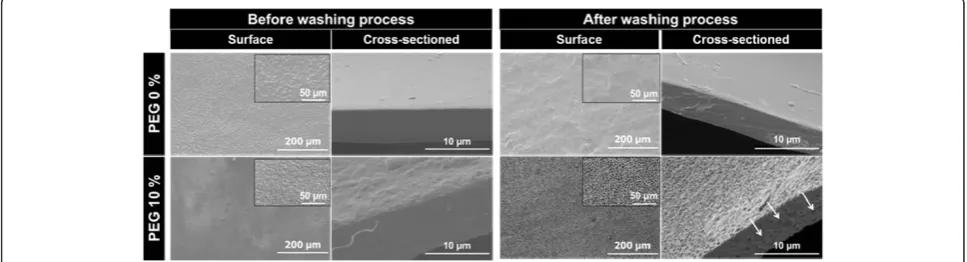
Figure 2Scanning electron micrographs of paclitaxel (PTX) incorporated PU membranes before and after washing process.
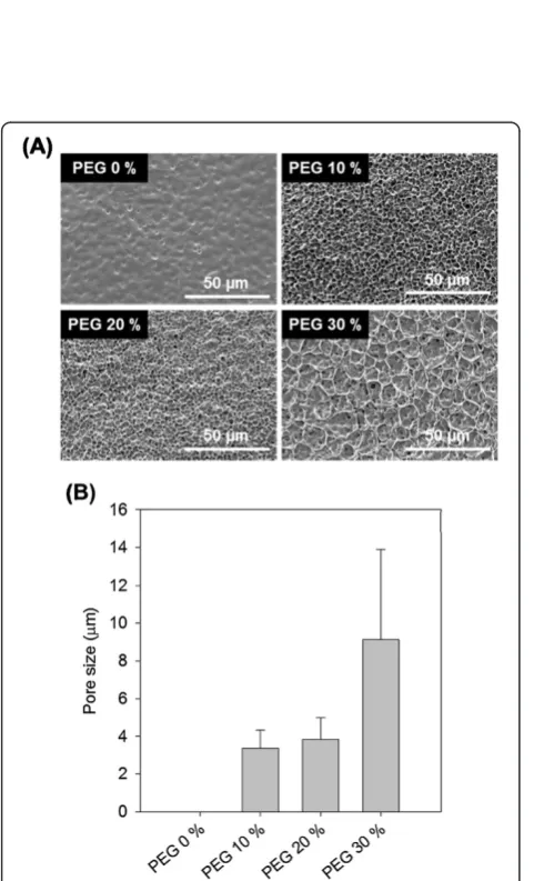
Figure 3Determination of pore size of PU membranes after washing process. (A)Scanning electron micrographs and(B)pore size distribution of PU membranes depending on PEG concentration.
16.0 ± 3.6μm, PEG 10% was 12.7 ± 1.2μm, PEG 20% was 11.3 ± 0.1 μm and PEG 30% was 14.0 ± 0.1 μm (Table 1). SEM observation of the PU membranes demonstrated that the surface and cross-sectioned was porous structure after PEG incorporating and washing out from PU membranes (Figure 1). On microscopic examination of the PTX-incorporated PU membranes with SEM, we could find ag-gregated and cracked PTX for PEG 0% PU membrane, but PTX crystallization wasn’t found on the surface of PEG 10% PU membrane which was a porous membrane (Figure 2). This PTX crystal and cracks allow limited release of PTX from membranes because of rate-limiting detachment of the drug from the PTX-incorporated mem-branes [17]. In other words, the porous PU membrane can inhibit PTX crystallization and then, PTX release was ex-pected to enhance.
PU membrane stent cover depending on PEG concentration
Each membrane type has uniform pore size (Figure 3A) and measurement of pore size was increased proportion-ally to PEG concentration (Figure 3B). PEG was incorpo-rated into PU membrane and washed out in water. The membranes increase in the equilibrium water uptake. This was attributed to the formation of a porous struc-ture in PU membranes. It was also evidenced by the ob-served increase in the diffusion coefficients. Generally, as diffusion is known to play a major role in the control of drug release [18,19].
According to our stent design, the porous PU mem-brane was designated as coating memmem-brane on bare metal stent. The PU membrane was formed between metal
(Figure 4A). The porous structure of PU membranes coat-ing on bare metal stents can be described like that PU membranes itself, as shown in Figure 4B.
In vitroPTX release test
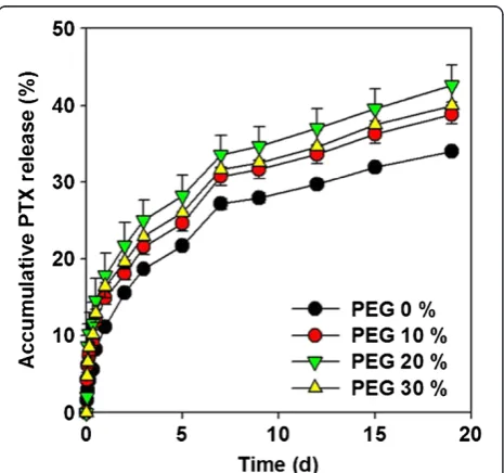
The PTX release behaviors of PU membranes under simulated physiological conditions (PBST, pH 7.4, 37°C) were investigated and compared (Figure 5). Overall PTX loading was approximately 0.461–0.467 mg/cm2. PTX
loading amounts was 0.465 mg/cm2in PEG 0% PU
mem-brane, 0.467 mg/cm2in PEG 10%, 0.466 mg/cm2in PEG
20% and 0.461 mg/cm2 in PEG 30% (Table 2). The
re-leased PTX, which was calculated based on the % rere-leased for 19 days, was 34.0% (released amount: 0.158 mg/cm2)
from PEG 0% PU membrane, 38.9% (0.182 mg/cm2) from
PEG 10%, 42.6% (0.198 mg/cm2) from PEG 20%, and
40.0% (0.185 mg/cm2) from PEG 30% (Figure 5). PEG 20% membrane showed the greatest release of PTX. Because PEG 30% membranes has too much larger pore size than PEG 10 and 20% membranes so, surface area of PEG 30% membranes is smaller than PEG 20% membranes [20]. As a result of the release properties associated with porous PU membranes, PTX crystallization was pro-tected and increased surface area could more release from PU membranes.
Conclusions
In this study, we investigated the effect of porous PU membrane as a bare metal stent coating material. The porous structure was formed by washing out of PEG from PU membranes. The release of PTX from porous PU membranes was increased for 19 days. This porous PU membrane could inhibit PTX crystallization and in-crease drug release because porous structure had larger surface area. The enhanced release of drug from porous PU membranes increases the potential usefulness of a bare metal stent cover to limited drug release of hydro-phobic anti-cancer drug, PTX.
Competing interests
The authors declare that they have no competing interests.
Authors’contributions
EHS made substantial contributions to conception and worked on the experiment. KN has provided guide lines and given final touch for the manuscript for publishing in biomaterials research. All authors read and approved the final manuscript.
Figure 5Accumulative PTX release profile depending on PEG concentration.
Table 2 PTX-incorporated PU membranes
Membrane type (w/w%) PTX amounts (mg/cm2)
PEG 0% 0.465 (±0.002)
PEG 10% 0.467 (±0.002)
PEG 20% 0.466 (±0.001)
Acknowledgments
This work was supported by the Technology Innovation Program (10044021, Development of nonvascular drug eluting stent for treatment of
gastrointestinal disease) funded by Ministry of Trade, industry & Energy (MOTIE, Korea).
Received: 30 July 2014 Accepted: 24 September 2014 Published: 8 October 2014
References
1. Jung G-S, Huh J-D, Lee SU, Han BH, Chang H-K, Cho YD:Bile duct: analysis of percutaneous transluminal forceps biopsy in 130 patients suspected of having malignant biliary obstruction 1.Radiology2002,224:725–730. 2. Seyama Y, Makuuchi M:Current surgical treatment for bile duct cancer.
World J Gastroenterol2007,13:1505.
3. Suk KT, Kim JW, Kim HS, Baik SK, Oh SJ, Lee SJ, Kim HG, Lee DH, Won YH, Lee DK:Human application of a metallic stent covered with a paclitaxel-incorporated membrane for malignant biliary obstruction: multicenter pilot study.Gastrointest Endosc2007,66:798–803.
4. Kwon C-I, Ko KH, Hahm KB, Kang DH:Functional self-expandable metal stents in biliary obstruction.Clin Endosc2013,46:515–521.
5. Kalinowski M, Alfke H, Kleb B, DüRFELD F, Wagner HJ:Paclitaxel inhibits proliferation of cell lines responsible for metal stent obstruction: possible topical application in malignant bile duct obstructions.Invest Radiol2002,37:399–404.
6. Lee DK:Drug‐eluting stent in malignant biliary obstruction.J Hepatobiliary Pancreat Surg2009,16:628–632.
7. Kwon HJ, Park S:Local delivery of antiproliferative agents via stents.
Polymers2014,6:755–775.
8. Lamba NM, Woodhouse KA, Cooper SL:Polyurethanes in biomedical applications.CRC press; 1997.
9. Heldman AW, Cheng L, Jenkins GM, Heller PF, Kim D-W, Ware M, Nater C, Hruban RH, Rezai B, Abella BS:Paclitaxel stent coating inhibits neointimal hyperplasia at 4 weeks in a porcine model of coronary restenosis.
Circulation2001,103:2289–2295.
10. Kang S-G, Lee SC, Choi SH, Park S, Jeong S, Lee DH, Kim M: Paclitaxel-polyurethane film for anti-cancer drug delivery: film characterization and preliminary in vivo study.Macromol Res2010,18:680–685.
11. Klose D, Siepmann F, Elkharraz K, Krenzlin S, Siepmann J:How porosity and size affect the drug release mechanisms from PLGA-based microparticles.
Int J Pharm2006,314:198–206.
12. Steele TW, Huang CL, Widjaja E, Boey FY, Loo JS, Venkatraman SS:The effect of polyethylene glycol structure on paclitaxel drug release and mechanical properties of PLGA thin films.Acta Biomater2011,7:1973–1983.
13. Huang CL, Steele TW, Widjaja E, Boey FY, Venkatraman SS, Loo JS:The influence of additives in modulating drug delivery and degradation of PLGA thin films.NPG Asia Materials2013,5:e54.
14. Andersson J, Rosenholm J, Areva S, Lindén M:Influences of material characteristics on ibuprofen drug loading and release profiles from ordered micro-and mesoporous silica matrices.Chem Mater2004,
16:4160–4167.
15. Moon S, Yang S-G, Na K:An acetylated polysaccharide-PTFE membrane-covered stent for the delivery of gemcitabine for treatment of gastrointestinal cancer and related stenosis.Biomaterials2011,32:3603–3610.
16. Park T-H, Jo E-A, Na K:Development of polymeric coating material for effective drug-eluting stent.Polymer (Korea)2011,35:483–487. 17. Lee DK, Kim HS, Kim K-S, Lee WJ, Kim HK, Won YH, Byun YR, Kim MY, Baik
SK, Kwon SO:The effect on porcine bile duct of a metallic stent covered with a paclitaxel-incorporated membrane.Gastrointest Endosc2005,
61:296–301.
18. Siepmann J, Göpferich A:Mathematical modeling of bioerodible, polymeric drug delivery systems.Adv Drug Deliv Rev2001,48:229–247.
19. Badiger MV, McNeill ME, Graham NB:Porogens in the preparation of microporous hydrogels based on poly (ethylene oxides).Biomaterials 1993,14:1059–1063.
20. Allen T:Particle Size Measurement. InVolume 2: Surface Area and Pore Size Determination.Springer; 1997 [Valverde Millán, José Manuel (Series Editor): Particle Technology, vol. 7].
doi:10.1186/2055-7124-18-15
Cite this article as:Seo and Na:Polyurethane membrane with porous surface for controlled drug release in drug eluting stent.Biomaterials Research201418:15.
Submit your next manuscript to BioMed Central and take full advantage of:
• Convenient online submission
• Thorough peer review
• No space constraints or color figure charges
• Immediate publication on acceptance
• Inclusion in PubMed, CAS, Scopus and Google Scholar
• Research which is freely available for redistribution