Original Research Article.
Cytopathological Evaluation of Various Thyroid Lesions Based on
Conventional Method and Bethesda System for Reporting in a
Tertiary Care Hospital
Sandeep Kumar Singh
1*, Faiyaz Ahmad
2, Seema Awasthi
3, Shyamoli Dutta
4, Himanshu Joshi
51*PG Resident (IIIrd Year), 2Associate Professor, 3Professor, 4Professor & Head, 5Assistant Professor,
Department of Pathology, Teerthanker Mahaveer Medical College & Research Centre, Moradabad, UP, India.
ABSTRACT
Aim and Objectives: To see the conventional and the Bethesda system for reporting thyroid cytopathology (TBSRTC), TBSRTC has standardized our diagnostic approach to cytomorphological criteria and reporting.
Material and Methods: A Total of 258 patients who presented with thyroid gland swelling were subjected to thyroid fine needle aspiration cytology (FNAC) and the smears were made followed by MGG staining and H&E staining with reporting was done. The conventional system used at our centre includes description of microscopic findings of the case along with an impression at the end and also classified according to the Bethesda system, found out the distribution of cases in each category, analysed the risk of malignancy in each category by the histological diagnosis wherever it was available and compared with other studies.
Results: Distribution of various categories from 258 FNAC of thyroid lesion was as follows: 1.2 % non-diagnostic, 94.2% benign, 1.9% atypia of undetermined significance (AUS/FLUS), 0.4% suspicious for follicular neoplasm, 0.4% suspicious for malignancy and 1.9% malignant. When the results of the conventional system were compared with the Bethesda adapted method was found to be more superior.
Conclusion: TBSRTC is an excellent reporting system in thyroid FNAC. It provides clear management guidelines to doctor to go for follow-up FNAC or surgery and the extent the surgery.
Key words: Fine Needle Aspiration Cytology, Bethesda
System, Histopathology, Thyroid Nodule.
*Correspondence to:
Dr. Sandeep Kumar Singh, PG Resident (IIIrd Year), Department of Pathology, TMMC & RC,
Moradabad, Uttar Pradesh, India. Article History:
Received: 05-12-2017, Revised: 28-12-2017, Accepted: 11-01-2018
Access this article online
Website:
www.ijmrp.com
Quick Response code
DOI:
10.21276/ijmrp.2018.4.1.056
INTRODUCTION
Fine needle aspiration cytology (FNAC) has been widely accepted as diagnostic of choice in the evaluation of patients presenting with non-toxic thyroid nodules.1,2 The technique is a safe, minimally invasive, easily performed with minimal patient discomfort, efficient, and an excellent cost effective method of evaluating thyroid lesions.3,4 However, due to lack of standardized system of reporting. The cytologists have used different terminologies thus creating confusion among clinicians in the interpretation of reports and further management. In the year 2007, a conference was hosted by the National Cancer Institute (NCI) at Bethesda, Maryland. The culmination of the meeting led to the Bethesda Thyroid Atlas Project and subsequently led to evolution of The Bethesda System for Reporting Thyroid Cytology (TBSRTC). It is a six category scheme with individual risks of malignancy that influence management paradigms.5,6 Our study was main purpose is to provide rational approach to management and to determine the correct surgical procedure when it is
required. A standardized categorical system for FNAC reporting can make results easier to understand for clinicians and give clear indications for therapeutic action.7,8
AIMS AND OBJECTIVES
▪ To compare the conventional and the Bethesda system for reporting thyroid cytopathology (TBSRTC).
▪ To correlate the cases with histology wherever available.
MATERIALS AND METHODS
obtained. These are helpful in reaching a probable clinical diagnosis as well as in cytohistological evaluation and formulations of the pathological diagnosis. Histopathology was used as a gold standard to compare the sensitivity of both systems.
Conventional Method
As per conventional method of reporting, the cases were diagnosed and placed under the following categories.7,9
▪ Non diagnostic/ Unsatisfactory: Smears were hemorrhagic
or containing less than six groups of well-preserved follicular cells on each of at least two slides.
▪ Colloid cyst: When follicular cells, thin or thick colloid in the background and hemosiderin laden macrophages were seen in smears.
▪ Follicular lesions/ Neoplasm: smears contained many
follicular cells without or scanty colloid background or when smears contain predominant population of Hurthle cells, the differential diagnosis would include hyperplasic adenomatoid nodule with Hurthle cell change, Hurthle cell adenoma, and Hurthle cell carcinoma.
▪ Indeterminate smears: containing cells with findings that were not clearly benign but were not diagnostic of a neoplasm or malignant lesions.
▪ Suspicious for malignancy: when aspirates suggested a follicular neoplasm, i.e., hyper cellular sample with scant colloid and a significant proportion of microfollicules, trabeculae, or crowded overlapping clusters of follicular cells (also includes lesions consisting of oncocytic (Hurthle cell) neoplasm).
▪ Malignant lesions:
Papillary Carcinoma; Medullary carcinoma; Anaplastic Carcinoma; Lymphoma; Metastatic
TBSRTC
The same cases were re-screened and reported as per the Bethesda system of reporting having the following six categories.10
▪ Non Diagnostic/ Unsatisfactory: fluid only virtually a cellular specimen other (obscuring blood, clotting artifact, etc). For a thyroid FNA specimen to be satisfactory for evaluation (and benign), at least six groups of benign follicular cells were required, each composed of at least 10 cells.11,12
▪ Benign: Consistent with a benign follicular nodule (includes adenomatoid nodule, colloid nodule etc). Consistent with
lymphocytic thyroiditis or Hashimoto’s thyroiditis in the proper
clinical context. Consistent with granulomatous (sub-acute) thyroiditis and others.
▪ Atypia of Undetermined Significance (AUS)/ Follicular
Lesion of Undetermined Significance: Result was
obtained in 3% to 6% of thyroid FNAC.
▪ Follicular Neoplasm/ Suspicious for a Follicular
Neoplasm: Specify if Hürthle cell (oncocytic) type.
▪ Suspicious for Malignancy: suspicious for papillary
carcinoma, suspicious for medullar carcinoma, suspicious of metastatic carcinoma, suspicious for lymphoma.
▪ Malignant: Papillary thyroid carcinoma, poorly differentiated carcinoma, Medullary thyroid carcinoma, Undifferentiated (anaplastic), carcinoma. Carcinoma with mixed features (specify), carcinoma, Non-Hodgkin lymphoma, and others.
The data included 258 cases of thyroid FNAC and 50 cases of follow-up histopathological specimens. The smears were prepared using conventional methods and stained with papanicolaou stains. We also calculate the risk of malignancy for each category and compare it with other studies. Sensitivity, specificity, positive predictive value and negative predictive value were calculated using histopathology diagnosis as gold standard. Data was analyzed using Statistical Package for Social Sciences (SPSS) version 21.0. Qualitative Data has been presented as number and percentages. Continuous data has been presented as mean+ SD.
Chi-square test and Independent samples‘t’-test was used to
compare the data. A ‘p’ value less than 0.05 indicated a
statistically significant association.
Table 1: Final Diagnosis Based on FNAC as conventional method
SN Diagnosis No. of cases Percentage
1. Colloid goiter/nodular/ multinodular goiter 115 44.6
2. Benign thyroid lesion 80 31.0
3. Thyroglossal cyst 8 3.1
4. Hashimoto’s thyroiditis 37 14.3
5. Carcinoma 6 2.3
6. Others 4 1.6
7. Thyroid/nodular hyperplasia 7 2.7
8. Unsatisfactory 1 0.4
Table 2: FNAC Reporting based on Proposed Bethesda System
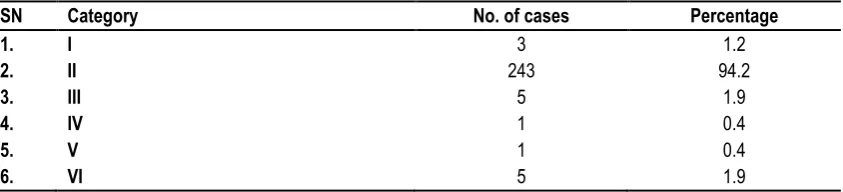
SN Category No. of cases Percentage
1. I 3 1.2
2. II 243 94.2
3. III 5 1.9
4. IV 1 0.4
5. V 1 0.4
Table 3: Distribution of subcategories in TBSRTC
S.No Cytological categories Subcategories No. of cases Total no. of cases
1 ND/UNS CYST FIUID 2 3(1.2%)
Acellular sample 0
Obscuring blood 1
2 Benign Adenomatoid nodule, colloid nodule 206 243(94.2%)
Lymphocytic thyroiditis 37
Granulomatous thyroiditis 0
3 AUS/FLUS - 5 5 (1.9%)
4 FN/SFN - 1 1(0.4%)
5 SFM Susp. For papillary carcinoma 0 1(0.4%)
Susp. For medullary carcinoma 1
6 Malignant Papillary thyroid carcinoma 5 5(1.9%)
Total 258
0 5 10 15 20 25 30 35 40 45
Colloid goiter/nodular/
multinodular goiter
Benign thyroid
lesion Thyroglossalcyst Hashimoto’s thyroiditis Carcinoma Others Thyroid/nodularhyperplasia Unsatisfactory
Fig 1: Final Diagnosis Based on FNAC as conventional method
0 10 20 30 40 50 60 70 80 90 100
I II III IV V VI
Table 4: Comparative analysis of study results of FNAC
Series No. of
cases
Sex Mean
age
Age range
FNAC RESULT
M(%) F(%) Benign Malignant Suspicious Non diagnostic
Chang et al (2006)
51 13(25) 38(76) 17 2-21 45(74) 6(10) 6(8) 4(7)
Kapila et al (2010)
792 68(9) 724(91) 17 4-21 699(88) 20(2.7) 26(3.5) 47(6)
Vidhya et al (2013)
284 25(9) 259(91) 17 7-21 243(86) 6(2) 12(4) 23(8)
Shanmuga et al (2016)
402 43(10.7) 359(89.3) 37.16 5-75 328(81.6) 11(2.7) 20(4.98) 43(10.7)
Present study 258 67(26) 191(74) 36.09 4-70 243(94.2) 5(1.9) 2(0.8) 3(1.2)
Table 5: Comparison of percentage of distribution of FNAC with other studies
Table 6: Comparison of percentage of risk of malignancy of present study with other studies
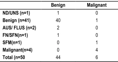
Table 7: Correlation of cytological and histopathological diagnosis
RESULTS
A total 258 patients (range 4-70 years) included in this study. The female to male ratio was 3:1. Majority of lesions were in 31-40 years of age group. The mean age of the study population was
36.09+13.95 years (SD) and median 36 years. Among the study population, a total of 50 patients underwent surgery and 208 patients did not undergo surgery. Distribution of cases as per conventional method of reporting was as per Table – 1 and figure
– 1. Distribution of cases as per Bethesda system of reporting was as per Table – 2 and figure - 2.
In our study AUS did not exceed the recommended target of 7%. Out of 258 cases, 50 cases were available for follow-up histopathology. We compared the original FNA diagnosis of these 50 cases with that of HPE and calculated the risk of malignancy for each category (Table - 5). None of the cases categorized as benign, or AUS/FLUS were reported to be malignant on follow-up (Table - 6). Thus malignancy risk for these categories is 0%.Out of 1 cases of FN/SFN, Thus malignancy risk for these categories is 0%. Out of 1 cases of SFM, 1 were malignant giving a risk of 100%. Out of 4 cases of malignant, thus malignancy risk for these categories is 100. The diagnostic accuracy of FNAC was 98.06% with sensitivity of 100% and specificity of 97.5%.
Diagnostic category Present
study
Shanmuga et al
Jo et al Yassa et al Nayar and
Inanovic
Payal M et al
Shagutta et al
ND/UNS 1.2 10.7 18.6 7 5 7.2 11.6
Benign 94.2 81.6 59 66 64 80 77.6
AUS/ FLUS 1.9 1.24 3.4 4 18 4.9 0.8
SFN 0.4 1.74 9.7 9 6 2.2 4
SFN 0.4 2 2.3 9 2 3.6 2.4
Malignant 1.9 2.7 7 5 5 2.2 3.6
Diagnostic category Present
study
Shanmuga et al
Jo et al Yassa et al Nayar and
Inanovic
Payal M et al
Shagutta et al
ND/UNS 0 0 8.9 10 9 0 20
Benign 2.5 0 11 0.3 2 13 3.1
AUS/ FLUS 0 0 17 24 6 100 50
SFN 0 28.6 25.4 28 14 25 20
SFM 80 71.4 70 60 53 50 80
Malignant 100 80 98.1 97 97 100 100
Benign Malignant
ND/UNS (n=1) 1 0
Benign (n=41) 40 1
AUS/ FLUS (n=2) 2 0
FN/SFN(n=1) 1 0
SFM(n=1) 0 1
Malignant(n=4) 0 4
Figure.3: Colloid goitre with cystic change Figure.4: Colloid goitre – scattered bare nuclei colloidophags with abundance of colloid
Figure.5: Benign thyroid lession Figure.6: Follicular epithelial along with micro follicle
Figure.7: Benign thyroid lesion – Follicular epithelial cells in mono layered sheet in fire flame
Figure.8: Hashimato’s thyroiditis
Figure.11: Medullary carcinoma with scattered plasmacytoid round to oval cell with binucleation
DISCUSSION
FNAC is the accurate, rapid, safe, reliable and cost- effective method for the evaluation of thyroid nodule. Ultrasound guided FNAC is recommended for non-palpable nodules, on-diagnostic aspirate and technically difficult location.
In pre-Bethesda group, the original cytological diagnoses could be summed up in 8 categories as depicted in Table 1. It included categories such as Inadequate/ Unsatisfactory (0.40%), Colloid goiter/nodular/ multinodular goiter (44.6%), Benign thyroid lesion (31%), Thyroglossal cyst (3.1%), Lymphocytic thyroiditis/
Hashimoto’s thyroiditis (14.3%), Thyroid/nodular hyperplasia
(2.7%), Carcinoma (suspicious of malignancy, suspicious of follicular neoplasm, follicular neoplasm, hurthle cell neoplasm, and other) (3.9%). Majority of the diagnostic labels used were broad and descriptive. They did not imply any specific treatment modalities. Such reports created confusion for treating clinicians and affected patient care.
The TBSRTC system is a simple, systematic universal reporting system with good clarity; thus creating understanding between pathologists and clinicians and helping in predicting prognosis and management of thyroid nodules.13,14 Managing pediatric patients with thyroid nodules can be challenging. Accurate pre-operative diagnosis is necessary to avoid thyroidectomy for benign lession. We compared the results in our study with Jo et al.,15 Chang et al.,16 kapila et al.,17 Yassa et al.,18 Payal M et al.,19 Nayar and Ivanovic20 and Shanmuga et al.21 The distribution of cases as per six-tier Bethesda system is different from other studies, with the percentage of cases in benign category being higher and that of non-diagnostic, malignant and AUS/FLUS category is lower (table 5). As per the guidelines of the Bethesda system, only aspirates with features of atypia, microfollicles and focal occurrence of hurthle cell that could not be categorized as benign, SFN, SFM and malignancy were described as AUS/FLUS. In our study the distribution of AUS is 0.4% that did not exceed the target 7%. The risk of malignancy for different categories in our study correlated well with the risks mentioned in the Bethesda system and with studies of Shanmuga Priya Shankar et al., Jo et al, Yassa et al. and Nayar and Ivanovic and differences with the studies of Payal M et al. and Shagutta et al.22 In our study number of cases under non-diagnostic, AUS, SFN and SFM category are less when compared to studies of Nayar & Ivanovic et al. and Yassa et al. hence the malignancy risk cannot be accurately compared (table 6). The recommended management for AUS/FLUS is clinical
correlation and repeat FNAC at an appropriate interval thus reducing the incidence of surgery.
In our study the sensitivity was 100%, Specificity 97.5% and accuracy 98.06%.Similar results were observed by Shanmuga Priya Shankar et al. and Kessler et al.23 in the false positive case, the presence of nuclear grooves and papillary fragments misled to the diagnosis of papillary carcinoma. The presence of nuclear grooves is also seen in cases of Hashimoto’s thyroiditis, nodular
hyperplasia and follicular adenoma. If suspicious lesions are considered positive, the sensitivity increases while the specificity decreases. If suspicious lesions are excluded, then the sensitivity decreases and the false negative rates increase.19,24
CONCLUSION
Fine needle aspiration cytology is gold standard diagnostic test for the diagnosis of thyroid lesions, in children and adult with a high diagnostic yield, accuracy, sensitivity & specificity. FNAC is a cost effective procedure that provides specific diagnosis rapidly with minimum complications. In conjuration with TBSRTC if provides good clarity thus creating understanding between pathologist and clinician helpful predicting prognosis and management. Detection of suspicious lesions or malignant cells is a definite indication of surgery. However; a negative FNAC from a thyroid lesions must be viewed with caution and should have a close follow-up.
REFERENCES
1. Mazzaferri EL. Management of a solitary thyroid nodule. N Engl J Med 1993; 328: 553-559.
2. Cooper DS, et al. guidelines for patients with thyroid nodules and differentiated thyroid cancer. Thyroid, 2006; 16(2): 109-142. 3. Cappelli C, Pirola I, Gandossi E, De Martino E, Agosti B, et al. Fine aspiration cytology of thyroid nodule:Does the needle matter? South Med J, 2009; 102: 498-501.
4. Moslavac S, Matesa N, Kusic Z. Thyroid fine needle aspirationcytology in children and adolescents Coll Antropol, 2010; 34: 197-200.
5. Cibas ES. Fine -needle aspiration in the work-up of thyroid nodules.Otolaryngol Clin North Am.2010; 43:257-71.
6. OzlukY, Pehlivan E et al. The use of the Bethesda terminology in thyroid fine-needle aspiration results in a lower rate of surgery for non-malignant nodules: A report from a reference centre in Turkey. Int J Surg Pathol.2011;19:761-71.
7. Basharat R, Bukhari MH, Saeed S, Hamid T. Comparison of fine needle aspiration cytology and thyroid scan in solitary thyroid nodule. Patholog Res Int, 2011; 7540-41.
8. Bongiovanni M, Spitale A, Faquin WC, Mazzucchelli L, Baloch ZW. The Bethesda System for Reporting Thyroid Cytopathology: A meta-analysis. Acta Cytol, 2012; 56: 333-339.
9. Bukhari MH, Niazi S, Hanif G, Qureshi SS, Munir M, et al. An updated audit of fine needle aspiration cytology procedure of solitary thyroid nodule. Diagn Cytopathol, 2008; 36: 104-112. 10. Langer JE, Baloch ZW et al. Thyroid nodule fine-needle aspiration. Semin Ultrasound CT MR, 2012; 33: 158-165
11. Goellner, et al. Fine needle aspiration cytology of the thyroid, 1980 to 1986. Acta Cytolo., 1987; 31: 587.
13. Wang HH. Reporting thyroid fine-needle aspiration: Literature review and a proposal. Diagnostic Cytopathology.2006; 34:67-76. 14. Cibas ES and Sanchez MA. The National cancer institute thyroid fine-needle aspiration state of the science conference: inspiration for a uniform terminology linked to management guidelines. Cancer cytopathology.2008; 114:71-73.
15. Jo VY, Stelow EB, Dustin SM, Hanley KZ. Malignancy risk for fine-needle aspiration of thyroid lesions according to the Bethesda system for reporting thyroid cytopathology. Am J Clin Pathol.2010;134:450-6.
16. Chang SH, Joe M, Kim H. Fine needle aspiration biopsy of thyroid nodule in children and adolescents. J Korean Med Sci.2006;21:469-73.
17. Kapila K, Pathan SK, Hali BE, Das DK. Fine- needle aspiration cytology of the thyroid in children and adolescents. Experience with 792 aspirates. Acta Cytologica.2010; 54:569-74.
18. Yassa L, Cibas ES, Benson CB, Frates MC, Doubilet PM, Gawande AA, et al. Long-term assessment of a multidisciplinary approach to thyroid nodule diagnostic evaluation. Cancer.2007;111:508-16.
19. Mehra P, Verma AK. Thyroid cytopathology reporting by the Bethesda system: A two-year prospective study in an academic institution. Pathology Research International.2015:1-11.
20. Nayar R, Ivanovic M. The indeterminate thyroid fine-needle aspiration: Experience from an academic centre using terminology similar to that proposed in the 2007 national cancer institute thyroid fine-needle aspiration state of the science conference.Cancer.2009;117:195-202.
21. Shanmuga Priya Shankar, Meenakshisundaram. K, Rajalakshmi. V, Satish Selvakumar. A, Bhanumathi Giridharan.
The Bethesda System for reporting thyroid cytopathology: A two year retrospective review in a tertiary care hospital. Indian Journal of Pathology and Oncology, January - March 2016:3(1); 48-54. 22. Mufti ST, Molah R. The Bethesda system of reporting thyroid cytopathology: A five year retrospective review of one centre experience. International journal of Health Sciences, Qassim University.2012; 6:131-143.
23. Kessler A et al. Accuracy and consistency of fine-needle aspiration biopsy in the diagnosis and management of solitary thyroid nodules.Israel Medical Association Journal.2005;7:371-73. 24. Mondal SK, Sinha S, Basak B, Roy DN, Sinha SK. The Bethesda system for reporting thyroid fine needle aspirates: A cytologic study with histologic follow-up. J Cytol.2013;30:94-99. [
Source of Support: Nil. Conflict of Interest: None Declared.
Copyright: © the author(s) and publisher. IJMRP is an official publication of Ibn Sina Academy of Medieval Medicine & Sciences, registered in 2001 under Indian Trusts Act, 1882. This is an open access article distributed under the terms of the Creative Commons Attribution Non-commercial License, which permits unrestricted non-commercial use, distribution, and reproduction in any medium, provided the original work is properly cited.