Original Research Article.
Assessment of Myocardial Perfusion Scan in the Detection of Ischemia and
Accurately Measuring the Ejection Fraction in CAD Patients in
Western Saudi Arabia: A Retrospective Cohort Study
Marwan Albeshri
1*, Loay Khair
2, Soha Elmorsy
3, Bashair Melebari
4, Mohammed Alsallum
1,
Anas Alsolami
1, Abdalrhman Qazli
1, Anwar Melebari
4, Fatma Aboul-Enein
51Medical Student, College of Medicine, King Abdualziz University, Jeddah, Saudi Arabia. 2MD, Assistant Consultant of Cardiology, King Abdullah Medical City, Makkah, Saudi Arabia. 3MD, PHD, Associate Professor of Pharmacology, Faculty of Medicine, Cairo University, Research Consultant, King Abdullah Medical City, Makkah, Saudi Arabia.
4Medical Student, College of Medicine, Umm Al-Qura University, Makkah, Saudi Arabia. 5Consultant Cardiology, King Abdullah Medical City (KAMC), Makkah, Saudi Arabia.
ABSTRACT
Background: Cardiovascular diseases are one of the most common diseases in Saudi Arabia and one of the most common leading causes of death. Myocardial perfusion scan (MPI) is one of the most commonly used imaging techniques for detection of myocardial ischemia and cardiac tissue viability. Previous literature estimated that the sensitivity of MPI in detecting the diseased patients ranges from 67-79 % and with specificity that ranges between 74-83%. Such assessments of MPI perfusion and ejection fraction evaluation have not been proven or estimated in our population and with the wide use of MPI in our country, it is important that new assessments should be conducted.
Methods: The records of King Abdullah Medical City were searched to locate the files of 535 cardiac patients who had undergone MPI. For all patients, the following data were extracted: demographics, co-morbidities, MPI results, coronary angiography results, and echocardiography results. After collecting all the data, sensitivity, specificity, positive predictive value, and negative predictive value were calculated. For ejection fraction, the level of agreement was examined between both MPI and echocardiography EF results.
Results: Analysis included 531 patients, 340 males and 191 females, with a mean age of 59.94 years. 319 patients had
diabetes, 363 were hypertensive, and 227 had dyslipidemia. MPI had a sensitivity of 91.8% and a specificity of 23.08%, with a positive predictive value of 71.63% and negative predictive value of 57.14%. Kappa measure of agreement between the ejection fraction of MPI and echocardiography showed a statistically significant difference (P<0.001) and k=0.376. Conclusion: MPI has high sensitivity but low specificity in detecting coronary artery disease, and not reliable in measuring EF in CAD patients.
Key-words: CAD, Angiography, MPI, Diagnosis.
*Correspondence to:
Marwan Ahmad Albeshri, Medical Student,
College of Medicine,
King Abdulaziz University, Jeddah, Saudi Arabia. Article History:
Received: 05-09-2018, Revised: 22-09-2018, Accepted: 14-11-2018
Access this article online Website:
www.ijmrp.com
Quick Response code
DOI:
10.21276/ijmrp.2018.4.6.005
INTRODUCTION
Cardiovascular disease (CVD) are one of the most common diseases in Saudi Arabia and one of the most common leading causes of death.1 With the increase in CVD risk factors among our
population the need for screening and early diagnosis is needed.2
In 2004 Al Nozha et al., concluded that the prevalence of CAD reached 5.5% in Saudi population with age of 45 years and above, male gender, high BMI and elevated blood sugar as risk factors.1
There are different non-invasive radiological imaging choices for detecting myocardial ischemia.3
Single-photon emission computed tomography (SPECT) myocardial perfusion scan (MPI) is one of the most commonly
used imaging techniques for detection of myocardial ischemia and cardiac tissue viability. SPECT MPI measures the physiological blood flow to the heart by detecting cardiac muscle uptake of the radiotracer.4
However, its ability to detect anatomical abnormalities in coronary arteries and multi-vessel disease is limited.5,6 Specifically, locating
the site of the lesion or its severity are two of those limitations and that will make the physicians’ decision on determining the right candidates for coronary angiography and revascularization procedures even more difficult.7-9 All of which contributes
features of coronary arteries and accurately detect the ischemic regions in cardiac muscle.5,6,9-12 Previous literature estimated that
the sensitivity of SPECT MPI in detecting the diseased vessel or patient ranges from 56-66 % and 67-79% at vessel and patients level respectively, and with specificity in detecting non-diseased patient or vessel that ranges from 81-87% and 74-83% at vessel and patient levels respectively.13
Other than measuring myocardial blood perfusion, SPECT MPI also assesses the ejection fraction of the left ventricle.14
Echocardiography is the most frequently used technique for initial evaluation of left ventricular ejection fraction because it is easily used and more widely available.15,16 Recent literature shows wide
variability in evaluation of EF using both echocardiography and SPECT MPI in patients with CAD.17,18
Such assessments of SPECT MPI perfusion and ejection fraction evaluation have not been proven or estimated in our population and with the wide use of SPECT MPI it would be difficult to predict the population at risk of false negative or false positive in our population and to assess the risk factors of false negative SPECT MPI. The degree of accuracy of SPECT MPI in measuring the ejection fraction have not been well established.
PATIENTS AND METHODS Methods
We followed the Strengthening the Reporting of Observational studies in Epidemiology (STROBE) guidelines for reporting our study methods and results.
Design
A hospital-based retrospective cohort study was conducted in the department of cardiology at King Abdullah Medical City (KAMC), Makkah, Saudi Arabia between January 2011 to September 2017 using electronic and paper-based patients’ records.
Setting
All adult patients who attended at KAMC Cardiac Center OPD or have been in-patients and underwent MPI from January 2011 until 15th of June 2016 were included.
Participants
We included patients ≥18 years old who underwent SPECT MPI in KAMC nuclear cardiology department with or without doing coronary angiography for diagnosing CAD between January 2011 and June 2016 were included. We included all adult patients of both genders and if no CAG is available patients should have records of 3 months of follow-up after the last MPI result. Patients less than 18 years of age and those with no CAG and no available record of 3 months follow-up after MPI were excluded.
Variables and Data Measurements
Using a standardized and pre-tested data extraction sheet, we collected data from electronic and paper-based hospital records for all patients. Files of patients who underwent MPI were identified via KAMC Hospital Information System and through retrospective chart review of medical records and percutaneous coronary intervention (PCI) registry.
According to the MPI test results, the patients were classified into two groups:
i) Those who did go for CAG: for those the results of CAG will be extracted.
ii) Those who did not go for CAG: for those we reviewed their files to see an evidence of coronary ischemia in the subsequent 3 months since the last negative MPI result.
We extracted data on age, gender, date of perfusion, co-morbidities (diabetes mellitus (DM), hypertension, dyslipidemia, smoking), last resting ECG before MPI, types of stress of MPI, result of MPI, types of perfusion defect, SSS, SRS, SDS, percentage of myocardial ischemia and CAG results.
If MPI was positive, the date of 1st subsequent CAG and the
arteries affected was extracted. If MPI was negative, the date of last visit OPD and the date of 1st subsequent CAG were extracted.
Finally, echocardiography results including the date of last echocardiogram and the ejection fraction were also extracted for all patients.
Quantitative Variables
MPI was considered positive if SSS result was more than 2. Coronary angiography was considered positive if at least one artery showed at least 70% lesion in all arteries except if it was in the left main coronary artery, in which case 50% lesion was considered positive. TID results were considered significant if the value was above 1.36 on adenosine stress and above 1.22 on exercise stress.20,21 Multivessel disease was defined as 2 or more
arteries showing 70% or more lesion in CAG. Study Size
Considering a reported prevalence of CAD in Saudi Arabia of around 5.5%(1) and at a 2-sided alpha of 0.05 and a power of 80%, we required a minimum of 278 patients for our study. Statistical Methods
Statistical analysis was done on SPSS version 20.0. Categorical variables were presented as percentages and numeric variables were presented as the mean ± the standard deviation if normally distributed. If not normally distributed, numeric variables were presented by the median and interquartile range. The agreement between the two tests was assessed by Kappa coefficient. Sensitivity of MPI was determined as true positives/true positives + false negatives. Specificity of MPI was determined as true negatives/true negatives + false positives. Positive predictive value was defined as true positive/ true positive + false positive. Negative predictive value was defined as: true negative/ true negative + false negative. Where true positive were cases labeled as positive MPI with positive CAG. True negative were cases labeled as negative MPI with negative CAG. False negative cases labeled as negative MPI but subsequently discovered to be positive by CAG within 3 months. False Positive defined as positive MPI with negative CAG within 3 months. All percentages were presented with 95% confidence interval.
RESULTS
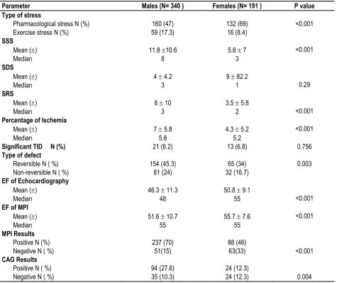
Analysis included 531 patients, in which 340 were males and 191 were females. Age shows no statistical difference (P=0.07). The frequency of co-morbidities in both males and females shows no statistically significant difference (P=0.157) nor each one of the DM, hypertension and dyslipidemia alone. Smoking was statistically significant (P<0.001) with 110 male patients were smokers compared to only 20 female patients that were smokers. (Table 1.1).
were significantly higher in males (11.8 10.6) when compared to females (5.6 7) with a p value of <0.001. SDS was not significantly different but SRS (P<0.001). The mean SRS in males was 8 ( 10) and females 3.5 ( 5.8).
Percentages of ischemia were higher in males than females (P<0.001). Males had a mean percentage of ischemia of 7 ( 5.8), where females had a mean of 4.3 ( 5.2). Reversible defects were found in 154 (45.3%) males and non-reversible in 81 (24%) compared to 65 (34%) females with reversible and 32 (16.7%) females with non-reversible defects (P=0.003).
The mean of ejection fraction was significantly different in males and females in both echocardiography and MPI (p<0.001). Males had a mean EF of 46.3 ( 11.3) using echocardiography and 51.6 ( 10.7) in MPI, where females had a mean EF of 50.8 ( 9.1) and 55.7 ( 7.6) in echocardiography and MPI respectively.
Males had more positive results in MPI and CAG compared to females. Two hundred and thirty-seven (70%) males had positive results in MPI, where only 94 (27.6%) of them had positive CAG results. On the other hand, 88 (46%) females had positive MPI results, where only 24 (12.3%) of them had positive CAG results.
Table 1.1: General Characteristics of the Study Population.
Parameter Males (N= 340 ) Females (N= 191 ) P value
Age
Mean () 59.3 11.7 61.1 9.2
0.07
Median 59 60
Co-morbidities N (%) 279 (82) 147 (77) 0.157
D.M. N (%) 204 (60) 115(60.2) 0.962
Hypertension N (%) 227 (66.7) 136 (71.2) 0.291
Dyslipidemia N (%) 154 (45.3) 73(38.2) 0.114
Smoking N (%) 110 (32.3) 20 (10.5) <0.001
Previous M.I. N (%) 83 (24.4) 29 (15.2) 0.012
Previous PCI N (%) 73 (21.5) 26 (13.6) 0.026
Previous CABG N (%) 34 (10) 13 (6.8) 0.214
Multivessel disease N(%) 54 (16) 14(7.3) 0.126
Table 1.2: MPI and CAG Results In Both Males And Female.
Parameter Males (N= 340 ) Females (N= 191 ) P value
Type of stress
Pharmacological stress N (%) 160 (47) 132 (69) <0.001
Exercise stress N (%) 59 (17.3) 16 (8.4)
SSS
Mean () 11.8 10.6 5.6 7 <0.001
Median 8 3
SDS
Mean () 4 4.2 9 82.2
0.29
Median 3 1
SRS
Mean () 8 10 3.5 5.8
<0.001
Median 3 2
Percentage of Ischemia
Mean () 7 5.8 4.3 5.2 <0.001
Median 5.8 5.2
Significant TID N (%) 21 (6.2) 13 (6.8) 0.756
Type of defect
Reversible N ( %) 154 (45.3) 65 (34) 0.003
Non-reversible N ( %) 81 (24) 32 (16.7)
EF of Echocardiography
Mean () 46.3 11.3 50.8 9.1
<0.001
Median 48 55
EF of MPI
Mean () 51.6 10.7 55.7 7.6 <0.001
Median 55 55
MPI Results
Positive N (%) 237 (70) 88 (46)
<0.001
Negative N ( %) 51(15) 63(33)
CAG Results
Positive N ( %) 94 (27.6) 24 (12.3)
0.004
Table 2: Sensitivity, Specificity, Negative Predictive Value And Positive Predictive.
Parameter Value 95% C.I.
Sensitivity % 91.8 85.04 – 96.19
Specificity % 23.1 12.53 – 36.84
Positive Predictive Value % 71.6 68.29 – 74.75
Negative Predictive Value % 57.1 37.49 – 74.77
Table 1.1: General Comparisons and Characteristics of Patients According To CAG Result.
Parameter CAG Positive (N=118) CAG Negative (N=59) P value
Age
Mean () 58.5 9.7 57.4 9.6 0.491
Median 58 58
Gender
Males 94 (80) 35 (59.3) 0.004
Females 24 (20.3) 24 (40.6)
Co-morbidities N (%) 99 (84) 45 (76.3) 0.219
D.M. N (%) 75 (63.6) 34 (57.6) 0.444
Hypertension N (%) 73 (62) 41 (69.5) 0.318
Dyslipidemia N (%) 47 (40) 31 (52.5) 0.108
Smoking N (%) 29 (24.6) 14 (24) 0.901
Previous M.I. N (%) 43 (36.4) 10 (17) 0.008
Previous PCI N (%) 37 (31.4) 9 (15.2) 0.021
Previous CABG 24 (20.3) 0 (0) <0.001
Multivessel disease N(%) 68 (57.6) 0 (0) <0.001
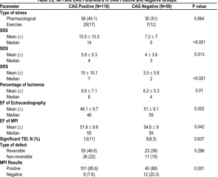
Table 3.2: MPI and CAG Parameters in CAG Positive and Negative Groups.
Parameter CAG Positive (N=118) CAG Negative (N=59) P value
Type of stress
Pharmacological 58 (49.1) 30 (51) 0.664
Exercise 20(17) 7(12)
SSS
Mean () 15.5 10.3 7.2 7
<0.001
Median 14 5
SDS
Mean () 5.8 5.3 4 3.6 0.013
Median 4 3
SRS
Mean () 10 10.1 3.5 5.8
<0.001
Median 7 2
Percentage of Ischemia
Mean () 9.5 7.1 6.2 5.3 0.01
Median 8 4
EF of Echocardiography
Mean () 46.1 9.7 51 9.1 0.002
Median 48 55
EF of MPI
Mean () 51.6 9.6 54.6 9 0.042
Median 55 55
Significant TID, N (%) 13(11) 5(8.5) 0.637
Type of defect
Reversible 55 (46.6) 23 (39) 0.296
Non-reversible 26 (22) 11 (19)
MPI Results
Positive 101 (85.6) 40 (68) 0.001
Negative 9 (7.6) 12 (20.3)
Table 2 shows the sensitivity, specificity, positive predictive value and negative predictive value of MPI after including all of the patients and using the definitions of each parameter as mentioned in the statistical methods section. MPI had a high sensitivity
In table 3.1, age shows no statistical difference but gender does (0.004) with males having more positive CAG results compared to females. However, when comparing comorbidities, DM, HTN, dyslipidemia, smoking and IHD, there were no statistically significant differences when comparing both groups. In CAG positive group, 24 patients had CABG compared to none of the CAG negative group (p<0.001), and 68 patients of CAG positive results had multivessel disease when underwent CAG compared to none of the CAG negative group (p<0.001).
In Table 3.2, SSS values were higher in CAG positive group with a mean of 15.5 ( 10.3) compared to 7.2 ( 7) in CAG negative group (p<0.001). SRS values were also significantly higher in CAG positive group when compared to CAG negative group (p<0.001). Percentages of ischemia and SDS values were not statistically significant. However, patients in CAG positive group showed lower EF (EF = 46.1 9.7) when compared to CAG negative group (EF = 51 9.1) (p=0.002). TID values and type of
defects in MPI were not significantly different in both groups. When comparing gated MPI left ventricular ejection fraction (LVEF) and echocardiography LVEF, the mean EF in our population was 53.07 by MPI and 47.87 by echocardiography. Paired sample T test showed that there is a statistically significant difference between the mean of LVEF of MPI and that of echocardiography (P<0.001). However, when we categorized patient according LV dysfunction: – Normal >50% – Mild- moderate 30-49% – Severe <30. There was low agreement between the two measurements, κ = 0.376 (p < 0.001).
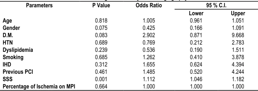
Multivariate regression analysis was done to examine the relation between the variables and CAG results. SSS score was significantly associated with positive CAG results (P<0.001, O.R. 1.112, 95%[CI] 1.046 to 1.182). DM had an odds ratio of 2.902 (95% CI 961-1051) when doing the regression analysis, however this result was not significant (P=0.083). Gender had an odds ratio of 0.425 (95% CI 0.166-1.091) (P=0.075)(Table 4)
Table 4: Multivariate Regression Analysis For Angiography Results.
Parameters P Value Odds Ratio 95 % C.I.
Lower Upper
Age 0.818 1.005 0.961 1.051
Gender 0.075 0.425 0.166 1.091
D.M. 0.083 2.902 0.871 9.668
HTN 0.689 0.769 0.212 2.783
Dyslipidemia 0.239 0.536 0.190 1.511
Smoking 0.685 1.262 0.410 3.878
IHD 0.312 1.655 0.624 4.394
Previous PCI 0.461 1.485 0.520 4.244
SSS 0.001 1.112 1.046 1.182
Percentage of Ischemia on MPI 0.664 1.000 1.000 1.000
Graph 1: Sensitivity, specificity, positive predictive value and negative predictive value of MPI.
DISCUSSION
After analyzing the results of 531 patients who had undergone SPECT MPI, our study revealed that SPECT MPI has a sensitivity and specificity of 91.8 % and 23.1 % in our patients, respectively. The sensitivity aligns with the findings of two notable meta-analyses where they concluded that the sensitivity of MPI is 87% and the specificity is 88%.22,23 However, other studies have found
that the sensitivity is considerably low. For example, one meta-analysis found that the sensitivity is only 78% in their patients24,
and in a prospective study funded by the British Heart Foundation (CE-MARC) where they found an even lower sensitivity.25
On the other hand, the specificity was significantly lower in our study than any other study with meta-analyses having a specificity
0 20 40 60 80 100
Sensitivity Specificity Postivie Predictive Value Negative Predictive Value
of 64 % and 61 % respectively compared to only 23.1 % in our study.22,23 Two other studies had a higher specificity (78% and
82.6% respectively).24,25 This can be attributed to the fact the SSS
threshold was set slightly lower than most papers26, being at 2
opposed to 3. It should be noted that a score of 2 in any individual segment means that perfusion is reduced moderately.
When looking at the mean SSS of CAG positive patients (15.5
10.3) in comparison to the patients who had CAG negative (7.2
7) (P<0.001), we find that there is a significant relationship between high risk of having cardiac event such as MI according to SSS score of the MPI and on the other hand, having at least one artery with 70% stenosis in CAG. Patients with SSS score of more than 13 are considered at high risk for developing a hard cardiac events.26
The negative predictive value (NPV) in our study was 57.1 %, which is very low compared to the NPV of another meta-analysis (NPV=98.8, C.I. 98.5-99.095) for the risk of MI and cardiac death27, proving that SPECT MPI is effective in detecting cardiac
ischemia in patients with low risk, where an invasive procedure such as CAG should be considered in patients with high risk only. CAG is commonly considered as a reference standard test for the evaluation and diagnosis of coronary artery disease (CAD). However, CAG may not provide some critical functional information such as the stenosis effect on the hemodynamics of the coronary artery and thus its perfusion. A study suggested that an unstable plaque may be the cause of an acute coronary syndrome instead of a severe coronary artery stenosis.28 In
addition, an emboli may cause an infarction even without chronic stenosis.29
The means of ejection fraction (EF) for both CAG positive and negative were 46.1 9.7 and 51 9.1, respectively on echocardiography and 51.6 9.6 and 54.6 9 respectively on rest SPECT MPI. EF was higher in the MPI groups which are contradictory to most studies measuring the differences between the two imaging modalities.30 One possible explanation is the fact
that we did not stratify the two groups based on transient ischemic dilation (TID) but based on CAG results in contrast to a study done by Emmett et al, which found a drop in LVEF in post-stress MPI in comparison to echocardiography, but they could not replicate their results, concluding to nearly similar results between the two modalities.31
It is believed that adenosine MPI normally induces hemodynamic alterations thus leading to abnormalities showing on the perfusion. The general consensus being that it is caused by reduced coronary flow reserve, not myocardial ischemia. Numerous studies showed that the reduction causes a subendocardial hypoperfusion, due to shunting of blood from the subendocardium to the subepicardium, taking into the account that coronary arteries are already having some stenosis.31,32
On Binary logistic regression, higher SSS score was significantly associated with positive CAG results (P<0.001, OR 1.112, 95% [CI] 1.046 to 1.182), this could be utilized by prioritizing patients with higher SSS score to undergo CAG. This could lead to lower cost and subsequently an increase in diagnostic accuracy. Our study is not without limitations, verification bias (when patients with positive screening test are referred more to the standard test in comparison to patients with negative screening test) could not be excluded. This is partly due to the invasive nature of the procedure and its cost, which would do more harm to the patients
than good if they had low risk of cardiac ischemia. Another potential limitation is the loss of follow up associated with this type of study and subsequently, the patients may have CAG done in another hospital. Another thing is that patients with negative results may have had subclinical obstructive atherosclerotic lesions, but were not detected because they did not have angiography done for them. The study contained a moderate to large sample size, but it was within one center. This could mask some weak but significant associations. There is a need for a multicenter study that includes the most important centers in Saudi Arabia's different regions.
CONCLUSION
SPECT MPI scan can be used as a screening test to detect myocardial ischemia for low to moderate risk patients; in addition, high SSS score can be used as an indicator for doing CAG in high risk patients.
ACKNOWLEDGMENTS
We would like to thank the medical records department at KAMC for their help in data collection.
REFERENCES
1. Al-Nozha MM, Arafah MR, Al-Mazrou YY, Al-Maatouq MA, Khan NB, Khalil MZ, et al. Coronary artery disease in Saudi Arabia. Saudi Med J. 2004;25(9):1165-71.
2. Al-Omran M. Atherosclerotic disease and risk factor modification in Saudi Arabia: a call to action.Vasc Health Risk Manag.2012;8:349-55. 3. Miller DD. Coronary flow studies for risk stratification in multivessel disease. A physiologic bridge too far? J Am Coll Cardiol. 2002;39(5):859-63.
4. Brindis RG, Douglas PS, Hendel RC, Peterson ED, Wolk MJ, Allen JM, et al. ACCF/ASNC appropriateness criteria for single-photon emission computed tomography myocardial perfusion imaging (SPECT MPI): a report of the American College of Cardiology Foundation Quality Strategic Directions Committee Appropriateness Criteria Working Group and the American Society of Nuclear Cardiology endorsed by the American Heart Association. J Am Coll Cardiol. 2005;46(8):1587-605.
5. Chamuleau SA, Meuwissen M, Koch KT, van Eck-Smit BL, Tio RA, Tijssen JG, et al. Usefulness of fractional flow reserve for risk stratification of patients with multivessel coronary artery disease and an intermediate stenosis. Am J Cardiol. 2002;89(4):377-80.
6. Christian TF, Miller TD, Bailey KR, Gibbons RJ. Noninvasive identification of severe coronary artery disease using exercise tomographic thallium-201 imaging.Am J Cardiol.1992;70(1):14-20. 7. Gibbons RJ, Chatterjee K, Daley J, Douglas JS, Fihn SD, Gardin JM, et al. ACC/AHA/ACP-ASIM guidelines for the management of patients with chronic stable angina: a report of the American College of Cardiology/American Heart Association Task Force on Practice Guidelines (Committee on Management of Patients With Chronic Stable Angina). J Am Coll Cardiol. 1999;33(7):2092-197.
8. Gibbons RJ, Abrams J, Chatterjee K, Daley J, Deedwania PC, Douglas JS, et al. ACC/AHA 2002 guideline update for the management of patients with chronic stable angina--summary article: a report of the American College of Cardiology/American Heart Association Task Force on practice guidelines (Committee on the Management of Patients With Chronic Stable Angina). J Am Coll Cardiol. 2003;41(1):159-68.
intervention (revision of the 1993 PTCA guidelines)-executive summary: a report of the American College of Cardiology/American Heart Association task force on practice guidelines (Committee to revise the 1993 guidelines for percutaneous transluminal coronary angioplasty) endorsed by the Society for Cardiac Angiography and Interventions. Circulation. 2001;103(24):3019-41.
10. Travin MI, Katz MS, Moulton AW, Miele NJ, Sharaf BL, Johnson LL. Accuracy of dipyridamole SPECT imaging in identifying individual coronary stenoses and multivessel disease in women versus men. J Nucl Cardiol. 2000;7(3):213-20.
11. Mahmarian JJ, Mahmarian AC, Marks GF, Pratt CM, Verani MS. Role of adenosine thallium-201 tomography for defining long-term risk in patients after acute myocardial infarction. J Am Coll Cardiol. 1995;25(6):1333-40.
12. Travin MI, Dessouki A, Cameron T, Heller GV. Use of exercise technetium-99m sestamibi SPECT imaging to detect residual ischemia and for risk stratification after acute myocardial infarction. Am J Cardiol. 1995;75(10):665-9.
13. Takx RA, Blomberg BA, El Aidi H, Habets J, de Jong PA, Nagel E, et al. Diagnostic accuracy of stress myocardial perfusion imaging compared to invasive coronary angiography with fractional flow reserve meta-analysis. Circ Cardiovasc Imaging. 2015;8(1).
14. Abidov A, Germano G, Hachamovitch R, Slomka P, Berman DS. Gated SPECT in assessment of regional and global left ventricular function: an update. J Nucl Cardiol. 2013;20(6):1118-43; quiz 44-6. 15. Hunt SA, Abraham WT, Chin MH, Feldman AM, Francis GS, Ganiats TG, et al. 2009 focused update incorporated into the ACC/AHA 2005 Guidelines for the Diagnosis and Management of Heart Failure in Adults: a report of the American College of Cardiology Foundation/American Heart Association Task Force on Practice Guidelines: developed in collaboration with the International Society for Heart andLungTransplantation.Circulation.2009;119(14):e391-479. 16. Patel MR, White RD, Abbara S et al. 2013 ACCF/ACR/ASE/ASNC/SCCT/SCMR appropriate utilization of cardiovascular imaging in heart failure: a joint report of the American College of Radiology Appropriateness Criteria Committee and the American College of Cardiology Foundation Appropriate Use Criteria Task Force. J Am Coll Cardiol. 2013;61(21):2207-31.
17. Garcia EV, Faber TL, Cooke CD, Folks RD, Chen J, Santana C. The increasing role of quantification in clinical nuclear cardiology: the Emory approach.J Nucl Cardiol.2007;14(4):420-32.
18. Germano G, Kavanagh PB, Slomka PJ, Van Kriekinge SD, Pollard G, Berman DS. Quantitation in gated perfusion SPECT imaging: the Cedars-Sinai approach. J Nucl Cardiol. 2007;14(4):433-54.
19. von Elm E, Altman DG, Egger M, Pocock SJ, Gøtzsche PC, Vandenbroucke JP. The Strengthening the Reporting of Observational Studies in Epidemiology (STROBE) statement: guidelines for reporting observational studies. The Lancet.370(9596):1453-7.
20. Mazzanti M, Germano G, Kiat H, Kavanagh PB, Alexanderson E, Friedman JD, et al. Identification of severe and extensive coronary artery disease by automatic measurement of transient ischemic dilation of the left ventricle in dual-isotope myocardial perfusion SPECT. J Am Coll Cardiol. 1996;27(7):1612-20.
21. Abidov A, Bax JJ, Hayes SW et al. Integration of automatically measured transient ischemic dilation ratio into interpretation of adenosine stress myocardial perfusion SPECT for detection of severe and extensive CAD. J Nucl Med. 2004;45(12):1999-2007.
22. Fleischmann KE, Hunink MM, Kuntz KM, Douglas PS. Exercise echocardiography or exercise spect imaging? A meta-analysis of diagnostic test performance. JAMA. 1998; 280 (10):913-20.
23. Jaarsma C, Leiner T, Bekkers SC, Crijns HJ, Wildberger JE, Nagel E, et al. Diagnostic Performance of Noninvasive Myocardial Perfusion Imaging Using Single-Photon Emission Computed Tomography, Cardiac Magnetic Resonance, and Positron Emission Tomography Imaging for the Detection of Obstructive Coronary Artery Disease: A Meta-Analysis. Journal of the American College of Cardiology. 2012;59(19):1719-28.
24. Dai N, Zhang X, Zhang Y, Hou L, Li W, Fan B, et al. Enhanced diagnostic utility achieved by myocardial blood analysis: A meta-analysis of noninvasive cardiac imaging in the detection of functional coronary artery disease. International Journal of Cardiology. 2016;221:665-73.
25. Greenwood JP, Maredia N, Younger JF, Brown JM, Nixon J, Everett CC, et al. Cardiovascular magnetic resonance and single-photon emission computed tomography for diagnosis of coronary heart disease (CE-MARC): a prospective trial. The Lancet. 2012;379(9814):453-60.
26. Dvorak RA, Brown RKJ, Corbett JR. Interpretation of SPECT/CT Myocardial Perfusion Images: Common Artifacts and Quality Control Techniques. RadioGraphics. 2011;31(7):2041-57.
27. Metz LD, Beattie M, Hom R, Redberg RF, Grady D, Fleischmann KE. The Prognostic Value of Normal Exercise Myocardial Perfusion Imaging and Exercise Echocardiography: A Meta-Analysis. Journal of the American College of Cardiology. 2007;49(2):227-37.
28. Virmani R, Burke AP, Farb A, Kolodgie FD. Pathology of the Vulnerable Plaque. Journal of the American College of Cardiology. 2006;47(8, Supplement):C13-C8.
29. Larsen AI, Galbraith PD, Ghali WA, Norris CM, Graham MM, Knudtson ML. Characteristics and outcomes of patients with acute myocardial infarction and angiographically normal coronary arteries. The American Journal of Cardiology. 2005;95(2):261-3.
30. Ward RP, Gundeck EL, Lang RM, Spencer KT, Williams KA. Overestimation of postischemic myocardial stunning on gated SPECT imaging: correlation with echocardiography. J Nucl Cardiol. 2006;13(4):514-20.
31. Emmett L, Ng A, Ha L, Russo R, Mansberg R, Zhao W, et al. Comparative assessment of rest and post-stress left ventricular volumes and left ventricular ejection fraction on gated myocardial perfusion imaging (MPI) and echocardiography in patients with transient ischaemic dilation on adenosine MPI: myocardial stunning or subendocardial hypoperfusion? J Nucl Cardiol. 2012;19(4):735-42. 32. Iskandrian AS. Adenosine myocardial perfusion imaging. J Nucl Med. 1994;35(4):734-6.
[
Source of Support: Nil. Conflict of Interest: None Declared.
Copyright: © the author(s) and publisher. IJMRP is an official publication ofIbn Sina Academy of Medieval Medicine & Sciences, registered in 2001 under Indian Trusts Act, 1882.
This is an open access article distributed under the terms of the Creative Commons Attribution Non-commercial License, which permits unrestricted non-commercial use, distribution, and reproduction in any medium, provided the original work is properly cited.