ISSN: 2319-6505
A COMPARATIVE ASSESSMENT OF TRACE METAL ACCUMULATION IN
OREOCHROMIS
MOSSAMBICUS
AND
LABEO ROHITA
EXPOSED TO PLANT NUTRIENT LIBRELTM
Sadekarpawar Shalaka, Pandya Parth, Upadhyay Ankur and Parikh Pragna*
Department of Zoology, Faculty of Science, The Maharaja Sayajirao University of Baroda, Vadodara- 390001, Gujarat, India
A R T I C L E I N F O A B S T R A C T
Metal pollution from multifarious sources like effluents from industries, agricultural runoff and untreated sewage system has adverse effects on aquatic ecosystem. Metallo-pesticides, including insecticides, fungicides, and herbicides are also known to contain various metals that can increase metal accumulation. The presence of metal pollutant in fresh water is known to disturb the delicate balance of the aquatic systems. Fish are often at the top of the aquatic food chain and may concentrate large amounts of some metals from the water. The study correlated the level of metal ions (Fe, Cu, Zn, Mn) in tissues and in water in a time dependent as well as dose dependent manner in both the species of teleost (O.mossambicus and L.rohita). Further Bioconcentration factor added metal based affinity towards tissue. Hence, the study gears up and proves that among both the fishes tested on exposure to micronutrient mixture, O.mossambicus was found to be more sturdy i.e it was able to with stand more metal load compared to L.rohita in a time dependent manner.
INTRODUCTION
The pollution of the aquatic environment with trace heavy metals has become a worldwide problem during recent years because they are indestructible and most of them have toxic effects on organisms (MacFarlane and Burchett, 2000). Among environmental pollutants, metals are of particular concern due to their potential toxic effect and ability to bio accumulate in aquatic ecosystems (Censi et. al., 2006). The presence of heavy metals in aquatic ecosystems is the result of two main sources of contamination; natural processes and anthropogenic activities (Goullé et al., 2012; Moore and Attar, 2011). Metal pollution from multifarious sources like effluents from industries, agricultural runoff and untreated sewage system has adverse effects on aquatic ecosystem. Intensification of agriculture practices to meet the demand has resulted in increased release of a wide range of agrochemical compounds to the environment (Desai and Parikh, 2013). Metallo-pesticides, including insecticides, fungicides, and herbicides are also known to contain various metals that can increase metal accumulation. (Senesi et al., 1999; Nicholson et al., 2003; Yabe et al., 2012). Metal concentration in aquatic ecosystems are usually monitored by measuring their concentrations in water, sediments and biota (Ergul et al., 2008) which generally exist in trace levels in water and attain considerable concentration in sediments and biota (Unlu et al., 2008). These pollutants when compared with other types of aquatic pollution are less visible but its effects on the ecosystem and humans are intensive and very extensive due to their toxicity and their ability to accumulate in the aquatic organisms (Edem et al., 2008). The presence of metal pollutant in fresh water is known to disturb the delicate
balance of the aquatic systems. Fish are often at the top of the aquatic food chain and may concentrate large amounts of some metals from the water (Mansour and Sidky, 2002).They are notorious for their ability to accumulate the metals in their muscles. Any of these metals can destroy life when they concentrate in the body above acceptable levels. Such a contaminated fish can cause health hazards when they enter into the human body through consumption (Ozuni et al., 2010). Hence, there is a need to carefully screen to ensure the unnecessary high level of some toxic trace metals that are being transferred to humans through fish consumption (Adeniyi and Yusuf, 2007).
Hence this study is geared towards determining the accumulation of the trace metals in the fish tissues as well as in the water with the view to establish the comprehensive evaluation of metals in various tissues i.e. liver, kidney, gills and muscles.
MATERIALS AND METHODS
Healthy O. mossambicus and L. rohita, were collected from local fresh water bodies of Baroda district and acclimatized at laboratory conditions for 10 days. Fishes were maintained in 25 ± 2 C, pH 7.4 ± 0.05, dissolved oxygen 8 ± 0.3 mg/L, and total hardness 188 mg/L CaCO3 with a 12:12 light: dark photoperiod accordance with the Guidelines of A.P.H.A., A.W.W.A., W.P.C.F (Jomova and Valko, 2011). Fishes were daily supplied with commercial food during acclimation and experimental period. Acclimatized fishes were exposed to water containing the test chemical at the concentration of 300 mg/L (1/20th of LC50 value) for 45 days at the semi static
Available Online at http://journalijcar.org
International Journal
of Current Advanced
Research
International Journal of Current Advanced Research Vol. 4, Issue, 10, pp. 441-449, October, 2015
Article History:
Received 19th, September, 2015
Received in revised form 27th,September,2015 Accepted 13th, October, 2015
Published online 28th, October, 2015
© Copy Right, Research Alert, 2015, Academic Journals. All rights reserved.
RESEARCH ARTICLE
ISSN: 2319 - 6475
Key words:
system. The Trace element mixture used was a commercial formulation of Librel TMX, Chelated Micronutrient mixture (Nutrient % by Wt. Min., Zn-4.0, Mn-0.5, Cu-0.3, Fe-2.0 and B-0.5). No test chemical was put into the aquarium containing the control fish. The water in the control and metal containing aquarium was renewed every day in order to maintain the concentration of the test mixture. At each interval of 15, 30 and 45 days of long-term exposure, fish were sampled from each group for determination of metals (Zn, Fe, Cu and Mn) in different organs (gills, liver, muscle and kidney). Water samples at every intervals were prepared using the method of APHA (1995) and different fish tissues were digested after drying according to the method described in APHA 3111B (Direct Acetylene Flame Method). The levels of Fe, Cu, Zn and Mn in digests as water were determined using atomic absorption spectrophotometer. Bioaccumulation factor was calculated according to using the following equation:
Bio-concentration factor (BCF) =Concentration of M in dry fish tissue (mg/kg)
Concentration of M in water (mg/L)
MPI = (CF1x CF2 x--- CFn)1/n
Where, Cfn is the contents for the metal n in the sample (Usero et al., 1997). Average concentrations and standard deviations were calculated for each element, tissues and fish species. The significance levels of the differences between element concentrations in the studied fish organs and between experimental groups were determined using the Mann-Whitney (Sokal and Rohlf, 1987) test. Heavy metal contents determined in water and fish tissue samples were evaluated statistically using analysis of correlation by SPSS (version number-21) statistical package. The statistical analyses were determined as 0.05.
Statistical analysis
The values of protein content were statistically calculated using one way ANOVA and post hoc Dunnetts’s t test was done to find the significance alterations if any between control and different exposure groups using SPSS software (version 21).
RESULTS
The result of metals determined in the water at different exposure periods are presented in Table I-VII along with the standard values. They were in the order Fe (18.83 mgL-1)
>Zn(6.34 mgL-1) >Mn (4.29 mgL-1) >Cu (0.7 mgL-1).
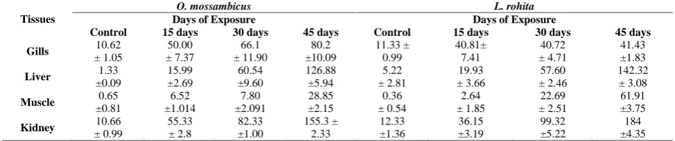
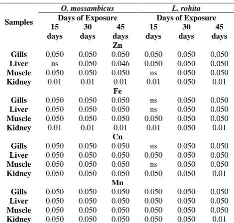
The alterations in the trace metal concentrations in water and tissue of O. mossambicus and L. rohita were determined and their means and S.D. are presented in Table (I-IV). Time dependent increase in the metal content of the tissues as well as water was observed. Amongst two fish species, L. rohita exhibited significantly higher ability to amass metals than O. mossambicus. The order of pattern of accumulation of metals in the tissues was liver>gills>kidney>muscle. Fe exhibited highest concentration in liver of both the fishes. Individual metal concentration assessment exhibited higher values of Fe in L. rohita (2108.20±771.67 mg/Kg) than O. mossambicus 1589.20±45.30 mg/Kg). Organ wise concentration that tracked the order for Fe was: L>G>K>M. The second highest trace metal in order was Zn in both the fishes, where L.rohita (184±4.35 mg/Kg) revealed higher concentration compared to O. mossambicus (155.3±2.33mg/Kg). Organ wise accumulation that followed the order for Zn concentration was K>G>M>L. Next in the order was Cu, and it was L. rohita (75.04±10.60 mg/Kg) which exhibited higher concentration compared to O. mossambicus (60.00±3.17 mg/Kg). Organ wise accumulation that followed the order for Cu accumulation was: L>G>M>K. Mn exhibited the least concentration, where L. rohita (131±3.60 mg/Kg) accumulated higher Mn compared to O. mossambicus 3.50±11.77 mg/Kg). Organ wise accumulation that followed the order for Mn accumulation was: M>L>K>G. To have an insight for the interspecific differences Mann-whitney test was performed (Table VI).
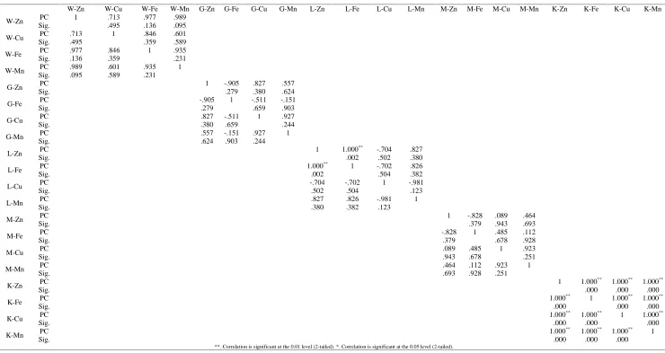
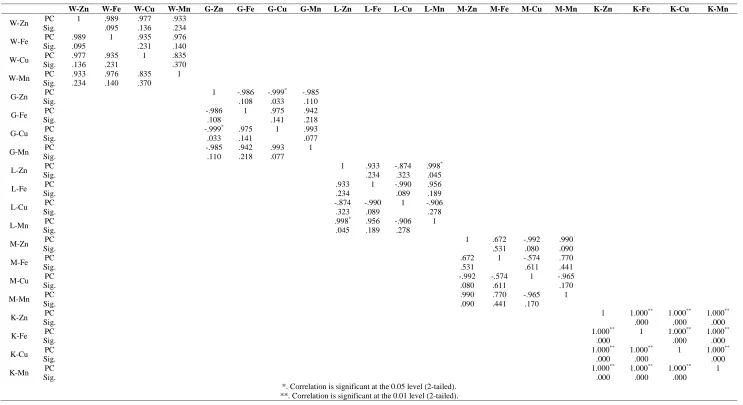
Concentration of trace metals (mean and standard deviations) in the tissues of both the teleost fish over the period of time are presented in Table I-IV where all trace element showed altered affinities in the studied fish tissues and did not differ significantly between the tissues except kidney. The overall relationship among the various elements was calculated by Pearson correlation coefficient and data is presented in the form of a matrix (Table VI&VII).
In water, all metals showed positive correlation with the other metals. Metal concentration for inter tissue correlation for O. mossambicus gills, Zn showed positive correlation with other three metals; while other three metals showed negative correlation with each other. In the Liver of showed negative correlation with all the other three metal ions, while Zn, Fe and Mn showed positive correlation with each other. In muscle Cu and Mn showed positive with each other as well as with Fe and Zn, on the other hand Zn and Fe showed a negative correlation with each other.
Table I Concentration of Zn in water and selected tissues of fish exposed to sub lethal concentration of plant nutrient (n = 6).
Tissues
O. mossambicus L. rohita
Days of Exposure Days of Exposure
Control 15 days 30 days 45 days Control 15 days 30 days 45 days Gills 10.62
± 1.05
50.00 ± 7.37
66.1 ± 11.90
80.2 ±10.09
11.33 ± 0.99
40.81± 7.41
40.72 ± 4.71
41.43 ±1.83 Liver 1.33
±0.09
15.99 ±2.69
60.54 ±9.60
126.88 ±5.94
5.22 ± 2.81
19.93 ± 3.66
57.60 ± 2.46
142.32 ± 3.08 Muscle 0.65
±0.81
6.52 ±1.014
7.80 ±2.091
28.85 ±2.15
0.36 ± 0.54
2.64 ± 1.85
22.69 ± 2.51
61.91 ±3.75 Kidney ± 0.9910.66 55.33± 2.8 82.33±1.00 155.3 ±2.33 12.33±1.36 36.15±3.19 99.32±5.22 ±4.35184
In kidney, a positive correlation amongst Zn, Fe and Cu and a negative correlation of MN towards all the three was reported. Whereas, in L. rohita: Zn showed negative correlation with other three metals; while other three metals showed positive correlation with each other.
In liver muscle and kidney Zn, Fe and Mn showed positive correlation with each other while negative correlation of these three metals was seen with Cu. In order to estimate the toxicity of trace metals accumulated in the experimental set up the BCF for each trace metal was calculated (Fig. II). The order of BCF for trace metals was Cu>Fe>Mn>Zn for both the fishes. However, when tissue BCF was compared the order was for O. mossambicus and L. rohita
Cu: liver>gills>muscle>kidney Fe: liver>gills>kidney>muscle Zn: liver>kidney>gills>muscle
Except for Mn which differed in both the fishes. For O. mossambicus it was liver>muscle>kidney>gills and L. rohita muscle>liver>kidney>gills
Table II Concentration of Fe in water and selected tissues of fish exposed to sub lethal concentration of plant nutrient (n = 6).
Tissues
O. mossambicus L. rohita
Days of Exposure Days of Exposure
Control 15 days 30 days 45 days Control 15 days 30 days 45 days Gills 4.13 ± 0.19 3.47 ±0.13 385.5 ±0.10 850.25
±111.54 2 ± 1 5 ± 5
151.41 ±26.06
330.50 ±28.30 Liver ±20.9216.49 93.16±8.34 ±140.95642.86 ±45.301589.2 ± 41.4441.44 178.40±9.66 1142.073±99.77 ±771.672108.20
Muscle 1.55 ±1.18 7.28 ±1.27 24.25 ±3.45 121.7 ±23.50 0.14 ± 0.16 0.87 ± 0.42 10.53 ± 0.72 51.77 ± 2.93 Kidney 14.33±2.98 47.33±2.87 ±3.2288.95 149.22±5.98 12.33±1.22 53.21±2.33 99.23±5.3 168.83±7.06
Values are presented in Means ± S.D.
Table III Concentration of Cu in water and selected tissues of fish exposed to sub lethal concentration of plant nutrient (n = 6).
Tissues
O. mossambicus L. rohita
Days of Exposure Days of Exposure
Control 15 days 30 days 45 days Control 15 days 30 days 45 days Gills 0.77 ±0.69 3.58 ±0.92 27.11 ±8.18 61.4 ±7.09 0.21 ± 0.11 0.97 ± 1.22 33.04 ± 20.24 46.01 ±6.10 Liver 2.58 ±0.15 6.26 ±0.20 59.25 ± 9.04 60.04 ±10.60 0.11 ± 0.18 1.48 ±0.76 15.75 ± 4.15 75.00 ± 3.17 Muscle 1.06 ±1.17 5.11 ±1.31 19.93 ±2.46 41.04 ±4.89 0.14 ± 0.16 0.92 ± 0.66 2.53 ± 0.64 27.94 ± 2.35 Kidney 1.22 ± 0.82 9.22 ±0.98 19.23 ±2.33 25.33
± 1.98 0.99 ± 0.19 15.32 ±0.33
20.32 ±1.22
27.52 ±2.03
Values are presented in Means ± S.D.
Table IV Concentration of Mn in water and selected tissues of fish exposed to sub lethal concentration of plant nutrient (n = 6).
Metals
O. mossambicus L. rohita
Days of Exposure Days of Exposure
Control 15 days 30 days 45 days Control 15 days 30 days 45 days Gills 0.95 ±0.61 6.99 ±2.08 10.37 ± 14.61 29.58 ±0.89 0.02 ± 0.02 0.34 ± 0.22 5.93 ± 1.39 15.52 ± 3.68 Liver 1.55 ±0.69 30.17 ±15.53 67.07 ±6.60 115.55 ±5.011 5.22 ± 1.15 15.96 ± 4.10 32.83 ± 6.21 64.86 ± 3.64 Muscle ±0.992.25 ±1.509.7 ±11.7723.50 ±1.5243.50 ± 17.6937.06 ± 3.6062.70 ± 3.5099.00 ± 2.51131
Kidney 5.32 ±0.87 15.33 ±2.39 25.38 ± 4.23 35.39 ±5.65 3.23 ±0.99 9.33 ±1.33 29.33 ±2.33 38.93 ±3.98
Values are presented in Means ± S.D.
Table V Time dependent significance of differences in the concentrations of metals in the organs of fish species
exposed to plant nutrient
Samples
O. mossambicus L. rohita Days of Exposure Days of Exposure 15 days 30 days 45 days 15 days 30 days 45 days Zn
Gills 0.050 0.050 0.050 0.050 0.050 0.050 Liver ns 0.050 0.046 0.050 0.050 0.050 Muscle 0.050 0.050 0.050 ns 0.050 0.050 Kidney 0.01 0.01 0.01 0.01 0.050 0.01
Fe
Gills 0.050 0.050 0.050 ns 0.050 0.050 Liver 0.050 0.050 0.050 ns 0.050 0.050 Muscle 0.050 0.050 0.050 0.050 0.050 0.050 Kidney 0.01 0.01 0.01 0.01 0.050 0.01
Cu
Gills 0.050 0.050 0.050 ns 0.050 0.050 Liver 0.050 0.050 0.050 0.050 0.050 0.050 Muscle 0.050 0.050 0.050 ns 0.050 0.050 Kidney 0.050 0.050 0.050 0.050 0.050 0.01
Mn
Gills 0.050 0.050 0.050 0.050 0.050 0.050 Liver 0.050 0.050 0.050 0.050 0.050 0.050 Muscle 0.050 0.050 0.050 0.050 0.050 0.050 Kidney 0.050 0.050 0.050 0.050 0.050 0.01
Table VII Interwater and inter tissue Pearson Correlation of O.mossambicus
W-Zn W-Cu W-Fe W-Mn G-Zn G-Fe G-Cu G-Mn L-Zn L-Fe L-Cu L-Mn M-Zn M-Fe M-Cu M-Mn K-Zn K-Fe K-Cu K-Mn W-Zn PC 1 .713 .977 .989
Sig. .495 .136 .095 W-Cu PC .713 1 .846 .601 Sig. .495 .359 .589 W-Fe Sig.PC .977.136 .846.359 1 .935.231
W-Mn PC .989 .601 .935 1 Sig. .095 .589 .231
G-Zn PC 1 -.905 .827 .557
Sig. .279 .380 .624
G-Fe PC -.905 1 -.511 -.151
Sig. .279 .659 .903
G-Cu PC .827 -.511 1 .927
Sig. .380 .659 .244
G-Mn PC .557 -.151 .927 1
Sig. .624 .903 .244
L-Zn PC 1 1.000
** -.704 .827
Sig. .002 .502 .380
L-Fe PC 1.000
** 1 -.702 .826
Sig. .002 .504 .382
L-Cu PC -.704 -.702 1 -.981
Sig. .502 .504 .123
L-Mn PC .827 .826 -.981 1
Sig. .380 .382 .123
M-Zn Sig.PC 1 -.828.379 .089.943 .464.693
M-Fe PC -.828 1 .485 .112
Sig. .379 .678 .928
M-Cu PC .089 .485 1 .923
Sig. .943 .678 .251
M-Mn PC .464 .112 .923 1
Sig. .693 .928 .251
K-Zn PC 1 1.000
** 1.000** 1.000**
Sig. .000 .000 .000
K-Fe PC 1.000
**
1 1.000** 1.000**
Sig. .000 .000 .000
K-Cu PC 1.000
** 1.000** 1 1.000**
Sig. .000 .000 .000
K-Mn PC 1.000
** 1.000** 1.000** 1
Sig. .000 .000 .000
Table VII: Interwater and inter tissue Pearson Correlation of L.rohita
W-Zn W-Fe W-Cu W-Mn G-Zn G-Fe G-Cu G-Mn L-Zn L-Fe L-Cu L-Mn M-Zn M-Fe M-Cu M-Mn K-Zn K-Fe K-Cu K-Mn W-Zn PC 1 .989 .977 .933
Sig. .095 .136 .234 W-Fe PC .989 1 .935 .976 Sig. .095 .231 .140 W-Cu PC .977 .935 1 .835 Sig. .136 .231 .370 W-Mn PC .933 .976 .835 1
Sig. .234 .140 .370
G-Zn PC 1 -.986 -.999
* -.985
Sig. .108 .033 .110
G-Fe PC -.986 1 .975 .942
Sig. .108 .141 .218
G-Cu PC -.999
* .975 1 .993
Sig. .033 .141 .077
G-Mn PC -.985 .942 .993 1
Sig. .110 .218 .077
L-Zn PC 1 .933 -.874 .998
*
Sig. .234 .323 .045
L-Fe PC .933 1 -.990 .956
Sig. .234 .089 .189
L-Cu PC -.874 -.990 1 -.906
Sig. .323 .089 .278
L-Mn PC .998
* .956 -.906 1
Sig. .045 .189 .278
M-Zn PC 1 .672 -.992 .990
Sig. .531 .080 .090
M-Fe PC .672 1 -.574 .770
Sig. .531 .611 .441
M-Cu Sig.PC -.992.080 -.574.611 1 -.965.170
M-Mn PC .990 .770 -.965 1
Sig. .090 .441 .170
K-Zn PC 1 1.000
** 1.000** 1.000**
Sig. .000 .000 .000
K-Fe PC 1.000
** 1 1.000** 1.000**
Sig. .000 .000 .000
K-Cu PC 1.000
** 1.000** 1 1.000**
Sig. .000 .000 .000
K-Mn PC 1.000
**
1.000** 1.000** 1
Sig. .000 .000 .000
DISCUSSION
The results of the present study strongly revealed that Librel exposure has resulted into time Dependent accumulation of metals in teleost. Although both the fishes showed the accumulation of the metals in tissues the most astonishing fact was that no mortality was reported during the experiment, presumably due to their function as co-factors for the activation of a number of enzymes and regulated to maintain a certain homeostatic status in fish. The results of several studies of metal accumulation in fish living in polluted waters showed that considerable amounts of metals may be deposited in fish tissues without causing mortality (Jezierska and
Witeska, 2001, Jezierska and Witeska 2006, Nimick et al., 2007, Akan et al., 2012, Naz and javed, 2013). In the present study of the two fishes, L. rohita was found to have more affinity to accumulate metals compared to O. mossambicus, which are in agreement with the comparative studies done by Voigt, 2004 Javed 2005) who observed significantly higher accumulation of metals in L. rohita than C. mrigala and C. catla. Moreover, a variety of species of fish from the same water body are also reported to accumulate different amounts of metals (Senthilkumar and Sajwan, 2007; Sajwanm et al., 2008). Metal distribution in various organs is time-associated (Eggleton and Thomas, 2004).
The effect of time on metal distribution within the organism is a complex issue due to different affinity of various metals to the tissues of various fish species. In the current study both fishes showed a time dependent significant increase in the metal concentration in all the tissues. These differences result from different affinity of metals to fish tissues, different uptake, deposition and excretion rates (Giguere et al., 2004). Of all the tissues, liver has shown the highest level of accumulation of metals followed by gills while kidney and muscle have shown the least level of accumulation. Significantly higher levels of all metals in fish liver can be related to the binding of metals to metallothionein that provide detoxification mechanism (sensi et al., 2010). As reported by Szarek-Gwiazda et al., (2006) that along with the species specificity the trace metal concentration and their binding capacity vary with tissues too. Liver accumulates high concentrations of metals, irrespective of the uptake route (Tsai et al., 2013).
The liver is considered a good monitor of water pollution with metals since their concentrations accumulated in this organ are often proportional to those present in the environment (Dural et al., 2006).Next in order of accumulation were the gills which are in direct contact with water and as the gill surface is negatively charged it provides a potential site for gill-metal interaction for positively charged metals. As reported earlier fish can absorb ions through gills, since they have special salt secreting cells and are involved in the secretion of metals, probably via the secretion of mucus, but when the metal accumulation crosses the excretion threshold limit bioaccumulation exceeds the excretion level. Hence the second highest metal burden observed in the gills of O. mossambicus and L. rohita is self-explanatory mechanism of metal accumulation. Our results are in agreement with earlier reported metal accumulation in tissues of freshwater fish C. gariepinus (Kusemiju et al., 2012) O.niloticus (Al-Nagaawy, 2008) in O.mossambicus (Naigaga, 2002) in Tor putitora (Shakoori, and Yousafzai, 2006). Metal concentrations in the kidneys rise slower than in liver, hence the low concentration of ions in the kidney is the present work is well justified. Kidney is also one of the active metabolically important organs next to liver. Metal uptake and binding has been reported to increase with increase in the metabolic rate (Green & Knutzen, 2003 and Voigt, 2004). The present data corroborates with the studies of metal accumulation in the kidney of O. niloticus (Abdel- Baki et al., 2011), Onchorynchus mykiss of Carassius auratus and Cyprinus carpio respectively (Boeck et al., 2004).
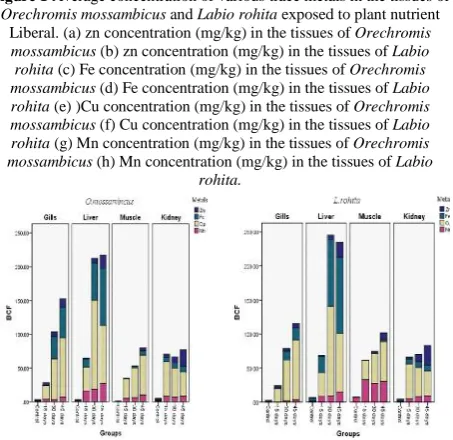
Figure 1 Average concentration of various trace metals in the tissues of
Orechromis mossambicus and Labio rohita exposed to plant nutrient
Liberal. (a) zn concentration (mg/kg) in the tissues of Orechromis
mossambicus (b) zn concentration (mg/kg) in the tissues of Labio rohita (c) Fe concentration (mg/kg) in the tissues of Orechromis mossambicus (d) Fe concentration (mg/kg) in the tissues of Labio rohita (e) )Cu concentration (mg/kg) in the tissues of Orechromis mossambicus (f) Cu concentration (mg/kg) in the tissues of Labio rohita (g) Mn concentration (mg/kg) in the tissues of Orechromis mossambicus (h) Mn concentration (mg/kg) in the tissues of Labio
rohita.
Figure 2 Bio concentration factor of tissues of Orechromis
mossambicus and Labio rohita exposed to plant nutrient Liberal.
(i)BCF of tissues of Orechromis mossambicus (j) BCF of the tissues of
Labio rohita
DISCUSSION
The results of the present study strongly revealed that Librel exposure has resulted into time Dependent accumulation of metals in teleost. Although both the fishes showed the accumulation of the metals in tissues the most astonishing fact was that no mortality was reported during the experiment, presumably due to their function as co-factors for the activation of a number of enzymes and regulated to maintain a certain homeostatic status in fish. The results of several studies of metal accumulation in fish living in polluted waters showed that considerable amounts of metals may be deposited in fish tissues without causing mortality (Jezierska and
Witeska, 2001, Jezierska and Witeska 2006, Nimick et al., 2007, Akan et al., 2012, Naz and javed, 2013). In the present study of the two fishes, L. rohita was found to have more affinity to accumulate metals compared to O. mossambicus, which are in agreement with the comparative studies done by Voigt, 2004 Javed 2005) who observed significantly higher accumulation of metals in L. rohita than C. mrigala and C. catla. Moreover, a variety of species of fish from the same water body are also reported to accumulate different amounts of metals (Senthilkumar and Sajwan, 2007; Sajwanm et al., 2008). Metal distribution in various organs is time-associated (Eggleton and Thomas, 2004).
The effect of time on metal distribution within the organism is a complex issue due to different affinity of various metals to the tissues of various fish species. In the current study both fishes showed a time dependent significant increase in the metal concentration in all the tissues. These differences result from different affinity of metals to fish tissues, different uptake, deposition and excretion rates (Giguere et al., 2004). Of all the tissues, liver has shown the highest level of accumulation of metals followed by gills while kidney and muscle have shown the least level of accumulation. Significantly higher levels of all metals in fish liver can be related to the binding of metals to metallothionein that provide detoxification mechanism (sensi et al., 2010). As reported by Szarek-Gwiazda et al., (2006) that along with the species specificity the trace metal concentration and their binding capacity vary with tissues too. Liver accumulates high concentrations of metals, irrespective of the uptake route (Tsai et al., 2013).
The liver is considered a good monitor of water pollution with metals since their concentrations accumulated in this organ are often proportional to those present in the environment (Dural et al., 2006).Next in order of accumulation were the gills which are in direct contact with water and as the gill surface is negatively charged it provides a potential site for gill-metal interaction for positively charged metals. As reported earlier fish can absorb ions through gills, since they have special salt secreting cells and are involved in the secretion of metals, probably via the secretion of mucus, but when the metal accumulation crosses the excretion threshold limit bioaccumulation exceeds the excretion level. Hence the second highest metal burden observed in the gills of O. mossambicus and L. rohita is self-explanatory mechanism of metal accumulation. Our results are in agreement with earlier reported metal accumulation in tissues of freshwater fish C. gariepinus (Kusemiju et al., 2012) O.niloticus (Al-Nagaawy, 2008) in O.mossambicus (Naigaga, 2002) in Tor putitora (Shakoori, and Yousafzai, 2006). Metal concentrations in the kidneys rise slower than in liver, hence the low concentration of ions in the kidney is the present work is well justified. Kidney is also one of the active metabolically important organs next to liver. Metal uptake and binding has been reported to increase with increase in the metabolic rate (Green & Knutzen, 2003 and Voigt, 2004). The present data corroborates with the studies of metal accumulation in the kidney of O. niloticus (Abdel- Baki et al., 2011), Onchorynchus mykiss of Carassius auratus and Cyprinus carpio respectively (Boeck et al., 2004).
Figure 1 Average concentration of various trace metals in the tissues of
Orechromis mossambicus and Labio rohita exposed to plant nutrient
Liberal. (a) zn concentration (mg/kg) in the tissues of Orechromis
mossambicus (b) zn concentration (mg/kg) in the tissues of Labio rohita (c) Fe concentration (mg/kg) in the tissues of Orechromis mossambicus (d) Fe concentration (mg/kg) in the tissues of Labio rohita (e) )Cu concentration (mg/kg) in the tissues of Orechromis mossambicus (f) Cu concentration (mg/kg) in the tissues of Labio rohita (g) Mn concentration (mg/kg) in the tissues of Orechromis mossambicus (h) Mn concentration (mg/kg) in the tissues of Labio
rohita.
Figure 2 Bio concentration factor of tissues of Orechromis
mossambicus and Labio rohita exposed to plant nutrient Liberal.
(i)BCF of tissues of Orechromis mossambicus (j) BCF of the tissues of
Labio rohita
DISCUSSION
The results of the present study strongly revealed that Librel exposure has resulted into time Dependent accumulation of metals in teleost. Although both the fishes showed the accumulation of the metals in tissues the most astonishing fact was that no mortality was reported during the experiment, presumably due to their function as co-factors for the activation of a number of enzymes and regulated to maintain a certain homeostatic status in fish. The results of several studies of metal accumulation in fish living in polluted waters showed that considerable amounts of metals may be deposited in fish tissues without causing mortality (Jezierska and
Witeska, 2001, Jezierska and Witeska 2006, Nimick et al., 2007, Akan et al., 2012, Naz and javed, 2013). In the present study of the two fishes, L. rohita was found to have more affinity to accumulate metals compared to O. mossambicus, which are in agreement with the comparative studies done by Voigt, 2004 Javed 2005) who observed significantly higher accumulation of metals in L. rohita than C. mrigala and C. catla. Moreover, a variety of species of fish from the same water body are also reported to accumulate different amounts of metals (Senthilkumar and Sajwan, 2007; Sajwanm et al., 2008). Metal distribution in various organs is time-associated (Eggleton and Thomas, 2004).
The effect of time on metal distribution within the organism is a complex issue due to different affinity of various metals to the tissues of various fish species. In the current study both fishes showed a time dependent significant increase in the metal concentration in all the tissues. These differences result from different affinity of metals to fish tissues, different uptake, deposition and excretion rates (Giguere et al., 2004). Of all the tissues, liver has shown the highest level of accumulation of metals followed by gills while kidney and muscle have shown the least level of accumulation. Significantly higher levels of all metals in fish liver can be related to the binding of metals to metallothionein that provide detoxification mechanism (sensi et al., 2010). As reported by Szarek-Gwiazda et al., (2006) that along with the species specificity the trace metal concentration and their binding capacity vary with tissues too. Liver accumulates high concentrations of metals, irrespective of the uptake route (Tsai et al., 2013).
The liver is considered a good monitor of water pollution with metals since their concentrations accumulated in this organ are often proportional to those present in the environment (Dural et al., 2006).Next in order of accumulation were the gills which are in direct contact with water and as the gill surface is negatively charged it provides a potential site for gill-metal interaction for positively charged metals. As reported earlier fish can absorb ions through gills, since they have special salt secreting cells and are involved in the secretion of metals, probably via the secretion of mucus, but when the metal accumulation crosses the excretion threshold limit bioaccumulation exceeds the excretion level. Hence the second highest metal burden observed in the gills of O. mossambicus and L. rohita is self-explanatory mechanism of metal accumulation. Our results are in agreement with earlier reported metal accumulation in tissues of freshwater fish C. gariepinus (Kusemiju et al., 2012) O.niloticus (Al-Nagaawy, 2008) in O.mossambicus (Naigaga, 2002) in Tor putitora (Shakoori, and Yousafzai, 2006). Metal concentrations in the kidneys rise slower than in liver, hence the low concentration of ions in the kidney is the present work is well justified. Kidney is also one of the active metabolically important organs next to liver. Metal uptake and binding has been reported to increase with increase in the metabolic rate (Green & Knutzen, 2003 and Voigt, 2004). The present data corroborates with the studies of metal accumulation in the kidney of O. niloticus (Abdel- Baki et al., 2011), Onchorynchus mykiss of Carassius auratus and Cyprinus carpio respectively (Boeck et al., 2004).
Figure 1 Average concentration of various trace metals in the tissues of
Orechromis mossambicus and Labio rohita exposed to plant nutrient
Liberal. (a) zn concentration (mg/kg) in the tissues of Orechromis
mossambicus (b) zn concentration (mg/kg) in the tissues of Labio rohita (c) Fe concentration (mg/kg) in the tissues of Orechromis mossambicus (d) Fe concentration (mg/kg) in the tissues of Labio rohita (e) )Cu concentration (mg/kg) in the tissues of Orechromis mossambicus (f) Cu concentration (mg/kg) in the tissues of Labio rohita (g) Mn concentration (mg/kg) in the tissues of Orechromis mossambicus (h) Mn concentration (mg/kg) in the tissues of Labio
rohita.
Figure 2 Bio concentration factor of tissues of Orechromis
mossambicus and Labio rohita exposed to plant nutrient Liberal.
(i)BCF of tissues of Orechromis mossambicus (j) BCF of the tissues of
Muscle tissue exhibited the least accumulation of all the tissues, This result are in agreement with many authors who reported that muscles is not an active organ in accumulation of most heavy metals (Yilmaz et al., 2007 and Kraiem 2007, Khalil and Faragallah, 2008;). Thus, plant nutrient exposure in the present study has probably led to the increase in the metabolic rate particularly for the metabolically important tissues such as liver. In the present study the order of accumulation of metals in water was found to be Fe >Zn>Mn>Cu.The mean concentration of Cu recorded in water in this investigation was below permissible limits, while the mean concentration of Zn, Mn and Fe were above limits (WHO, 2005). This higher concentration could be linked to the presence of synergistic or additive effects other metals. Metals are non-biodegradable, and once they enter the aquatic environment, bioconcentration may occur in fish tissue by means of metabolic and bio sorption processes (Yousafzai and Shakoori, 2008; Kaoud and El-Dahshan, 2010). From an environmental point of view, bio concentration is important because metal ions usually occur in low concentrations in the aquatic environment and subtle physiological effects go unnoticed until gross chronic reactions (e.g. changes in populations’ structure, altered reproduction, etc.) become apparent.
BCF in the present study has revealed alterations in the tissue specific and metal specific responses. Overall the BCF of liver tissue has resulted into the highest affinity for Cu, Fe and Zn, Whereas BCF of gills showed affinity for Cu and Fe, kidney showed affinity for Fe and Zn and muscle had affinity for Mn. Fish liver as a major detoxifying and storage organ differed from the concentrations detected in the gills, kidney and muscles. Significantly higher levels of all metals in fish liver can be related to the binding of metals to metallothionein that provide detoxification mechanism (sensi et al., 2010). Species difference in heavy metals bio concentration is linked to difference in feeding habits and behaviour of the species (Altindag and Yigit, 2005). The variability observed in the fish species is a reflection o
f different thresholds of metals which are a function of homeostasis. The thresholds of metals in fish can be considered as the concentration level where the metal starts to interfere with the variable physiology of the fish species in such manner that once a particular level of the metal has been sequestered in the body, equilibrium is established between the fish burden and the ambience. The high bioaccumulation factor for Cu and Fe suggests that the concentration of these metal ions serves as a harborage or the fish species have poor mechanisms for digesting and eliminating these heavy metals. Simkiss & Taylor (1989) in their studies have proposed various pathways of metal accumulation by aquatic organisms either through lipid permeation, complex permeation, and carrier mediated, through ion channels, ion pumps or endocytosis. The high metal concentration in the tissues and water reported in the present studies thus suggest that possibly few of the mechanism might be working simultaneously, however at this point it is difficult to conclude the exact mechanism for metal accumulation. Moreover depuration studies were not done and perhaps it will throw more light to validate our data. As there was no mortality reported in the present studies, possibly the fishes are having an inborn mechanism to counteract the potential toxicity of the metals in
the tissues, by synthesizing the stress proteins as they are assumed to play a role in the detoxification of heavy metals.
CONCLUSION
The overall results from the metal accumulation studies have proved that the metal accumulation was time dependent, species specific and organ specific. Moreover, of the two species it was observed that O.mossambicus was able to withstand the metal load more compared to L.rohita.
Acknowledgement
The authors are highly thankful to University Grant Commission (UGC) Major research Project for financial assistance which help the study to reach to the desired point.
Reference
1. Abdel-Baki, AS., Dkhil, MA. and Al-Quraishy, S. 2011. Bioaccumulation of some heavy metals in tilapia fish relevant to their concentration in water and sediment of Wadi Hanifah. Saudi Arabia African Journal of Biotechnology.,10(13): 2541-2547.
2. Adeniyi, A. and KA, Yusuf. 2007. Determination of heavy metals in fish tissues, water and bottom sediments from Epe and Badagry Lagoons, Lagos, Nigeria. Environ. Monitor Assess., 37: 451-458. 3. Akan, J. C., Mohmoud, S., Yikala, B. S. and Ogugbuja,
V. B. 2012. Bioaccumulation of some Heavy metals in fish samples from river benue in vinkilang, Adamawa state, Nigeria. American journal of analytical Chemistry, 3: 727-736.
4. Al-Nagaawy, A. M. 2008. Accumulation and Elimination of copper and lead From O. Niloticus fingerlings And Consequent influence On Their Tissue Residues and Some Biochemical Parameters. 8th International Symposium on Tilapia in Aquaculture. Egypt. 1:431-445.
5. Altndag, A., Yigit, S., 2005. Assessment of heavy metal concentrations in the food web of Lake Beysehir, Turkey. Chemosphere., 60: 552-556.
6. Anantigha Beach market in Calabar-Nigeria. Afr. J. Environ. Pollut. & Health., 6: 61-64.
7. APHA (American Public Health Association), (1995) Standard Methods for the Examination of water and Wastewater. Washington DC.
8. Boeck, GD., Meeus, W., Coen, WD. and Blust, R. 2004. Tissue specific Cu bioaccumulation patterns and differences in sensitivity to water borne Cu in three fresh water fish: rainbow trout (Oncorhyncus mykiss), common carp (Cyprinus carpio), and gibel carp (Carassius auratus) Aquat Toxicol., 70: 179–188. 9. Censi, P., Spoto, SE., Saiano, F., Sprovieri, M.,
Mazzola, S., Nardone, G., Di Geronimo, SI., Puntu, R. and Ottonello, D. 2006. Heavy metals in coastal water system. A case study from the north western Gulf of Thailand. Chemosphere., 64: 1167–1176.
Toxicology., 7(2): 53-59.
11. Dural, M., Goksu, M. Z., Ozak, A. A. and Bari, D. 2006. Bioaccumulation of some heavy metals in different tissues of Dicentrarchus labrax L, 1758, Sparusaurata L, 1758 and Mugilcephalus L, 1758 from the Camlik lagoon of the eastern cost of Mediterranean (Turkey). Environmental Monitoring and Assessment., 118 (1-3): 65-74.
12. Edem, CA., Akpan, B. and Dosunmu, MI. 2008. A comparative assessment of heavy metals and hydrocarbon accumulation in Sphyrena afra, Orechromis niloticus and lops lacerta from Anantigha Beach market in Calabar-Nigeria. Afr. J. Environ. Pollut.& Health., 6: 61-64.
13. Eggleton, J. and Thomas, KV. 2004. A review of factors affecting the release and bioavailability of contaminants during sediment disturbance events. Environ Int., 30(7): 973-80.
14.Ergül, H.A., Topçuoğlu, S., Ölmez, E. and Kırbaşoğlu, Ç. 2008. Heavy metals in sinking particles and bottom sediments from the eastern Turkish coast of the Black Sea. Estuarine, Coastal and Shelf Science, 78: 396-402.
15. Giguere, A., Campbell, PC., Hare, L., McDonald, DG. and Rasmussen, JB. 2004. Influence of lake chemistry and fish age on cadmium, copper, and zinc concentrations in various organs of indigenous yellow perch (Perca flavescens), Can. J. Fish. Aquat.Sci., 61: 1702–1716.
16. Goullé, JP., Saussereau, E., Mahieu, L., Cellier, D., Spiroux, J. and Guerbet, M. 2012. Importance of Anthropogenic Metals in Hospital and Urban Wastewater: Its Significance for theEnvironment. Bulletin of environmental contamination and toxicology., 89(6): 1220-1224.
17. Green, NW. and Knutzen, J. 2003. Organ halogens and metals in marine fish and mussels and some relationships to biological variables at reference localities in Norway. Marine Pollut. Bull., 46:362–374. 18. Indigenous yellow perch (Perca flavescens). Can. J.
Fish. Aquat. Sci., 61: 1702–1716.
19. Javed, M. 2005. Growth responses of Catla catla, Labeo rohita and Cirrhina mrigala and bioaccumulation of zinc during chronic exposure. Pak. J. Biol. Sci., 8:1357-1360.
20. Jezierska, B. and Witeska, M. 2004. The effect of metals on fish gill functions – gas and ion exchange (Review). Fresenius Environmental Bulletin., 13:1370-78.
21. Jezierska, B. and Witeska, M. 2006. The metal uptake and accumulation in fish living in polluted waters. Soil and Water Pollution Monitoring. Protection and Remediation., 69: 107-114.
22. Jomova, K and Valko, M., 2011. Advances in metal-induced oxidative stress and human disease. Toxicology., 283(2-3): 65-87.
23. Kaoud, HA. and El-Dahshan, AR. 2010. Bioaccumulation and histopathological alterations of the heavy metals in Oreochromis niloticus fish. Nature and science., 8(4):147-156.
24. Khalil, M. and Faragallah, H. 2008. The distribution of some leachable and total heavy metals in core sediments of Manzala lagoon, Egypt. Egypt. J. Aquat.
Res., 34(1): 1-11.
25. Kraiem, C.M., Njimi, W., Tissaoui, C.H., Thompson, J.R. and Flower, R.J. 2007. Seasonal their interactions in juvenile rainbow trout, Aquat. Toxicol., 72: 99–117. 26. Kusemiju, V., Patience, A. and Oluwatoyin, AJ. 2012.
Accumulation of lead in the tissues of freshwater catfish Clarias gariepinus exposed to static nominal concentrations of lead nitrate. Agric. Biol. J. N. Am., 3(12): 510-515.
27. Macfarlane, G.B and Burchett, M.D. 2000. Cellular distribution of Cu, Pb and Zn in the Grey Mangroove Avicemmia marina (Forsk). Vierh Aquatic Botanic., 68: 45–49.
28. Mansour, S. A. and Sidky, M. M. 2002. Ecotoxi-cological studies. 3: Heavy metals contaminating water and fish from Fayoum Governorate, Egypt. Food Chem., 78:15-22.
29. Moore, F., Attar, A. and Rastmanesh, F. 2011. Anthropogenic sources of heavy metals in deposited sediments from runoff and industrial effluents, Shiraz, SW Iran.In: 2nd International Conference on Environmental Science and Technology IPCBEE vol. 6.
30. Naigaga, I. 2002. Bioaccumulation and Histopathology of Copper in Oreochromis mossambicus. Submitted in fulfillment of the requirements for the degree of MASTER OF SCIENCE of Rhodes University.
31. Naz, S. and Javed, M. 2013. Evaluation of Acute Toxicity of Metals Mixture and Bioaccumualtion in Freshwater Fish. BM., 4(3): 11-18.
32. Nicholson, FA., Smith, SR., Alloway, BJ., Carlton-Simith, C. and Chambers, BJ. 2003. An inventory of heavy metal input to agricultural soil in England and Wales. Sci. Total Environ., 311: 205–219.
33. Nimick, D.A., Harper, D.D., Farag, A.M., Cleasby, T.E., MacConnell, E. and Skaar, D. 2007. Influence of in-stream diel concentration cycles of dissolved trace metals on acute toxicity to one-year-old cutthroat trout (Oncorhynchus clarki lewisi). Environ. Toxicol. Chem., 26: 2667–2678.
34. Ozuni, E., Dhaskali, L., Abeshi, J., Zogaj, M., Haziri, I., Beqiraj, D. and Latifi, F. 2010. Heavy metals in fish for public consumption and consumer protection. In Natura Montenegrina., 9(3): 843-851.
35. Reviews in Aquatic Sciences., 1:174-188.
36. Sajwan, KS., Senthil Kumar, K., Paramasivam, S., Compton, SS. and Richardson, JP. 2008. Elemental Status in Sediment and American Oyster Collected from Savannah Marsh/Estuarine
37. Senesi, G.S., Baldassarre, G., Senesi, N. and Raldina, B. 1999. Trace element inputs into soils by anthropogenic activities and implications for human health. Chemosphere., 39: 343–377.
38. Senesi, GS., Baldassarre, G., Senesi, N. and Raldina, B. 1999. Trace element inputs into soils by anthropogenic activities and implications for human health. Chemosphere., 39: 343–377.
40. Shakoori, AR. and Yousafzai, AM., 2006. Bioaccumulation of chromium, nickel, lead, copper and zinc in the skin of Tor putitora as an indicator of the presence of the heavy metal load in River Kabul, Pakistan. Pakistan J. Zool., 33: 341-347.
41. Sokal, RR. and Rohlf, FJ. 1987. Introduction to biostatistics. 2nd ed. New York: Freeman.
42. Szarek-Gwiazda, E. and Amirowicz, A., 2006. Bioaccumulation of trace elements in roach, silver bream, rudd, and perch living in an inundated opencast sulphur mine. Aquatic Ecology., 40: 221–236.
43. Taylor M. G. and Simkiss K. 1989. Metal fluxes across membranes of aquatic organisms.
44. Tsai, J. W., Ju, YR., Huang, YH., Deng, YS., Chen, WY., Wu, CC. and Liao, CM. 2013. Toxico kinetics of tilapia following high exposure to waterborne and dietary copper and implications for coping mechanisms. Environmental Science and Pollution Research., 20(6): 3771-3780.
45. Unlu, S., Topcuoglu, S., Alpar, B., Kirbasoglu, Ç. and Yilmaz, YZ. 2008. Heavy metal pollution in surface sediment and mussel samples in the Gulf of Gemlik. Environ .Monit. Assess., 144: 169-178.
46. Voigt, HR. 2004. Concentrations of mercury (Hg) and cadmium (Cd), and the condition of some coastal Baltic fishes, Environ mentalica Fennica., 21: 26.
47. Yabe, J., Nakayama, SM., Ikenaka, Y., Muzandu, K., Ishizuka, M. and Umemura, T. 2012. Accumulation of Metals in the Liver and Kidneys of Cattle from Agricultural Areas in Lusaka, Zambia. The Journal of veterinary medical science/the Japanese Society of Veterinary Science., 74: 1345-1347.
48.Yılmaz, M., Ersan, Y., Karaman, M., Özen, H., Evren, K. and Hacali, N. 2008. Toxic effects of cobalt parahydroxy- benzoate on tissue histopathology and serum proteins in Capoeta capoeta capoeta. Fresenius Environmental Bulletin., 17(9a): 1322–1327.
49. Yousafzai, AM. and Shakoori, AR. 2008. Heavy metal accumulation in gills of an endangered south Asian freshwater fish as an indicator of aquatic pollution. Pakistan Journal of Zoology., 40(6):423-430.