IMAGE COMPRESSION OF
RADIOGRAPH USING NEURAL
NETWORK AND WAVELET
ANCHAL RANI
Electronics and Communication Engineering, Punjab Technical University, Distt. Ambala, Shahzadpur, Haryana, India
MOHIT MEHTA
Electronics and Communication, Punjab Technical University, Derabassi, Derabassi, Punjab, India
Abstract :
Bandwidth conservation is an important issue in case of multimedia communication. Uncompressed multimedia (graphical, audio and video) data requires considerable storage capacity and transmission bandwidth. Despite rapid progress in mass-storage density, processor speeds and digital communication system performance, it demands for data storage capacity and data-transmission bandwidth continuously to outstrip the capabilities of available technologies. So to solve this problem an efficient multimedia communication scheme is proposed which is based on Wavelet. Image compression is the technique of reducing the size of image file without degrading the quality of the image. There are many techniques available in the lossy image compression in which Wavelet transform based image compression is the best technique. Various types of Wavelets are used for image compression. This paper shows Better image compression by using different wavelet with the help of Neural network. The paper defines the progress made towards calculating different parameter for Wavelet and after that determines the wavelet which gives minimum value of mean square error and maximum value of peak signal to noise ratio. By this best compression Wavelet is obtained. For Analysis considered MSE value should be a minimum and peak signal to noise ratio value should be a maximum. By implementing neural network, the optimum image compression system use a supervised neural network based on the back propagation learning algorithm, due to its implementation, simplicity and the availability of sufficient target database for training the supervised learner is obtained.
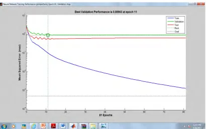
The paper present the idea of image compression based on hierarchical back propagation neural network and results are analyzed. The further analysis is conducted in the network model and tested training algorithm. Finally image compression and image reconstruction are accomplished respectively, a minimum accuracy of 89% was considered as accepted. The neural network yielded 98.65% correct recognition rate of optimum compression ratios,
This concludes that a high compression ratio is achieved with Bi-orthogonal Wavelet functions. The results are obtained with a Bi-orthogonal 6.8 Reconstruction Wavelet function and proved the best. Then Neural Network is implemented to prove the best result and hence achieved. Experimental results suggest that the proposed system can be efficiently used to compress while maintaining high image compression.
Keywords: X-Ray; Image Compression; Wavelet Transform; Back Propagation. 1. Introduction
Uncompressed multimedia (graphics, audio and video) data requires considerable storage capacity and transmission bandwidth. Despite rapid progress in mass-storage density, processor speeds, and digital communication system performance, demand for data storage capacity and data-transmission bandwidth continues to outstrip the capabilities of available technologies. The recent growth of data intensive multimedia-based web applications have not only sustained the need for more efficient ways to encode signals and images but have made compression of such signals central to storage and communication technology.
2. Wavelets and wavelet choice
A wave is an oscillating function of time or space and is periodic. In contrast, wavelets are localized waves. They have their energy concentrated in time or space and are suited to analysis of transient signals.
Basically wavelets may be classified in to two basic classes: (a) Orthogonal
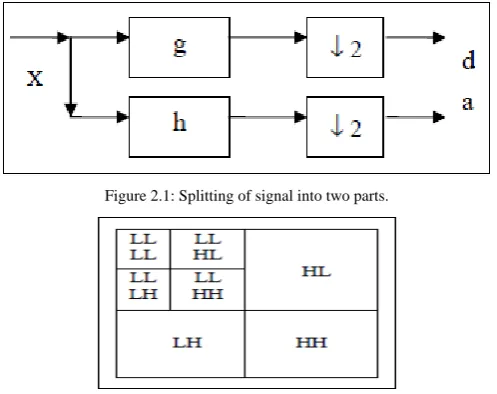
The coefficients of orthogonal filters are real numbers. The filters are of the same length and are not symmetric. The Discrete Wavelet Transform (DWT), which is based on sub-band coding, is found to yield a fast computation of Wavelet Transform. It is easy to implement and reduces the computation time and resources required. Sampled input image is decomposed into various frequency sub-bands or sub-band signals. Splitting of signal into two parts shown in Figure 2.1. A two dimensional decomposition can be applied over the image. A simple example of level 2 decomposing is shown in Figure 2.2. [4]
Figure 2.1: Splitting of signal into two parts.
Figure 2.2: Two levels of 2-D DWT decomposition.
3. Picture Quality Measures
Picture quality is measure by calculating different parameter[3].Also calculating compression ratio for each image. It should be near about same for all the image.[6]
Mean Square Error (MSE) =
Peak Signal to Noise Ratio (PSNR) = =
Normalized Cross-Correlation (NK) =
Average Difference (AD) =
Structural Content (SC) = \
Maximum Difference (MD) =
Picture Quality Scale (PQS) =
4. Neural Network
The term neural network was traditionally used to refer to a network or circuit of biological neurons. The modern usage of the term often refers to artificial neural networks, which are composed of artificial neurons or nodes.[1]
Backpropagation
It is a supervised learning method, and is a generalization of the delta rule. It requires a dataset of the desired output for many inputs, making up the training set.
multi-method for measuring the discrepancy between the expected output and the actual output is using the squared error measure:
,
where is the discrepancy or error.
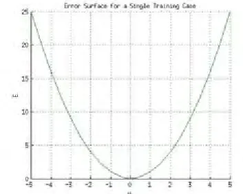
As an example, consider the network on a single training case: (1, 1, 0), thus the input and are 1 and 1 respectively and the correct output, is 0. Now if the actual output is plotted on the x-axis against the error
on the y-axis, the result is a parabola.
Figure 4(a) Error surface of a linear neuron for a single training case.
However, the output of a neuron depends on the weighted sum of all its inputs:
,
where and are the weights on the connection from the input units to the output unit. Therefore, the error also depends on the incoming weights to the neuron,
5. Methodology 5.1 Methodology Used
1) Selection of best wavelet for medical Radiographs.
Compress input image with each wavelet with fixed CR. Find the following parameters to make comparison
2) MSE, PSNR, Correlation, Average difference, Normalized absolute error, Structural content. 3) Make database using selected wavelet.
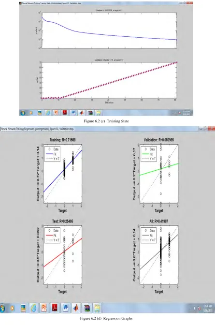
4) Save each with its optimum compression ratio using subjective and objective evaluation. 5) Train and testing of neural network.
6) Apply input image to neural network with unknown CR. 7) Compress this image with CR defined by neural network.
8) Comparison with implemented paper based upon following parameters CR
PSNR MSE 6. Experimental Results
Choice of wavelet
Table 6.1: Analysis of Radiograph image compression using various wavelets
Wavelet THR CR MSE PSNR MD SC NAE CC
db2 0.7 50.4 0.0469 141.4959 1.0959 1 0.0019 1
db3 0.6 49.205 0.0339 144.7469 0.9525 1 0.0017 1
db4 0.6 50.46 0.034 144.5272 0.9071 1 0.0017 1
db5 0.6 50.62 0.0345 144.5651 0.9014 1 0.0017 1
db6 0.6 50.78 0.0351 144.3925 0.9168 1 0.0017 1
db7 0.6 50.81 0.0349 144.4441 1.0017 1 0.0017 1
db8 0.6 50.87 0.0357 144.2412 0.9414 1 0.0018 1
bior1.1 2.6 50.1245 0.5303 117.2472 3.75 1 0.0064 1
bior1.3 2.6 51.995 0.5066 117.7048 3.7109 1 0.0064 1
bior1.5 2.4 51.6671 0.4339 119.2527 3.2912 1 0.006 1
bior2.2 0.8 49.38 0.0794 136.2351 1.6523 1 0.0025 1
bior2.4 0.8 50.7235 0.0791 136.2747 1.55 1 0.0026 1
bior2.6 0.8 51.313 0.0792 136.2574 1.4348 1 0.0026 1
bior2.8 0.7 49.4246 0.0636 138.4601 1.1896 1 0.0023 1
bior3.1 0.5 51.8102 0.064 138.374 1.1602 1 0.0024 1
bior3.3 0.4 48.2939 0.0332 144.9532 0.7946 1 0.0017 1
bior3.5 0.4 48.6704 0.0313 145.5413 0.7535 1 0.0016 1
bi0r3.7 0.4 48.9488 0.0307 145.7351 0.7542 1 0.0016 1
bior4.4 0.6 51.87 0.0372 143.809 0.9595 1 0.0017 1
bior6.8 0.5 49.163 0.0254 147.6272 0.7523 1 0.0015 1
rbio1.3 0.6 48.5924 0.0336 144.8497 0.8555 1 0.0016 1
rbio2.2 1 49.61 0.0719 137.2215 1.3125 1 0.0024 1
rbio2.4 0.9 47.54 0.0577 139.425 1.082 1 0.0021 1
rbio2.6 0.8 49.91 0.0477 141.3389 0.9793 1 0.002 1
rbio2.8 0.8 51.54 0.0487 141.1204 0.9848 1 0.002 1
rbio3.3 1 42.36 0.0682 137.736 1.2757 1 0.0023 1
rbio3.5 1 49.194 0.068 137.7805 1.4611 1 0.0024 1
rbio3.7 1 50.85 0.0684 137.7259 1.2606 1 0.0024 1
rbio4.4 0.7 50.9181 0.0446 141.9998 1.0022 1 0.0019 1
Original image Using bior 6.8
Figure 6.1(a): Show the comparison between radiograph image using bior 6.8 wavelets.
7. Conclusion
When we have done selection of best wavelet for medical Radiographs. Then to compress input image with each wavelet with fixed CR. After that to find the following parameters to make comparison MSE, PSNR, Correlation, Average difference, normalized absolute error, Structural content. Then we have database using selected wavelet. And then to save each with its optimum compression ratio using subjective and objective evaluation. To train and testing of neural network. And then apply input image to neural network with unknown CR. To compress this image with CR defined by neural network. It is clearly seen that, for bi-orthogonal 6.8 wavelet, we are getting least MSE and highest PSNR of 147.6272 dB with fixed CR. From the above observation table it is more cleared with mathematical formulae support, that bi-orthogonal type 6.8 wavelet is best suitable for medical image.
Future Scope
In future we can apply JPEG (Joint Photographic Expert Group) compression method instead of threshold for finding better compression. Another scope is to optimized the compression method by using AI (Artificial Intelligence) algorithm
References
[1] Adnan Khashman and Kamil Dimililer, “Medical Radiographs Compression Using Neural Networks” has published by IEEE members in 2009.
[2] V. K. Bhairagi and A. M. Sapkal, “Selection of Wavelets for Medical Image Compression.” Proceeding in 2009 International Conference on Advance in Computing, Control, and Telecommunication Technologies.
[3] Marta Mrak, Sonja Grgic and Mishav Grgic, “Picture Quality Measures in Image Compression Systems.” Proceeding in EUROCON 2003 Ljubljana, Slovenia.
[4] Kamrul Hasan Talukder and Koichi Harada, “Wavelet Based Approach for Image Compression and Quality Assessment of Compressed Image.” Proceeding in IAENG International Journal of Applied Mathematics in Feb 2007.
[5] Adnan Khashman and Kamil Dimililer, “Comparison Criteria for Optimum Image Compression.” Proceeding in EUROCON 2005 Serbia & Montenegro, Belgrade, Nov 22-24, 2005.
[6] Elham Shahhoseini, Hamid Behnam, Nasrin Ahmadi Nejad and Amir Shahhoseini, “A New Approach to Compression of Medical Ultrasound Images using Wavelet Transform.” Proceeding in 2010 Third International Conference on Advance in Circuits, Electronics and Micro-electronics.