1535-9778/08/$08.00⫹0 doi:10.1128/EC.00427-07
Copyright © 2008, American Society for Microbiology. All Rights Reserved.
Neurospora crassa heat shock factor 1
Is an Essential Gene; a Second
Heat Shock Factor-Like Gene,
hsf2
, Is Required for
Asexual Spore Formation
䌤
Seona Thompson, Nirvana J. Croft, Antonis Sotiriou, Hugh D. Piggins, and Susan K. Crosthwaite*
Faculty of Life Sciences, University of Manchester, Manchester M13 9PT, United Kingdom
Received 27 November 2007/Accepted 17 June 2008
Appropriate responses of organisms to heat stress are essential for their survival. In eukaryotes, adaptation to high temperatures is mediated by heat shock transcription factors (HSFs). HSFs regulate the expression of heat shock proteins, which function as molecular chaperones assisting in protein folding and stability. In many
model organisms a great deal is known about the products of hsf genes. An important exception is the
filamentous fungus and model eukaryoteNeurospora crassa. Here we show that twoNeurospora crassagenes
whose protein products share similarity to known HSFs play different biological roles. We report thatheat
shock factor 1(hsf1) is an essential gene and thathsf2is required for asexual development. Conidiation may be
blocked in thehsf2knockout (hsf2KO
) strain because HSF2 is an integral element of the conidiation pathway or because it affects the availability of protein chaperones. We report that genes expressed during conidiation,
for examplefluffy,conidiation-10, andrepressor of conidiation-1show wild-type levels of expression in ahsf2KO
strain. However, consistent with the lack of macroconidium development, levels of eas are much reduced.
Cultures of thehsf2KO
strain along with two other aconidial strains, thefluffyandaconidial-2strains, took
longer than the wild type to recover from heat shock. Altered expression profiles ofhsp90 and a putative
hsp90-associated protein in thehsf2KO
strain after exposure to heat shock may in part account for its reduced ability to cope with heat stress.
The heat shock response is a protective mechanism against sudden temperature change and other forms of physical and chemical stress. The response is brought into effect by heat shock proteins (HSPs), molecular chaperones that assist pro-tein folding and stability (35). Several highly conserved families of HSPs, named according to their molecular masses, are ex-pressed in response to heat shock and include HSP110, HSP100, HSP90, HSP70, HSP60, and a number of small HSPs. Some of the family members are expressed in non-heat shock conditions, but the expression of others is induced only upon exposure to stress due to the action of heat shock transcription factors (HSFs). HSFs are a functionally conserved group of molecules. They were first identified as regulators of the heat shock response (reviewed in reference 28), but it is now evi-dent that certain HSFs also regulate developmental pathways (for a review, see reference 36).
Drosophila melanogasterand Saccharomyces cerevisiaeeach
have one essential HSF (7, 48). In S. cerevisiae, while the
induction of genes encoding chaperones and associated pro-teins is under the control of its HSF, a subset of genes induced by a variety of environmental stresses and encoding metabolic enzymes, components of the ubiquitin-proteosome degrada-tion pathway, and antioxidant defense proteins are under the control of the transcription factors Msn2, Msn4, Yap1, and
Skn7 (5, 17, 6, 25). InDrosophila, temperature-dependent
al-ternative splicing of dHSF gives rise to four forms of dHSF
mRNA which encode HSF isoforms with different transcrip-tional activities (16). Although the functranscrip-tional significance of the HSF isoforms is not yet understood clearly, one possibility is that they activate different subsets of HSP or
stress-respon-sive genes. Moreover, in species with multiple hsf genes, a
functional specialization of certain HSFs toward developmen-tal roles has occurred. For example, of the four vertebrate HSFs, HSF1 mediates heat shock gene expression, whereas HSF2 is reported to be unresponsive to temperature, while its activity changes throughout differentiation. In addition,
alter-native splicing of hsf1, hsf2, and hsf4transcripts generates a
variety of HSF isoforms (13, 18, 45). Since these splice forms show tissue-specific expression, it is likely that they also have distinct roles.
In contrast, no Neurospora HSFs have yet been isolated,
although HSPs are expressed in this organism during heat shock and cell differentiation (14, 19) suggesting that, as seen
for other organisms, the Neurospora HSF(s) is required for
development. Neurospora crassa undergoes both sexual and
asexual developmental cycles. When nitrogen is limiting, fe-male sexual development ensues, and when nuclei of opposite mating types fuse numerous asci are produced, each containing eight haploid spores (42). In addition, two asexual develop-mental pathways are described: macroconidiation and
micro-conidiation. Macroconidial development inNeurosporacan be
induced by desiccation, nutrient deprivation (47), light (23), oxidative stress (29), and signals relayed from the organism’s circadian clock (37). Once induced, aerial hyphae are formed which branch and eventually form chains of macroconidia, each containing between one and five nuclei (44). In a dis-tinctly different developmental process, microconidia
contain-* Corresponding author. Mailing address: Faculty of Life Sciences, University of Manchester, Oxford Road, Manchester M13 9PT, United Kingdom. Phone: 44-161-275-5996. Fax: 44-161-275-5082. E-mail: susan [email protected].
䌤Published ahead of print on 27 June 2008.
1573
on September 8, 2020 by guest
http://ec.asm.org/
ing one or rarely two nuclei are formed from smaller branched microconidiophores (30).
Several phases in the development of macroconidia have been recorded (31, 44). The first step is the production of branching aerial hyphae. Minor constrictions then develop, followed by the production of proconidia via budding from the terminal cells of each branch. Finally, proconidia are separated by major constrictions and mature into conidia, which are easily separated and dispersed by wind or water. A number of mutants affecting this developmental pathway are known. The
fluffyoid (fld) and aconidial-2 (acon-2) mutants are blocked
before and thefluffy(fl) mutant is blocked after the formation
of minor constrictions (31, 44). Theacon-3mutant is blocked
after the maturation of minor constrictions. conidial
separa-tion-1(csp-1) and csp-2 are required for conidial separation
(41). Genetic and molecular studies placeacon-2upstream of
fl, withacon-3,csp-1, andcsp-2being downstream (9, 1).
We report here thatNeurospora hsf1is an essential gene and
that a deletion strain can be maintained only as a
hetero-karyon. In strains withhsf2deleted, no dramatic alteration of
the heat shock response is detected. However,
macroconidia-tion inhsf2is defective; aerial hyphae emerge but no
macro-conidia are produced. Consistent with this, differences are seen
between the wild-type andhsf2knockout (hsf2KO) strains in the
expression of two conidiation-associated transcripts,flandeas.
MATERIALS AND METHODS
Bioinformatic analysis.Genes were identified by searching the Broad Institute Neurospora crassa database. Protein prediction was performed using NCBI BLAST at http://www.ncbi.nlm.nih.gov/BLAST/ and COILS at http://www.ch .embnet.org/software/COILS_form.html. Sequence alignments were performed using ClustalW at http://www.ebi.ac.uk/clustalw/.
Strains and growth conditions.mus-51a(FGSC9718),hsf2 a(FGSC 11671), acon-2 a(FGSC3263),fl a (FGSC818), and thehsp90a(NCU01792),hsp30 (NCU09364),hsp78(NCU02630),hsp101(NCU00104),hsp70(NCU08683), and hsp90(NCU04142) strains were obtained from the Fungal Genetics Stock Center (http://www.fgsc.net/) and crossed into either 30-7bd Aor 87-3bd ato introduce thebdmutation. 87-3bd awas obtained from Christian Heintzen (University of Manchester, United Kingdom).fl,bd was a kind gift from D. Bell-Pedersen (Texas A&M University). The hsf1KO strain was made by amplifying 1-kbp regions 5⬘and 3⬘of thehsf1open reading frame (ORF) and fusing these to the hygromycin phosphotransferase(hph) gene.mus-51awas transformed with this fragment and transformants were selected on hygromycin-containing agar plates (8). Strains were maintained on minimal medium (1⫻Vogel’s salts [10], 2% sucrose, 500 ng/ml biotin, 1.5% agar). Crossing medium contained 1⫻ Wester-gaard’s salts, 2% sucrose, 500 ng/ml biotin, and 1.5% agar (10). Spores were picked onto slants containing Vogel’s minimal medium, heat shocked at 60°C for 1 h to induce germination, and then incubated at 30°C. Progeny were screened for hygromycin resistance on minimal medium containing 200g/ml of hygro-mycin B and/or for rhythmic conidiation on race tubes.
Race tube medium contained 1⫻Vogel’s salts, 0.1% glucose, 0.17% arginine, 500 ng/ml biotin, and 1.5% agar. After inoculation with 5- to 10-day-old macro-conidia, race tubes were placed in constant light (LL) at 25°C for 48 h and then transferred to constant darkness (DD). Growth fronts were marked daily. Heat shocks were given by immersing race tubes in a 50°C water bath for 1 h after 24 h in the dark at 25°C. The race tube cultures were then returned to 25°C to recover and examined daily for regrowth.
Microconidiation was induced according to the protocol of Ebbole and Sachs (11). Conidia were harvested by vortexing in 2 ml of sterile distilled water and filtered through 5-m filters (Millipore) into sterile Eppendorf tubes. Micro-conidia were plated onto minimal (1⫻Vogel’s salts, 1% sorbose, 0.005% fruc-tose, 0.005% glucose, 1 M sorbitol) or minimal hygromycin-containing plates (200g/ml). Plates were incubated at 30°C until single colonies were detected (3 or 4 days later). Colonies were picked onto minimal agar slants, and this was followed by the transfer of conidia onto hygromycin-containing (200g/ml) slants.
Liquid culture medium for the growth of mycelial mats and shaking cultures contained 1⫻Vogel’s salts, 2% glucose, and 500 ng/ml biotin. One hundred milliliters of liquid culture medium was inoculated with 2.3-cm-diameter discs cut from mycelial mats. These cultures were put into LL at 25°C with shaking at 150 rpm. After 24 h, cultures were placed into a shaking water bath at 45°C. Flasks were removed at regular intervals and their contents were immediately harvested by vacuum filtration onto filter paper and frozen in liquid nitrogen.
The experiment examining nitrogen starvation-induced conidial development was first described in the work of Bailey-Shrode and Ebbole (1). Strains cultured on Vogel’s minimal medium slants (2% sucrose) were used to inoculate culture dishes of modified Vogel’s minimal medium (1.5% sucrose) with 50 mM NH4Cl
in place of NH4NO3. After 3 days at 30°C, a cork borer was used to punch discs
from the resulting mycelial mats. These discs were then transferred to 100-ml flasks containing 50 ml of modified Vogel’s minimal medium and incubated for 22 h at 34°C in LL plus shaking at 150 rpm. The cultures were then washed in sterile distilled water and transferred to flasks containing Vogel’s minimal me-dium lacking any nitrogen source. Tissue was harvested before and 1 h, 6 h, 12 h, and 24 h after transfer.
Induction of conidiation by exposure to air was carried out essentially as described in the work of Lee and Ebbole (26), with the following modifications. Disks were cut from mycelial mats and placed into Vogel’s minimal medium with 2% glucose. After 24 h in DD at 25°C, disks were blotted dry and placed on agar blocks (Vogel’s minimal medium, 2% glucose, 2% agar) in open petri dishes containing 15 ml of fresh culture medium.
RT-PCR.The RevertAid H⫺first-strand cDNA synthesis kit (Fermentas) was used for all reverse transcription-PCR (RT-PCR). Primers are listed in Table 1. For semiquantitative RT-PCR, 1g of total RNA was reverse transcribed using the poly(T) primer supplied with the kit, following the manufacturer’s instruc-tions. A limiting number of cycles were used to stop the subsequent PCRs during the exponential phase of amplification. After agarose gel electrophoresis, quan-tification was performed using Bio-Rad QuantityOne software and data were normalized againstactintranscript levels.
Construction of thehsf1knockout strain.Ahsf1deletion cassette was made containing thehphgene (confers resistance to hygromycin) flanked by approximately 1.6 kbp 5⬘and 2 kbp 3⬘of the NCU08512 ORF. One hundred microliters of amus-51 conidial cell suspension (2⫻109conidia/ml) in a 2-mm gap cuvette (Bio-Rad) was
electroporated with 0.5g of the knockout construct. A Bio-Rad gene pulser was used with the following settings: 12.5 kV/cm, 400⍀, 25F. Transformants were selected on hygromycin plates (200g/ml) incubated at 30°C for 4 days. Transfor-mants were crossed with 87-3bd ato obtain homokaryons.
RNA extraction and Northern blotting.RNA was extracted using Trizol (In-vitrogen) following the manufacturer’s protocol. Equal amounts of RNA were run through a 1% agarose formaldehyde gel and blotted onto Hybond N⫹ nitrocellulose membrane (GE Healthcare). Blots were probed with randomly primed32
P-labeled (Roche labeling kit; GE Healthcare radionucleotide) cDNA or32P-labeled RNA (MAXIscript kit; Ambion) and detected using
phosphorim-ager screens (Fuji). Blots were prehybridized (NorthernMax; Ambion) and hy-bridized at 42°C for DNA probes and at 68°C for RNA probes. Blots were washed twice for 10 min in 2⫻SSC (1⫻SSC is 0.15 M NaCl plus 0.015 M sodium citrate) plus 0.1% sodium dodecyl sulfate (SDS), once for 15 min in 1⫻SSC plus 0.1% SDS at room temperature, and once for 15 min in 0.1⫻SSC plus 0.1% SDS at either 42 or 68°C.
DNA extraction and Southern blotting.Genomic DNA was extracted from homogenized tissue by use of a cetyltrimethylammonium bromide protocol. DNA was digested with NdeI (hsf2) or NdeI and BamHI (hsf1), separated through 0.8% agarose gels, and transferred onto Hybond N⫹nitrocellulose membrane. The DNA was cross-linked to membranes with UV light. Blots were hybridized to digoxigenin (DIG)-labeled (Roche) probes against the 5⬘-flanking region of the knockout cassettes (http://www.dartmouth.edu/⬃neurosporagenome/knockouts_completed .html). Primers for the amplification of probe DNA are listed in Table 1. Each blot was incubated with prehybridization solution (NorthernMAX; Ambion) at 42°C for 1 h before the addition of 300 ng of denatured probe in 4 to 5 ml of fresh hybrid-ization solution and incubation at 42°C overnight. After hybridhybrid-ization, the blots were washed according to the manufacturer’s protocol, with the final washes carried out at 42°C. Detection of bound DIG-labeled probe was also carried out according to the manufacturer’s protocol, and the blots were exposed to X-ray film for 5 to 25 min.
RESULTS
Identification of putative HSFs. Two putative Neurospora
HSFs encoded by hsf1 (NCU08512) and hsf2 (NCU08480)
were identified in a BLAST search of theNeurosporagenome.
on September 8, 2020 by guest
http://ec.asm.org/
We cloned RNAs expressed from these genes and analyzed
their sequences to determine the details of any introns.hsf1
contains one intron and encodes a 787-amino-acid (aa)
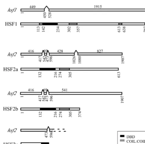
pro-tein. Alternative splicing ofhsf2 RNA gives rise to at least
three different transcripts encoding polypeptides of 613 aa, 374 aa, and 173 aa or of 579 aa, 340 aa, and 139 aa depending at which of the three possible in-frame ATGs translation is initi-ated (Fig. 1). HSFs from different species, though binding to similar DNA consensus sequences, are not highly conserved. However, all contain a DNA-binding domain at the amino terminus and an adjacent cluster of hydrophobic amino acids organized into heptad repeats required for oligomerization, and they often also contain a heptad repeat close to the car-boxy terminus (49). In order to become functionally active, HSF protein monomers form homotrimers through the inter-action of coiled-coil domains found toward the N terminus. HSF1 and the large HSF2 isoforms contain putative DNA-binding and trimerization domains. In addition, a carboxy hep-tad repeat is present in HSF1. In contrast, the 173-aa isoform of HSF2 contains only a partial DNA-binding domain (Fig. 1). Although the presence of these alternatively spliced transcripts in the five cDNAs we cloned and sequenced suggests that they are present at a high level relative to what is seen for the fully
spliced transcript, further investigation revealed that in⬎99%
of the transcripts, introns 1 to 3 have been removed (data not shown).
hsf1is an essential gene;hsf2is required for asexual
devel-opment.To knock outhsf1, at least 1.5 kb of DNA 5⬘and 3⬘of
the ORF was ligated to thehygromycin phosphotransferasegene
(Fig. 2A). This construct was electroporated into conidia of a
mus-51::barstrain which lacks an enzyme required for
nonho-mologous DNA joining. Due to the absence of mus-51,
tar-geted transformation is almost 100% efficient (33). DNA from 20 transformants was extracted and recombination events at
thehsf1gene were checked by Southern blot analysis (Fig. 2B).
All transformants were heterokaryons harboring both nuclei
withhsf1 intact and nuclei withhsf1 deleted. To obtain
ho-mokaryons, microconidiation was induced in two of the het-erokaryons (11). Only five colonies derived from microconidia were hygromycin resistant, and once again these were hetero-karyons (data not shown). In parallel, two of the transformants
were backcrossed to the bd strain. Sixty-five spores derived
from these crosses were selected and all 39 that germinated were inoculated onto hygromycin slants. None of these strains
FIG. 1. RNA splice forms and protein domain structures of HSF1 and HSF2 isoforms. For each pair of diagrams the top schematic represents RNA and the bottom schematic represents protein. Open rectangles in mRNAs represent exons. Nucleotide numbers below transcripts start from the ATGs of the respective ORFs. Amino acid numbers are indicated below proteins. DBD, DNA-binding domain. TABLE 1. Primers used
Gene Usea Primer
b
Forward Reverse
hsf1 RT/cloning/cDNA probe CGGGATCCCGCATGTCTTCTCCTAACC CGGGATCCCGCGACAGAAGCAACAGAT RNA probe/sqRT-PCR (1; 35) CGGGATCCCGCATGTCTTCTCCTAACC TAATACGACTCACTATAGGGACGTGA
GGGCCTGCTGGAATAAGGTCG hsf2L RT/cloning/cDNA probe TAGGATCCTACAGTGCAGATGGCTACC TAGGATCCACATAACGTCTCAAGATGC
RNA probe/sqRT-PCR (2; 30) TAGGATCCTACAGTGCAGATGGCTACC TAATACGACTCACTATAGGGATCCTC CATCTGGCGCTGAACCTGC
hsf2S RT/cloning TAGGATCCTACAGTGCAGATGGCTACC TAGGATCCCAAGCTAACCAGGTCTCCG
hsf25⬘flank Southern blot DNA probe GTAGCCTCTCCATACCTACC GTAGAGTCCATTCTACCTCG
hsp70 DNA probe GTCTCGACAAGAAGGTCG TCTCGGAGGTGGACTTGG
hsp90 DNA probe GGATCCTTGTTGCCGACAGAGTTA GGATCCCATCAATGGGGTCGACAAGG
NCU01792 DNA probe AGCGCTCTTCTGCTACCG CATGGATCTTGGGTATGC
fluffy RNA probe/sqRT-PCR (1; 30) TGTGATGGCCAGATGCCATGC TAATACGACTCACTATAGGGACGAGT TATCGCTGCCCTCACG
actin sqRT-PCR (0.5; 20) GGCTGGCCGTGATCTTACCGACTA GAGAGCGAGGCGAGAATGGAACC con-10 sqRT-PCR (1; 26) CGTCAACATGGCTGGCACTGGTAACG TTGTAATACGACTCACTATAGGGCAA
ATTGTCAAAGCATTCAGTTGC
rco-1 sqRT-PCR (1; 35) GTACGAGGATGAGATTGCGCT TCTGATGAGCTTGTCCTCGGCTC
aFor semiquantitative RT-PCR (sqRT-PCR), the numbers in parentheses indicate the amounts of cDNA inl followed by the cycle numbers. bModifications are in lightface, and annealing portions are in boldface.
on September 8, 2020 by guest
http://ec.asm.org/
were resistant to hygromycin, supporting our conclusion that
hsf1is an essential gene.
A hsf2 deletion strain (8) was obtained from the Fungal
Genetics Stock Center (University of Missouri, Kansas City) and checked by Southern analysis (data not shown). The
veg-etative hyphal growth of thehsf2mutant appears normal, as
does the growth rate, but the deletion strain has a striking phenotype; it produces very few macroconidia (Fig. 3). Only after several weeks are a few proconidial chains present, per-haps induced by starvation or desiccation.
The heat shock response in thehsf2,bd strain.We first
as-sayed the phenotypic response of thehsf2mutant to heat shock
by monitoring its growth along race tubes after exposure to high temperature. In order to assess the effect of heat shock on
conidiation as well as on growth ahsf2,bdstrain was initially
used in these experiments. Thebdmutation allows conidiation
to occur in the presence of the high carbon dioxide levels which accumulate in the enclosed race tube. In response to a 1-h, 50°C heat shock during the third day in DD at 25°C, growth of bothbdand hsf2,bdstrains stopped. On the second day after
the heat shock 100% of thebdcultures had resumed growth,
while only 4 days after the heat shock did 50% of thehsf2,bd
cultures resume growth (Fig. 4A). We wondered if the delayed
recovery was correlated with the aconidial state of thehsf2,bd
strain, and we therefore repeated the experiment, this time
including theacon-2,bdand fl,bdstrains. Of note is that the
acon-2,bdstrain is a temperature-sensitive strain and no spores develop at 34°C or above. Therefore, all of the strains were cultured at either 25°C or 34°C before and after a 50°C heat shock. In both sets of conditions, the aconidial strains took
longer to recover from heat shock than did thebdstrain (Fig.
4B and C). These data suggest either that the germination of conidia that fall to the agar surface after heat shock accounts
for the faster regrowth of the bd strain and/or that all the
aconidial strains are inherently temperature sensitive. If the latter of these possibilities is true, then these results implicate HSF2 in the heat shock response. Therefore, we
followed levels of bothhsf1and hsf2and of three HSP
tran-scripts in control and heat-shocked cultures. In abdstrain, the
levels ofhsfIandhsf2mRNA declined during the first 45 min
of a prolonged heat shock. In thehsf2,bdstrain,hsf1
transcrip-tion also decreased and, as expected,hsf2RNA was not
ex-pressed (Fig. 5A). In spite of this, the three HSP transcripts we
assayed, namely, those for hsp70 (NCU09602) and hsp90
(NCU04142) and a transcript that arises from NCU01792
which we have named heat shock protein 90-associated
(hsp90a), were upregulated in response to heat shock in both
strains (Fig. 5B). Interestingly, twohsp70 transcripts can be
distinguished after transfer to high temperatures in both the
wild-type and hsf2,bd strains. The lower-molecular-weight
FIG. 2. Analysis of mus-51::bar, ‚hsf1 transformants. (A) Sche-matic diagram showing the location of SacI restriction enzyme sites in the region ofhsf1. (Left) Wild-typehsf1locus; (right) locus with the hsf1 gene deleted and replaced with hph. (B) Southern blot of mus-51::bar, ‚hsf1 strains. Genomic DNA digested with SacI and probed with DIG-labeled DNA homologous to a region 5⬘of thehsf1 ORF. Lanes: 1, 7, and 12, DNA ladder; 8, control parentalmus-51 genomic DNA; 2 to 6, 9 to 11, 13, and 14, DNA from different transformants.
FIG. 3. Thehsf2KOstrain is aconidial. Light microscopy showing growth ofbd(left) andhsf2KO,bd(right) strains on minimal agar plates (low magnification) and at higher magnification (inset; magnification,⫻80) at the growth front. Macroconidia (asexual spores) are bright orange.
on September 8, 2020 by guest
http://ec.asm.org/
transcript is most likely another member of thehsp70family,
ssb1(NCU02075), which shares 87% identity over 239
nucle-otides to our 901-nucleotidehsp70probe. Of note is thathsp90
transcripts were significantly more abundant 15 min after the
onset of heat shock in thehsf2,bdstrain than in thebdstrain.
One other significant difference was detected after 45 min of
exposure to heat shock between levels ofhsp90aRNA in thebd
andhsf2,bdstrains, with the levels being higher in thebdstrain. RNA levels of genes associated with conidiation are affected
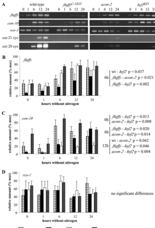
in the hsf2 strain. To test where HSF2 acts in the known
conidiation pathway, we investigated the expression ofhsf1,
hsf2(Fig. 6), fl, conidiation-10 (con-10),repressor of
conidia-tion-1(rco-1), andeas(Fig. 7) in the wild-type,fl,acon-2, and
hsf2strains. We assayed gene expression upon the transfer of
cultures to medium lacking nitrogen, conditions known to in-duce the conidiation pathway (1). Although there was a
gen-eral increase in hsf1 mRNA as development progressed, no
significant difference in the levels of eitherhsf1orhsf2RNA
was detected among the four strains. As previously reported,
levels offland con-10RNA in the wild type increase from 6
hours after the transfer of cultures to medium lacking nitrogen
(1). Among the aconidial strains,fltranscript levels are higher
in theacon-2andhsf2strains at 6 h, whereascon-10transcripts
show significantly increased levels of expression in the hsf2
strain (similar tocon-10levels in the wild-type) compared to
those in the acon-2 and fl strains at 0, 6, and 12 h. To our
surprise, no differences in the transcript levels ofrco-1, a
re-pressor ofcon-10, were detected between any of the strains at
the time points sampled. In our study, by far the most striking difference between the aconidial strains and the wild type is the
greatly reduced levels ofeasin thefl,acon-2, andhsf2strains.
Conidiation can be induced by a number of environmental cues, for example, carbon and nitrogen deprivation and light (47). Synchronous conidiation can also be induced by the
ex-FIG. 4. Response of wild-type and aconidial strains to heat shock. Strains inoculated onto race tubes were germinated and grown in LL at 25°C (A and B) or 34°C (C) for 48 h before transfer to DD at 25°C (A and B) or 34°C (C). After 24 h in DD, cultures were exposed to a 1-h 50°C heat shock and then transferred back to DD at 25°C (A and B) or 34°C (C). Growth was monitored twice daily over the following 9 days. The graph shows the percentages of recovery for the following strains against time (h) after heat shock. (A) 30-7bd(n⫽21), 16-31 bd(n⫽18), 16-21hsf2KO,bd(n⫽17), and 16-6hsf2KO,bd(n⫽18). Numbers preceding the strain names refer to the cross and progeny identifiers, e.g., 16-31 indicates cross number 16 and progeny number 31. (B) 16-21hsf2KO,bd(n⫽17), 16-6hsf2KO,bd(n⫽18),acon-2,bd (n⫽17), andfl,bd(n⫽21). (C) 16-31bd(n⫽17), 16-21hsf2KO,bd (n⫽18),fl,bd(n⫽15), andacon-2,bd(n⫽18). The average results of three independent experiments are plotted. HS, heat shock.
FIG. 5. Gene expression in wild-type andhsf2,bdknockout strains. Northern blots ofbd andhsf2KO,bdRNA probed forhsf1 andhsf2 RNA (A) andhsp70,hsp90, andhsp90aRNA (B). Cultures transferred from LL at 25°C to LL at 45°C were harvested for RNA extraction after 0, 15, 30, 45, and 60 min. There are two sets of data for thehsf1 transcript and three for all other transcripts. Theyaxes represent the relative amounts of each transcript (percentages of the maximum). HS, heat shock.
on September 8, 2020 by guest
http://ec.asm.org/
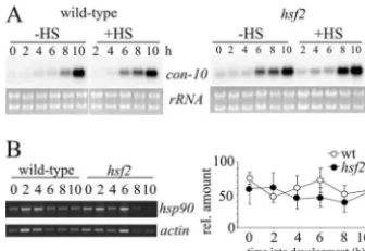
posure of mycelia to air (44). Interestingly, although develop-mentally induced genes are expressed at similar times in cul-tures induced to conidiate by either nitrogen starvation or exposure to air, evidence suggests that their sensitivity to other stresses, such as heat shock, may change. For example, it is
reported that in air-exposed cultures,con-10is induced by heat
shock, whereas in liquid cultures it is not (26). However, in our hands, no significant difference in the developmental induction of con-10 expression between the wild-type and hsf2 strains during air-induced development was revealed, and in neither
strain wascon-10 induced by heat shock (Fig. 8).con-10 has
been shown to be regulated by numerous factors e.g., carbon and nitrogen deprivation and light, and it is probable that slight differences in experimental conditions account for the discrep-ancy between our results and those of Lee and Ebbole (26).
In an effort to determine whether development in the hsf2
strain is indirectly affected through the misregulation of HSPs, we
also assayed expression levels of thehsp90transcript. Again, no
significant differences in expression were detected (Fig. 7B).
Fur-thermore, strains withhsp90a(NCU01792),hsp30(NCU09364),
hsp78(NCU02630), andhsp101(NCU00104) deleted showed no overt defect in conidiation (data not shown). While we can con-clude that these HSPs are not essential for normal asexual
devel-opment,hsp70(NCU08683) andhsp90(NCU04142) are essential
genes, and thus, as forhsf1, their role in development could not be
assessed by simple phenotypic observation of the respective gene knockout strains.
DISCUSSION
The following three genes encoding proteins with domains
sharing similarity to known HSFs are present inNeurospora(4):
hsf1andhsf2, which are discussed in this paper, and a third gene
not included in our study, NCU02413, which encodesresponse
regulator-2(rrg-2).rrg-2contains a truncated HSF DNA-binding
domain and is involved in Neurospora’s response to oxidative
stress (2). HSF1, HSF2a, and HSF2b all contain conserved HSF binding domains and coiled-coil regions that in other HSFs are required for trimerization (43). HSF binding domains recognize and bind to heat shock elements (HSE), consensus sequences
found in the promoters ofhspgenes (28). The basic HSE shows
similarity across a wide range of organisms and is based around GAA repeats, the spacing and orientation of which vary and may influence which sites are recognized by different HSFs or
differ-ently phosphorylated HSFs (20). ForNeurospora, HSE are not yet
well defined, but nGAAn .. nTTCn motifs (where “n” can be any
nucleotide) are present in the promoters ofhsp30(38) andhsp70
(21). Additionally, a factor inNeurosporaprotein extracts binds
specifically to labeled oligonucleotides containing the classic yeast HSF binding sequence nTTCnnGAAnnTTCn (32). Band shift
assays with purifiedNeurosporaHSF(s) should aid in the
identi-fication of the discriminating consensus binding sites for HSF1 and HSF2.
Our finding that various splice forms ofhsf2exist is
consis-tent with recent publications from other laboratories. For
ex-ample, alternative splicing ofhsfRNA is developmental stage
specific inSchistosoma mansoni(22), and inDrosophila
differ-ent splice forms are made depending on the stress to which the
organism is exposed (16). Moreover, in bothDrosophilaand
humans, alternative splicing ofhsf RNA produces either an
activator or a repressor of transcription (45). The repressors retain the DNA-binding and trimerization domains but lack
the C-terminal activation domain, similar to Neurospora
HSF2b and HSF2c. Nonetheless, further work is required to elucidate the conditions under which the three splice variants ofNeurospora hsf2are expressed and their purpose. Intrigu-ingly, a possible function for HSF2b and HSF2c arises from a study reporting that in response to heat shock, proteins bind to
putative HSE and/or stress elements in theglycogen synthase
gene of Neurosporaand repress transcription (15). Although
we found only low levels of alternatively splicedhsftranscripts
and observed that these did not correlate with reduced levels of glycogen synthase mRNA (data not shown), this does not rule out the possibility that forms of HSF2 protein change with time and in response to heat shock and/or development.
Whereas hsf2KO strains are viable but have an aconidial
phenotype, we report that hsf1 is an essential gene. No
ho-mokaryonhsf1KOtransformants were obtained, and no viable
hsf1KO progeny were obtained from crosses of hsf1KO and
hsf1⫹heterokaryons with the wild type. The singleS.cerevisiae
andDrosophilaHSF genes are also essential. In yeast, evidence suggests that HSF is required for the expression of both the constitutive and heat-inducible forms of Hsp90 (Hsc82 and
Hsp82), and in theS.cerevisiae hsf-82mutant, in which Hsp82
is no longer induced upon heat shock, spindle pole body for-mation is affected and completion of the cell cycle at 37°C is blocked (51). Moreover, indirect effects of Hsp90 misexpres-sion on cell wall integrity, for example, occurs via a reduction
FIG. 6. Semiquantitative RT-PCR ofhsfand hsf2RNA in wild-type,flC-1835, acon-2, and hsf2KO mutants. Cultures were harvested before and at various times after transfer to medium lacking nitrogen, conditions that initiate conidiation in the wild-type strain. (A) Repre-sentative gels showing RT-PCR products of each transcript. (B and C) Relative amounts of each transcript graphed after normalization againstactin. % max, percentages of the maximum.
on September 8, 2020 by guest
http://ec.asm.org/
in Slt2 kinase activity and a consequent lack of transcriptional activation of a subset of cell wall genes (46). The essential
nature ofNeurospora hsf1would be easily explained if future
experiments to elucidate its targets reveal a subset of genes that encode constitutive as well as heat shock-induced protein chaperones.
Our results suggest that HSF2 may have a role in regulating
the level of HSP transcripts. We looked at the levels ofhsp70,
hsp90, andhsp90aspecifically, because in other organisms the
interaction of chaperoneshsp70andhsp90with hormone
re-ceptors, kinases, and other signaling molecules is well known
(reviewed in reference 34) and the inhibition or overexpression of these chaperones is associated with defects in development inDrosophilaand in yeast (40, 12, 51, 46). The putative protein encoded by NCU01792 contains a CS domain, a domain that binds to Hsp90 and which is found in p23, a Hsp90 cochaper-one (27). In our experiments, there were no significant
differ-ences between hsp70 transcript levels in the bd and hsf2,bd
strains. However, immediately following heat shock, hsp90a
RNA is significantly lower in the hsf2,bdstrain compared to
levels in the wild type, andhsp90transcript levels are higher
than those seen for the wild type 45 min after exposure to heat
FIG. 7. Comparison offl,con-10,rco-1, andeasexpression in wild-type and conidiation-defective strains ofNeurospora. Semiquantitative RT-PCR offl,con-10,rco-1, andeasRNA in the wild type and theflC-1835,acon-2, andhsf2KOmutants. Cultures were harvested before and at various times after transfer to medium lacking nitrogen, conditions that initiate conidiation in the wild-type strain. (A) Representative gels showing RT-PCR products of each transcript. (B, C, and D) Relative amounts of each transcript graphed after normalization againstactin. % max, percentages of the maximum; wt, wild type.
on September 8, 2020 by guest
http://ec.asm.org/
shock. Both of these departures from wild-type levels of gene expression could be directly due to the lack of HSF2.
Deletinghsf2had a dramatic effect on asexual development;
aerial hyphae developed but no conidial chains formed. The following six mutants blocked at different stages of conidial development have been placed in a conidiation pathway: the
fld(linkage group IV [LGIV]),fl(LGII),acon-2(LGIII), and
acon-3(LGIV) mutants and two conidial separation mutants, thecsp-1andcsp-2mutants. In addition, several recently gen-erated knockout strains have been recorded as aconidial, in-cluding strains with mutations in vegetative growth and asexual
development (thevad-2[LGI] andvad-4[LGIII] strains).hsf1
andhsf2are both located on LGII, separated by approximately
100 kbp, and do not map to any of the previously described
aconidial mutants. Bailey-Shrode and Ebbole placeacon-2
up-stream offlandacon-3 in the pathway to conidiation (1). In
this scheme, fl regulates acon-3 and eas, a gene encoding a
hydrophobin found on the surfaces of macroconidia (3, 24),
whileacon-3regulatescon-6andcon-10. Therco-1gene
prod-uct represses the expression ofeas,con-6, andcon-10(50, 26,
1). We were interested to find out if HSF2 is an integral element of the proposed pathway or whether it acts as a facil-itator ensuring that the necessary chaperones are present to ensure the correct conformation of pathway components.
After initiation of development induced by the transfer of cultures to medium lacking nitrogen we found, in agreement
with previous reports, thatfltranscript levels in the wild type
increase after 6 hours (1).flis a transcriptional activator and a
major regulator of conidiation in Neurospora (1, 39), whose
expression has previously been reported to be low in theacon-2
strain (1). However, we found thatflwas increased in all three
aconidial strains, data which suggest that ACON2 regulates
some posttranscriptional aspect offlgene expression. In
keep-ing with the lack of spore formation in theacon-2 and fluffy
mutants, levels ofcon-10 are low in these strains. With the
exception ofeas, and the higher levels offl at 6 h, all of the
transcripts assayed throughout development showed similar
levels of expression in thehsf2and wild-type strains.
One scenario that could account for the aconidial phenotype
of thehsf2KOstrain is that the induction of HSPs that interact
with components of the conidiation pathway are no longer sufficiently abundant for conidiation to proceed. Alternatively, HSF2 or a HSF2-induced protein is an integral component of the developmental pathway. We were unable in this study to distinguish conclusively between these possibilities. On the one hand, asexual development is normal in strains with different
hspgenes deleted, and no significant differences inhsp90
ex-pression over the course of development were detected. More-over, levels of many of the transcripts we assayed show
wild-type expression patterns in thehsf2strain, which would be not
be expected if protein folding is affected. On the other hand,
somehspgenes are essential for survival, and their effect on
asexual development could not be easily tested. However, if HSF2 or a HSF2-induced protein is indeed an integral part of the conidiation program, the gene expression data we have
place HSF2 in a pathway parallel to that offluffy. In support of
this conclusion, wild-typefl and con-10 (repressed in a fluffy
mutant) transcript levels are seen throughout development in
thehsf2strain. Hence, whileacon-2andfluffycan regulate the
level of bothcon-10andeas, thehsf2strain must regulate levels
ofeasand other as yet unidentified genes essential for normal
development via an independent pathway.
ACKNOWLEDGMENTS
We thank Christian Heintzen for helpful discussions, Christian Heintzen and Suzanne Hunt for critical reading of the manuscript, and the reviewers for helpful suggestions.
This work was supported by a grant from The Leverhulme Trust, F/00120Z.
REFERENCES
1.Bailey-Shrode, L., and D. J. Ebbole.2004. Thefluffygene ofNeurospora crassais necessary and sufficient to induce conidiophore development. Ge-netics166:1741–1749.
2.Banno, S., R. Noguchi, K. Yamashita, F. Fukumori, M. Kimura, I. Yamaguchi, and M. Fujimura.2007. Roles of putative His-to-Asp signaling modules HPT-1 and RRG-2, on viability and sensitivity to osmotic stress and oxidative stresses inNeurospora crassa. Curr. Genet.51:197–208.
3.Bell-Pedersen, D., J. C. Dunlap, and J. J. Loros.1992. TheNeurospora circadian clock-controlled gene,ccg-2, is allelic toeasand encodes a fungal hydrophobin required for formation of the conidial rodlet layer. Genes Dev.
6:2382–2394.
4.Borkovich, K. A., L. A. Alex, O. Yarden, M. Freitag, G. E. Turner, N. D. Read, S. Seiler, D. Bell-Pedersen, J. Paietta, N. Plesofsky, M. Plamann, M. Goodrich-Tanrikulu, U. Schulte, G. Mannhaupt, F. E. Nargang, A. Radford, C. Selitrennikoff, J. E. Galagan, J. C. Dunlap, J. J. Loros, D. Catcheside, H. Inoue, R. Aramayo, M. Polymenis, E. U. Selker, M. S. Sachs, G. A. Marzluf, I. Paulsen, R. Davis, D. J. Ebbole, A. Zelter, E. R. Kalkman, R. O’Rourke, F. Bowring, J. Yeadon, C. Ishii, K. Suzuki, W. Sakai, and R. Pratt.2004. Lessons from the genome sequence ofNeurospora crassa: tracing the path from genomic blueprint to multicellular organism. Microbiol. Mol. Biol. Rev.
68:1–108.
5.Boy-Marcotte, E., G. Lagniel, M. Perrot, F. Bussereau, A. Boudsocq, M. Jacquet, and J. Labarre.1999. The heat shock response in yeast: differential regulations and contributions of the Msn2p/Msn4p and Hsf1p regulons. Mol. Microbiol.33:274–283.
6.Causton, H. C., B. Ren, S. S. Koh, C. T. Harbison, E. Kanin, E. G. Jennings, T. I. Lee, H. L. True, E. S. Lander, and R. A. Young.2001. Remodeling of yeast genome expression in response to environmental changes. Mol. Biol. Cell12:323–327.
7.Clos, J., J. T. Westwood, P. B. Becker, S. Wilson, K. Lambert, and C. Wu.
1990. Molecular cloning and expression of a hexameric Drosophila heat shock factor subject to negative regulation. Cell63:1085–1097.
8.Colot, H. V., G. Park, G. E. Turner, C. Ringelberg, C. M. Crew, L. Litvinkova, R. L. Weiss, K. A. Borkovich, and J. C. Dunlap.2006. A high-throughput gene knockout procedure forNeurosporareveals functions for multiple transcription factors. Proc. Natl. Acad. Sci. USA103:10352–10357. 9.Correa, A., and D. Bell-Pedersen.2002. Distinct signaling pathways from the circadian clock participate in regulation of rhythmic conidiospore develop-ment inNeurospora crassa. Eukaryot. Cell1:273–280.
FIG. 8.con-10 and hsp90expression during development in air-exposed cultures. (A) Northern blots of wild-type andhsf2total RNA probed forcon-10. Mycelial mats were taken out of liquid cultures and exposed to air at time zero. Control mats (no heat shock [⫺HS]) were harvested at 2-h intervals. Heat-shocked samples (⫹HS) were exposed to 50°C for 1 hour before being harvested. (B) (Left) Representative gel showing semiquantitative RT-PCR products ofhsp90 andactin transcripts; (right) quantitation ofhsp90RNA from the same samples (n⫽3). rel., relative; wt, wild type.
on September 8, 2020 by guest
http://ec.asm.org/
10.Davis, R. H., and F. J. deSerres.1970. Genetic and microbiological research techniques for Neurospora. Methods Enzymol.17A:79–143.
11.Ebbole, D., and M. S. Sachs.1990. A rapid and simple method of isolation of Neurospora crassa homokaryons using microconidia. Fungal Genet. Newsl.37:17–18.
12.Feder, J. H., J. M. Rossi, J. Solomon, N. Solomon, and S. Lindquist.1992. The consequences of expressing hsp70 in Drosophila cells at normal tem-peratures. Genes Dev.6:1402–1413.
13.Fiorenza, M. T., T. Farkas, M. Dissing, D. Kolding, and V. Zimarino.1995. Complex expression of murine heat shock transcription factors. Nucleic Acids Res.23:467–474.
14.Fracella, F., C. Scholle, A. Kallies, T. Ha¨fker, T. Schro¨der, and L. Rensing.
1997. Differential HSC70 expression during asexual development of Neuro-spora crassa. Microbiology143:3615–3624.
15.Freitas, F. Z., and M. C. Bertolini. 2004. Genomic organization of the Neurospora crassa gsngene: possible involvement of the STRE and HSE elements in the modulation of transcription during heat shock. Mol. Genet. Genomics272:550–561.
16.Fujikake, N., Y. Nagai, H. A. Popiel, H. Kano, M. Yamaguchi, and T. Toda.
2005. Alternative splicing regulates the transcriptional activity of Drosophila heat shock transcription factor in response to heat/cold stress. FEBS Lett.
579:3842–3848.
17.Gasch, A. P., P. T. Spellman, C. M. Kao, O. Carmel-Harel, M. B. Eisen, G. Storz, D. Botstein, and P. O. Brown.2000. Genomic expression programs in the response of yeast cells to environmental changes. Mol. Biol. Cell11:
2241–2257.
18.Goodson, M. L., and K. D. Sarge.1995. Regulated expression of heat shock factor 1 isoforms with distinct leucine zipper arrays via tissue-dependent alternative splicing. Biochem. Biophys. Res. Commun.211:943–949. 19.Hafker, T., D. Techel, G. Steier, and L. Rensing.1998. Differential
expres-sion of glucose-regulated (grp78) and heat-shock-inducible (hsp70) genes during asexual development ofNeurospora crassa. Microbiology144:37–43. 20.Hashikawa, N., N. Yamamoto, and H. Sakurai.2007. Different mechanisms are involved in the transcriptional activation by yeast heat shock transcrip-tion factor through two different types of heat shock elements. J. Biol. Chem.
282:10333–10340.
21.Kapoor, M., C. A. Curle, and C. Runham.1995. Thehsp70gene family of Neurospora crassa: cloning, sequence analysis, expression, and genetic map-ping of the major stress-inducible member. J. Bacteriol.177:212–221. 22.Lantner, F., E. Ziv, D. Ram, and I. Schechter.1998. Different forms of the
mRNA encoding the heat-shock transcription factor are expressed during the life cycle of the parasitic helminthSchistosoma mansoni. Eur. J. Biochem.
253:390–398.
23.Lauter, F., and V. E. Russo.1991. Blue light induction of conidiation-specific genes inNeurospora crassa. Nucleic Acids Res.19:6883–6886.
24.Lauter, F. R., V. E. Russo, and C. Yanofsky.1992. Developmental and light regulation ofeas, the structural gene for the rodlet protein ofNeurospora. Genes Dev.12A:2373–2381.
25.Lee, J., C. Godon, G. Lagniel, D. Spector, J. Garin, J. Labarre, and M. B. Toledano.1999. Yap1 and Skn7 control two specialized oxidative stress response regulons in yeast. J. Biol. Chem.274:16040–16046.
26.Lee, K., and D. J. Ebbole.1998. Tissue-specific repression of starvation and stress responses of theNeurospora crassa con-10gene is mediated by RCO1. Fungal Genet. Biol.23:269–278.
27.Lee, Y.-T., J. Jacob, W. Michowski, M. Nowotny, J. Kuznicki, and W. J. Chazin.2004. Human Sgt1 binds HSP90 through the CHORD-Sgt1 domain and not the tetratricopeptide repeat domain. J. Biol. Chem. 279:16511– 16517.
28.Lindquist, S.1986. The heat shock response. Annu. Rev. Biochem.55:1151– 1191.
29.Lledı´as, F., P. Rangel, and W. Hansberg.1999. Singlet oxygen is part of a hyperoxidant state generated during spore germination. Free Radic. Biol. Med.26:1396–1404.
30.Lowry, R., T. L. Durkee, and A. S. Springer.1967. Ultrastructural studies of microconidium formation inNeurospora crassa. J. Bacteriol.94:1757–1763. 31.Matsuyama, S. S., R. E. Nelson, and R. W. Siegel.1974. Mutations specifi-cally blocking differentiation of macroconidiaNeurospora crassa. Dev. Biol.
41:278–287.
32.Meyer, U., C. Monnerjahn, D. Techel, and L. Rensing.2000. Interaction of theNeurospora crassaheat shock factor with the heat shock element during heat shock and different developmental stages. FEMS Microbiol. Lett.185:
255–261.
33.Ninomiya, Y., K. Suzuki, C. Ishii, and H. Inoue.2004. Highly efficient gene replacements inNeurosporastrains deficient for nonhomologous end-join-ing. Proc. Natl. Acad. Sci. USA101:12248–12253.
34.Nollen, E. A., and R. L. Morimoto.2002. Chaperoning signaling pathways: molecular chaperones as stress-sensing ‘heat shock’ proteins. J. Cell Sci.
115:2809–2816.
35.Parsell, D., and S. Lindquist.1993. The function of heat-shock proteins in stress tolerance: degradation and reactivation of damaged proteins. Annu. Rev. Genet.27:437–496.
36.Pirkkala, L., P. Nykanen, and L. Sistonen.2001. Roles of the heat shock transcription factors in regulation of the heat shock response and beyond. FASEB J.15:1118–1131.
37.Pittendrigh, C., V. Bruce, N. Rosensweig, and M. Rubin.1959. Growth patterns in Neurospora. Nature184:169–170.
38.Plesofsky-Vig, N., and R. Brambl.1987. Two developmental stages of Neu-rospora crassautilize similar mechanisms for responding to heat shock but contrasting mechanisms for recovery. Mol. Cell. Biol.7:3041–3048. 39.Rerngsamran, P., M. B. Murphy, S. A. Doyle, and D. J. Ebbole.2005.Fluffy,
the major regulator of conidiation inNeurospora crassa, directly activates a developmentally regulated hydrophobin gene. Mol. Microbiol.56:282–297. 40.Rutherford, S., and S. Lindquist.1998. Hsp90 as a capacitor for
morpho-logical evolution. Nature396:336–342.
41.Selitrennikoff, C. P., R. E. Nelson, and R. W. Siegel.1974. Phase-specific genes for macroconidiation inNeurospora crassa. Genetics78:679–690. 42.Shear, C. L., and B. O. Dodge.1927. Life histories and heterothallism of the
red bread mold fungi of theMonlia sitipholoiagroup. J. Agric. Res.34:1019– 1042.
43.Sorger, P. K., and H. C. M. Nelson.1989. Trimerization of a yeast transcrip-tional activator via a coil-coil motif. Cell59:807–813.
44.Springer, M., and C. Yanofsky.1989. A morphological and genetic analysis of conidiophore development inNeurospora crassa. Genes Dev.3:559–571. 45.Tanabe, M., N. Sasai, K. Nagata, X. D. Liu, P. C. Liu, D. J. Thiele, and A. Nakai.1999. The mammalian HSF4 gene generates both an activator and a repressor of heat shock genes by alternative splicing. J. Biol. Chem.274:
27845–27856.
46.Truman, A. W., S. H. Millson, J. M. Nuttall, M. Mollapour, C. Prodromou, and P. W. Piper.2007. In the yeast heat shock response, Hsf1-directed induction of Hsp90 facilitates the activation of the Slt2 (Mpk1) mitogen-activated protein kinase required for cell integrity. Eukaryot. Cell6:744–752. 47.Turian, G., and D. E. Bianchi.1972. Conidiation inNeurospora. Bot. Rev.
38:119–153.
48.Wiederrecht, G., D. Seto, and C. S. Parker.1988. Isolation of the gene encoding theS.cerevisiaeheat shock transcription factor. Cell54:841–853. 49.Wu, C.1995. Heat shock transcription factors: structure and regulation.
Annu. Rev. Cell Dev. Biol.11:441–469.
50.Yamashiro, C., D. Ebbole, B.-U. Lee, R. E. Brown, C. Bourland, L. Madi, and C. Yanofsky.1996. Characterization ofrco-1ofNeurospora crassa, a pleio-tropic gene affecting growth and development that encodes a homolog of Tup1 ofSaccharomyces cerevisiae. Mol. Cell. Biol.16:6218–6228. 51.Zarzov, P., H. Boucherie, and C. Mann.1997. A yeast heat shock
transcrip-tion factor (Hsf1) mutant is defective in both Hsc82/Hsp82 synthesis and spindle pole body duplication. J. Cell Sci.110:1879–1891.