Copyright © 2002, American Society for Microbiology. All Rights Reserved.
A New, Expressed Multigene Family Containing a Hot Spot for
Insertion of Retroelements Is Associated with Polymorphic
Subtelomeric Regions of
Trypanosoma brucei
Frédéric Bringaud,
1* Nicolas Biteau,
1Sara E. Melville,
2Stéphanie Hez,
1Najib M. El-Sayed,
3,4Vanessa Leech,
2Matthew Berriman,
5Neil Hall,
5John E. Donelson,
6and Théo Baltz
1Laboratoire de Parasitologie Moléculaire, Université Victor Segalen Bordeaux II, UMR-5016 CNRS, 33076 Bordeaux cedex,
France1; Molteno Institute for Parasitology, Department of Pathology, University of Cambridge, Cambridge CB2 1QP,2and
The Wellcome Trust Sanger Institute, Wellcome Trust Genome Campus, Hinxton, Cambridgeshire CB10 1SA,5
United Kingdom; The Institute for Genomic Research, Rockville, Maryland 108503; George Washington
University, Department of Microbiology and Tropical Medicine, Washington, D.C.4; and
Department of Biochemistry, University of Iowa, Iowa City, Iowa 522426
Received 6 August 2001/Accepted 21 November 2001
We describe a novel gene family that forms clusters in subtelomeric regions ofTrypanosoma brucei chromo-somes and partially accounts for the observed clustering of retrotransposons. Theingiand ribosomal inserted mobile element (RIME) non-LTR retrotransposons share 250 bp at both extremities and are the most abundant putatively mobile elements, with about 500 copies per haploid genome. From cDNA clones and subsequently in the T. brucei genomic DNA databases, we identified 52 homologous gene and pseudogene sequences, 16 of which contain a RIME and/or ingi retrotransposon inserted at exactly the same relative position. Here these genes are called theRHSfamily, for retrotransposon hot spot. Comparison of the protein sequences encoded byRHSgenes (21 copies) and pseudogenes (24 copies) revealed a conserved central region containing an ATP/GTP-binding motif and the RIME/ingiinsertion site. The RHS proteins share between 13 and 96% identity, and six subfamilies,RHS1toRHS6, can be defined on the basis of their divergent C-terminal domains. Immunofluorescence and Western blot analyses using RHS subfamily-specific immune sera show that RHS proteins are constitutively expressed and occur mainly in the nucleus. Analysis of Genome Survey Sequence databases indicated that theTrypanosoma bruceidiploid genome contains about 280RHS (pseudo)-genes. Among the 52 identifiedRHS(pseudo)genes, 48 copies are in threeRHSclusters located in subtelomeric regions of chromosomes Ia and II and adjacent to the active bloodstream form expression site inT. bruceistrain TREU927/4 GUTat10.1.RHSgenes comprise the remaining sequence of the size-polymorphic “repetitive region” described forT. bruceichromosome I, and a homologous gene family is present in theTrypanosoma cruzigenome.
African trypanosomes, including Trypanosoma brucei, are unicellular protists that are responsible for diseases affecting humans and livestock. The nuclear chromosomes ofT. brucei
can be grouped into three size classes based on their migration in pulsed-field gel electrophoresis: 11 pairs of diploid mega-base chromosomes (1 to 6 Mb) that contain the housekeeping genes and represent about 80% of the nuclear DNA content, a few intermediate-sized chromosomes (200 to 900 kb), and an undetermined number of minichromosomes (in the range of 100 that were 50 to 150 kb in size) (27, 44, 68). The interme-diate and minichromosomes, whose ploidy is uncertain, play a role in antigenic variation. The genome sizes of differentT.
bruceiisolates can vary by up to 30% (30, 32, 44, 45).T. brucei
TREU927/4 GUTat10.1, the reference strain for genome se-quencing, contains an estimated 62-Mb diploid nuclear ge-nome, including 53.4 Mb of diploid megabase chromosomal DNA (44).
T. bruceihas a life cycle that alternates between the tsetse fly
and the mammal. In the bloodstream of their mammalian hosts,
the parasites evade the immune response by antigenic varia-tion, a continual switching of the variant surface glycoprotein (VSG) that constitutes the surface coat. Although each blood-stream trypanosome has a single VSG species on its surface, the parasite genome has a repertoire of several hundred to 1,000 differentVSG genes that are expressed in a mutually exclusive manner from about 20 potential bloodstream form expression sites (B-ESs), invariantly located near telomeres (see references 5, 10, 19, 53, and 66 for recent reviews). Only one B-ES at a time is activated by an unknown mechanism. These expression sites are long polycistronic transcription units in which the VSG is cotranscribed with several intervening expression site-associated genes (ESAGs) from a promoter located about 45 to 60 kb upstream (34, 54) and are separated from the rest of the chromosome by a 10- to 40-kb region of 50-bp repeats. VSGs are also expressed during the metacyclic stage of the life cycle in the salivary glands of the tsetse fly as a preadaptation to life in the mammal. The genome contains 20 to 30 telomere-linked metacyclic expression sites (M-ESs) containingVSGs that are transcribed into monocistronic pre-cursor RNAs from a proximal promoter located within 2 kb upstream (see references 4 and 22 for recent reviews).
The genome is highly plastic, as revealed by pulsed-field gel electrophoresis (PFGE) and analysis of the recombination * Corresponding author. Mailing address: Laboratoire de
Parasi-tologie Moléculaire, Université Victor Segalen Bordeaux II, UMR-5016 CNRS, 146 rue Léo Saignat, 33076 Bordeaux cedex, France. Phone: (33) 5 57 57 46 32. Fax: (33) 5 57 57 10 15. E-mail: bringaud@u -bordeaux2.fr.
137
on September 8, 2020 by guest
http://ec.asm.org/
Downloaded from
on September 8, 2020 by guest
http://ec.asm.org/
Downloaded from
on September 8, 2020 by guest
http://ec.asm.org/
events associated withVSGswitching. It also contains a large number of putative non-long terminal repeat (LTR) retro-transposons: ingi’s and ribosomal inserted mobile elements (RIMEs) (29, 33, 47). Non-LTR retrotransposons, exemplified by the human short interspersed nucleotide elements (SINE) and long interspersed nucleotide elements (LINE), are repli-cating retroelements of a type that are ubiquitous in nature and may constitute as much as 14% of host genomes (60). Retroelements replicate by copying their RNA transcript into DNA by using a reverse transcriptase. The DNA copy then in-tegrates into the genome (35). All the non-LTR retroelements are flanked by target site duplications of variable length, have variable length poly(A) or A-rich 3⬘tails, and are devoid of the LTRs present in retroviruses and LTR retrotransposons. As reported for mammals (60) and plants (58), the non-LTR ret-rotransposons constitute the most abundant repeat elements described for the genome ofT. brucei(ingi, RIME, and SLACS) (3, 47). The ingielements (5.2 kb) has the characteristics of LINE elements, while the RIME (500-bp) elements are similar to the nonautonomous SINE elements.ingi’s are composed of a 4.7-kb fragment bordered by two separate halves of RIME and, if their reading frames are not mutated to possess termi-nation codons, they may encode a single large protein con-taining a central reverse transcriptase domain, a C-terminal DNA-binding domain (52), and an N-terminal apurinic-apyri-midinic-like endonuclease domain (48). SLACS are site-spe-cific retroelements found only in the spliced leader RNA genes (3), butingi’s and RIMEs were previously thought to be ran-domly distributed in the host genome (47). Individualingiand RIME are associated with rRNA genes (29) and tubulin gene arrays (1) and precede or are within most of the B-ESs and M-ESs characterized so far (4, 7, 13, 36, 39, 43, 54, 55, 59).
Recently, Melville et al. showed that a large region (about 200 kb) of uncharacterized repeated sequences is present up-stream of the 50-bp repeats preceding the B-ES of chromo-some I (ChrI) (43). Interestingly, this region also contains a high number ofingi’s and RIMEs and is very size polymorphic between strains, and similar sequences are present in many of the megabase chromosomes ofT. brucei(43; unpublished data). We have characterized a novel, large multigene family (about 128 copies per haploid nonminichromosomal genome) encod-ing mainly nuclear proteins, multiple copies of which are also located in the RIME/ingi-rich region. Approximately 60% of the identified members of this gene family are pseudogenes. The gene family can be divided into six subfamilies, called
RHS1throughRHS6(for retrotransposon hot spot), based on deduced amino acid sequences. About one-third of theRHS
(pseudo)genes contain RIME and/oringiretroelement(s) in-serted in frame and at exactly the same relative nucleotide position. Analysis of the ChrIa sequence indicates that the
RHSgenes are clustered upstream of the 50-bp repeats pre-ceding the bloodstream expression site (B-ES). They account for most of the unknown sequences present in the RIME/ingi -rich repeated region described previously (43).
MATERIALS AND METHODS
Trypanosomes.Cells of the bloodstream form ofT.bruceiAnTat1 were used to infect rats and then were isolated by ion exchange chromatography (37). Procyclic form ofT. bruceiEATRO1125, TREU927/4, and 427 were cultured at
27°C in SDM-79 medium (15) containing 10% fetal calf serum and 5 mg of hemin liter⫺1.
Construction and screening of genomic and cDNA libraries.ZAP II clones containing cDNA-004, cDNA-005, cDNA-040, and cDNA-132 (accession num-bers AF403385, AF403388, AF403386, and AF403387, respectively) were ran-domly isolated from aT. bruceiAnTat1 cDNA library (derived from the blood-stream form). The cDNA was synthesized from poly(A)⫹mRNA as described
previously (12) and was inserted into theEcoRI site ofZAP II cloning vector (Stratagene). Recombinant pBluescript II plasmids containing cDNA fragments were excised from theZAP II clones according to the manufacturer’s instruc-tions (Stratagene). The genomic DNA library of theT. bruceiAnTat1 strain was constructed in the c2X75 cosmid vector (17). Large DNA fragments generated bySau3A partial digestion of genomic DNA were inserted into theBamHI site of the vector as previously described (14), and the cosmid library (20,000 clones) was screened with␣-32P-labeled cDNA-132. We have selected and partially
sequenced five cosmid clones, three containing a full-length and apparently functionalRHS1gene (Cos-02, Cos-03, and Cos-17 with the accession num-bers AY046893, AY046894, and AY046895, respectively) and two containing oneRHS1pseudogene inactivated by a RIME/ingiinsertion (Cos-12 [acces-sion number AY046896] and Cos-23 [acces[acces-sion numbers AY046897S1 and AY046897S2]).
DNA sequencing, alignments, and phylogenetic analysis.Inserts of recombi-nant pBluescript II plasmids and c2X75 cosmids were sequenced by the dideoxynucleotide chain termination method, using AmpliTaq DNA polymerase, as described by the manufacturer (ABI PRISM, Perkin-Elmer). DNA and amino acid sequences were analyzed using the DNA STRIDER and Artemis programs (The Wellcome Trust Sanger Institute), and database searches were done with BLAST. Multiple alignments of amino acid sequences were obtained using MacVector 6.0.1. For the phylogenetic analysis, multiple alignments of DNA and amino acid sequences were obtained using CLUSTAL W version 1.6 (64). For DNA alignments, all the available full-lengthRHS1 orRHS2(pseudo)gene sequences located downstream of the RIME/ingiinsertion site were used. For amino acid alignments, the full-length protein sequence encoded by functional genes and pseudogenes (RHS1c,RHS3b, andRHS3c), corrected to remove frame shifts or premature stop codons, were analyzed. The phylogenetic trees were constructed using version 3.5c of the PHYLIP program package of J. Felsenstein (CLUSTAL W and PHYLIP were obtained through Bisance and Infobiogen facilities). The matrix of pairwise sequence distances were calculated by the Dayhoff’s method using DNADIST or PROTDIST. The unrooted phy-logenetic trees were constructed from the distance matrix using the neighbor or Fitch methods and were drawn with TREEVIEW version 1.3 (50). The statistical robustness of the resulting phylogenetic trees was assessed with the SEQBOOT program by bootstrap resampling analysis generating 100 reiterated data sets. The resulting bootstrap values were added manually at each corresponding node. Southern blot analysis.Approximately 2.5g of genomic DNA fromT. brucei TREU927/4 and 427, extracted as described elsewhere (8), was subjected to endonuclease digestion (HincII for RHS1 and RHS5,ClaI for RHS2 and RHS6. AseI for RHS3, andHpaII andKpnI for RHS4), electrophoresed in 0.6% agarose gel, blotted onto neutral membrane (Quantum-Appligene), and hybridized with ␣-32P-labeled RHS-specific probes at 65°C in 6⫻SSPE (1⫻SSPE is 0.18 mM
NaCl, 10 mM NaH2PO4, 1 mM EDTA, pH 7.0)–0.1% sodium dodecyl sulfate
(SDS). The probes specific for eachRHS1toRHS6multigene subfamily were obtained by PCR from the most divergent 3⬘region of the (pseudo)genes, which corresponds to box 2 in Fig. 2. The membranes were washed at 65°C using 0.1⫻ SSPE–0.1% SDS, before autoradiography. Probes were removed by boiling in a solution of 0.5% SDS, before rehybridizing blots.
Estimation ofRHS1toRHS6(pseudo)gene and RIME/ingicopy numbers.The copy numbers per haploid nonminichromosomal genome (T. bruceiTREU927/4) of eachRHSsubfamily and the RIME andingiretrotransposons were estimated by BLAST analysis of a Genome Survey Sequence (GSS) database and hy-dridization to a P1 genomic DNA library. For the BLAST analysis, the calcula-tion of the copy number per haploid nonminichromosomal genome (CN) in-cludes the number of GSSs (GSS) homologous to the probe, the size of the probe (GS), the size of the haploid nonminichromosomal genome (HGS⫽30 Mb), and the number of GSSs contained in the library (TGSS), using the following equa-tion: CN⫽(GSS⫻HGS)/(TGSS⫻GS). For P1 library hybridization, the copy number per haploid nonminichromosomal genome (CN) is calculated from the number of positive P1 clones (PC), the total number of P1 clones (TC⫽1,819 clones), the average size of the P1 DNA inserts (PIS⫽65 kb), and the size of the haploid genome without mini and intermediate chromosomes (HGS⫽26.7 Mb): CN⫽(PC⫻HGS)/(TC⫻PIS). Since all completeingiretroelements contain a full-length RIME sequence, theingicopy number was deduced from the RIME copy number.
138 BRINGAUD ET AL. EUKARYOT. CELL
on September 8, 2020 by guest
http://ec.asm.org/
Production of recombinant proteins inEscherichia coliand antibody produc-tion.PCR fragments encoding the C-terminal subfamily-specific domain of RHS1 (372 amino acids [aa]), RHS2 (260 aa), RHS4 (289 aa), RHS5 (285 aa), and RHS6 (286 aa), preceded by a methionine and six histidine residues, were obtained using the respective 5⬘primers (5⬘-GCCTCACATATGcaccatcaccatca ccatTTGAAGGATTTGGAAGCCA-3⬘, 5⬘-AATTTACATATGcatcaccatcaccatc acGAAGAATGCAGAAACAGAGC-3⬘, 5⬘-TATTTACATATGcatcaccatcaccat cacCGAGATGCCGGAGAGAGCGT-3⬘, 5⬘-AATTTACATATGcatcaccatcacca tcacAAAGCTCGAGAAGGAAACT-3⬘and 5⬘-AATTTACATATGcatcaccatca ccatcacGTACCTCACTCTGAATCCAT-3⬘) and 3⬘primers (5⬘-TCCTTCGGA TCCCTATGCATTGTTACCACC-3⬘, 5⬘-TTTATTGGATCCTCAGTCAGCGG GGCCACCAG-3⬘, 5⬘-AATTAAGGATCCTCACCCTCCTTGCGCTCCCG-3⬘, 5⬘-TTTAAA GGATCCTTACCTTCGGCCCGCAGCAG-3⬘ and 5⬘-TTTAAA GGATCCTTATTCGTTATTCGCCACTT-3⬘). The 5⬘primers contain anNdeI restriction site (italicized), a start codon (italicized and bold), and six histidine codons (lower case). The 3⬘primers contain aBamHI restriction site (italicized) and a stop codon (bold). DNA isolated from Cos-02 (RHS1) and BAC-25N24 (RHS2, RHS4, RHS5, and RHS6) clones was used as template for PCR. The resulting DNA fragments were cloned into the pET3a expression vector (Nova-gen) and expressed inE. coliBL21 cells. Expression and affinity purification of the recombinant proteins were performed as described by the manufacturer (Novagen). The affinity-purified recombinant proteins were separated by SDS-polyacrylamide gel electrophoresis (PAGE), electroeluted, and emulsified with complete (first injection) or incomplete Freund adjuvants. Antisera were raised in rabbits (RHS1) or rats (RHS2, RHS4, RHS5, and RHS6) by five injections at 2-week intervals by using 100 or 30g of protein per injection, respectively.
Western blot analysis.Total extracts of trypanosomes were boiled for 5 min in 2% (wt/vol) SDS. Sample preparation, migration in SDS–8% PAGE, immuno-blotting on Immobilon-P membranes (Millipore), and immunodetection using as secondary antibody goat anti-rabbit or anti-goat antibody conjugated to horse-radish peroxidase (SIGMA) were achieved as previously described (28, 57). The antisera were diluted 1:100 in phosphate-buffered saline (PBS)–0.05% (vol/vol) Tween 20 containing 5% (wt/vol) nonfat milk, and blots were developed with 3,3⬘-diaminobenzidine.
Immunolocalization of RHS proteins.For immunofluorescence microscopy, trypanosomes were fixed in PBS–1% (vol/vol) formaldehyde for 30 min, perme-abilized for 10 min by adjusting the solution to 0.1% (vol/vol) Triton X-100, and finally 0.1 M glycine was added for 10 min to neutralize active aldehyde groups. Cells were washed once in PBS, and trypanosomes were resuspended in PBS and allowed to adhere to glass slides until completely dry before incubation with antibodies. Rabbit or rat antisera raised against the RHS recombinant proteins were diluted 1:100, whereas secondary goat anti-rabbit fluorescein isothiocyanate (FITC) or anti-rat FITC were used at a 1:10,000 or 1:1,000 dilution, respectively. All incubations were carried out for 30 min at room temperature, and all dilu-tions were performed with PBS containing 0.1% (vol/vol) Triton X-100 and 0.1% (wt/vol) bovine serum albumin. At the end of the immunofluorescence assay, cells were incubated for 5 min with PBS containing 1g of the fluorescent DNA dye DAPI (4⬘,6⬘-diamino-2-phenylindole; SIGMA) ml⫺1. Observations were
made after mounting in Vectashield (Valbiotech) mounting medium using a Zeiss epifluorescence microscope fitted with FITC and UV filters. Images were captured by camera (Princeton) and MetaView software (Universal Imaging Corporation) and were processed in Adobe Photoshop (Adobe Systems, Moun-tain View, Calif.) on a Macintosh iMac computer.
Nucleotide sequence accession numbers.The sequences have been deposited in GenBank and assigned accession numbers as follows: cDNA-004, AF403385; cDNA-005, AF403388; cDNA-040, AF403386; cDNA-132, AF403387; Cos-02, AY046893; Cos-03, AY046894; Cos-12, AY046896; Cos-17, AY046895; Cos-23, AY046897S1 and AY046897S2;RHS1a,AY046887;RHS2a, AY046888;RHS3a, AY046889;RHS4a, AY046890;RHS5a, AY046891;RHS6a, AY046892.
RESULTS
Characterization of new multigene family containing poten-tial retroelement insertion site.We previously observed that cDNA can be synthesized fromT. bruceimRNA by self-prim-ing, probably through the presence of a poly(U) stretch located a few dozen nucleotides upstream of the poly(A) tail (11, 12). With the aim of identifying novel genes, we produced sequence tags from both ends of 114 new cDNAs derived from a library
ofT. brucei(AnTat1) bloodstream form cDNAs and ranging
from 0.3 to 2.2 kb. Among the 40 sequenced pairs that did not have any significant matches in databases, 004, cDNA-040, cDNA-132, and the 5⬘end of cDNA-005 shared an over-lap with over 80% sequence identity. Only the 5⬘end of cDNA-005 is similar to the three other cDNA sequences, due to the presence of a non-LTR retrotransposon (ingi) sequence (Fig. 1A). These four cDNA sequences are sufficiently divergent to indicate that they may originate from four different genes and could be members of a new multigene family.
To obtain full-length sequences of members of this novel, putative multigene family, we screened a cosmid library of AnTat1 genomic DNA with an␣-32P-labeled cDNA-132 frag-ment. Of 18,000 cosmid clones, the probe hybridized to 319 (1.8%). Comparison of the nucleotide sequences of five genes isolated from different cosmid clones revealed a high degree of conservation (from 63 to 87% identity). However, the 5⬘ cod-ing sequences of two of these genes, in Cos-17 and Cos-23, are unrelated to the same regions in the three other sequences (Fig. 1B). In addition, both the Cos-12 and Cos-23 coding sequences are interrupted by two tandemly arranged non-LTR retrotransposons (a RIME followed byingi) inserted at exactly the same relative position in each sequence (Fig. 1B). These elements are flanked by short duplicated sequences as shown in Fig. 1B and described previously (29, 33, 47). In addition, theingiretroelement present in cDNA-005 is inserted at the same relative position with identical flanking sequences (Fig. 1A), suggesting that members of this new multigene family, calledRHS for retrotransposon hot spot, may contain a hot spot for non-LTR retrotransposon insertion.
Identification of sixRHSmultigene subfamilies.To further our analyses of this multigene family, we studied theT. brucei
(TREU927/4 GUTat10.1 strain) sequence databases that contain the 1.1-MbChrIa sequence (http://www.sanger.ac.uk /Projects/T_brucei/ [The Wellcome Trust Sanger Institute]) and about 30 sequenced bacterial artificial chromosome (BAC) clones containing genomic DNA fragments of ca. 140 kb (http://www.tigr.org/tdb/mdb/tbdb/index.shtml [The In-stitute of Genome Research {TIGR}]). Ten of these BAC sequences have been assembled to generate a contig cover-ing chromosome II (ChrII) (unpublished data). To date, the
T. brucei databases contain about 5 Mb of large, fully
se-quenced genomic DNA fragments (about 17% of the 30-Mb haploid nonminichromosomal genome). A BLAST computer search performed on these databases, using the RHS amino acid sequence of Cos-02 as the query, identified 52 different sequences. Schematic maps of these sequences, omitting seven that are highly degenerate, are presented in Fig. 2. Among these 52 related sequences, 31 are pseudogenes (60%) due to the presence in the coding sequence of RIME and/oringi(16 sequences [⬃31%]), frame shift(s), unexpected stop codon(s), and/or deletion(s), whereas the other 21 gene sequences (40%) may code for functional proteins. Interestingly, RIME andingi
were invariably inserted at exactly the same relative nucleotide position in the 16 differentRHSpseudogenes analyzed, strong-ly suggesting that the (pseudo)genes contain a site-specific hot spot for insertion of non-LTR retrotransposons.
The N-terminal halves of the proteins encoded by these genes contain a highly conserved domain (box 1 in Fig. 2 and Fig. 3) whose coding region includes the retrotransposon in-sertion site. In contrast, the C-terminal sequences are very
on September 8, 2020 by guest
http://ec.asm.org/
divergent (Fig. 2, box 2) and constitute the best criterion to tentatively group these proteins: six RHS subfamilies have been defined and namedRHS1toRHS6(Fig. 2). These groups were formed to maximize the number of genes contained within each group; however, sequence analysis of the N-termi-nal regions detected 16 sequences that are the result of chi-meric formations between four different sequences
constitut-ing RHS1, RHS2, RHS3/4, and RHS5. To determine if the
proposed subdivision is significant, we compared three full-length and nonchimeric proteins of each subfamily, with the exception of RHS6, which presently has only one full-length member. The construction of a phylogenetic tree clearly showed six clusters corresponding to the sixRHSsubfamilies (Fig. 4A). Calculation of the amino acid identity between all members of the same or different subfamilies supports this analysis: the intragroup alignments reveal a high level of con-servation (81 to 97% identity), while the intergroup alignments show 13 to 39% identity with the exception of theRHS3/RHS4
(43%) andRHS5/RHS6(50%) comparisons, which overlap by about one-half and one-third, respectively (Fig. 2). The four cDNA and five cosmid clones containingRHS(pseudo)genes isolated from the AnTat1 strain (Fig. 1) all belong to theRHS1
subfamily.
A BLAST search revealed no significant homology between
any deducedRHSsequences and any sequences in the SWISS-PROT database. However, using the Profile program (Infobio-gen), they all contain a predicted ATP/GTP-binding motif specified by a coding sequence located five codons upstream of the RIME/ingiinsertion site (Fig. 3).
To investigate the presence of conserved sequences associ-ated withRHS(pseudo)genes, the flanking regions of all iden-tifiedRHScopies were compared (data not shown). The orga-nization of conserved regions upstream ofRHS(pseudo)genes, ranging from 0.4 to 20 kb, is complex. However, two main groups of 1.4- and 0.4-kb sequence tracts located upstream of the initiation codon are associated withRHS1andRHS2and withRHS3toRHS6N-terminal coding sequences, respectively. In contrast, the sequence downstream ofRHS(pseudo)genes has a less complicated organization. These sequences are 0.8 to 5.2 kb in length depending on the subfamily, they are specific to each RHSsubfamily, and they lack interfamily conserved sequences. No gene has been identified in the conservedRHS
flanking regions upstream or downstream (up to 7 kb) of the
RHSgenes.
RHS (pseudo)genes contain hot spot of homologous recom-bination.About one-third of theRHSgenes analyzed (16 out of 49 copies) are chimeric between copies belonging to the different subfamilies defined above, such as the two, three, and FIG. 1. New multigene family containing a hot spot for retroelement insertion. (A) Map of homologous cDNAs (cDNA-004, cDNA-005, cDNA-040, and cDNA-132 with accession numbers AF403385, AF403388, AF403386, and AF403387, respectively) isolated from aT. brucei
AnTat1 library. The large and small black boxes represent coding and noncoding sequences, respectively, and the white box shows the 5⬘end of a non-LTR retrotransposoningi. The 12-bp sequence upstream of the retrotransposon is shown below the cDNA-005 map. (B) Schematic map of theRHSgenes and pseudogenes sequenced from five different cosmid clones of AnTat1 genomic DNA (Cos-02, -03, -12, -17, and -23 with accession numbers AY046893, AY046894, AY046896, AY046895, and AY046897S1, and AY046897S2, respectively). Coding and noncoding sequences are represented by large and small boxes, respectively. The black boxes correspond to sequences presenting at least 80% identity, while the hatched boxes correspond to unrelated sequences. Premature stop codons (S) and retroelement RIME (䡬) oringi(䢇) insertion are indicated above the maps. The RIME andingiretroelements inserted in theRHSpseudogene of Cos-12 and Cos-23 clones are shown below the maps. The 12-bp repetitive duplication sequences flanking the retroelements are shown below the RIME/ingimap.
140 BRINGAUD ET AL. EUKARYOT. CELL
on September 8, 2020 by guest
http://ec.asm.org/
FIG. 2. Schematic representation ofRHS(pseudo)genes present inT. brucei(TREU927/4) database. The name of each gene or pseudogene is on the left; those in a grey box are potentially functional, while the unboxed names correspond to nonfunctional pseudogenes. Arrows, chimeric sequences;ⴱ, sequences (RHSgenes and conserved flanking regions) present in the database under accession numbers AY046887 (RHS1a), AY046888 (RHS2a), AY046889 (RHS3a), AY046890 (RHS4a), AY046891 (RHS5a), and AY046892 (RHS6a). The amino acid sequences of box 1 encoded by theRHS1atoRHS6agenes which are labeled by an asterisk are compared in Fig. 3. Chromosome and/or BAC clones containing the sequence are indicated on the right. The color code for each subfamily is as follows:RHS1( ),RHS2( ),RHS3(ⵦ),RHS4( ),RHS5
( ), andRHS6( ). In the coding sequences, the positions of frame shifts (F), premature stop codons (S), and RIME (E) and/oringi(●)
insertions are indicated. Multiple retroelement insertions are shown by a corresponding number of open and filled circles. Horizontal lines in the middle ofRHS1handRHS2fcoding sequences represent a deletion of a part of theRHScoding sequence. The most conserved (box 1) and most divergent (box 2) coding regions betweenRHSsubfamilies are shaded.
on September 8, 2020 by guest
http://ec.asm.org/
four (pseudo)genes of theRHS2,RHS3, andRHS4subfamilies, respectively, which contain the 5⬘extremity ofRHS1 (pseudo)-genes (Fig. 2). These chimera probably result from homolo-gous recombination between two copies from different subfam-ilies, the crossing over taking place in the N-terminal region upstream of the retroelement insertion site. The probable site of homologous recombination was determined by comparing chimeric RHS nucleotide sequences with the corresponding
RHS (pseudo)genes to find the region of overlap (data not shown). Analysis of the 16 chimeric RHS (pseudo)genes re-veals eight different sites of recombination clustered in a 140-bp fragment that includes the 5⬘end of conserved box 1 (Fig. 2), suggesting that there is one or more hot spots of ho-mologous recombination in this region.
Insertion of RIME and/oringielements to formRHS pseu-dogenes. The analysis of the TREU927/4 databases showed that about one-third of theRHS(pseudo)genes (16 out of 52 copies) contain a RIME and/oringiretrotransposon inserted at exactly the same relative position. This observation suggests thatRHS(pseudo)genes contain a hot spot for retroelement insertion. However, at this stage of the analysis, we cannot rule out the possibility that most, if not all, of theRHSpseudogenes containing RIME/ingiretroelement(s) are derived by gene du-plication from a common RHS ancestor inactivated by ran-dom insertion of a retroelement. This hypothesis implies that
RHS(pseudo)genes should form separate monophylogenetic
groups, depending on the presence or the absence of retroele-ment(s). The phylogenetic analysis of the RHS1-6 proteins shows that each RHS subfamily forms a monophylogenetic group, suggesting that the retroelement insertions in RHS1,
RHS2, andRHS3-4subfamilies occurred after the differentia-tion of each subfamily from a common ancestor (Fig. 4A). To further address this question for theRHS1andRHS2 subfam-ilies, we constructed phylogenetic trees with the RHS1 or
RHS2(pseudo)gene nucleotide sequences located downstream of the retroelement insertion site (12 and 9 sequences, respec-tively). This analysis shows thatRHS2g and RHS2j, which contain one retroelement, are more closely related to several
RHS2(pseudo)genes without RIME/ingithan toRHS2dand
RHS2k, which also contain one retrotransposon (Fig. 4B).
Similarly,RHS1(pseudo)genes appear to be randomly distrib-uted in the tree regardless of the presence or absence of ret-rotransposons (Fig. 4C). Furthermore, based on the number, the type (RIME oringi), and the organization of the inserted retroelement(s) (Fig. 1 and 2), at least half of the retroele-ment-containingRHS pseudogenes were generated by inde-pendent retroelement insertion. In summary, these data show that theRHS(pseudo)genes do indeed contain a hot spot for retroelement insertion.
The insertion of retrotransposons, such as RIMEs oringi’s, generates a duplication of the target site sequence to form a direct repeat of a few base pairs flanking the inserted retro-FIG. 3. Amino acid alignment of box 1 domain from RHS1a-6a proteins and relatedT. cruziprotein. The aligned box 1 sequences indicated by an asterisk in the left margin of Fig. 2 are representative of each RHS subfamily. Dashes were introduced to maximize the alignment. Identical amino acids are shaded and in bold. The positions of the ATP/GTP-binding motif and the duplicated insertion site sequence associated with non-LTR retroelement (RIME and/oringi) insertion are indicated above the alignment. The last row (T.cr.) represents the chimeric RHS-related protein identified in theT. cruzidatabase, as shown in Fig. 10. All boxed and bold residues of theT. cruzisequence are identical to residues located at the same relative position in theT. bruceiRHS proteins.
142 BRINGAUD ET AL. EUKARYOT. CELL
on September 8, 2020 by guest
http://ec.asm.org/
element. This is exemplified in theRHS2jpseudogene, which contains aningiretroelement flanked by 12 conserved nucle-otides (Fig. 5). The analysis ofRHSpseudogenes inactivated by retrotransposon insertion shows thatingi’s are often associated with a RIME to form a doublet of retroelements. The RIME may follow (RHS4d) or precede (RHS1in 23 and Cos-12)ingi(Fig. 5). In the case of Cos-12RHS1, Cos-23RHS1,
andRHS4d, the 12-bp sequence located between the RIME
and ingiis identical to the 12-bp duplicated target site. This strict conservation of length and sequence is unlikely to occur by chance and may be involved in the mechanism of retrotrans-poson insertion. Thus, we propose that after the insertion of a first retroelement (RIME or ingi), a second one (RIME or
ingi) recognizes the same target site (the original one or the duplicated one) for insertion upstream or downstream of the first retrotransposon, resulting in the formation of another copy of the target site. The presence of RHS pseudogenes inactivated by insertion of three tandemly arranged retroele-ments flanked by 12-bp conserved sequences, such asRHS1e
andRHS1g(Fig. 5), supports this hypothesis of multiple
in-sertion events at the same target site. Alternatively, we cannot rule out the possibility that tandem arrangement of retroele-ments is the result of a single event by an unknown mechanism. The two other RHS pseudogenes (RHS1f and RHS2g) inactivated by retrotransposon insertion differ in that one du-plicated 12-bp sequence is different from the other(s). It is possible that genetic exchange by homologous recombination between retrotransposons occurred to generate chimeric trans-posons flanked by unrelated regions (Fig. 5). This hypothesis was previously considered to explain the loss of a conserved region located upstream of aVSGgene family member, due to the presence of an ingi 22 bp upstream of the VSG gene without a flanking repeat sequence (59). The presence of un-known sequences (not related toRHS[pseudo]genes) down-stream or updown-stream of the divergent 12-bp duplicated sequence
inRHS1fandRHS2g, respectively, supports this hypothesis.
Expression and subcellular localization of RHS proteins.A BLAST search with RHS subfamily-specific DNA fragments detected a total of 14 matches among ca. 4,500T. brucei ex-pressed sequence tags (ESTs) (http://www.ebi.ac.uk/blast2/ parasites.html) (21, 23), suggesting that some of theRHSgenes may be expressed (data not shown). Interestingly, as was ob-served for cDNA-005 (Fig. 1), one EST (AQ657854) homolo-gous to RHS2 (pseudo)genes contains the 5⬘ extremity of a RIME/ingi sequence, with the boundary between the RHS2
and RIME sequence corresponding exactly to the insertion site described above. Northern blot analyses performed with RHS subfamily-specific probes on RNA fromT. bruceibloodstream (AnTat1) and procyclic (EATRO1125) forms indicate that the
RHSmultigene subfamilies are constitutively transcribed (data not shown). The mRNA detected by the RHS probes range between 3 and 3.5 kb, depending on the probe, which is con-sistent with the size ofRHS(pseudo)genes (1.8 to 2.5 kb).
To study the expression of RHS protein, we raised antibod-ies against each RHS protein subfamily C-terminal domain, defined as the RHS subfamily-specific domain. The specificity of each immune serum was determined by testing the absence of cross-reaction with the other RHS recombinant proteins. All were found to be specific for the corresponding RHS re-combinant protein except for the anti-RHS3 immune serum, which did not recognize the RHS3 recombinant protein, prob-ably due to the weak immunogenicity of the 10-kDa recombi-nant protein (data not shown). Western blot analysis showed that all RHS proteins are constitutively expressed, although they are more abundant in the procyclic form than in the bloodstream form ofT. brucei(Fig. 6). In addition, the differ-ent anti-RHS immune sera produced differdiffer-ent protein profiles, confirming the absence of cross-reactivity. The detected pro-teins ranged from 85 to 110 kDa, which corresponds to the molecular mass calculated from theRHSgenes. RHS proteins appear to be present in both life cycle stages with the exception of the highest band (110 kDa), which is only detected in the FIG. 4. Phylogenetic analysis of RHS proteins andRHS(pseudo)
genes. (A) The phylogenetic tree was constructed with three full-length RHS amino acid sequences (a, b, and c) specific for each RHS sub-family except RHS6, for which only a single full-length sequence has been identified. Premature stop codons and frame shifts present in threeRHSpseudogenes (RHS1c,RHS3b, andRHS3c) were cor-rected to obtain a chimeric full-length RHS protein. The two other phylogenetic trees were constructed using the full-lengthRHS2(B) or
RHS1(C) (pseudo)gene sequences located downstream of the RIME/
ingiinsertion site. The grey boxes includeRHS(pseudo)genes with RIME (E) and/oringi(●) retroelement(s). In each panel, the scale bar
represents a genetic distance of 0.1 or 0.01 amino acid or nucleotide substitutions per site, and numbers given beside the nodes represent the percentage of bootstrap replicas yielding these trees.
on September 8, 2020 by guest
http://ec.asm.org/
bloodstream form with the RHS4, RHS5, and anti-RHS6 immune sera (Fig. 6).
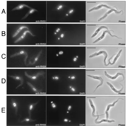
Immunofluorescence analysis of theT. bruceiprocyclic form (Fig. 7) and bloodstream form (data not shown) revealed that the RHS1, RHS4, RHS5, and RHS6 proteins colocalize with the DAPI-stained nuclear DNA with no visible label in the
nucleolus (Fig. 7A and C to E). In contrast, the anti-RHS2 immune serum showed a perinuclear signal, whereby fluores-cence intensity was higher around the nucleus (Fig. 7B).
RHS gene copy number. InTrypanosoma cruzi, GSS data-bases proved to be an extremely powerful and accurate tool to study repeated sequences and particularly to estimate their FIG. 5. Comparison of 12-bp sequences flanking RIME oringielements inserted intoRHSpseudogenes. The RIME andingiretroelements (䊐), theRHSflanking pseudogenes (u), and the unknown flanking region (^) are schematically represented. The 12 bp located at the junction
between the RIME/ingiretroelements and theRHS/unknown flanking regions, and also between retroelements, are indicated by numbered black boxes, and the corresponding sequence is indicated on the right. The nucleotides in bold correspond to the duplicated region associated with the RIME/ingiinsertion, and the boxes define the conserved region. The AAAAAA and CCCTGG sequences correspond to the end and the beginning of the RIME/ingielements, respectively, and dots represent theRHSor unknown sequences. The crosses (✖) in the middle of retroelements indicate in which RIME oringielement homologous recombination probably occurred. The accession numbers ofRHS2j,RHS4d, andRHS1
in Cos-12 and Cos-23,RHS1e,RHS1g,RHS1f, andRHS2gare AL359782 (ChrIa), AC008146 (BAC-30P15), AY046896, AY046897S1 and AY046897S2, AC079606 (BAC-3B10), AC087701 (BAC-26P8), AL359782 (ChrIa), and AC087701 (BAC-26P8), respectively.
FIG. 6. Western blot analysis of RHS proteins. Lysates (4⫻107cells) ofT. bruceiprocyclic form EATRO1125 (PF) and bloodstream form AnTat1 (BF) were analyzed by Western blotting with the immune sera specific for tubulin, RHS1, RHS2, RHS4, RHS5, and RHS6. The positions of the molecular mass markers (in kilodaltons) are indicated on the left and right, and the names of the immune sera is given under each blot.
144 BRINGAUD ET AL. EUKARYOT. CELL
on September 8, 2020 by guest
http://ec.asm.org/
copy number per genome (2). We used the same approach to estimate the copy number of each RHS subfamily in the
T. bruceigenome. A BLAST analysis was performed on the
T. bruceiGSS sequences (http://www.ebi.ac.uk/blast2/parasites
.html [TIGR and The Wellcome Trust Sanger Institute]) using specific 500-bp sequences located in the 3⬘end of each multi-gene subfamily. The GSS represent about 1.8-fold coverage of the haploid nonminichromosomal genome (ca. 30 Mb in strain TREU927/4). The gene copy number per haploid genome was found to range between 6 and 31 depending on the subfamily, with a total of 128 copies for theRHS(pseudo)gene family. TheRHS1(28 copies),RHS2(26 copies),RHS3(31 copies), andRHS4(27 copies) (pseudo)genes are the most abundantly represented, whileRHS5(10 copies) andRHS6(6 copies) are less abundant. The same BLAST computer analysis was con-ducted withingiand RIME sequences. TheingiandRHS (pseu-do)gene copy numbers are similar (140 versus 128 copies per
haploid nonminichromosomal genome), while the RIME copy number is about two to three times higher (380 copies). A previous Southern blot analysis estimated theingicopy number in the range of 200 per haploid total genome (47).
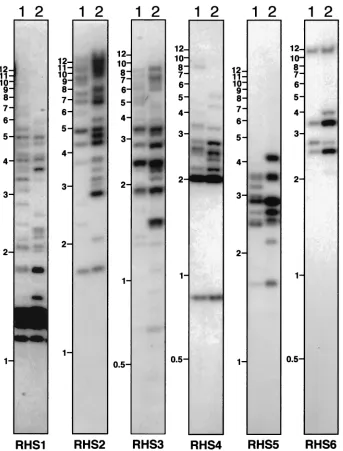
After comparing the availableRHS(pseudo)genes and their conserved flanking regions, a Southern blot analysis of geno-mic DNA was conducted using restriction enzymes selected for their capacity to generate (i) relatively small DNA fragments that separate on a 0.6% agarose gel, (ii) a single DNA frag-ment for eachRHS(pseudo)gene (the enzymes do not cleave the DNA fragments hybridizing with the probes), and (iii) size-polymorphic DNA fragments due to restriction site poly-morphism in the different subfamily members. Using subfam-ily-specific probes (Fig. 2, box 2), ca. 100 different bands were detected in the genome ofT. bruceiTREU927/4 for the whole
RHSmultigene family (Fig. 8), indicating that at least 50RHS
(pseudo)genes are present in the haploid genome. This value is FIG. 7. Immunolocalization of RHS proteins.T. brucei procyclic cells (EATRO1125) were stained with anti-RHS1 (A), anti-RHS2 (B), anti-RHS4 (C), anti-RHS5 (D), and anti-RHS6 (E) immune sera (first column) and with DAPI (second column). Respective phase contrast (phase) images are shown in the third column. Bar⫽5m.
on September 8, 2020 by guest
http://ec.asm.org/
two to three times lower than obtained by GSS database anal-ysis due to the comigration of fragments, as is clear from the variable intensity of hybridization to restriction fragments on the Southern blot (Fig. 8). Comparison of five differentT. bru-ceistrains showed a moderate DNA fragment polymorphism but the overall copy number for eachRHSmultigene subfamily appeared to be in the same range (Fig. 8 and data not shown).
RHS(pseudo)genes are clustered in genome.A P1 library of
T. brucei(TREU927/4) genomic DNA containing average
in-serts of 65 kb (46) was screened with RHS subfamily-specific probes. Only 134 P1 clones (7.4% of the P1 library) are rec-ognized by RHS probe(s), representing approximately 2 Mb of the haploid genome and yielding an estimatedRHS1toRHS6
copy number of only 35 copies per haploid
nonminichromo-somal genome. The latter figure is 3.3 times lower than that obtained by BLAST computer analysis of the GSS databases, suggesting that positive P1 clones contain severalRHS (pseu-do)genes. Indeed, more than 70% of the RHS-positive P1 clones contain at least two representatives of different subfam-ilies, since 29, 38, 21, 14, 15, and 17 P1 clones are recognized by one, two, three, four, five, and six different RHS probes, respectively. However, the estimated copy numbers of the less abundant RHSsubfamilies (RHS5and RHS6) are about the same using both approaches (10 versus 10 and 10 versus 6, respectively).
To determine the extent ofRHS(pseudo)gene clustering, we analyzed several fully or almost fully sequenced BAC clones containing TREU927/4 genomic DNA fragments. Among the FIG. 8. Southern blot analysis ofRHS(pseudo)genes. Genomic DNA fromT. bruceiTREU927/4 (lanes 1) and 427 (lanes 2) was digested to completion withHincII (RHS1 and RHS5),ClaI (RHS2 and RHS6),AseI (RHS3), orHpaII andKpnI (RHS4) and was analyzed by hybridization with the RHS1- to RHS6-specific probes to a Southern blot. The names of the probes and molecular size markers (in kilobases) are indicated below and on the left of each panel, respectively.
146 BRINGAUD ET AL. EUKARYOT. CELL
on September 8, 2020 by guest
http://ec.asm.org/
30 sequenced BACs, we found clones containing 2 (BAC-45I2 and BAC-30P15), 5 26P8), 12 25N24), or 16 (BAC-3B10)RHS(pseudo)genes (Fig. 2). BAC-3B10 (163 kb) and BAC-25N24 (115 kb) constitute one end ofChrII (unpublished data) and contain a 250-kb region mainly composed of 28RHS
(pseudo)genes and their conserved flanking regions, as defined above, with some RIME oringiretroelements inserted in the
RHS1toRHS4coding sequences. This clearly indicates that
RHS(pseudo)genes and their conserved flanking regions are often tandemly arranged. BAC-30P15 and BAC-26P8 clones contain only two and five RHS (pseudo)genes, respectively, which are also tandemly arranged (Fig. 9B and data not shown).
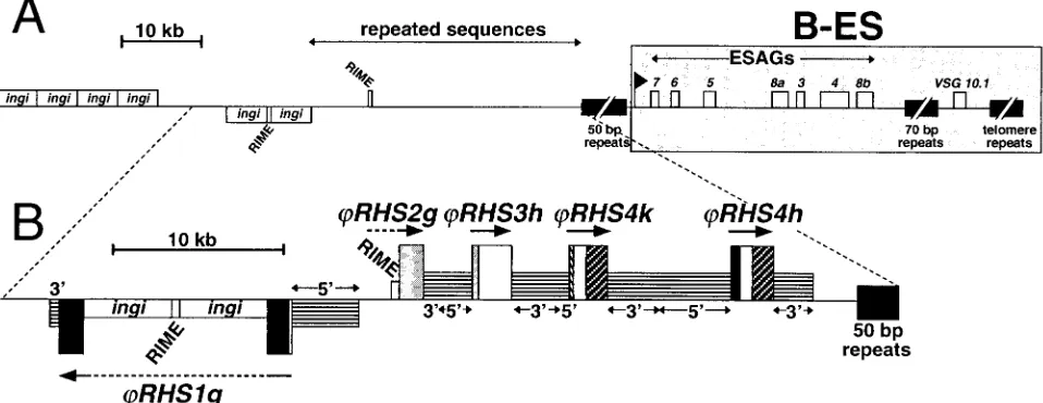
RHS (pseudo)genes are clustered upstream of B-ES.VSGs are expressed inT. bruceibloodstream forms in 1 of about 20
VSG expression sites (B-ESs) located upstream of the telo-mere repeats. Upstream of the B-ES promoter there is a large array of 50-bp repeats (up to 15 kb) that is specific to B-ES (44, 51). In ChrI, and probably in most of the other megabase chromosomes containing a B-ES, the 50-bp repeats are pre-ceded by a large region (100 to 300 kb) composed of RIME/
ingi retroelements and previously uncharacterized repeats (43). Analysis of theChrIa sequence (1.1 Mb), recently com-pleted by The Wellcome Trust Sanger Institute (data not shown), revealed the presence of 15RHS(pseudo)genes clus-tered upstream of the B-ES in a 150-kb DNA region corre-sponding to the RIME/ingi-rich region (43). The RIME/ingi -rich region ofChrIa contains only five full-length and probably functionalRHSgenes (21%), and of the 10RHSpseudogenes, four are highly degenerate. As was observed forChrII, all the RIME/ingi retroelements present in the RHS-rich region of
ChrIa (seven elements) are inserted intoRHS-related
(pseu-do)genes, suggesting that the RIME/ingirichness of this repeti-tive region is directly related to the presence of the RHS
(pseudo)genes. RIME,ingi, and theRHS(pseudo)genes with their conserved flanking regions constitute most (if not all) of the repetitive sequences in this region.
Recently, LaCount et al. sequenced a BAC genomic DNA insert (BAC-26P8) containing the active B-ES expressing the
VSG10.1gene of strain TREU927/4 GUTat10.1 and the region
(90 kb) upstream of the 50-bp repeats (36). As previously observed for the B-ES flanking region of ChrIa, this 90-kb DNA region is RIME/ingi-rich (sixingis and two RIMEs) and contains uncharacterized repeats (Fig. 9A). We found that this 90-kb repeated region is composed of fiveRHSpseudogenes
(RHS2g andRHS1g contain one and three RIME/ingi
ret-roelements, respectively) and conserved flanking sequences, as shown in Fig. 9B. The extent of theRHS(pseudo)gene cluster flankingVSG10.1-ES may be longer since the region upstream of the BAC-26P8 is not yet sequenced.
To determine if the presence of anRHS(pseudo)gene clus-ter is a general feature of the regions upstream of B-ESs, we studied the locations ofRHS(pseudo)genes and B-ES-associ-ated sequences in theT. brucei(TREU927/4) P1 library. All B-ESs described to date contain ESAG7 and ESAG6 genes downstream of the B-ES-specific promoter region and are separated from the remainder of the chromosome by a 50-bp repeat cluster (Fig. 9). All these sequences are considered to be B-ES specific (44, 51). Interestingly, 72, 73, and 86% of the P1 clones recognized by the 50-bp repeat, B-ES promoter and/or ESAG6/7 probes, respectively, contain RHS sequences. These data strongly support the hypothesis that most, if not all, B-ESs are preceded by RHS sequences.
FIG. 9. Gene organization of the BAC-26P8 clone which contains the B-ES ofVSG 10.1. (A) Map of the B-ES ofVSG 10.1and upstream regions in TREU927/4 GUTat10.1, as previously described (36). The locations of the genes and retrotransposons (RIME andingi) in BAC-26P8 are shown (䊐). Genes shown above the line are oriented towards the telomere, whereas those shown below the line are oriented away from the telomere.䡲, 50-bp, 70-bp, and telomere repeats. Expressions site-associated genes (ESAGs) are numbered 1 to 8 and the black flag indicates the position of the B-ES promoter. The B-ES, starting at the promoter, is highlighted by a large grey box. The region containing uncharacterized repeated sequences located upstream of the B-ES is indicated. (B) Detailed analysis of the region containingRHSpseudogenes in BAC-26P8. Sequences encodingRHS1toRHS4pseudogenes (large boxes) are shown using the same color code as in Fig. 2. The name and orientation of each
RHSpseudogene are indicated above or below the boxes. The upstream (5⬘) and downstream (3⬘) sequences conserved between the differentRHS
(pseudo)genes of the same or different subfamilies are indicated by intermediate size boxes containing horizontal lines. All the RIME andingi
retroelements that are inserted intoRHSgenes are indicated by small white boxes.
on September 8, 2020 by guest
http://ec.asm.org/
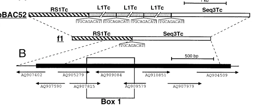
Presence ofRHS-related genes inT. cruzi.A BLAST search of the trypanosomatid databases (http://www.ebi.ac.uk/blast2 /parasites.html) revealed that RHS-related sequences are also abundant in the T. cruzi genome. Recently, Olivares et al. showed that the non-LTR retrotransposon L1Tc, theT. cruzi
homologue of ingi, is frequently inserted between a RS1Tc (1.5-kb) and Seq3Tc (1.9-kb) fragment (49) (Fig. 10A). We have determined that these could encode RHS-related pro-teins. Since, the RS1Tc and Seq3Tc sequences contain numer-ous frame shifts and stop codons, a chimeric full-lengthRHS -related gene was assembled in silico using the T. cruzi GSS database (http://www.ebi.ac.uk/parasites/paratable.html) (Fig. 10B). This database contains 11,459 sequences and represents about 10% of the 40-Mb haploid genome of theT. cruziCL strain (2). The T. cruzi chimeric protein has 16.5 to 23.4% identity with the differentT. bruceiRHS proteins, and most of the residues conserved between theT. brucei RHS proteins, including the ATP/GTP-binding motif, are also conserved in
theT. cruziprotein (Fig. 3). The RS1Tc and Seq3Tc sequences
detected 133 and 167, respectively, significantly similar DNA fragments in theT. cruzi GSS database by BLAST analysis, which corresponds to 348 and 304 copies per haploid genome, respectively. Furthermore, we detected 49 sequences related to the Seq3Tc sequence among approximately 5,000T. cruziESTs (http://www.genpat.uu.se/tryp/tryp.html [Uppsala University]). In summary, these computer analyses indicate that T. cruzi
contains an expressed RHS-related mutigene family, which may also contain a hot spot for retroelement insertion.
DISCUSSION
We have characterized a new, large multigene family encod-ing nuclear and perinuclear proteins inT. brucei.We analyzed a total of 61 differentRHSgenes and pseudogenes detected in
four cDNA clones, two BAC clones fromChrII, the contiguous sequence of ChrIa, and three BACs and five cosmids of un-known genomic location. Analysis of the C-terminal DNA se-quence allowed us to subdivide the family into six multigene subfamilies, RHS1 to RHS6. More than half of the RHS
copies described here are pseudogenes. To estimate the num-ber of RHS (pseudo)genes in the nuclear genome of strain TREU927/4, we took advantage of theT. bruceiGSS databases at TIGR and The Wellcome Trust Sanger Institute, which provide about 1.8-fold coverage of the haploid DNA (exclud-ing minichromosomes). We estimate that there are 128RHS
(pseudo)gene fragments per nonminichromosomal haploid ge-nome. RHS (pseudo)genes also appear to be present in a subset of minichromosomes (hybridization data not shown).
The computational analysis of DNA sequences from TIGR and The Wellcome Trust Sanger Institute, selected cosmids and cDNAs revealed that this multigene family contains a hot spot for insertion of the RIME and ingiretrotransposons: (i) approximately one-third of theRHS (pseudo)genes con-tain RIME and/oringiretrotransposons (16 out of 51 copies), (ii) the retroelements are always inserted at exactly the same relative position in theRHSpseudogenes, even though these genes display up to 50% variation in nucleotide sequence in the vicinity of the insertion site (data not shown), (iii) of the 16
RHSpseudogenes containing RIME/ingielement(s), 25% con-tain two or three retroelements while only 1 of the 10 non-RHS
sequences in the databases containing RIME/ingi retroele-ments has tandemly arranged eleretroele-ments (data not shown), (iv) a phylogenetic analysis shows that most were generated by inde-pendent insertion events, and (v) among the 10 RIME/ingi
retroelements present in the sequenced ChrIa of strain TREU927/4, 7 are inserted intoRHSpseudogenes. Many eu-karyotes contain site-specific non-LTR retrotransposons (3, 9, 16, 26, 38, 63, 67). Also, non-LTR retrotransposons that ap-FIG. 10. Characterization of anRHS-related multigene family inT. cruzi. (A) Map of the pBAC52 and f1 genomic clones fromT. cruzi
CL-Brener and Maracay strains, respectively, as previously reported by Olivares et al. (49). Grey, hatched, and white boxes represent the L1Tc retrotransposons (5 kb) and the RS1Tc (1.4 kb) and Seq3Tc (1.9 kb) repetitive elements, respectively. The conserved duplicated 9-bp sequences (TGCAGACAT) flanking the L1Tc elements and the corresponding L1Tc insertion site in the f1 sequences are shown under the map. (B) Chimeric RS1Tc/Seq3Tc sequence coding for an RHS-related protein. To generate an RS1Tc/Seq3Tc-related sequence containing a large open reading frame (black box on the map), a selection of nine representativeT. cruziGSS were assembled. The relative positions and accession numbers of the selected GSSs are indicated under the map. Box 1 indicates the amino acid sequence, aligned with theT. bruceiRHS proteins in Fig. 3.
148 BRINGAUD ET AL. EUKARYOT. CELL
on September 8, 2020 by guest
http://ec.asm.org/
pear to be randomly distributed in the host genome in fact show a bias of recognition for insertion sites, as exemplified by the TTAAAA sequence of human LINEs (31). The exact site specificity of retroelement insertion intoRHS genes leads to the observed tandem arrays of elements. Interestingly, the tan-dem arrangement of theT. brucei(RIME andingi) andT. cruzi
(L1Tc) non-LTR retrotransposons is unique since, to our knowledge, none of the site-specific or randomly distributed retroelements show this organization in other organisms.
It appears that all the RIME/ingielements present inRHS
genes are inserted in frame with the RHS gene. When the retroelement is unmutated, this results in the generation of long open reading frames encoding putative chimeric proteins composed of the RHS N-terminal half followed by a peptide encoded by the retroelement. However, it is noteworthy that only a fewingi elements contain a single long open reading frame encoding a putative multifunctional protein (data not shown). Most, including those originally described (33, 47), are probably not able to encode functional mRNAs due to the presence of frame shifts or premature stop codons. Conse-quently, the putative RHS/ingichimeric proteins may exhibit an important size and sequence polymorphism due to the
ingi polymorphism. At least seven different chimeric pro-teins formed between cellular and mobile element genes are expressed in humans (24, 56, 60, 65). Thus, it is tempting to consider that some of theRHS/ingichimeric proteins may be expressed and that the proteins may have a cellular role. This would provide a functionalraison d’êtrefor the presence and conservation of a RIME/ingiinsertion hot spot within theRHS
genes. This hypothesis is supported by the characterization of
RHS/retrotransposon chimeric cDNA molecules in which the boundary between the RHS pseudogene and the RIME se-quence corresponds exactly to the conserved RIME/ingi inser-tion site observed in genomic DNA. Producinser-tion of antibodies against the N-terminal region of theingiproducts will allow us to determine if the RHS/ingichimeric proteins are expressed. Analysis of theT. cruzidatabases revealed that the genome
ofT. cruzialso contains polymorphic repeated sequences that
potentially code for proteins homologous to theT. bruceiRHS proteins. Interestingly, these DNA sequences were initially characterized as non-LTR retrotransposon (L1Tc) flanking se-quences (49), suggesting that such elements also frequently insert into the putativeT. cruzi RHS-like genes. In contrast, a BLAST analysis of theLeishmaniaGSS and cosmid sequence databases, which contain at least as many sequences as the
T. bruceidatabases, does not reveal the presence of anyRHS
homologue. The absence of these sequences is probably cor-related with the apparent absence of mobile elements, includ-ing retrotransposons, as revealed by the ongoinclud-ing sequence analysis of this highly related genome (http://www.ebi.ac.uk/ parasites/leish.html).
Comparison ofChrI homologues in differentT. bruceistrains indicates that the large RIME/ingi-rich repetitive region pre-sents a polymorphism with an important size (43). The RIME/
ingi richness observed for this large section of ChrIa in TREU927/4 (43), but also inChrII and BAC-26P8, is entirely due to insertion into the clustered RHS (pseudo)genes. De-tailed analysis of theRHSmultigene family shows that they are subject to frequent homologous recombination. Where this occurs within and between nonhomologous chromosomes may
explain not only the size of the polymorphism of the RIME/
ingi-rich repetitive area (43) but also the variation in number and location of B-ESs observed in different strains (43, 44, 45). Our analysis reveals that among 23 retroelements present in 14
RHSpseudogenes within the large clusters described here, 8 are flanked by oneRHSsequence and one unknown sequence. The latter were probably generated by homologous recombi-nation between two retroelements, one inserted in an RHS
pseudogene and another inserted into the unknown sequence. In addition, approximately one-third of the RHS (pseudo)-genes studied are chimeric, and we suggest that these probably result from homologous recombination in conserved regions of
RHS copies belonging to different subfamilies. These sus-pected homologous recombination events are probably the tip of the iceberg, since numerous undetectable events probably occur between the abundant homologous sequences clustered in large sections of multiple chromosomes.
The 52RHS(pseudo)genes identified so far in theT. brucei
(TREU927/4) databases are located in five different clusters that are almost exclusively composed ofRHScopies and their large conserved flanking regions: 28 copies (15 genes, 13 pseu-dogenes) in a 250-kb area of ChrII (unpublished data), 15 copies (5 genes, 10 pseudogenes) in the 150-kb RIME/ingi-rich region inChrIa (42, 43), five pseudogenes in BAC-26P8 (36), two pseudogenes in BAC-45I2 (36), and two pseudogenes in BAC-30P15 (unpublished data). The three largestRHS clus-ters are located upstream of the TTAGGG telomere repeats (ChrII) or upstream of a 45- to 60-kb B-ES that is adjacent to the telomere repeats (ChrIa and BAC-26P8). The tandemly arranged RHS pseudogenes in BAC-45I2 are located 30 kb upstream of a region with the characteristics of a telomeric M-ES. Similarly, the M-ES active in T. brucei rhodesiense
WRATat1.1-MVAT5 (41) and present in T. brucei AnTat1 (13) is preceded by aRHS1pseudogene (Cos-12) located 10 kb upstream of the telomere repeats (unpublished data). Al-though the chromosomal positions of the DNA sequences de-rived from the other BACs and the cosmids are not known, it appears from this analysis that the RHS (pseudo)genes are located in subtelomeric regions of chromosomes, upstream of ESs (B-ESs or M-ESs) or directly adjacent to the telomere repeats. However, in the fully sequencedChrI andChrII, the large clusters are found only at one end, indicating that not all telomeres are separated from the central coding regions by
RHSclusters. Nevertheless, it is interesting that the P1 geno-mic library analysis showed that most of the B-ESs, maybe all of them, are flanked byRHS(pseudo)genes.
The subtelomeric localization of the RHS (pseudo)genes may be related to their function. In most eukaryotes, subtelo-meric regions are large and repetitive, and poorly transcribed sequences are located at both ends of chromosomes and di-rectly adjacent to the short telomere repeats (69). Although subtelomeres are essentially composed of noncoding se-quences, expressed genes are found embedded in subtelomeric repeats, such as the PAU, SUC, MAL, and MEL multigene families in yeast (40), and surface antigen gene families in
Plasmodium(6, 18, 20, 61, 62). Apparently there is a selective
advantage for the Plasmodiumsurface antigen genes, which are involved in antigenic variation, to be located within lomeric regions. The high recombination frequencies in subte-lomeric domains seem to create a favorable environment for
on September 8, 2020 by guest
http://ec.asm.org/
the rapid generation of novel genes encoding surface proteins (25). Interestingly, inPlasmodium vivax, a large cluster of 35vir
genes and pseudogenes encoding immunovariant surface pro-teins is located directly upstream of the telomere repeats (20), exactly as observed for theRHScluster inChrII. In addition,
T. brucei VSGs are expressed in the telomeric ESs (B-ESs and
M-ESs) and homologous recombination is required to mediate antigenic variation. These observations suggest that the diver-sity observed for theRHS multigene family, probably gener-ated by the high rate of recombination in subtelomeric regions, may be advantageous for the parasite. Our experiments indi-cate that the RHS proteins are loindi-cated inside the cell, not on the cell surface, and it is now a priority to investigate the function of this diverse and potentially rapidly evolving gene family.
In summary, we describe for the first time a gene family with conserved flanking regions that constitutes about 5% of theT.
brucei genome. This multigene family is associated with the
most abundant putative mobile elements (about 5% of the genome content) and may be undergoing rapid evolution by recombination and sequence divergence. TheRHSgenes are clustered in defined regions of chromosomes inT. bruceiand are probably always found upstream of B-ESs, although also present on chromosomes not carrying B-ESs. A homologous family is present inT. cruzi, and for both of these organisms the data presented here will be very significant to the finishing stages of the genome sequencing projects.
ACKNOWLEDGMENTS
The contributions of the two first coauthors, F.B. and N.B., are equivalent.
We are grateful to D. Baltz and A. Ambit for technical help and T. Heidmann, S. Litvak, M. Pages and D. R. Robinson for critical reading of the manuscript.
This work was supported by the CNRS, the Conseil Régional d’Aquitaine, the GDR Parasitologie (CNRS), the Ministère de l’Edu-cation Nationale de la Recherche et de la Technologie (Action Micro-biologie), the Programme Alliance Franco-Britannique 2001, UNDP/ World Bank/WHO-TDRT. bruceiGenome Project and the Wellcome Trust Beowulf Genomics Initiative.
REFERENCES
1.Affolter, M., L. Rindisbacher, and R. Braun.1989. The tubulin gene cluster ofTrypanosoma bruceistarts with an intact beta-gene and ends with a trun-cated beta-gene interrupted by a retrotransposon-like sequence. Gene80: 177–183.
2.Aguero, F., R. E. Verdun, A. C. Frasch, and D. O. Sanchez.2000. A random sequencing approach for the analysis of theTrypanosoma cruzigenome: general structure, large gene and repetitive DNA families, and gene discov-ery. Genome Res.10:1996–2005.
3.Aksoy, S., T. M. Lalor, J. Martin, L. H. Van der Ploeg, and F. F. Richards. 1987. Multiple copies of a retroposon interrupt spliced leader RNA genes in the African trypanosome Trypanosoma gambiense. EMBO J.6:3819–3826. 4.Barry, J. D., S. V. Graham, M. Fotheringham, V. S. Graham, K. Kobryn, and B. Wymer.1998. VSG gene control and infectivity strategy of metacyclic stageTrypanosoma brucei. Mol. Biochem. Parasitol.91:93–105.
5.Barry, J. D., and R. McCulloch.2001. Antigenic variation in trypanosomes: enhanced phenotypic variation in a eukaryotic parasite. Adv. Parasitol.49: 1–70.
6.Baruch, D. I., B. L. Pasloske, H. B. Singh, X. Bi, X. C. Ma, M. Feldman, T. F. Taraschi, and R. J. Howard.1995. Cloning theP. falciparumgene encoding PfEMP1, a malarial variant antigen and adherence receptor on the surface of parasitized human erythrocytes. Cell82:77–87.
7.Beals, T. P., and J. C. Boothroyd.1992. Genomic organization and context of a trypanosome variant surface glycoprotein gene family. J. Mol. Biol. 225:961–971.
8.Bernards, A., L. H. Van der Ploeg, A. C. Frasch, P. Borst, J. C. Boothroyd, S. Coleman, and G. A. Cross.1981. Activation of trypanosome surface glycoprotein genes involves a duplication-transposition leading to an altered 3⬘end. Cell27:497–505.
9.Besansky, N. J., S. M. Paskewitz, D. M. Hamm, and F. H. Collins.1992. Distinct families of site-specific retrotransposons occupy identical positions in the rRNA genes ofAnopheles gambiae. Mol. Cell. Biol.12:5102–5110. 10.Borst, P., W. Bitter, P. A. Blundell, I. Chaves, M. Cross, H. Gerrits, F. van
Leeuwen, R. McCulloch, M. Taylor, and G. Rudenko.1998. Control of VSG gene expression sites inTrypanosoma brucei. Mol. Biochem. Parasitol.91: 67–76.
11.Bringaud, F., D. Baltz, and T. Baltz.1998. Functional and molecular char-acterization of a glycosomal PPi-dependent enzyme in trypanosomatids: pyruvate, phosphate dikinase. Proc. Natl. Acad. Sci. USA95:7963–7968. 12.Bringaud, F., and T. Baltz.1992. A potential hexose transporter gene
ex-pressed predominantly in the bloodstream form ofTrypanosoma brucei. Mol. Biochem. Parasitol.52:111–121.
13.Bringaud, F., N. Biteau, J. E. Donelson, and T. Baltz.2001. Conservation of metacyclic variant surface glycoprotein expression sites among different try-panosome isolates. Mol. Biochem. Parasitol.113:67–78.
14.Bringaud, F., C. Vedrenne, A. Cuvillier, D. Parzy, D. Baltz, E. Tetaud, E. Pays, J. Venegas, G. Merlin, and T. Baltz.1998. Conserved organization of genes in trypanosomatids. Mol. Biochem. Parasitol.94:249–264.
15.Brun, R., and M. Schonenberger.1979. Cultivation and in vitro cloning or procyclic culture forms ofTrypanosoma bruceiin a semi-defined medium. Acta Trop.36:289–292.
16.Burke, W. D., F. Muller, and T. H. Eickbush.1995. R4, a non-LTR retro-transposon specific to the large subunit rRNA genes of nematodes. Nucleic Acids Res.23:4628–4634.
17.Campbell, D. A. 1989. c2X75, a derivative of the cosmid vector c2XB. Nucleic Acids Res.17:458.
18.Cheng, Q., N. Cloonan, K. Fischer, J. Thompson, G. Waine, M. Lanzer, and A. Saul. 1998. stevor and rif arePlasmodium falciparummulticopy gene families which potentially encode variant antigens. Mol. Biochem. Parasitol. 97:161–176.
19.Cross, G. A., L. E. Wirtz, and M. Navarro.1998. Regulation of vsg expression site transcription and switching inTrypanosoma brucei. Mol. Biochem. Para-sitol.91:77–91.
20.del Portillo, H. A., C. Fernandez-Becerra, S. Bowman, K. Oliver, M. Preuss, C. P. Sanchez, N. K. Schneider, J. M. Villalobos, M. A. Rajandream, D. Harris, L. H. Pereira da Silva, B. Barrell, and M. Lanzer.2001. A super-family of variant genes encoded in the subtelomeric region ofPlasmodium vivax. Nature410:839–842.
21.Djikeng, A., C. Agufa, J. E. Donelson, and P. A. Majiwa.1998. Generation of expressed sequence tags as physical landmarks in the genome of Trypano-soma brucei. Gene221:93–106.
22.Donelson, J. E., K. L. Hill, and N. M. El-Sayed.1998. Multiple mechanisms of immune evasion by African trypanosomes. Mol. Biochem. Parasitol.91: 51–66.
23.El-Sayed, N. M., C. M. Alarcon, J. C. Beck, V. C. Sheffield, and J. E. Donelson. 1995. cDNA expressed sequence tags ofTrypanosoma brucei rhodesienseprovide new insights into the biology of the parasite. Mol. Bio-chem. Parasitol.73:75–90.
24.Esposito, T., F. Gianfrancesco, A. Ciccodicola, L. Montanini, S. Mumm, M. D’Urso, and A. Forabosco.1999. A novel pseudoautosomal human gene encodes a putative protein similar to Ac-like transposases. Hum. Mol. Genet.8:61–67.
25.Freitas-Junior, L. H., E. Bottius, L. A. Pirrit, K. W. Deitsch, C. Scheidig, F. Guinet, U. Nehrbass, T. E. Wellems, and A. Scherf.2000. Frequent ectopic recombination of virulence factor genes in telomeric chromosome clusters of P. falciparum. Nature407:1018–1022.
26.Gabriel, A., T. J. Yen, D. C. Schwartz, C. L. Smith, J. D. Boeke, B. Sollner-Webb, and D. W. Cleveland.1990. A rapidly rearranging retrotransposon within the miniexon gene locus ofCrithidia fasciculata. Mol. Cell. Biol. 10:615–624.
27.Gottesdiener, K., J. Garcia-Anoveros, M. G. Lee, and L. H. Van der Ploeg. 1990. Chromosome organization of the protozoanTrypanosoma brucei. Mol. Cell. Biol.10:6079–6083.
28.Harlow, E., and D. Lane (ed.).1988. Antibodies: a laboratory manual. Cold Spring Harbor Laboratory Press, Cold Spring Harbor, N.Y.
29.Hasan, G., M. J. Turner, and J. S. Cordingley.1984. Complete nucleotide sequence of an unusual mobile element from Trypanosoma brucei. Cell 37:333–341.
30.Hope, M., A. MacLeod, V. Leech, S. Melville, J. Sasse, A. Tait, and C. M. Turner.1999. Analysis of ploidy (in megabase chromosomes) in Trypano-soma bruceiafter genetic exchange. Mol. Biochem. Parasitol.104:1–9. 31.Jurka, J.1997. Sequence patterns indicate an enzymatic involvement in
integration of mammalian retroposons. Proc. Natl. Acad. Sci. USA94:1872– 1877.
32.Kanmogne, G. D., M. Bailey, and W. C. Gibson.1997. Wide variation in DNA content among isolates ofTrypanosoma bruceissp. Acta Trop.63:75– 87.
33.Kimmel, B. E., O. K. ole-MoiYoi, and J. R. Young.1987. Ingi, a 5.2-kb dispersed sequence element fromTrypanosoma bruceithat carries half of a smaller mobile element at either end and has homology with mammalian LINEs. Mol. Cell. Biol.7:1465–1475.
150 BRINGAUD ET AL. EUKARYOT. CELL