Effects of miRNA-143 and the non-coding
RNA MALAT1 on the pathogenesis and
metastasis of HeLa cells
L. Zhang1, H.E.X.D. Niyazi1, H.R. Zhao1, X.P. Cao2, M.N.S. Abudula1,
W.J. Ye2, S.A. Zhang1, R.H.M. Yiming1, Y. Zhang1, W.P. Su1, R. Chen1,
Y. Ouyang2, N. Miao3 and Y.X. Bao1
1Department of Oncology,
The First Affiliated Hospital of Xinjiang Medical University, Urumqi, Xinjiang, China
2Central Laboratory, Xinjiang Medical University, Urumqi, Xinjiang, China
3Department of Pathology,
The First Affiliated Hospital of Xinjiang Medical University, Urumqi, Xinjiang, China
Corresponding author: Y.X. Bao E-mail: [email protected] Genet. Mol. Res. 16 (1): gmr16019269 Received September 13, 2016
Accepted January 19, 2017 Published February 23, 2017
DOI http://dx.doi.org/10.4238/gmr16019269
Copyright © 2017 The Authors. This is an open-access article distributed under the terms of
the Creative Commons Attribution ShareAlike (CC BY-SA) 4.0 License.
ABSTRACT. Cervical cancer is a common female malignancy of global dimensions. MicroRNAs (miRNAs) play crucial roles in the development, differentiation, proliferation, and apoptosis of tumors. The non-coding RNA MALAT1 participates in various physiological processes that are important for proper functioning of the body. Here, we analyzed the expression of miRNA-143 and MALAT1 in HeLa cells to evaluate their roles in the occurrence and metastasis of cervical cancer. HeLa cells were divided into five groups depending on the treatment conditions, namely, transfected with miRNA-143,
untreated control. Reverse transcription-polymerase chain reaction was used to analyze the expression of miRNA-143 and MALAT1, the 3-(4,5-dimethylthiazol-2-yl)-2,5-diphenyltetrazolium bromide (MTT) assay to assess proliferation, the trans-well assay to study cell invasion and migration, and western blot to analyze the levels of E-cadherin and vimentin. The proliferation of HeLa cells increased upon treatment with the miRNA-143 inhibitor and decreased when treated with the
MALAT1 inhibitor, compared to the proliferation of the groups that were
transfected with miRNA-143 and MALAT1, respectively (P < 0.05). Thus, miRNA-143 decreased cell invasion and migration potency, downregulated vimentin and upregulated E-cadherin expression, while MALAT1 had the opposite effects. In conclusion, the low expression
of miRNA-143 and high expression of MALAT1 in cervical cancer cells
could possibly potentiate cell invasion/migration and alter the levels of vimentin and E-cadherin.
Key words: miRNA-143; MALAT1; Cervical cancer; Cell proliferation; Cell invasion and migration
INTRODUCTION
Cervical cancer is the second-most common malignancy regarding incidence and mortality in women from developing countries that severely affects public health (Huang et al., 2014b). MicroRNAs (miRNAs) are single stranded non-coding RNAs of 19-25 nucleotides that occur in all eukaryotes and negatively regulate gene expression at the post-transcriptional level. miRNAs can regulate the occurrence and progression of tumors similar to oncogenes and tumor suppressor genes (Zhang et al., 2012; Ahmad et al., 2013). Currently, the advancements in sequencing technology has enabled the discovery of the long-chain non-coding RNAs that can modulate both transcriptional and post- transcriptional processes (Gupta et al., 2010). The expression of several non-coding RNAs is altered in malignant tumors, which can therefore be used as diagnostic markers for cancer progression. Among these, MALAT1 has been shown to be related to the occurrence and progression of various human tumors (Gutschner and Diederichs, 2012). Therefore, we investigated the relationship between the expression of miRNA-143 and MALAT1 with the occurrence and metastasis of cervical cancer using HeLa cells as the model system.
MATERIAL AND METHODS
Cells
The human cervical cancer cell line, HeLa, was obtained from the Xinjiang Medical University.
Reagents and equipment
reverse primers were designed and synthesized by the Sangon Biotech. Co. Ltd. (Sangon, Shanghai, China). The lipofectamine TM2000 and the Trizol kit were purchased from Invitrogen (Invitrogen, Waltham, MA, USA). The fluorescent real time quantitative PCR kit was purchased from Sangon Biotech. Co. Ltd. (Sangon, Shanghai, China). Trans-well chambers were purchased from Millipore (Millipore, Billerica, Massachusetts, USA). Mouse anti-human vimentin monoclonal antibody, rabbit human E-cadherin polyclonal antibody, goat rabbit or mouse secondary antibody conjugated to alkaline phosphatase, and mouse anti-human β-actin antibody were purchased from Santa Cruz (Santa Cruz Biotechnology, Dallas, TX, USA). The incubation chamber was purchased from Thermo (Waltham, MA, USA), CO2 incubator and -80°C refrigerator from Sanyo, inverted microscope from Nikon, PCR cycler from Biometra, and high-speed micro-centrifuge from Beckman (Brea, CA, USA).
Cell culture
HeLa cells were cultured in Roswell Park Memorial Institute (RPMI) 1640 medium at 37°C in a chamber with 5% CO2 perfusion.
Vector transfection
Vectors for miRNA-143, MALAT1, the miRNA-143 inhibitor, and MALAT1 inhibitor were synthesized by Sangon Biotech Co. Ltd. as shown in Table 1. Following EcoRI digestion of the target gene, the linearized vector fragments were purified from agarose gel and treated with alkaline phosphatase to prevent auto-ligation. Cells at log-phase of growth were seeded in 6-well plates and were divided into the miRNA-143 transfection group, MALAT1 transfection group, miRNA-143 inhibitor transfection group, MALAT1 inhibitor transfection group, and the control group. Vector transfection was performed with Lipofectamine 2000 (Invitrogen) according to manufacturer’s instructions.
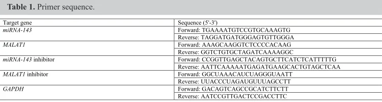
Table 1. Primer sequence.
Target gene Sequence (5'-3')
miRNA-143 Forward: TGAAAATGTCCGTGCAAAGTG Reverse: TAGGATGATGGGAGTGTTGGGA
MALAT1 Forward: AAAGCAAGGTCTCCCCACAAG Reverse: GGTCTGTGCTAGATCAAAAGGC
miRNA-143 inhibitor Forward: CCGGTTGAGCTACAGTGCTTCATCTCATTTTTG Reverse: AATTCAAAAATGAGATGAAGCACTGTAGCTCAA MALAT1 inhibitor Forward: GGCUAAACAUCUAGGGUAATT
Reverse: UUACCCUAGAUGUUUAGCCTT GAPDH Forward: GACAGTCAGCCGCATCTTCTT
Reverse: AATCCGTTGACTCCGACCTTC
Reverse transcription quantitative PCR (RT-qPCR)
HeLa cells at log-phase were collected from all the groups and RNA was extracted using the Trizol kit according to the manufacturer’s instruction.
3-(4,5-dimethylthiazol-2-yl)-2,5-diphenyltetrazolium bromide (MTT) assay
HeLa cells at the log-phase of growth were counted and inoculated in culture plates for attachment and overnight growth. The cells were first incubated in DMEM medium containing 2% fetal bovine serum for 24 h, followed by addition of DMEM containing 10% FBS. Then, the cells were treated with 20 µL MTT solution (5 mg/mL) for 4 h, followed by incubation with 150 µL dimethyl sulfoxide for 10 min and vortexing to dissolve the crystals. Absorbance values at 570 nm were measured.
Trans-well assay
Invasion assay: Matrigel was added in the trans-well chamber and incubated at 4°C overnight, followed by hydration in serum-free medium at 37°C for 1 h. Post-transfection, the cells were cultured in the upper part of the trans-well chamber, the lower part of which was perfused with RPMI1640 medium. Giemsa staining was used for microscopic observation and quantification.
Migration assay: The same procedure as that used in the invasion assay was followed, however, in the absence of the artificial basal membrane after inoculation of the cells into the chamber.
Western blotting
HeLa cells at log-phase were separated on an 8% SDS-PAGE with 40 µg lysates per well. The proteins were then transferred onto a PVDF membrane (Thermo Fisher Scientific), which had been blocked with 5% BSA (Cell Signaling Technology, Danvers, MA, USA) at room temperature for 1 h. Next, the membrane was incubated with the primary antibody (anti-E-cadherin or Vimentin, all at 1: 200 or 1: 500 for β-actin) for 30 min under warm conditions, followed by overnight incubation at 4°C. TBST (tris-buffered saline and Tween 20, pH 7.5) buffer was used to rinse the membrane, followed by incubation with the secondary antibody (1: 2,000 dilution) for 1 h. Chromogenic substrates A and B were added sequentially, followed by ECL development and imaging. The Quantity One software was used to analyze the optical densities.
Data analysis
The SPSS17.0 software was used for data processing. The enumeration data were analyzed by the chi-square test, while the measurement data were analyzed by the analysis of variance (ANOVA) and were presented as mean ± standard deviation (SD). A statistical significance was defined when P < 0.05.
RESULTS
Analysis of
miRNA-143
and
MALAT1
expression in HeLa cells
MALAT1 were also significantly higher in the MALAT1 transfection group compared to the control group (P < 0.05, Table 2). However, the transfection of inhibitors reduced the levels of miRNA-143 and MALAT1 (P < 0.05, Table 2).
*P < 0.05 compared to the control group.
Table 2. Expression of miRNA-143 and MALAT1 in HeLa cells.
Group miRNA-143 MALAT1
miRNA-143 3.65 ± 0.82* 1.14 ± 0.17
MALAT1 1.29 ± 0.41 2.89 ± 0.73*
miRNA-143 inhibitor 0.56 ± 0.12* 1.13 ± 0.22 MALAT1 inhibitor 1.32 ± 0.27 0.35 ± 0.11*
Control 1.31 ± 0.32 1.12 ± 0.25
Proliferation of HeLa cells under different transfection conditions
The MTT assay was used to test the proliferation of cells in all the groups. With longer incubation time, the proliferation of cells in the miRNA-143 and the MALAT1- inhibitor- transfected group gradually decreased (P < 0.05, Table 3), while those of the cells transfected with MALAT1 or the miRNA-143 inhibitor progressively increased (P < 0.05, Table 3), compared to the proliferation of cells in the control group. Similarly, compared to the proliferation of cells in the control group, HeLa cells transfected with MALAT1 or the
miRNA-143 inhibitor had elevated proliferation while those transfected with miRNA-143 or
the MALAT1 inhibitor had lower proliferation (P < 0.05, Table 3).
Table 3. Proliferation of HeLa cells under different transfection conditions.
*P < 0.05 compared to the control group; #P < 0.05 compared to the 24 h group; ∆P < 0.05 compared to the 48 h
group.
Group 24 h 48 h 72 h
miRNA-143 0.189 ± 0.242* 0.153 ± 0.178* 0.125 ± 0.112* MALAT1 0.517 ± 0.383* 0.712 ± 0.423*# 0.897 ± 0.621*#
miRNA-143 inhibitor 0.525 ± 0.352* 0.753 ± 0.445*# 0.973 ± 0.551*#
MALAT1 inhibitor 0.196 ± 0.233* 0.142 ± 0.168*# 0.101 ± 0.098*#
Control 0.235 ± 0.215 0.287 ± 0.216 0.296 ± 0.221
Analysis of cell invasion and migration under different transfection conditions
The trans-well assay was used to detect the invasion and migration of HeLa cells. The results indicated that the invasion and migration of HeLa cells were significantly increased in the miRNA-143 inhibitor-transfected group compared to miRNA-143-transfected group (Figure 1). However, the invasion and migration abilities of HeLa cells were significantly decreased in the MALAT1 inhibitor-transfected group (Figure 1).
Expression of E-cadherin and vimentin in HeLa cells transfected with
miR-143
.
MALAT1
, and their inhibitors
group (P < 0.05, Figure 2 and Table 4). E-cadherin level was decreased in the miRNA-143
inhibitor-transfected group compared to the levels of the miRNA-143-transfected group; the levels were increased in the MALAT1 inhibitor-treated group compared to the levels of the
MALAT1-transfected group (P < 0.05, Figure 2 and Table 4).
Figure 1. Analysis of HeLa cell invasion and migration under various transfection conditions. Matrigel was added in the trans-well chamber and incubated at 4°C overnight, followed by hydration for 1 h. Post-transfection. Then the cells were cultured in the upper part of the trans-well chamber, the lower part of which was perfused with RPMI1640 medium. Giemsa staining was used for microscopic observation and quantification of the cell numbers.
Figure 2. Analysis of E-cadherin and vimentin levels by western blot. Loading order from left to right: lysates of HeLa cells (20 µL) transfected with miRNA-143, MALAT1, miRNA-143 inhibitor, MALAT1 inhibitor, and control
transfected cells. β-Actin was used as a loading control. The molecular weight of E-cadherin and vimentin were 135 and 54 kDa respectively. The name of the protein ladder was Precision Plus ProteinTM (Bio-Rad, Hercules, CA, USA).
Table 4. Analysis of levels of E-cadherin and Vimentin in HeLa cells under different transfection conditions.
*P < 0.05 compared to the control group.
Group E-cadherin Vimentin
miRNA-143 3.57 ± 1.63* 0.86 ± 0.08*
MALAT1 1.06 ± 0.11* 2.15 ± 1.05*
miRNA-143 inhibitor 1.02 ± 0.12* 2.34 ± 1.16* MALAT1 inhibitor 3.78 ± 1.75* 0.78 ± 0.07*
Control 1.38 ± 0.22 1.26 ± 0.18
DISCUSSION
et al., 2014; Huang et al., 2014a). The proliferation and migration of tumor cells are major aspects of malignant transformation (Yasui et al., 2013). Metastasis is a multi-step complex process that involves tumor invasion of adjacent tissues, dissemination of metastatic cancerous cells into the blood circulation, extravasation from the blood vessels, colonization at peripheral tissues and further proliferation (Gupta and Massagué, 2006). miRNAs participate in various biological processes and regulate gene expression in a wide spectrum of diseases (Calin et al., 2004), including cancer, where they regulate the occurrence and progression of tumors similar to oncogenes and tumor suppressor genes (Zhang et al., 2010). Results from a previous study implied that miR-143 might facilitate tumor cell apoptosis (Calin et al., 2004). MALAT1 was first discovered in non-small cell lung cancer. As a long non-coding RNA, it is expressed in various tumors and tissues, and may affect proliferation, apoptosis, invasion, metastasis, and drug resistance of tumors (Gutschner et al., 2013a).
Here, we selected the human cervical cancer line, HeLa, as the model system. After normal incubation, the cells were transfected with miRNA-143, MALAT1, the miRNA-143
inhibitor, and the MALAT1 inhibitor and we found elevated levels of miRNA-143 and MALAT1 in the cells transfected with the respective RNA-encoding genes, whereas the levels of miRNA-143 and MALAT1 were low in the respective inhibitor-transfected groups. These results suggested that the expression of miRNA-143 and MALAT1 was successfully manipulated. Further, the MTT assay showed that with longer time of incubation, the proliferation of cells with miRNA-143 or MALAT1 inhibitor gradually decreased (P < 0.05), while that of cells transfected with MALAT1 or the miRNA-143 inhibitor progressively increased (P < 0.05). For the same duration of incubation, HeLa cells transfected with MALAT1 or the miRNA-143
inhibitor had elevated proliferative activity, and those transfected with miRNA-143 or the
MALAT1 inhibitor showed lower proliferation, compared to that of the control transfected
cells. These results suggested that the high levels of miRNA-143 and/or low levels of MALAT1 decreased the proliferation of HeLa cells, while low levels of miRNA-143 and high levels of MALAT1 might facilitate proliferation, consistent with previous studies revealing the negative regulation of miRNA-143 in cell proliferation (Li et al., 2011; Liu et al., 2016). Lajer et al. (2012) suggested that miRNA-143 might participate in the pathogenesis of the disease in a way that is similar to that observed for human papilloma virus infection in patients with cervical cancer. Wang et al. (2008) found lower levels of miRNA-143 in cervical cancer tissues, while the proliferation of HeLa cells was significantly inhibited by miRNA-143 over-expression. MALAT1 has also been shown to regulate the metastatic ability of tumor cells, and to facilitate proliferation and migration of cells (Boardman, 2009). All of the above findings are consistent with our results.
The trans-well method was used to analyze the invasiveness and migratory ability of the cells under various treatment conditions. The results showed significantly high invasive/ migration potency of HeLa cells transfected with MALAT1 or the miRNA-143 inhibitor. A previous study (Chen et al., 2009) showed that over-expression of miRNA-143 could inhibit cell invasion. In another study, the MALAT1 sequence was infused into mice pulmonary carcinoma and the tumor growth and metastasis was inhibited (Gutschner et al., 2013b). Compared to primary tumors, the level of MALAT1 was further elevated in metastatic tumors, suggesting a correlation between the levels of MALAT1 and malignant tumors (Ren et al., 2016).
pathways, which causes degradation of the extracellular matrix (ECM), thereby promoting infiltration by tumor cells and distal metastasis of tumors (Fu et al., 2011). E-cadherin, an epithelial marker, is a Ca2+-dependent intracellular transmembrane glycoprotein adhesion molecule found in epithelial tissues, which participates in establishing cellular connections. Vimentin is a cytoskeleton component and a biomarker for cells of mesenchymal origin. Abnormal expression of vimentin may induce alteration of the cytoskeleton that may facilitate the migration of metastatic cells (Matsuoka et al., 2013). We found elevated levels of vimentin and reduced levels of E-cadherin in MALAT1 and miRNA-143 inhibitor-transfected cells, and the reverse in miRNA-143 and MALAT1 inhibitor-transfected cells. These results suggested that low expression of miRNA-143 and high expression of MALAT1 could regulate the expression of vimentin and E-cadherin. A previous study (Ying et al., 2012) showed that MALAT1 upregulates the expression of E-cadherin and β-catenin, facilitates mesenchymal transition of epithelial cells, and regulates proliferation and metastasis of colorectal carcinoma. Our results are consistent with the conclusions of Ying et al. (2012).
Although the present study revealed certain interesting findings, it has few limitations. The Giemsa staining method used in the trans-well assay is not optimal as it requires use of fixed cells, and the processes of decolorization and staining affects the cells on the Matrigel. Therefore, in subsequent studies, we would use the crystal violet staining method, which might circumvent the problems that were caused by Giemsa staining. In addition, due to only one cervical cancer cell line used in the present study, we plan to investigate the role of miRNA-143 and MALAT1 in other cervical cancer cell lines to confirm the findings obtained from the current study.
In summary, low levels of miRNA-143 and high levels of MALAT1 could potentiate proliferation of the cervical cancer cell line HeLa, and accelerate its invasion and migration via enhancement of vimentin and reduction of E-cadherin levels, respectively. Thus, miRNA-143 and MALAT1 may work as novel markers for metastasis of cervical cancer, although further investigations are required to understand its detailed mechanism.
Conflicts of interest
The authors declare no conflict of interest.
ACKNOWLEDGMENTS
Research supported by the Natural Science Foundation of the First Affiliated Hospital of the Xinjiang Medical University Hospital, Youth Fund Project (#2015ZRQN29) and the Xinjiang Urumqi Infection and Tumor Key Laboratory Open Topic (#WIT- 2013-01).
REFERENCES
Ahmad I, Singh LB, Yang ZH, Kalna G, et al. (2013). Mir143 expression inversely correlates with nuclear ERK5 immunoreactivity in clinical prostate cancer. Br. J. Cancer 108: 149-154. http://dx.doi.org/10.1038/bjc.2012.510
Banno K, Iida M, Yanokura M, Kisu I, et al. (2014). MicroRNA in cervical cancer: OncomiRs and tumor suppressor miRs
in diagnosis and treatment. Sci. World J. 2014: 178075. http://dx.doi.org/10.1155/2014/178075
Boardman LA (2009). Overexpression of MACC1 leads to downstream activation of HGF/MET and potentiates metastasis
and recurrence of colorectal cancer. Genome Med. 1: 36. http://dx.doi.org/10.1186/gm36
sites and genomic regions involved in cancers. Proc. Natl. Acad. Sci. USA 101: 2999-3004. http://dx.doi.org/10.1073/ pnas.0307323101
Chen X, Guo X, Zhang H, Xiang Y, et al. (2009). Role of miR-143 targeting KRAS in colorectal tumorigenesis. Oncogene
28: 1385-1392. http://dx.doi.org/10.1038/onc.2008.474
Fu J, Qin L, He T, Qin J, et al. (2011). The TWIST/Mi2/NuRD protein complex and its essential role in cancer metastasis.
Cell Res. 21: 275-289. http://dx.doi.org/10.1038/cr.2010.118
Gupta GP and Massagué J (2006). Cancer metastasis: building a framework. Cell 127: 679-695. http://dx.doi.org/10.1016/j. cell.2006.11.001
Gupta RA, Shah N, Wang KC, Kim J, et al. (2010). Long non-coding RNA HOTAIR reprograms chromatin state to
promote cancer metastasis. Nature 464: 1071-1076. http://dx.doi.org/10.1038/nature08975
Gutschner T and Diederichs S (2012). The hallmarks of cancer: a long non-coding RNA point of view. RNA Biol. 9: 703-719. http://dx.doi.org/10.4161/rna.20481
Gutschner T, Hämmerle M and Diederichs S (2013a). MALAT1 - a paradigm for long noncoding RNA function in cancer.
J. Mol. Med. (Berl.) 91: 791-801. http://dx.doi.org/10.1007/s00109-013-1028-y
Gutschner T, Hämmerle M, Eissmann M, Hsu J, et al. (2013b). The noncoding RNA MALAT1 is a critical regulator of
the metastasis phenotype of lung cancer cells. Cancer Res. 73: 1180-1189. http://dx.doi.org/10.1158/0008-5472. CAN-12-2850
Huang J, Lyu H, Wang J and Liu B (2014a). MicroRNA regulation and therapeutic targeting of survivin in cancer. Am. J.
Cancer Res. 5: 20-31.
Huang JT, Wang J, Srivastava V, Sen S, et al. (2014b). MicroRNA Machinery Genes as Novel Biomarkers for Cancer.
Front. Oncol. 4: 113. http://dx.doi.org/10.3389/fonc.2014.00113
Lajer CB, Garnæs E, Friis-Hansen L, Norrild B, et al. (2012). The role of miRNAs in human papilloma virus (HPV)-associated cancers: bridging between HPV-related head and neck cancer and cervical cancer. Br. J. Cancer 106: 1526-1534. http://dx.doi.org/10.1038/bjc.2012.109
Li H, Zhang Z, Zhou X, Wang Z, et al. (2011). Effects of microRNA-143 in the differentiation and proliferation of bovine
intramuscular preadipocytes. Mol. Biol. Rep. 38: 4273-4280. http://dx.doi.org/10.1007/s11033-010-0550-z
Liu J, Mao Y, Zhang D, Hao S, et al. (2016). MiR-143 inhibits tumor cell proliferation and invasion by targeting STAT3 in esophageal squamous cell carcinoma. Cancer Lett. 373: 97-108. http://dx.doi.org/10.1016/j.canlet.2016.01.023
Matsuoka J, Yashiro M, Doi Y, Fuyuhiro Y, et al. (2013). Hypoxia stimulates the EMT of gastric cancer cells through
autocrine TGFβ signaling. PLoS One 8: e62310. http://dx.doi.org/10.1371/journal.pone.0062310
Ren D, Li H, Li R, Sun J, et al. (2016). Novel insight into MALAT-1 in cancer: Therapeutic targets and clinical applications.
Oncol. Lett. 11: 1621-1630.
Wang X, Tang S, Le SY, Lu R, et al. (2008). Aberrant expression of oncogenic and tumor-suppressive microRNAs in cervical cancer is required for cancer cell growth. PLoS One 3: e2557. http://dx.doi.org/10.1371/journal.pone.0002557
Yasui K, Shimamura M, Mitsutake N, Nagayama Y. (2013). SNAIL induces epithelial-to-mesenchymal transition and cancer stem cell-like properties in aldehyde dehydroghenase-negative thyroid cancer cells. Thyroid: Official Journal
of the American Thyroid Association. 23: 989-996.
Ying L, Chen Q, Wang Y, Zhou Z, et al. (2012). Upregulated MALAT-1 contributes to bladder cancer cell migration by
inducing epithelial-to-mesenchymal transition. Mol. Biosyst. 8: 2289-2294. http://dx.doi.org/10.1039/c2mb25070e
Zhang H, Cai X, Wang Y, Tang H, et al. (2010). microRNA-143, down-regulated in osteosarcoma, promotes apoptosis and suppresses tumorigenicity by targeting Bcl-2. Oncol. Rep. 24: 1363-1369.
Zhang Y, Wang Z, Chen M, Peng L, et al. (2012). MicroRNA-143 targets MACC1 to inhibit cell invasion and migration