R E V I E W
Open Access
Genetically engineered mouse models and
human osteosarcoma
Alvin JM Ng
1,2†, Anthony J Mutsaers
1,2,3†, Emma K Baker
1,2and Carl R Walkley
1,2*Abstract
Osteosarcoma is the most common form of bone cancer. Pivotal insight into the genes involved in human osteosarcoma has been provided by the study of rare familial cancer predisposition syndromes. Three kindreds stand out as predisposing to the development of osteosarcoma: Li-Fraumeni syndrome, familial retinoblastoma and RecQ helicase disorders, which include Rothmund-Thomson Syndrome in particular. These disorders have
highlighted the important roles ofP53andRBrespectively, in the development of osteosarcoma. The association of OS withRECQL4mutations is apparent but the relevance of this to OS is uncertain as mutations inRECQL4are not found in sporadic OS. Application of the knowledge or mutations ofP53andRBin familial and sporadic OS has enabled the development of tractable, highly penetrant murine models of OS. These models share many of the cardinal features associated with human osteosarcoma including, importantly, a high incidence of spontaneous metastasis. The recent development of these models has been a significant advance for efforts to improve our understanding of the genetics of human OS and, more critically, to provide a high-throughput genetically modifiable platform for preclinical evaluation of new therapeutics.
Keywords:Osteosarcoma, p53, Rb, Mouse models
Review Osteosarcoma
Osteosarcoma (OS) is the most common primary tumour of bone. It is most frequent in children and ado-lescents with an incidence of 7.3 per 1 million of the population [1]. Although OS is mainly classified as a childhood disease, a second peak of incidence is reported in the elderly population [1]. The majority of OS tumours are situated in the long bones with a small proportion located in the pelvis and axial skeleton [2,3]. OS has a relatively high metastatic rate, with the lung being the most common site of spread.
The current treatment for OS revolves around the use of chemotherapy, radiotherapy and the surgical removal of the tumour. The chemotherapeutic regimen for OS patients combines cisplatin, doxorubicin and high doses of methotrexate [4]. Surgical resection is coupled with
limb salvage procedures to remove malignant tissue and minimize the impact on quality of life.
The lack of new therapeutic options for the manage-ment of OS has translated to a stagnation of patient out-comes [5,6]. Survival and prognosis rates have remained largely unchanged in two decades despite increased de-tection and monitoring afforded by advances in clinical imaging modalities [7-9]. Furthermore, there are difficul-ties associated with the study of OS in humans, such as recruiting sufficient patients to allow clinical insights in trialing new treatment options. A key component to im-proving patient outcome will be the development and application of faithful experimental models of human OS. Such models can serve as a preclinical platform for the identification of new therapeutic targets and the in vivotesting and triaging of those proposed for human trials. Experimentally derived interventions could then be developed in in vivo models where therapies can be rigorously evaluated side by side prior to human evalu-ation. Equally importantly, experimental OS models serve as a means to further understand the genetics and biology of OS with an emphasis on metastatic disease. * Correspondence:[email protected]
†Equal contributors 1
St Vincent’s Institute of Medical Research, 9 Princes Street, Fitzroy, VIC 3065, Australia
2
Department of Medicine, University of Melbourne, St. Vincent’s Hospital, Fitzroy, VIC 3065, Australia
Full list of author information is available at the end of the article
Animal models of osteosarcoma
Robust animal models have the capacity to preclinically evaluate therapeutic interventions derived from the ex-tensive basic research efforts underway in OS. To date, the major species used to deliberately generate experi-mental OS are the mouse and the rat [10,11]. The lineage and temporal specificity afforded by murine gen-etic engineering has lead to a rapid increase in the qual-ity and fidelqual-ity of murine OS models when compared to the human condition. Spontaneous disease arising in large breed pet dogs is also of note as a model of human OS and is useful to understanding OS in humans and veterinary practice. It is also gaining prominence in the research environment as a validated model of spontan-eous OS [12-14].
Rodent models of OS have been established for many decades and were originally generated through the ex-posure to chemical and radioactive carcinogens. [15-17]. These models demonstrated the principle of high-penetrance OS models that histologically resemble human OS. However, they possessed several caveats regarding their application to preclinical studies. The majority of OS in humans is sporadic, while the carcinogen-induced mur-ine OS are more representative of therapy-induced disease rather than the primary lesions arising in the majority of human OS [18,19]. Radiation induced OS models gener-ally have a longer latency than alternate strategies and can result in a range of non-mesenchymal tumours due to its non-specific nature. Furthermore it has not been clearly defined what genetic lesions occur during the initiation and maintenance of these tumours. Nonetheless, these radiation-induced OS models have yielded robust experi-mental data and gave rise to valuable reagents such as cell lines to complement human OS studies. Further characterization of these tumours would enable the ra-tional application of these alongside the recently generated tractable genetically engineered models.
Human hereditary disorders: insight into the genetics of human OS
Rare human hereditary disorders offer powerful insights into genes that play critical roles in human cancer biol-ogy in vivo. This is because they offer unequivocal evidence of defined genetic lesions and their importance in human disease pathogenesis. There is a cluster of familial syndromes that predispose to the development of OS and are of relevance to understanding the under-lying genetics of OS. Li-Fraumeni syndrome, familial Retinoblastoma and RecQ helicase disorders such as Rothmund-Thomson Syndrome (RTS) are caused by germ-line mutations of P53, RB and RECQL4 respect-ively. These three kindreds have a greatly enhanced inci-dence of OS compared to the general population as documented in a range of clinical studies in affected
families. In particular, Li-Fraumeni Syndrome patients are highly prone to develop OS, while OS is the second most common tumour type in Retinoblastoma patients [20-22]. OS tumours are a frequent feature of the tumour spectrum affecting RTS patients, however unlike mutations in p53 and the Rb pathway, RECQL4 muta-tions are not observed in sporadic OS [23].
A range of approaches has been used to incorporate information from clinical human OS to model the dis-ease in the mouse. In particular, transgenic and germ-line loss of function alleles have demonstrated important roles for p53 mutations in generating experimental OS. More recently, lineage-restricted somatic deletion mod-els that generate high penetrant metastatic disease have been described [24,25]. These models will provide a de-finitive assessment on the roles of genes in the initiation and maintenance of OS. Furthermore they can be exploited to reveal new therapeutic avenues that can be targeted for the development of new therapies, with a particular emphasis on metastatic disease.
Human hereditary disorders and osteosarcoma Li-fraumeni syndrome (LFS)
Li-Fraumeni syndrome is an autosomal dominant dis-order with germ-line heterozygous mutation inP53. It is characterized by a predisposition to a range of cancers [26,27]. LFS patients have a highly elevated risk of devel-oping soft tissue sarcoma and osteosarcoma [28], and mutations in the“p53-pathway”are thought to be essen-tial for the formation of human cancer.
Mutations in components of the p53 pathway are found in both familial and sporadic OS. Interestingly, the P53 allele itself is found to be mutated in human OS, most commonly as missense mutations [29,30].P53 mutations are not associated with therapeutic response or metastatic status [31,32]. Other reported lesions in the p53 pathway in human OS include amplification of MDM2 and loss of p19ARF[33-37].
Hereditary retinoblastoma
Patients with familial retinoblastoma possess germline mutations in the Retinoblastoma(RB) gene [38]. Rb is a critical co-ordinator of G1-S phase cell cycle progression through its interaction with E2F and has been implicated in a wide range of cellular processes [39].
radiation induced OS has observed mutation ofP53and retention of the intactRBallele in hereditary retinoblast-oma patients [43]. As with the p53 pathway, mutations in the members of the Rb pathway occur frequently in OS with known mutations including amplifications of Cyclin E and CDK4 [44-48].
OS mouse models based on p53 and Rb mutations
The majority of murine OS models to date have been developed based on knowledge of the mutation of p53 and Rb pathways in both familial and sporadic human OS. Mice with germline mutations of p53 developed OS, but also succumbed to a wide range of tumours [49,50]. Mice with tumour-associated p53 variants presented with a higher incidence of OS than germ-line p53 null animals, amongst the tumour spectrum these animals develop [51]. Mice with homozygous deletions ofRBare embryonic lethal and their heterozygous counterparts are not predisposed to OS [50,52]. The role of genetic compensation by other family members is apparent with the Rb related p107 and p130 in certain circumstance [53]. However, neither p107−/− nor p130−/− mice (or compound mutants that are viable) have a reported sus-ceptibility to OS and these genes are not frequently mutated in human cancers based on data available through the COSMIC database [54].
The move to conditional lineage-restricted alleles of both p53 and pRb has allowed the development of new and more faithful models of OS. Utilising Prx1-Cre, which deletes LoxP flanked alleles in the early budding mesenchymal tissue of the limbs, 22% of mice withp53 heterozygosity develop OS. Homozygous deletion ofp53 had a three-fold increase in OS occurrence. However, the deletion of Rb alone in mesenchymal progenitors failed to produce OS tumours [55]. Interestingly the conditional deletion of both p53 and RbusingPrx1-Cre resulted in approximately 70% of animals developing a poorly differentiated soft tissue sarcoma (PD-STS). This result suggests that the cell of origin is strongly influen-cing the arising tumour phenotype, with primitive multi-potential cells favoring the development of PD-STS whilst committed osteoblast precursors give rise to OS at high incidence.
A separate group utilized the same transgenic system and yielded similar results. Over 60% ofPrx1-Cre-p53fl/fl mice developed OS, while the homozygous deletion of Rb in isolation again yielded no tumours. The com-pound deletion of one Rb allele with homozygous p53 deletion increased the OS incidence rate to 92%. How-ever, homozygous deletion of both genes yielded only 18% of OS tumours with a strong preference for hiber-nomas [56].
Rb has been proposed to have a role in influencing late osteoblast differentiation by interacting with Runx2 [57].
However, the removal ofRbalone is not sufficient to in-duce OS in a number of independent studies.Rb muta-tion does show a profound synergy withp53mutation in the induction of experimental OS [24,25]. Similarly, shRNAs that reduced Rb expression in p53-deficient OS cell lines (prior to allografts) gave rise to more ag-gressive and multilineage tumours [56]. The experimen-tal approaches strongly suggest that mutation on the p53 pathway can serve as an initiating event in OS with mutation in the Rb pathway strongly synergizing in the immortalisation of osteoblastic cells.
Rothmund Thomson syndrome (RTS) and RecQ disorders
RTS is a rare autosomal disorder that consists of epithe-lial features (skin atrophy, hyper/hypo-pigmentation), congenital skeletal malformations (leading to short stat-ure), premature ageing and increased malignant disease [58]. Most RTS patients have germ-line mutations in the RECQL4 DNA helicase [59-63]. RTS patients often present with multiple malignancies. In two separate studies, significant portions of RTS patients developed OS with median ages below 11 yrs [23,64]. Conversely, overexpression of Recql4 was reported in human OS tumours with chromosomal abberations and instabilities in the 8q24 locus, which also contains c-Myc [65,66]. RTS patients with truncatingRecql4mutations associate with a higher risk of developing OS as compared to non-truncated mutations [67,68].
RECQL4is a member of a family of DNA helicases in-cluding Bloom (BLM) and Werner (WRN) helicases, All three members are associated with familial cancer pre-disposition syndromes with high frequencies of mesen-chymal derived tumours, with RTS in particular developing OS at approximately 30% frequency. As an ATP-dependent DNA helicase, Recql4 is recruited at the G1and S phases of the cell cycle and plays a critical role in regulating DNA replication.Recql4deficiency in mice is associated with karyotypic abnormalities and increased rates of aneuploidy [69,70]. Strikingly in contrast top53 and Rb mutations, Recql4 mutations are not associated with sporadic human OS and appear restricted to famil-ial RTS OS. The failure to find RECQL4 mutations in sporadic OS raises several questions regarding the na-ture of the disease and whether it represents a distinct entity or subtype of OS. Further efforts characterizing the RTS- related OS are needed to clarify this and efforts to model RTS mutations in mouse may be informative. The contribution of prior chemotherapy/radiotherapy for other cancers arising in RTS patients may be a con-founding factor in RTS-associated OS.
Recql4 Mutation in the mouse
inverse relationship with Rb, although telomere-lengthening activities are enhanced in cells lacking both genes [71,72]. Interestingly, Recql4 expression plays a role in osteoblast proliferation but its reduction is reported to be needed for full differentiation [73].
The attempts at modeling of Recql4 deficiency in mice has led to confounding results. Three non-conditional alleles have been reported. The first allele replaced exons 5 through 8 with a LacZ cassette. The homozygous defi-cient animals were reported as very early embryonic le-thal between embryonic days 3–6 [74]. The second reported allele involved deletion of exon 13. The homo-zygous mutants were viable but exhibited severe growth retardation and multiple abnormalities and 95% of the mice died within 2 weeks of birth [75]. Hetrozygous Recql4 mutants were viable and had a decreased bone mass [73]. The third allele involved replacement of part of exon 9 through to exon 13 with a PGK-Hprt mini gene cassette [76]. These mice were viable and homozy-gous Recql4deficient animals presented with a range of defects reminiscent of the human RTS alleles. Approxi-mately 16% of mice with homozygous Recql4mutations died within 24hrs of birth. 5.8% of animals displayed skeletal defects of the animals that survived past 24hrs. Cancers were detected in 5% ofRecql4−/−animals in an aged cohort of 100 animals compared to 43 age matched controls, and of these 2 animals developed OS and 3 animals developed lymphoma. This low rate of tumour formation contrasts with the clinical presentation of RTS. The development and characterization of new tar-geted alleles will be needed to resolve the role of Recql4 in the initiation and maintenance of OS.
Werner & bloom syndromes
Werner syndrome is characterized by premature ageing and cancer predisposition that occurs during adoles-cence, whereas Bloom syndrome is characterized by short statures and photosensitive skin [77]. Both disor-ders are inherited in an autosomal recessive manner, and are attributed to germ-line mutations of the WRN and BLMgenes respectively.
BLM plays a major role in maintaining genomic stabil-ity in cells [78]. Likewise, WRN acts against DNA breakages during chromatin structural modifications [79]. It is interesting to note that the expression of BLM and WRN is induced by the loss of Rb. Also, cells that lack the normal expression of all 3 genes presented with enhanced telomere lengthening [71,72]. When treated with chemotherapeutics, cells that were deficient for BLM or WRN had decreased cell proliferation with impaired cell viability [80].
Werner Syndrome patients present with a range of cancers including OS [81,82]. Similarly, patients with Bloom Syndrome are predisposed to various cancers,
coupled with an early onset of these tumours [83,84]. As for RTS, the relevance of these mutations to sporadic OS is also unclear and further work is needed to clarify the relationship between these OS and their sporadic counterpart.
BLM & WRN mouse models
Genetically engineered mice that habour null mutations ofBLMwere generated by 3 separate groups. Mice with homozygous deletion of BLMwere embryonic lethal by day 13.5 and presented with an increased level of apop-tosis and anaemia [85]. However, viable BLM-null mice were generated with the removal of neomycin plasmid sequence, of which 30% of these mice presented with a wide spectrum of spontaneous tumours [86]. Heterozy-gous mutant mice were also viable, with a predisposition to develop tumours [87].
Mice with homozygous deficiency for WRN were vi-able and developed tumours by 2 years of age. Interest-ingly, the combined deletions of p53 and WRN in mice resulted in various soft tissue sarcomas, where half of these mice developed tumours by 3 months of age [88]. However, its strongest link to OS was evident when WRN and Telomerase RNA Component (Terc) defi-ciency were combined in mice, with 50% of these mice developing OS [89]. Of note, these were not lineage-restricted alleles suggesting that these pathways co-operate specifically in osteoblasts and strongly synergise in the development of OS.
Paget’s Disease and p62
Paget’s disease of the bone is characterized by abnormal-ities in bone growth and destruction, resulting in limb deformities [90]. It is autosomal dominant in nature, and affects mainly adults over the age of 55 [91,92]. It is also often asymptomatic until patients present with fracture or bone pain [93].
Sequestosome1 (SQSTM1) is the only gene currently identified and associated with Paget’s disease of the bone [94]. Also known as p62, this gene contributes to autop-hagy and removal of abnormal cells [95]. Interestingly, p62 expression needs to be repressed to suppress tumourigenesis [96].
The fraction of patients with Paget’s disease presenting with OS does not exceed 1% [97-101]. This cohort coin-cide with the second peak of OS incoin-cidence rates in the elderly [1,102]. The survival rate of Paget’s disease-associated OS is 5% at 5 years [103].
Insights from p62 mouse models
from both groups presented with increased osteoclasts in response to RANKL stimulation, reminiscent of Paget’s disease patients [104,105]. No OS was reported in these mice.
Other genes associated with osteosarcoma
A range of other genes have been implicated in OS pathogenesis based on studies of human OS samples and cell lines (Table 1). These mutations appear to be cooperative to the defects in the p53 and Rb pathways. Their involvement in OS pathogenesis is also supported by evidence derived from a range of genetically engi-neered mouse approaches.
c-Fos
The overexpression of c-Fos was first noted in human OS tumour samples, particularly in metastasized tumours [124,125]. Its expression was also detected in mouse sporadic and radiation-induced OS [123]. In addition, genetically engineered mice that overexpressed c-Fos developed OS, thus suggestive of its role in OS pathogenesis [126,127]. However, the overexpression of c-Fos in humans is linked to fibrous dysplasia, of which less than 2% of patients develop OS [143,144]. Also, a recent study detected no change in c-Fos gene expres-sion between human osteoblasts and OS tumours, which is in conflict with findings from Gamberi and Wu [66]. Therefore, the role of c-Fos in OS requires further
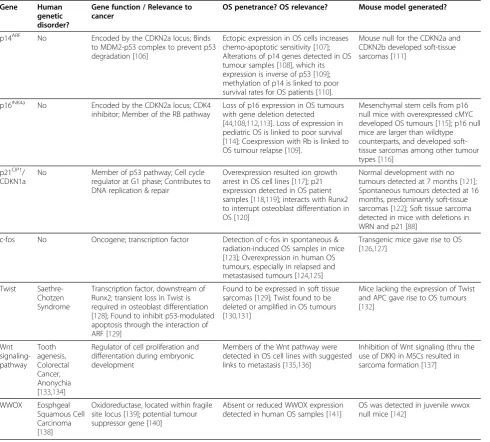
Table 1 Additional genes implicated in osteosarcoma (not discussed in text)
Gene Human
genetic disorder?
Gene function / Relevance to cancer
OS penetrance? OS relevance? Mouse model generated?
p14ARF No Encoded by the CDKN2a locus; Binds to MDM2-p53 complex to prevent p53 degradation [106]
Ectopic expression in OS cells increases chemo-apoptotic sensitivity [107]; Alterations of p14 genes detected in OS tumour samples [108], which its expression is inverse of p53 [109]; methylation of p14 is linked to poor survival rates for OS patients [110].
Mouse null for the CDKN2a and CDKN2b developed soft-tissue sarcomas [111]
p16INK4a No Encoded by the CDKN2a locus; CDK4 inhibitor; Member of the RB pathway
Loss of p16 expression in OS tumours with gene deletion detected [44,108,112,113]. Loss of expression in pediatric OS is linked to poor survival [114]; Coexpression with Rb is linked to OS tumour relapse [109].
Mesenchymal stem cells from p16 null mice with overexpressed cMYC developed OS tumours [115]; p16 null mice are larger than wildtype counterparts, and developed soft-tissue sarcomas among other tumour types [116]
p21CIP1/ CDKN1a
No Member of p53 pathway; Cell cycle regulator at G1 phase; Contributes to DNA replication & repair
Overexpression resulted ion growth arrest in OS cell lines [117]; p21 expression detected in OS patient samples [118,119]; interacts with Runx2 to interrupt osteoblast differentiation in OS [120]
Normal development with no tumours detected at 7 months [121]; Spontaneous tumours detected at 16 months, predominantly soft-tissue sarcomas [122]; Soft tissue sarcoma detected in mice with deletions in WRN and p21 [88]
c-fos No Oncogene; transcription factor Detection of c-fos in spontaneous & radiation-induced OS samples in mice [123]; Overexpression in human OS tumours, especially in relapsed and metastasised tumours [124,125]
Transgenic mice gave rise to OS [126,127]
Twist Saethre-Chotzen Syndrome
Transcription factor, downstream of Runx2; transient loss in Twist is required in osteoblast differentiation [128]; Found to inhibit p53-modulated apoptosis through the interaction of ARF [129]
Found to be expressed in soft tissue sarcomas [129]; Twist found to be deleted or amplified in OS tumours [130,131]
Mice lacking the expression of Twist and APC gave rise to OS tumours [132]
Wnt signaling-pathway
Tooth agenesis, Colorectal Cancer, Anonychia [133,134]
Regulator of cell proliferation and differentation during embryonic development
Members of the Wnt pathway were detected in OS cell lines with suggested links to metastasis [135,136]
Inhibition of Wnt signaling (thru the use of DKK) in MSCs resulted in sarcoma formation [137]
WWOX Eosphgeal Squamous Cell Carcinoma [138]
Oxidoreductase, located within fragile site locus [139]; potential tumour suppressor gene [140]
Absent or reduced WWOX expression detected in human OS samples [141]
studies to close the gap between transgenic mouse biol-ogy and human clinical studies.
c-MYC
Amplification of thec-MYC gene is more prominent in Paget’s disease-related OS as compared to primary OS, although genetic rearrangement does not appear to be the cause [145,146]. Clinically, c-MYC expression levels in OS tumour samples was linked to resistance to methotrexate, with high c-MYC expression correlating to worse outcomes in OS patients [147].
A small cohort of transgenic mice developed OS when c-MYC expression was turned on with a tetracycline regulated transgene in haematopoietic cells [148]. The OS arising in these studies was most likely a result of ec-topic expression of the transgene in osteoblastic cells. When c-MYCexpression was inactivated by doxycycline administration, tumours transplanted into syngeneic mice regressed as OS cells differentiated into mature osteocytes [149]. In a subsequent report from the same group, the tumour regression from c-MYC inactivation in OS cells was attributed to the induction of senescence [150]. The development of OS was also reported in ret-rovirally transdcued c-MYC-overexpressing mesenchy-mal progenitor cells derived from Ink4a/Arf mutant mice [115].
Osteoblast lineage restricted expression of Simian Virus 40 (SV40) T antigen
Antigens of the SV40 virus interact with and inactivate tumour suppressor genes including both Rb and p53 [151,152]. Interestingly, the SV40 gene was detected in a portion of human OS tumours, of which the sequence revealed viral integration in half of these tumours [153]. Early studies of transgenic mice that expressed SV40 antigens presented with OS and other tumours [154,155]. A recent study of mice that expressed the SV40 T antigen in mature osteoblasts using the osteocalcin promoter pre-sented with bone tumours and were morbid by 21 weeks of age. This timeframe for tumour development is strik-ingly similar to that observed withOsx-Crep53fl/flpRbfl/fl animals. The tumours inOcn-SV40Tag animals were his-tologically confirmed as OS and possessed various levels of calcification. Also, the OS tumours metastasized at high frequency and were found predominantly in the lung and spleen [156].
Further analysis of tumours derived in this model revealed a recurrent genomic deletion of thePrkar1agene [156]. Correspondingly, deletion of 1 allele ofPrkar1a dra-matically accelerated OS formation in mice withOcn-SV40 T antigen with tumours arising within 5 weeks of birth. The analysis of human tumours found a subset of human OS also habour a Prkar1a deletion, demonstrating the
power of mouse models to uncover new information into the complex genetics of human OS.
Cell cycle genes: p15INK4b, p16INK4a
Several negative regulators of the G1-S cell cycle phase transition have been implicated in human OS. These fall into the “Rb pathway” and provide further support to the near obligate nature of this pathway disruption in the genesis of OS. p15INK4b was demonstrated to be repressed by c-MYC expression [157]. Mice deficient for
p15INK4b (along with p14ARF and p16INK4a) developed a
wide spectrum of cancers, including soft tissue sarcomas [111]. Genetic alterations were found in human patient-derived OS cell lines in the p15INK4b locus [112]. Dele-tions of thep16genomic locus were apparent in samples from OS patients [158]. Loss of p16INK4Aexpression was found in pediatric OS samples, with its expression level correlating to survival rates [114].
Translating human cancer into animal models: issues & challenges
Human cell lines vs animal models?
Experimental studies of OS have involved the use of cell lines and animal disease models [159,160]. However, cytogenetic complexity in human OS has confounded the efforts [161]. In particular, some human OS cell lines such as U2OS and SAOS-2 have been in use and pas-saged for many decades [162,163]. The extended passage and tissue culture can result in the acquisition of adap-tive mutations from cell-culture conditions, as seen in long-term culturing of embryonic stem cells and lung cancer cell lines [164-166]. As such, the drift in gene ex-pression signatures may make it less representative of the original tumour tissue and also lead to heterogeneity of the cell line populations held by different investigators [167,168]. The recent establishment and description of new OS cell lines opens up new avenues of study and hopefully improves the fidelity of tissue culture studies when referenced back to the human disease.
possible from available human samples and canine OS cell lines.
The recent study in the identification of the Prkar1a gene performed by Khokha and colleagues highlights the power of genetically engineered murine models to gain new insights into human OS genetics [156]. In particular, the use of high-resolution comparative genomic hybridization (cGH) in primary tumours among other complementary analytical techniques was utilized in this project. This allows biologically relevant genetic changes during OS pathogenesis to be isolated, defined and validated from an-euploidy- associated“noise”. Such approaches coupled with the developed murine models may allow significant advances in our understanding of the complexity of OS.
The comparison of primary and metastatic disease from as many of these models as possible would be a novel approach to develop a better understanding of metastatic disease. This will be very useful for under-standing the genetics and cell biology of metastatic OS, and the epigenetic processes that drive these mechan-isms. The experimental approach focused on by analysis of paired primary and metastatic tumours and cell lines derived from the same animal should provide a strong basis for identifying key drivers of the progression and maintenance of metastatic disease. Such an approach could be a starting point to develop better therapeutic strategies for treating metastatic disease, the primary cause of mortality in OS patients.
Different mouse models for different OS conditions
Various technological advancements have been incorpo-rated into generating transgenic cancer mouse models. This includes germline & conditional knockouts, alleles bearing point mutations and tissue/region-specific gene expression [171,172]. These technologies have allowed for multiple paradigms in exploring targeted gene ex-pression and its role in OS pathogenesis. For instance, the Cre-Lox system is widely used to turn off the expres-sion of targeted genes [173]. The turning off of desired genes using Cre-Lox is most often an irreversible step and is useful for modelling OS related to the partial and complete loss of gene function. For instance, the occur-rence of OS in mice with homozygous p53 and Rb dele-tions mimics the clinical scenario of patients with autosomal-dominant hereditary disorders as well as lesions found in the sporadic OS population [24,25].
The mouse models employed by two separate groups produced varying OS incidence rates, which was corre-lated with pRb and p53 status [24,25]. This observation is concordant with various sporadic-OS patient reports where allelic alterations for both genes were reported retrospectively [42,174-176]. The murine models have suggested strongly that deficiency for p53 is a strong
initiating event for the development of OS and that dis-ruption of the Rb pathway is a strongly synergistic muta-tion. The recent work from the Lees group provides an elegant model for the interaction and relative contribu-tion of the p53 and pRb pathway mutacontribu-tions to the bio-logical aspects of OS [56]. An unresolved question which will require analysis of human OS is to determine if the genetic alterations in OS could be different be-tween sporadic and those associated with hereditary disorders.
An outstanding question is do mutations in all mem-bers of the p53 and Rb pathways contribute equally to tumour formation? For example, null mutation of the cyclin-dependent kinase p27Kip1, which results in de-regulation of the “Rb pathway” did not result in OS in these mice [122,177]. When coupled with a p53 muta-tion would p27Kip1 or p21Cip1deficiency recapitulate all or only partial aspects of the loss of Rb? This is intri-guing in light of the spectrum of mutations that have been reported in human OS. It provides an opportunity to compare mutations in distinct components of these pathways directly in the murine models that have been developed.
The emerging use of RNA interference (RNAi) in transgenic cancer models presents an exciting avenue to explore OS genetics and therapeutics. This is because the expression of targeted genes can be manipulated re-versibly in a temporally controlled fashion to elucidate its biological purpose [178-180]. Also, this model pro-vides the attractive prospect of exploring therapeutic tar-get inhibition and resistance. As siRNA/shRNA represents a loss of function allele that are efficient but rarely complete this technology could be harnessed for the rapid and large scale in vivo screening of putative therapeutic targets. As small molecule inhibitors, like siRNA/shRNA, provide efficient but rarely complete get inactivation the testing of candidate therapeutic tar-gets is highly suited to this approach.
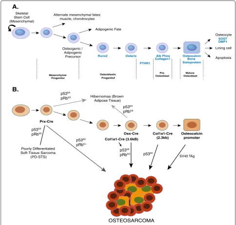
The OS cell of origin
poorly differentiated tissue sarcoma [55]. Also, soft-tissue sarcomas seem most likely to arise from mesenchy-mal stem/progenitor cells [191,192]. As the multipotent mesenchymal/skeletal stem cells can give rise to bone, car-tilage and adipose cell lineages, perhaps it plays a more realistic purpose as a pan-sarcoma cell of origin.
Data derived from a range of genetic approaches most strongly favours the OS cell of origin to be found within the committed osteoblast lineage. In particular,
accumulating experimental evidence is most consistent with OS arising from the osteoblastic progenitor population [24,25]. For instance, the deletion of p53 in pre-osteoblasts and osteoblast progenitors resulted in significantly higher OS incidence rates than early multi-lineage potential cells (Figure 1 and Table 2). As osteoblast progenitors are more committed than their mesenchymal counterparts, this would correlate to decreased occurrence of other sarcoma types. As such, these studies strongly propose that OS
A.
B.
Hibernomas (Brown Adipose Tissue)
OSTEOSARCOMA
Skeletal Stem Cell (Mesenchymal)
Alternate mesenchymal fates: muscle, chondrocytes
Osteogenic / Adipogenic
Precursor
Adipogenic Fate
Runx2 Alk Phos
Collagen1
Osteocalcin Bone Sialoprotein
Osteocyte
Lining cell
Apoptosis
Osterix
SOST DMP1
PTHR1
Differentiation
Osteoblastic Progenitor
Mature Osteoblast
Pre-Osteoblast Mesenchymal
Progenitor
Col1a1-Cre (2.3kb)
Osteocalcin promoter Osx-Cre
p53fl/fl
pRbfl/fl
SV40 TAg
Col1a1-Cre (3.6kB) Prx-Cre
p53fl/fl
pRbfl/fl
Poorly Differentiated Soft-Tissue Sarcoma
(PD-STS) p53
fl/fl
p53fl/fl
pRbfl/fl
p53fl/fl
pRbfl/+
p53fl/fl
pRbfl/fl
arises from the osteoblast lineage-committed progenitor population and that the resulting tumour phenotype is a re-sult of the accumulated genetic mutations that are present.
Metastatic disease–high fidelity and high penetrant models
The use of cancer mouse models with high penetrance allows a substantial population of mice with metastatic disease to be established. In particular, the mice gener-ated by 3 separate groups developed OS with significant metastasis to soft tissues [24,25,156]. These models will be valuable in pre-clinical studies, as primary and metas-tasized tumours could be procured for the comparative studies. Advances in small animal imaging techniques such as μPET and μCT coupled with serology for alka-line phosphatase make possible the establishment of cohorts of animals with primary and a small metastatic disease burden. This strategy makes possible an assess-ment of therapeutic interventions in the context of primary and metastatic disease which are the most pressing clinical need. Longitudinal studies using such approaches would be an effective means to test and tri-age candidate therapeutic approaches in a controlled and reproducible manner. When coupled with xeno-grafts of human material it may facilitate translation into rational clinical trials. Also, untreated paired tumour tis-sue will be useful as it is not readily collected in humans.
Conclusion
Li-Fraumeni, Retinoblastoma and Rothmund-Thomson Syndrome are three human familial cancer syndromes that present with the strongest association to OS. Amongst sporadic OS, a wider range of genes and mem-bers of the p53 and Rb pathways are also implicated in OS pathogenesis. These mutations fulfill a range of the prerequisite requirements associated with the hallmarks of cancer, however the genes do not carry equal
importance in tumour biology nor fully account for the pathogenesis of OS [194]. The integration of genetically engineered murine models based on familial human gen-etics of OS and additional experimental models such as the spontaneous OS arising in large breed dogs combine to form the basis of a preclinical platform that can serve to translate the extensive basic research efforts asso-ciated with OS to a clinically meaningful advantage. The use of primary human xenografts, in contrast to approaches using established human OS cell lines, adds an important component to the preclinical assessment phase of any new therapeutic options [195]. The under-lying genetics in OS covers a wide spectrum, ranging from complete loss of gene function to hypomorphic mutations and gain of function. Various genetically modified mouse models of OS are now available and have demonstrated clearly that these are able to recap-itulate the clinical spectrum of human OS.
Abbreviations
BLM: Bloom; LFS: Li-Fraumeni Syndrome; L-MTP-PE: Liposomal Muramyl-Tripeptide Phosphatidyl Ethanolamine; Ocn: Osteocalcin; OS: Osteosarcoma; PD-STS: Poorly differentiated soft tissue sarcoma; Rb: Retinoblastoma; shRNA: Short hairpin RNA; siRNA: Small interfering RNA;
SQSTM1: Sequestosome1; SV40: Simian Virus 40; RNAi: RNA interference; Tag: T antigen; Terc: Telomerase RNA Component; WRN: Werner.
Competing interests
The authors would like to declare that there are no competing financial, professional or personal interests that might have influenced the performance or presentation of the work described in this manuscript.
Authors' contributions
AJN, AJM, and EKB acquired data and drafted the manuscript. CRW conceived, participated in the design, coordination and helped to draft the manuscript. All authors read and approved the final manuscript.
Acknowledgements
This work is supported by an NHMRC Career Development Award (C.R.W) and NHMRC Project grants (C.R.W); a CRC for Cancer Therapeutics (CTx) graduate student award (A.J.M.N). Research was supported in part by the Victorian Government’s Operational Infrastructure Support Program (to SVI); C.R.W. is the Leukaemia Foundation Philip Desbrow Senior Research Fellow.
Author details
1St Vincent’s Institute of Medical Research, 9 Princes Street, Fitzroy, VIC 3065, Australia.2Department of Medicine, University of Melbourne, St. Vincent’s Hospital, Fitzroy, VIC 3065, Australia.3Ontario Veterinary College, University of Guelph, 50 Stone Road, Guelph, ON N1G 2W1, Canada.
Received: 18 August 2011 Accepted: 30 November 2011 Published: 4 October 2012
References
1. Mirabello L, Troisi RJ, Savage SA:International osteosarcoma incidence patterns in children and adolescents, middle ages and elderly persons. Int J Cancer2009,125:229–234.
2. Klein MJ, Siegal GP:Osteosarcoma: anatomic and histologic variants. Am J Clin Pathol2006,125:555–581.
3. Lamoureux F, Trichet V, Chipoy C, Blanchard F, Gouin F, Redini F:Recent advances in the management of osteosarcoma and forthcoming therapeutic strategies.Expert Rev Anticancer Ther2007,7:169–181. 4. Chou AJ, Gorlick R:Chemotherapy resistance in osteosarcoma: current
challenges and future directions.Expert Rev Anticancer Ther2006, 6:1075–1085.
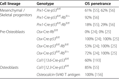
Table 2 OS Incidence rates, murine genotypes & its associated cell lineages
Cell lineage Genotype OS penetrance
Mesenchymal / Skeletal progenitors
Prx1-Cre-p53fl/fl 61% [55]; 62% [56]
Prx1-Cre-p53fl/fl-Rbfl/+ 92% [56]
Prx1-Cre-p53fl/fl-Rbfl/fl 18% [55]; 29% [56]
Pre-Osteoblasts Osx-Cre-Rbfl/fl 0% [24]; 0% [25]
Osx-Cre-p53fl/fl 100% [24]; 100% [25]
Osx-Cre-p53fl/fl-Rbfl/fl 53% [24]; 100% [25]
Osx-Cre-p53fl/fl-Rbfl/+ 72% [24]; 100% [25]
Col1h13.6-Cre-p53fl/fl 60% [193]
Osteoblasts Col1h12.3-Cre-p53fl/fl 85% [55]
5. Ferguson PC, McLaughlin CE, Griffin AM, Bell RS, Deheshi BM, Wunder JS: Clinical and functional outcomes of patients with a pathologic fracture in high-grade osteosarcoma.J Surg Oncol2010,102:120–124. 6. Jawad MU, Cheung MC, Clarke J, Koniaris LG, Scully SP:Osteosarcoma:
improvement in survival limited to high-grade patients only.J Cancer Res Clin Oncol2011,137:597–607.
7. Arndt V, Lacour B, Steliarova-Foucher E, Spix C, Znaor A, Pastore G, Stiller C, Brenner H:Up-to-date monitoring of childhood cancer long-term survival in Europe: tumours of the sympathetic nervous system, retinoblastoma, renal and bone tumours, and soft tissue sarcomas.Ann Oncol2007, 18:1722–1733.
8. Gatta G, Capocaccia R, Stiller C, Kaatsch P, Berrino F, Terenziani M: Childhood cancer survival trends in Europe: a EUROCARE Working Group study.J Clin Oncol2005,23:3742–3751.
9. Mirabello L, Troisi RJ, Savage SA:Osteosarcoma incidence and survival rates from 1973 to 2004: data from the Surveillance, Epidemiology, and End Results Program.Cancer2009,115:1531–1543.
10. Heymann D, Ory B, Blanchard F, Heymann MF, Coipeau P, Charrier C, Couillaud S, Thiery JP, Gouin F, Redini F:Enhanced tumor regression and tissue repair when zoledronic acid is combined with ifosfamide in rat osteosarcoma.Bone2005,37:74–86.
11. Ko SC, Cheon J, Kao C, Gotoh A, Shirakawa T, Sikes RA, Karsenty G, Chung LW:Osteocalcin promoter-based toxic gene therapy for the treatment of osteosarcoma in experimental models.Cancer Res1996,
56:4614–4619.
12. Johnson AS, Couto CG, Weghorst CM:Mutation of the p53 tumor suppressor gene in spontaneously occurring osteosarcomas of the dog. Carcinogenesis1998,19:213–217.
13. Lascelles BD, Dernell WS, Correa MT, Lafferty M, Devitt CM, Kuntz CA, Straw RC, Withrow SJ:Improved survival associated with postoperative wound infection in dogs treated with limb-salvage surgery for osteosarcoma. Ann Surg Oncol2005,12:1073–1083.
14. Paoloni M, Davis S, Lana S, Withrow S, Sangiorgi L, Picci P, Hewitt S, Triche T, Meltzer P, Khanna C:Canine tumor cross-species genomics uncovers targets linked to osteosarcoma progression.BMC Genomics 2009,10:625.
15. Ingleton PM, Coulton LA, Preston CJ, Martin TJ:Alkaline phosphatase in serum and tumour of rats bearing a hormone-responsive transplantable osteogenic sarcoma.Eur J Cancer1979,15:685–691.
16. Martin TJ, Ingleton PM, Underwood JC, Michelangeli VP, Hunt NH, Melick RA:Parathyroid hormone-responsive adenylate cyclase in induced transplantable osteogenic rat sarcoma.Nature1976, 260:436–438.
17. Underwood JC, Melick RA, Loomes RS, Dangerfield VM, Crawford A, Coulton L, Ingleton PM, Martin TJ:Structural and functional correlations in parathyroid hormone responsive transplantable osteogenic sarcomas. Eur J Cancer1979,15:1151–1158.
18. Jones KB:Osteosarcomagenesis: modeling cancer initiation in the mouse. Sarcoma2011,2011:694136.
19. McHugh JB, Thomas DG, Herman JM, Ray ME, Baker LH, Adsay NV, Rabah R, Lucas DR:Primary versus radiation-associated craniofacial osteosarcoma: Biologic and clinicopathologic comparisons.Cancer2006, 107:554–562.
20. Birch JM, Blair V, Kelsey AM, Evans DG, Harris M, Tricker KJ, Varley JM:Cancer phenotype correlates with constitutional TP53 genotype in families with the Li-Fraumeni syndrome.Oncogene1998,17:1061–1068.
21. Garber JE, Goldstein AM, Kantor AF, Dreyfus MG, Fraumeni JF Jr, Li FP: Follow-up study of twenty-four families with Li-Fraumeni syndrome. Cancer Res1991,51:6094–6097.
22. Gurney JG, Severson RK, Davis S, Robison LL:Incidence of cancer in children in the United States. Sex-, race-, and 1-year age-specific rates by histologic type.Cancer1995,75:2186–2195.
23. Wang LL, Levy ML, Lewis RA, Chintagumpala MM, Lev D, Rogers M, Plon SE: Clinical manifestations in a cohort of 41 Rothmund-Thomson syndrome patients.Am J Med Genet2001,102:11–17.
24. Berman SD, Calo E, Landman AS, Danielian PS, Miller ES, West JC, Fonhoue BD, Caron A, Bronson R, Bouxsein ML,et al:Metastatic osteosarcoma induced by inactivation of Rb and p53 in the osteoblast lineage.Proc Natl Acad Sci U S A2008,105:11851–11856.
25. Walkley CR, Qudsi R, Sankaran VG, Perry JA, Gostissa M, Roth SI, Rodda SJ, Snay E, Dunning P, Fahey FH,et al:Conditional mouse osteosarcoma,
dependent on p53 loss and potentiated by loss of Rb, mimics the human disease.Genes Dev2008,22:1662–1676.
26. Li FP, Fraumeni JF Jr, Mulvihill JJ, Blattner WA, Dreyfus MG, Tucker MA, Miller RW:A cancer family syndrome in twenty-four kindreds.Cancer Res1988, 48:5358–5362.
27. Malkin D, Li FP, Strong LC, Fraumeni JF Jr, Nelson CE, Kim DH, Kassel J, Gryka MA, Bischoff FZ, Tainsky MA,et al:Germ line p53 mutations in a familial syndrome of breast cancer, sarcomas, and other neoplasms. Science1990,250:1233–1238.
28. Schneider K, Garber J:Li-Fraumeni Syndrome. InGene Reviews [Internet]. Edited by Pagon R, Bird T, Dolan C. Seattle: University of Washington, Seattle; 1999.
29. Gokgoz N, Wunder JS, Mousses S, Eskandarian S, Bell RS, Andrulis IL: Comparison of p53 mutations in patients with localized osteosarcoma and metastatic osteosarcoma.Cancer2001,92:2181–2189.
30. Overholtzer M, Rao PH, Favis R, Lu XY, Elowitz MB, Barany F, Ladanyi M, Gorlick R, Levine AJ:The presence of p53 mutations in human osteosarcomas correlates with high levels of genomic instability.Proc Natl Acad Sci U S A2003,100:11547–11552.
31. Pakos EE, Kyzas PA, Ioannidis JP:Prognostic significance of TP53 tumor suppressor gene expression and mutations in human osteosarcoma: a meta-analysis.Clin Cancer Res2004,10:6208–6214.
32. Wunder JS, Gokgoz N, Parkes R, Bull SB, Eskandarian S, Davis AM, Beauchamp CP, Conrad EU, Grimer RJ, Healey JH,et al:TP53 mutations and outcome in osteosarcoma: a prospective, multicenter study.J Clin Oncol 2005,23:1483–1490.
33. Ladanyi M, Cha C, Lewis R, Jhanwar SC, Huvos AG, Healey JH:MDM2 gene amplification in metastatic osteosarcoma.Cancer Res1993, 53:16–18.
34. Miller CW, Aslo A, Won A, Tan M, Lampkin B, Koeffler HP:Alterations of the p53, Rb and MDM2 genes in osteosarcoma.J Cancer Res Clin Oncol1996, 122:559–565.
35. Miller CW, Yeon C, Aslo A, Mendoza S, Aytac U, Koeffler HP:The p19INK4D cyclin dependent kinase inhibitor gene is altered in osteosarcoma. Oncogene1997,15:231–235.
36. Rauch DA, Hurchla MA, Harding JC, Deng H, Shea LK, Eagleton MC, Niewiesk S, Lairmore MD, Piwnica-Worms D, Rosol TJ,et al:The ARF tumor suppressor regulates bone remodeling and osteosarcoma development in mice.PLoS One2011,5:e15755.
37. Smida J, Baumhoer D, Rosemann M, Walch A, Bielack S, Poremba C, Remberger K, Korsching E, Scheurlen W, Dierkes C,et al:Genomic alterations and allelic imbalances are strong prognostic predictors in osteosarcoma.Clin Cancer Res2010,16:4256–4267.
38. Lohmann DR, Gerick M, Brandt B, Oelschlager U, Lorenz B, Passarge E, Horsthemke B:Constitutional RB1-gene mutations in patients with isolated unilateral retinoblastoma.Am J Hum Genet1997,61:282–294. 39. Hiebert SW:Regions of the retinoblastoma gene product required for its
interaction with the E2F transcription factor are necessary for E2 promoter repression and pRb-mediated growth suppression.Mol Cell Biol 1993,13:3384–3391.
40. Wong FL, Boice JD Jr, Abramson DH, Tarone RE, Kleinerman RA, Stovall M, Goldman MB, Seddon JM, Tarbell N, Fraumeni JF Jr, Li FP:Cancer incidence after retinoblastoma. Radiation dose and sarcoma risk.JAMA1997, 278:1262–1267.
41. Hansen MF, Koufos A, Gallie BL, Phillips RA, Fodstad O, Brogger A, Gedde-Dahl T, Cavenee WK:Osteosarcoma and retinoblastoma: a shared chromosomal mechanism revealing recessive predisposition.Proc Natl Acad Sci U S A1985,82:6216–6220.
42. Wadayama B, Toguchida J, Shimizu T, Ishizaki K, Sasaki MS, Kotoura Y, Yamamuro T:Mutation spectrum of the retinoblastoma gene in osteosarcomas.Cancer Res1994,54:3042–3048.
43. Gonin-Laurent N, Hadj-Hamou NS, Vogt N, Houdayer C, Gauthiers-Villars M, Dehainault C, Sastre-Garau X, Chevillard S, Malfoy B:RB1 and TP53 pathways in radiation-induced sarcomas.Oncogene2007,26:6106–6112. 44. Benassi MS, Molendini L, Gamberi G, Ragazzini P, Sollazzo MR, Merli M, Asp
J, Magagnoli G, Balladelli A, Bertoni F, Picci P:Alteration of pRb/p16/cdk4 regulation in human osteosarcoma.Int J Cancer1999,84:489–493. 45. Janeway KA, Walkley CR:Modeling human osteosarcoma in the mouse:
From bedside to bench.Bone2010,47:859–865.
amplified and overexpressed in osteosarcoma.J Mol Diagn2011, 13:289–296.
47. Molendini L, Benassi MS, Magagnoli G, Merli M, Sollazzo MR, Ragazzini P, Gamberi G, Ferrari C, Balladelli A, Bacchini P, Picci P:Prognostic significance of cyclin expression in human osteosarcoma.Int J Oncol1998,
12:1007–1011.
48. Wunder JS, Eppert K, Burrow SR, Gokgoz N, Bell RS, Andrulis IL: Co-amplification and overexpression of CDK4, SAS and MDM2 occurs frequently in human parosteal osteosarcomas.Oncogene1999, 18:783–788.
49. Jacks T, Remington L, Williams BO, Schmitt EM, Halachmi S, Bronson RT, Weinberg RA:Tumor spectrum analysis in p53-mutant mice.Curr Biol 1994,4:1–7.
50. Williams BO, Remington L, Albert DM, Mukai S, Bronson RT, Jacks T: Cooperative tumorigenic effects of germline mutations in Rb and p53. Nat Genet1994,7:480–484.
51. Lavigueur A, Maltby V, Mock D, Rossant J, Pawson T, Bernstein A:High incidence of lung, bone, and lymphoid tumors in transgenic mice overexpressing mutant alleles of the p53 oncogene.Mol Cell Biol1989, 9:3982–3991.
52. Lee EY, Chang CY, Hu N, Wang YC, Lai CC, Herrup K, Lee WH, Bradley A: Mice deficient for Rb are nonviable and show defects in neurogenesis and haematopoiesis.Nature1992,359:288–294.
53. Viatour P, Somervaille TC, Venkatasubrahmanyam S, Kogan S, McLaughlin ME, Weissman IL, Butte AJ, Passegue E, Sage J: Hematopoietic stem cell quiescence is maintained by compound contributions of the retinoblastoma gene family.Cell Stem Cell2008, 3:416–428.
54. Forbes SA, Bindal N, Bamford S, Cole C, Kok CY, Beare D, Jia M, Shepherd R, Leung K, Menzies A,et al:COSMIC: mining complete cancer genomes in the Catalogue of Somatic Mutations in Cancer.Nucleic Acids Res2011, 39:D945–D950.
55. Lin PP, Pandey MK, Jin F, Raymond AK, Akiyama H, Lozano G:Targeted mutation of p53 and Rb in mesenchymal cells of the limb bud produces sarcomas in mice.Carcinogenesis2009,30:1789–1795.
56. Calo E, Quintero-Estades JA, Danielian PS, Nedelcu S, Berman SD, Lees JA: Rb regulates fate choice and lineage commitment in vivo.Nature2010, 466:1110–1114.
57. Thomas DM, Carty SA, Piscopo DM, Lee JS, Wang WF, Forrester WC, Hinds PW:The retinoblastoma protein acts as a transcriptional coactivator required for osteogenic differentiation.Mol Cell2001, 8:303–316.
58. Simon T, Kohlhase J, Wilhelm C, Kochanek M, De Carolis B, Berthold F: Multiple malignant diseases in a patient with Rothmund-Thomson syndrome with RECQL4 mutations: Case report and literature review.Am J Med Genet A2010,152A:1575–1579.
59. Anbari KK, Ierardi-Curto LA, Silber JS, Asada N, Spinner N, Zackai EH, Belasco J, Morrissette JD, Dormans JP:Two primary osteosarcomas in a patient with Rothmund-Thomson syndrome.Clin Orthop Relat Res2000, 378:213–223.
60. Der Kaloustian VM, McGill JJ, Vekemans M, Kopelman HR:Clonal lines of aneuploid cells in Rothmund-Thomson syndrome.Am J Med Genet1990, 37:336–339.
61. Kitao S, Lindor NM, Shiratori M, Furuichi Y, Shimamoto A: Rothmund-thomson syndrome responsible gene, RECQL4: genomic structure and products.Genomics1999,61:268–276.
62. Siitonen HA, Kopra O, Kaariainen H, Haravuori H, Winter RM, Saamanen AM, Peltonen L, Kestila M:Molecular defect of RAPADILINO syndrome expands the phenotype spectrum of RECQL diseases.Hum Mol Genet 2003,12:2837–2844.
63. Vennos EM, Collins M, James WD:Rothmund-Thomson syndrome: review of the world literature.J Am Acad Dermatol1992,27:750–762.
64. Stinco G, Governatori G, Mattighello P, Patrone P:Multiple cutaneous neoplasms in a patient with Rothmund-Thomson syndrome: case report and published work review.J Dermatol2008,35:154–161. 65. Maire G, Yoshimoto M, Chilton-MacNeill S, Thorner PS, Zielenska M, Squire
JA:Recurrent RECQL4 imbalance and increased gene expression levels are associated with structural chromosomal instability in sporadic osteosarcoma.Neoplasia2009,11:260–268. 263p following 268. 66. Sadikovic B, Thorner P, Chilton-Macneill S, Martin JW, Cervigne NK, Squire J,
Zielenska M:Expression analysis of genes associated with human
osteosarcoma tumors shows correlation of RUNX2 overexpression with poor response to chemotherapy.BMC Cancer2010,10:202.
67. Wang LL, Gannavarapu A, Kozinetz CA, Levy ML, Lewis RA, Chintagumpala MM, Ruiz-Maldanado R, Contreras-Ruiz J, Cunniff C, Erickson RP,et al: Association between osteosarcoma and deleterious mutations in the RECQL4 gene in Rothmund-Thomson syndrome.J Natl Cancer Inst2003, 95:669–674.
68. Wang W, Seki M, Narita Y, Nakagawa T, Yoshimura A, Otsuki M, Kawabe Y, Tada S, Yagi H, Ishii Y, Enomoto T:Functional relation among RecQ family helicases RecQL1, RecQL5, and BLM in cell growth and sister chromatid exchange formation.Mol Cell Biol2003,23:3527–3535.
69. Kitao S, Ohsugi I, Ichikawa K, Goto M, Furuichi Y, Shimamoto A:Cloning of two new human helicase genes of the RecQ family: biological significance of multiple species in higher eukaryotes.Genomics1998, 54:443–452.
70. Thangavel S, Mendoza-Maldonado R, Tissino E, Sidorova JM, Yin J, Wang W, Monnat RJ Jr, Falaschi A, Vindigni A:Human RECQ1 and RECQ4 helicases play distinct roles in DNA replication initiation.Mol Cell Biol2010, 30:1382–1396.
71. Garcia-Cao M, Gonzalo S, Dean D, Blasco MA:A role for the Rb family of proteins in controlling telomere length.Nat Genet2002, 32:415–419.
72. Liu Y, El-Naggar S, Clem B, Chesney J, Dean DC:The Rb/E2F pathway and Ras activation regulate RecQ helicase gene expression.Biochem J2008, 412:299–306.
73. Yang J, Murthy S, Winata T, Werner S, Abe M, Prahalad AK, Hock JM:Recql4 haploinsufficiency in mice leads to defects in osteoblast progenitors: Implications for low bone mass phenotype.Biochem Biophys Res Commun 2006,344:346–352.
74. Ichikawa K, Noda T, Furuichi Y:[Preparation of the gene targeted knockout mice for human premature aging diseases, Werner syndrome, and Rothmund-Thomson syndrome caused by the mutation of DNA helicases].Nippon Yakurigaku Zasshi2002,119:219–226.
75. Hoki Y, Araki R, Fujimori A, Ohhata T, Koseki H, Fukumura R, Nakamura M, Takahashi H, Noda Y, Kito S, Abe M:Growth retardation and skin abnormalities of the Recql4-deficient mouse.Hum Mol Genet2003, 12:2293–2299.
76. Mann MB, Hodges CA, Barnes E, Vogel H, Hassold TJ, Luo G:Defective sister-chromatid cohesion, aneuploidy and cancer predisposition in a mouse model of type II Rothmund-Thomson syndrome.Hum Mol Genet 2005,14:813–825.
77. German J:Bloom's syndrome.Dermatol Clin1995,13:7–18.
78. Ellis NA, Groden J, Ye TZ, Straughen J, Lennon DJ, Ciocci S, Proytcheva M, German J:The Bloom's syndrome gene product is homologous to RecQ helicases.Cell1995,83:655–666.
79. Turaga RV, Massip L, Chavez A, Johnson FB, Lebel M:Werner syndrome protein prevents DNA breaks upon chromatin structure alteration.Aging Cell2007,6:471–481.
80. Mao FJ, Sidorova JM, Lauper JM, Emond MJ, Monnat RJ:The human WRN and BLM RecQ helicases differentially regulate cell proliferation and survival after chemotherapeutic DNA damage.Cancer Res2010, 70:6548–6555.
81. Goto M, Miller RW, Ishikawa Y, Sugano H:Excess of rare cancers in Werner syndrome (adult progeria).Cancer Epidemiol Biomarkers Prev1996, 5:239–246.
82. Ishikawa Y, Miller RW, Machinami R, Sugano H, Goto M:Atypical osteosarcomas in Werner Syndrome (adult progeria).Jpn J Cancer Res 2000,91:1345–1349.
83. Bachrati CZ, Hickson ID:RecQ helicases: suppressors of
tumorigenesis and premature aging.Biochem J2003,374:577–606. 84. German J:Bloom syndrome: a mendelian prototype of somatic
mutational disease.Medicine (Baltimore)1993,72:393–406.
85. Chester N, Kuo F, Kozak C, O'Hara CD, Leder P:Stage-specific apoptosis, developmental delay, and embryonic lethality in mice homozygous for a targeted disruption in the murine Bloom's syndrome gene.Genes Dev 1998,12:3382–3393.
86. Luo G, Santoro IM, McDaniel LD, Nishijima I, Mills M, Youssoufian H, Vogel H, Schultz RA, Bradley A:Cancer predisposition caused by elevated mitotic recombination in Bloom mice.Nat Genet2000,26:424–429. 87. Goss KH, Risinger MA, Kordich JJ, Sanz MM, Straughen JE, Slovek LE,
in mice heterozygous for Blm mutation.Science2002, 297:2051–2053.
88. Lebel M, Cardiff RD, Leder P:Tumorigenic effect of nonfunctional p53 or p21 in mice mutant in the Werner syndrome helicase.Cancer Res2001, 61:1816–1819.
89. Chang S, Multani AS, Cabrera NG, Naylor ML, Laud P, Lombard D, Pathak S, Guarente L, DePinho RA:Essential role of limiting telomeres in the pathogenesis of Werner syndrome.Nat Genet2004, 36:877–882.
90. Roodman GD, Windle JJ:Paget disease of bone.J Clin Invest2005, 115:200–208.
91. Eekhoff ME, van der Klift M, Kroon HM, Cooper C, Hofman A, Pols HA, Papapoulos SE:Paget's disease of bone in The Netherlands: a population-based radiological and biochemical survey–the Rotterdam Study.J Bone Miner Res2004,19:566–570.
92. Hocking L, Slee F, Haslam SI, Cundy T, Nicholson G, van Hul W, Ralston SH: Familial Paget's disease of bone: patterns of inheritance and frequency of linkage to chromosome 18q.Bone2000,26:577–580.
93. Selby PL, Davie MW, Ralston SH, Stone MD:Guidelines on the management of Paget's disease of bone.Bone2002,31:366–373. 94. Laurin N, Brown JP, Lemainque A, Duchesne A, Huot D, Lacourciere Y,
Drapeau G, Verreault J, Raymond V, Morissette J:Paget disease of bone: mapping of two loci at 5q35-qter and 5q31.Am J Hum Genet2001, 69:528–543.
95. Komatsu M, Waguri S, Koike M, Sou YS, Ueno T, Hara T, Mizushima N, Iwata J, Ezaki J, Murata S,et al:Homeostatic levels of p62 control cytoplasmic inclusion body formation in autophagy-deficient mice.Cell2007, 131:1149–1163.
96. Mathew R, Karp CM, Beaudoin B, Vuong N, Chen G, Chen HY, Bray K, Reddy A, Bhanot G, Gelinas C,et al:Autophagy suppresses tumorigenesis through elimination of p62.Cell2009,137:1062–1075.
97. Hansen MF, Seton M, Merchant A:Osteosarcoma in Paget's disease of bone.J Bone Miner Res2006,21(Suppl 2):P58–P63.
98. Mangham DC, Davie MW, Grimer RJ:Sarcoma arising in Paget's disease of bone: declining incidence and increasing age at presentation.Bone2009, 44:431–436.
99. Rousiere M, Michou L, Cornelis F, Orcel P:Paget's disease of bone.Best Pract Res Clin Rheumatol2003,17:1019–1041.
100. Wermers RA, Tiegs RD, Atkinson EJ, Achenbach SJ, Melton LJ 3rd:Morbidity and mortality associated with Paget's disease of bone: a population-based study.J Bone Miner Res2008,23:819–825.
101. Yochum TR:Paget's sarcoma of bone.Radiologe1984,24:428–433. 102. Longhi A, Errani C, Gonzales-Arabio D, Ferrari C, Mercuri M:Osteosarcoma
in patients older than 65 years.J Clin Oncol2008,26:5368–5373. 103. Huvos AG, Butler A, Bretsky SS:Osteogenic sarcoma associated with
Paget's disease of bone. A clinicopathologic study of 65 patients.Cancer 1983,52:1489–1495.
104. Hiruma Y, Kurihara N, Subler MA, Zhou H, Boykin CS, Zhang H, Ishizuka S, Dempster DW, Roodman GD, Windle JJ:A SQSTM1/p62 mutation linked to Paget's disease increases the osteoclastogenic potential of the bone microenvironment.Hum Mol Genet2008,17:3708–3719.
105. Kurihara N, Hiruma Y, Zhou H, Subler MA, Dempster DW, Singer FR, Reddy SV, Gruber HE, Windle JJ, Roodman GD:Mutation of the sequestosome 1 (p62) gene increases osteoclastogenesis but does not induce Paget disease.J Clin Invest2007,117:133–142.
106. Bates S, Phillips AC, Clark PA, Stott F, Peters G, Ludwig RL, Vousden KH:p14ARF links the tumour suppressors RB and p53.Nature1998,395:124–125. 107. Yuan XW, Zhu XF, Huang XF, Sheng PY, He AS, Yang ZB, Deng R, Feng GK,
Liao WM:P14ARF sensitizes human osteosarcoma cells to cisplatin-induced apoptosis in a p53-independent manner.Cancer Biol Ther2007,6:1074–1080. 108. Tsuchiya T, Sekine K, Hinohara S, Namiki T, Nobori T, Kaneko Y:Analysis of
the p16INK4, p14ARF, p15, TP53, and MDM2 genes and their prognostic implications in osteosarcoma and Ewing sarcoma.Cancer Genet Cytogenet 2000,120:91–98.
109. Benassi MS, Molendini L, Gamberi G, Magagnoli G, Ragazzini P, Gobbi GA, Sangiorgi L, Pazzaglia L, Asp J, Brantsing C, Picci P:Involvement of INK4A gene products in the pathogenesis and development of human osteosarcoma.Cancer2001,92:3062–3067.
110. Oh JH, Kim HS, Kim HH, Kim WH, Lee SH:Aberrant methylation of p14ARF gene correlates with poor survival in osteosarcoma.Clin Orthop Relat Res 2006,442:216–222.
111. Krimpenfort P, Ijpenberg A, Song JY, van der Valk M, Nawijn M, Zevenhoven J, Berns A:p15Ink4b is a critical tumour suppressor in the absence of p16Ink4a.Nature2007,448:943–946.
112. Miller CW, Aslo A, Campbell MJ, Kawamata N, Lampkin BC, Koeffler HP: Alterations of the p15, p16, and p18 genes in osteosarcoma.Cancer Genet Cytogenet1996,86:136–142.
113. Nielsen GP, Burns KL, Rosenberg AE, Louis DN:CDKN2A gene deletions and loss of p16 expression occur in osteosarcomas that lack RB alterations.Am J Pathol1998,153:159–163.
114. Maitra A, Roberts H, Weinberg AG, Geradts J:Loss of p16(INK4a)
expression correlates with decreased survival in pediatric osteosarcomas. Int J Cancer2001,95:34–38.
115. Shimizu T, Ishikawa T, Sugihara E, Kuninaka S, Miyamoto T, Mabuchi Y, Matsuzaki Y, Tsunoda T, Miya F, Morioka H,et al:c-MYC overexpression with loss of Ink4a/Arf transforms bone marrow stromal cells into osteosarcoma accompanied by loss of adipogenesis.Oncogene2010,29:5687–5699. 116. Sharpless NE, Bardeesy N, Lee KH, Carrasco D, Castrillon DH, Aguirre AJ, Wu
EA, Horner JW, DePinho RA:Loss of p16Ink4a with retention of p19Arf predisposes mice to tumorigenesis.Nature2001,413:86–91. 117. Agiostratidou G, Derventzi A, Gonos ES:Over-expression of CDKIs
p15INK4b, p16INK4a and p21CIP1/WAF1 genes mediate growth arrest in human osteosarcoma cell lines.In Vivo2001,15:443–446.
118. Kawaguchi K, Oda Y, Sakamoto A, Saito T, Tamiya S, Iwamoto Y, Tsuneyoshi M:Molecular analysis of p53, MDM2, and H-ras genes in osteosarcoma and malignant fibrous histiocytoma of bone in patients older than 40 years.Mod Pathol2002,15:878–888.
119. Liao WM, Zhang CL, Li FB, Zeng BF, Zeng YX:p21WAF1/CIP1 gene DNA sequencing and its expression in human osteosarcoma.Chin Med J (Engl) 2004,117:936–940.
120. Thomas DM, Johnson SA, Sims NA, Trivett MK, Slavin JL, Rubin BP, Waring P, McArthur GA, Walkley CR, Holloway AJ,et al:Terminal osteoblast differentiation, mediated by runx2 and p27KIP1, is disrupted in osteosarcoma.J Cell Biol2004,167:925–934.
121. Deng C, Zhang P, Harper JW, Elledge SJ, Leder P:Mice lacking p21CIP1/ WAF1 undergo normal development, but are defective in G1 checkpoint control.Cell1995,82:675–684.
122. Martin-Caballero J, Flores JM, Garcia-Palencia P, Serrano M:Tumor susceptibility of p21(Waf1/Cip1)-deficient mice.Cancer Res2001, 61:6234–6238.
123. Schon A, Michiels L, Janowski M, Merregaert J, Erfle V:Expression of protooncogenes in murine osteosarcomas.Int J Cancer1986,38:67–74. 124. Gamberi G, Benassi MS, Bohling T, Ragazzini P, Molendini L, Sollazzo MR,
Pompetti F, Merli M, Magagnoli G, Balladelli A, Picci P:C-myc and c-fos in human osteosarcoma: prognostic value of mRNA and protein expression.Oncology1998,55:556–563.
125. Wu JX, Carpenter PM, Gresens C, Keh R, Niman H, Morris JW, Mercola D:The proto-oncogene c-fos is over-expressed in the majority of human osteosarcomas.Oncogene1990,5:989–1000.
126. Sunters A, McCluskey J, Grigoriadis AE:Control of cell cycle gene expression in bone development and during c-Fos-induced osteosarcoma formation.Dev Genet1998,22:386–397.
127. Wang ZQ, Liang J, Schellander K, Wagner EF, Grigoriadis AE:c-fos-induced osteosarcoma formation in transgenic mice: cooperativity with c-jun and the role of endogenous c-fos.Cancer Res1995,55:6244–6251.
128. Bialek P, Kern B, Yang X, Schrock M, Sosic D, Hong N, Wu H, Yu K, Ornitz DM, Olson EN,et al:A twist code determines the onset of osteoblast differentiation.Dev Cell2004,6:423–435.
129. Maestro R, Dei Tos AP, Hamamori Y, Krasnokutsky S, Sartorelli V, Kedes L, Doglioni C, Beach DH, Hannon GJ:Twist is a potential oncogene that inhibits apoptosis.Genes Dev1999,13:2207–2217.
130. Entz-Werle N, Lavaux T, Metzger N, Stoetzel C, Lasthaus C, Marec P, Kalifa C, Brugieres L, Pacquement H, Schmitt C,et al:Involvement of MET/TWIST/APC combination or the potential role of ossification factors in pediatric high-grade osteosarcoma oncogenesis.Neoplasia 2007,9:678–688.
131. Entz-Werle N, Stoetzel C, Berard-Marec P, Kalifa C, Brugiere L, Pacquement H, Schmitt C, Tabone MD, Gentet JC, Quillet R,et al:Frequent genomic abnormalities at TWIST in human pediatric osteosarcomas.Int J Cancer 2005,117:349–355.
double-mutant mice: a new model of spontaneous osteosarcoma that mimics the human disease.Transl Oncol2010,3:344–353.
133. Bergmann C, Senderek J, Anhuf D, Thiel CT, Ekici AB, Poblete-Gutierrez P, van Steensel M, Seelow D, Nurnberg G, Schild HH,et al:Mutations in the gene encoding the Wnt-signaling component R-spondin 4 (RSPO4) cause autosomal recessive anonychia.Am J Hum Genet2006,79:1105–1109. 134. Blaydon DC, Ishii Y, O'Toole EA, Unsworth HC, Teh MT, Ruschendorf F,
Sinclair C, Hopsu-Havu VK, Tidman N, Moss C,et al:The gene encoding R-spondin 4 (RSPO4), a secreted protein implicated in Wnt signaling, is mutated in inherited anonychia.Nat Genet2006,38:1245–1247. 135. Chen K, Fallen S, Abaan HO, Hayran M, Gonzalez C, Wodajo F, MacDonald T,
Toretsky JA, Uren A:Wnt10b induces chemotaxis of osteosarcoma and correlates with reduced survival.Pediatr Blood Cancer2008,51:349–355. 136. Cleton-Jansen AM, Anninga JK, Briaire-de Bruijn IH, Romeo S, Oosting J,
Egeler RM, Gelderblom H, Taminiau AH, Hogendoorn PC:Profiling of high-grade central osteosarcoma and its putative progenitor cells identifies tumourigenic pathways.Br J Cancer2009,101:1909–1918.
137. Matushansky I, Hernando E, Socci ND, Mills JE, Matos TA, Edgar MA, Singer S, Maki RG, Cordon-Cardo C:Derivation of sarcomas from mesenchymal stem cells via inactivation of the Wnt pathway.J Clin Invest2007, 117:3248–3257.
138. Kuroki T, Trapasso F, Shiraishi T, Alder H, Mimori K, Mori M, Croce CM: Genetic alterations of the tumor suppressor gene WWOX in esophageal squamous cell carcinoma.Cancer Res2002,62:2258–2260.
139. Ried K, Finnis M, Hobson L, Mangelsdorf M, Dayan S, Nancarrow JK, Woollatt E, Kremmidiotis G, Gardner A, Venter D,et al:Common chromosomal fragile site FRA16D sequence: identification of the FOR gene spanning FRA16D and homozygous deletions and translocation breakpoints in cancer cells.Hum Mol Genet2000,9:1651–1663.
140. Paige AJ, Taylor KJ, Taylor C, Hillier SG, Farrington S, Scott D, Porteous DJ, Smyth JF, Gabra H, Watson JE:WWOX: a candidate tumor suppressor gene involved in multiple tumor types.Proc Natl Acad Sci U S A2001, 98:11417–11422.
141. Kurek KC, Del Mare S, Salah Z, Abdeen S, Sadiq H, Lee SH, Gaudio E, Zanesi N, Jones KB, DeYoung B,et al:Frequent attenuation of the WWOX tumor suppressor in osteosarcoma is associated with increased tumorigenicity and aberrant RUNX2 expression.Cancer Res2010,
70:5577–5586.
142. Aqeilan RI, Trapasso F, Hussain S, Costinean S, Marshall D, Pekarsky Y, Hagan JP, Zanesi N, Kaou M, Stein GS,et al:Targeted deletion of Wwox reveals a tumor suppressor function.Proc Natl Acad Sci U S A2007,104:3949–3954. 143. Candeliere GA, Glorieux FH, Prud'homme J, St-Arnaud R:Increased
expression of the c-fos proto-oncogene in bone from patients with fibrous dysplasia.N Engl J Med1995,332:1546–1551.
144. Ruggieri P, Sim FH, Bond JR, Unni KK:Malignancies in fibrous dysplasia. Cancer1994,73:1411–1424.
145. Ladanyi M, Park CK, Lewis R, Jhanwar SC, Healey JH, Huvos AG:Sporadic amplification of the MYC gene in human osteosarcomas.Diagn Mol Pathol1993,2:163–167.
146. Ueda T, Healey JH, Huvos AG, Ladanyi M:Amplification of the MYC Gene in Osteosarcoma Secondary to Paget's Disease of Bone.Sarcoma1997, 1:131–134.
147. Scionti I, Michelacci F, Pasello M, Hattinger CM, Alberghini M, Manara MC, Bacci G, Ferrari S, Scotlandi K, Picci P, Serra M:Clinical impact of the methotrexate resistance-associated genes C-MYC and dihydrofolate reductase (DHFR) in high-grade osteosarcoma.Ann Oncol2008, 19:1500–1508.
148. Felsher DW, Bishop JM:Reversible tumorigenesis by MYC in hematopoietic lineages.Mol Cell1999,4:199–207.
149. Jain M, Arvanitis C, Chu K, Dewey W, Leonhardt E, Trinh M, Sundberg CD, Bishop JM, Felsher DW:Sustained loss of a neoplastic phenotype by brief inactivation of MYC.Science2002,297:102–104.
150. Wu CH, van Riggelen J, Yetil A, Fan AC, Bachireddy P, Felsher DW:Cellular senescence is an important mechanism of tumor regression upon c-Myc inactivation.Proc Natl Acad Sci U S A2007,104:13028–13033.
151. Ahuja D, Saenz-Robles MT, Pipas JM:SV40 large T antigen targets multiple cellular pathways to elicit cellular transformation.Oncogene2005, 24:7729–7745.
152. Chen W, Possemato R, Campbell KT, Plattner CA, Pallas DC, Hahn WC: Identification of specific PP2A complexes involved in human cell transformation.Cancer Cell2004,5:127–136.
153. Mendoza SM, Konishi T, Miller CW:Integration of SV40 in human osteosarcoma DNA.Oncogene1998,17:2457–2462.
154. Knowles BB, McCarrick J, Fox N, Solter D, Damjanov I:Osteosarcomas in transgenic mice expressing an alpha-amylase-SV40 T-antigen hybrid gene.Am J Pathol1990,137:259–262.
155. Wilkie TM, Schmidt RA, Baetscher M, Messing A:Smooth muscle and bone neoplasms in transgenic mice expressing SV40 T antigen.Oncogene1994, 9:2889–2895.
156. Molyneux SD, Di Grappa MA, Beristain AG, McKee TD, Wai DH, Paderova J, Kashyap M, Hu P, Maiuri T, Narala SR,et al:Prkar1a is an osteosarcoma tumor suppressor that defines a molecular subclass in mice.J Clin Invest 2010,120:3310–3325.
157. Staller P, Peukert K, Kiermaier A, Seoane J, Lukas J, Karsunky H, Moroy T, Bartek J, Massague J, Hanel F, Eilers M:Repression of p15INK4b expression by Myc through association with Miz-1.Nat Cell Biol2001,3:392–399. 158. Mohseny AB, Tieken C, van der Velden PA, Szuhai K, de Andrea C,
Hogendoorn PC, Cleton-Jansen AM:Small deletions but not methylation underlie CDKN2A/p16 loss of expression in conventional osteosarcoma. Genes Chromosomes Cancer2010,49:1095–1103.
159. Ek ET, Dass CR, Choong PF:Commonly used mouse models of osteosarcoma.Crit Rev Oncol Hematol2006,60:1–8.
160. Fan TM:Animal models of osteosarcoma.Expert Rev Anticancer Ther2010, 10:1327–1338.
161. Fletcher JA, Gebhardt MC, Kozakewich HP:Cytogenetic aberrations in osteosarcomas. Nonrandom deletions, rings, and double-minute chromosomes.Cancer Genet Cytogenet1994,77:81–88.
162. Fogh J, Fogh JM, Orfeo T:One hundred and twenty-seven cultured human tumor cell lines producing tumors in nude mice.J Natl Cancer Inst1977,59:221–226.
163. Lind M, Eriksen EF, Bunger C:Bone morphogenetic protein-2 but not bone morphogenetic protein-4 and−6 stimulates chemotactic migration of human osteoblasts, human marrow osteoblasts, and U2-OS cells.Bone 1996,18:53–57.
164. Baker DE, Harrison NJ, Maltby E, Smith K, Moore HD, Shaw PJ, Heath PR, Holden H, Andrews PW:Adaptation to culture of human embryonic stem cells and oncogenesis in vivo.Nat Biotechnol2007,
25:207–215.
165. Daniel VC, Marchionni L, Hierman JS, Rhodes JT, Devereux WL, Rudin CM, Yung R, Parmigiani G, Dorsch M, Peacock CD, Watkins DN:A primary xenograft model of small-cell lung cancer reveals irreversible changes in gene expression imposed by culture in vitro.Cancer Res2009,
69:3364–3373.
166. Imreh MP, Gertow K, Cedervall J, Unger C, Holmberg K, Szoke K, Csoregh L, Fried G, Dilber S, Blennow E, Ahrlund-Richter L:In vitro culture conditions favoring selection of chromosomal abnormalities in human ES cells.J Cell Biochem2006,99:508–516.
167. Fernando A, Glaysher S, Conroy M, Pekalski M, Smith J, Knight LA, Di Nicolantonio F, Cree IA:Effect of culture conditions on the chemosensitivity of ovarian cancer cell lines.Anticancer Drugs2006, 17:913–919.
168. Lisle JW, Choi JY, Horton JA, Allen MJ, Damron TA:Metastatic osteosarcoma gene expression differs in vitro and in vivo.Clin Orthop Relat Res2008,466:2071–2080.
169. Esquenet M, Swinnen JV, Heyns W, Verhoeven G:LNCaP prostatic adenocarcinoma cells derived from low and high passage numbers display divergent responses not only to androgens but also to retinoids. J Steroid Biochem Mol Biol1997,62:391–399.
170. Wenger SL, Senft JR, Sargent LM, Bamezai R, Bairwa N, Grant SG: Comparison of established cell lines at different passages by karyotype and comparative genomic hybridization.Biosci Rep2004,24:631–639. 171. Frese KK, Tuveson DA:Maximizing mouse cancer models.Nat Rev Cancer
2007,7:645–658.
172. Sahin E, Depinho RA:Linking functional decline of telomeres, mitochondria and stem cells during ageing.Nature2010, 464:520–528.
173. Bouvier J, Cheng JG:Recombineering-based procedure for creating Cre/ loxP conditional knockouts in the mouse.Curr Protoc Mol Biol2009, Chapter 23:Unit 23 13.