Published online June 30, 2014 (http://www.sciencepublishinggroup.com/j/ab) doi: 10.11648/j.ab.20140203.11
Interrelationship between testosterone level and
aldo-keto reductase activity in the blood of different ages
rats
Vadim Vyacheslavovich Davydov
1, *, Evgeniya Romanovna Grabovetskaya
21Department of Biochemistry, V.N. Karazin Kharkov National University, Kharkov, Ukraine
2Department of Medical and Bioorganic Chemistry, Kharkov National Medical University, Kharkov, Ukraine
Email address:
[email protected] (V. V. Davydov)
To cite this article:
Vadim Vyacheslavovich Davydov, Evgeniya Romanovna Grabovetskaya. Interrelationship between Testosterone Level and Aldo-Keto Reductase Activity in the Blood of Different Ages Rats.Advances in Biochemistry. Vol. 2, No. 3, 2014, pp. 40-44.
doi: 10.11648/j.ab.20140203.11
Abstract:
Aldo-keto reductases play an important role in cell protection against oxidative stress and in the modulation of resistance to the effect of stress damaging factors during ontogenesis. However, there are no clear concepts about changing of their catalytic properties in the process of individual development to date. The reason may be age-dependent changes in hormonal regulation. Still there is no information about the hormonal regulation of aldo-keto reductases. Taking this into account, the present study investigated the effect of testosterone on aldo-keto reductase activity of the blood of the rats of different ages. It has been found that an increased level of testosterone in the blood of rats from 1.5 to 26 months of age was accompanied by an increased aldo-keto reductase activity and the modulation in the structure of isozymes spectrum of aldo-keto reductases. Intramuscular injections of testosterone to pubertal and adult rats increased aldo-keto reductase activity of the blood, which did not occur in immature and aged animals. The obtained data suggest that testosterone is involved in the regulation of the synthesis of individual aldo-keto reductase isozymes.Keywords:
Testosterone, Aldo-Keto Reductase, Blood, Ontogenesis1. Introduction
The incidence of pathology of cardiovascular, central nervous, endocrine and other systems increases at certain stages of ontogenesis (pubertal age and aging) [1, 2, 3]. All these diseases are of stressor etiology [4, 5]. Therefore, one of the major reasons for the increasing incidence of diseases at these stages of ontogenesis may be age-dependent decrease in resistance of the organism to the effects of stressors. According to modern concepts the stimulation of free radical processes in the cells of the visceral organs is considered as the central nonspecific factor of the stress-induced injury [6, 7]. It should be noted, that pubertal organisms as well as aged ones are more sensitive to free radical damage [7- 9].
Stimulation of free radical processes is accompanied by accumulation of cytotoxic carbonyl metabolic products including aldehydes in the cells. They are formed in the course of metabolism of products of lipids, amino acids, and monosaccharides free radical oxidation [10-12]. Aldehydes are stable molecules which are characterized by
high reactivity. They are capable to form adducts with proteins, nucleic acids, phospholipids, etc. [12-14]. That’s
why they act as damaging messengers of oxidative
stress [15].
Protection of cells from carbonyl products of free radical oxidation is provided by aldehydes scavenger enzymes [16, 17]. These include aldo-keto reductases which catalyze reduction of endogenous aldehydes into less toxic alcohols [17, 18]. Aldo-keto reductases family consists of numerous representatives. Subfamily 1 (ALKR1) is of greatest importance. It includes aldose reductases, aldehyde reductases and carbonyl reductases [19, 20].
Taking into account the changes in production of sex hormones in the organism at the stage of puberty and aging, the testosterone can be expected to participate in the regulation of synthesis of aldo-keto reductases and modulation of this effect in the organism at the given stages of ontogenesis. However, nowadays there is still not enough data about the effect of this hormone on the aldo-keto reductase activity of the blood.
Taking this fact into account, the aim of the work was to study the effect of testosterone on aldo-keto reductase activity of the blood of rats at different stages of ontogenesis.
2. Materials and Methods
66 Male Wistar rats of four different age groups were employed in the study: 1 – 3-weeks-old (immature), 2 – 1.5-months-old (pubertal); 3 – 3-months-old (adult); 4 – 24 – 26-months-old (old) rats.
Euthanasia was performed by decapitation under light etherization. Aldo-keto reductase activity was measured in blood serum [21]. For this purpose, 0.1 ml of blood serum was added to a spectrophotometric cuvette containing (final concentrations): 0.05 moles of potassium phosphate buffer (pH 6.5), 0.01 moles of glutaric aldehyde and 0.0001 moles of reduced NAD (nicotinamide adenine dinucleotide). The rate of reaction was measured according to decrease of optical density at 340 nm on SF-46 (LOMO, Russia). The enzymatic activity was expressed in nmol of reduced NAD / mg protein ˙ min.
Determination of total testosterone content in the serum was performed by ELISA technique.
Fractionation of aldo-keto reductases from serum of intact rats was carried out in special experiments using the method of analytical electrophoresis on agarose plates. A set of plates and reagents for proteins separation Cormay Gel Protein 100 (Cormay) was used for this purpose. Serum portion of 5 µL was placed on the plate. Fractionation of aldo-keto reductases was performed for 30 minutes at 100 V.
Dyeing of plates after electrophoresis was carried out in a special staining solution for 30 minutes at 37 0C. Staining solution was prepared by dissolving 30 mg of NAD, 17.5 mg of nitro blue tetrazolium, and 1 mg of phenazine methosulfate in 45 ml of 0.1 M glycine-NaOH buffer (pH 10,0). After filtration, 0.486 ml of benzyl alcohol dissolved in 0.75 ml of methanol was added to the solution [22]. Stained plates were thoroughly washed, air dried and scanned on densitometer DM 2120 (Solar, Belarus).
In special experiments rats of given age groups were divided into 2 subgroups: 1 – rats that were intramuscularly injected with solution of testosterone in sterile vegetable oil at a dose of 0.07 mg / 100 g body weight within three days (5 rats in each age group), and 2 – rats that were intramuscularly injected with equivalent volume of sterile vegetable oil (control group; 5 rats in each age group). Aldo-keto reductase activity and testosterone concentration were measured in the serum.
Statistical calculations were done by Wilcoxon-Mann-Witney test. Differences between the data were considered significant at P <0.05.
3. Results and Discussion
Studies have shown gradual elevating of testosterone level in the blood of control animals with increasing age. A similar trend is typical for total aldo-keto reductase activity in the blood (Fig. 1).
Thus in the stage of puberty which is characterized by a minimal level of enzymatic activity, animals have limited efficiency of utilization of carbonyl cytotoxic metabolites. As a consequence, organism in pubertal age becomes more sensitive to endogenous aldehydes produced in the body during stress. This can serve as the reason of increased
susceptibility of adolescents to diseases of stress etiology [1].
In order to evaluate the effect of testosterone on the aldo-keto reductase activity, further investigations have been conducted on animals intramuscularly injected with
the hormone. Investigations have shown that after
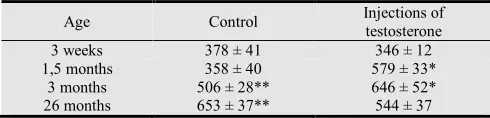
injections of testosterone aldo-keto reductase activity in 1.5- and 3-month-old rats increases by 62 % and 28 % respectively as compared with that of control group (Tabl.1). At the same time testosterone injection is not accompanied by increasing of aldo-keto reductase activity of the blood of 3-weeks-old and 26-months-old rats.
Table 1. Aldo-keto reductase activity of the serum of rats of different ages in control group and in animals after testosterone intramuscular injection, nmol/(min • mg protein), Мe±Se
Age Control Injections of
testosterone
3 weeks 378 ± 41 346 ± 12
1,5 months 358 ± 40 579 ± 33*
3 months 506 ± 28** 646 ± 52*
26 months 653 ± 37** 544 ± 37
* -The data are positively distinguished from intact rats of same age group (P < 0.05)
** - The data are positively distinguished from intact1.5-months-old rats (P < 0.05)
Analysis of the results allows to suggest that animals of different age groups are characterized by different isozyme composition of aldo-keto reductases of blood. These differences predetermine the probability of occurrence of age-dependent features in the manifestation of the testosterone effect on the synthesis of definite isozymes. In order to confirm this hypothesis further research of aldo-keto reductases spectrum from the blood of animals of different age groups was done.
Investigation of the spectrum of aldo-keto reductases allowed to reveal four invariable fractions in the blood of rats (Fig. 2). Fraction 4 is a predominant one in the
spectrum. Fractions 5 and 6 have the greatest
Figure 2. Densitogram and photograph of the plate with separated fractions of aldo-keto reductases of the blood of 3-month-old rat. Numerals indicate the fractions numbers on electrophoregram.
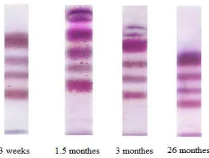
Spectrum of aldo-keto reductases in the blood alters during ontogenesis (Fig. 3, 4). Only four invariable electrophoretic fractions of isozymes are found in 3-weeks-old and 3-weeks-old rats during electrophoresis. Pubertal and adult animals show increasing of the proportion of the 3rd fraction in the aldo-keto reductases spectrum. Moreover, two additional fractions with maximal electrophoretic mobility appear there.
Figure 3. Photographs of the plates with separated fractions of aldo-keto reductases of the blood of various age groups rats
The obtained results show the change of blood aldo-keto reductases spectrum during ontogenesis. This indicates arising of peculiarities in the regulation of expression of genes of enzymes which catalyze the reductive pathway of endogenous aldehydes scavenging in the organism at certain stages of individual development. It is significant that compositions of blood aldo-keto reductases spectra are similar in early immature age (3 weeks) and in aging (26 months). We can assume that this is due to the peculiarities of the endocrine system functioning at these stages of ontogenesis, as hormones act as natural regulators of gene expression. It should be noted that unidirectional changes in endocrine regulation system arise in early postnatal development and in aging: child (immature) age is characterized by its functional immaturity, and late ontogenesis is characterized by manifestations of its involution [23, 24]. Generally it is regarding production of sex steroids including testosterone [25, 26]. This suggests the involvement of testosterone in the regulation of genes expression of some aldo-keto reductases isozymes.
Figure 4. Spectrum of aldo-keto reductases (point out the number of fraction on electrophoregram) in the blood of rats of different ages (in % from total aldo-keto reductase activity). The figure represents the average data from 5 investigations.
Analysis of the obtained results indicates a certain parallelism between the age dynamics in aldo-keto reductase activity of blood and content of testosterone. This may confirm involvement of testosterone in the regulation of aldo-keto reductase synthesis. In order to verify the above assumption, experiments with the introduction of exogenous testosterone to animals were performed.
Studies have shown that increasing the concentration of testosterone in the blood by its parenteral administration is accompanied by elevated aldo-keto reductase activity in 1.5- and 3-months-old rats. Old and young impuberal animals do not show the same change. These data indicate that the effect of testosterone on aldo-keto reductase activity has age-dependent manner. Taking this into account, a comparative analysis of the relationship between found changes in blood aldo-keto reductase activity after testosterone injections and change in the spectrum of aldo-keto reductases in animals of different age groups was carried out.
The results of this analysis allow to regard testosterone as an inductor of synthesis of only certain fractions of aldo-keto reductases isozymes (for instance, the 3rd invariable, or variable the 5th, or the 6th fractions). It can be concluded that testosterone acts as an inductor of synthesis of aldo-keto reductases isozymes through regulation of their genes expression. In addition, there are literature data about the participation of this hormone in the regulation of genes expression of individual proteins [27, 28]. This fact predetermines the appearance of age-related changes in the spectrum of aldo-keto reductases in the blood at certain stages of ontogenesis.
However, the stated assumption requires experimental verification, which our future researches will be devoted to.
4. Conclusion
1. Increased testosterone level in the blood of rats aged from 1.5 to 26 months is accompanied by a parallel elevation of aldo-keto reductase activity of blood.
2. Change in the level of testosterone in the blood during
ontogenesis is accompanied by modulation of spectrum of aldo-keto reductases of the blood. 3. Intramuscular injections of testosterone to pubertal
and adult rats are accompanied by increasing of aldo-keto reductase activity of the blood, which is not typical for young immature and old animals.
References
[1] Korenev N.M. and Nosova E.M. (2002) Parameters Clinical-hemodynamic parameters of cerebral disorders formation at adolescent with primary arterial hypertony. Pediatry, Obstetrics and Gynecology, 2, 15 – 18 (in Ukranian).
[2] Andrawes W. F., Bussy C. and Belmin J. (2005) Prevention of cardiovascular events in elderly people. Drugs Aging, 22, 859–876.
[3] Müller-Werdan U., et al. (2007) Elderly patients with cardiovascular diseases. Internis, 48, 1211–1219.
[4] Saner H. (2005) Stress as a cardiovascular risk factor. Ther. Umsch., 62, 597–602.
[5] Giallauria F., et al. (2007) Psychosocial risk factors in cardiac practice. Monaldi Arch. Chest Dis, 68, 74–80.
[6] Sahin E. and Gumuslu S. (2007) Immobilization stress in rat tissues: alteration of protein oxidation, lipid peroxidation and antioxidant defense system. Comp. Biochem. Physiol. C. Toxicol. Pharmacol., 144, 324–347.
[7] Davydov V.V. and Shvets V.N. (2003) Age-dependent differences in the stimulation of lipid peroxidation in the heart of rats during immobilization stress. Exp. Gerontol., 38, 693 – 698.
[8] Volkova Yu.V. et al. (2011) Activity of the first line antioxidant defense enzymes in the liver of pubertal rats during stress. Biochemistry (Moscow) Supplement Series B: Biomedical Chemistry., 5, 389 – 391.
[9] Davydov V.V. and Shvets V.N. (2001) Lipid peroxidation in the heart of adult and old rats during immobilization stress. Exp. Gerontol., 36, 1155 – 1160.
[10] Spiteller G. (2001) Lipid peroxidation in aging and age-dependent diseases. Exp. Gerontol., 36, 1425 – 1457.
[11] Scheider C. et al. (2001) Two distinct pathways of formation of 4-hydroxynonenal. J. Biol. Chem., 276, 20831 – 20838.
[12] Esterbauer H., Schaur R. J. and Zollner H. (1991) Chemistry and biochemistry of 4-hydroxynonenal, malonaldehyde and related aldehydes. Free Radic. Biol. Med., 11, 81 – 128.
[13] Stone M. P. et al. (2008) Interstrand DNA cross-links induced by α,β-unsaturated aldehyde-derived from lipid peroxidation and environmental sources. Acc. Chem. Res., 41, 793 – 804.
[14] Pizzimenti S. et al. (2013) Interaction of aldehydes derived from lipid peroxidation and membrane proteins. Front Physiol., 4, 242 – 262 .
[15] Uchida K. (2000) 4-hydroxy-2-nonenal: a product and mediator of oxidative stress. Prog. Lipid Res., 42, 318 – 343.
[16] Davydov V. V., Dobaeva N. M. and Bozhkov A. I. (2004) Possible role of aldehyde,s scavenger enzymes during aging. Exp. Gerontol., 39, 11-16.
[17] O’Brein P. J. O., Siraki A. G., and Shangari N. (2005) Aldehyde sources metabolism, molecular toxicity mechanisms, and possible effects on human health. Critical Reviews in Toxicology, 35, 609 – 662.
[18] Srivastava S., et al. (1998) Identification of cardiac oxidoreductase (s) involved in the metabolism of the lipid peroxidation-derived aldehyde 4-hydroxynonenal. Biochem. J., 329, 469 – 475.
[19] Handrman D., et al. (2003) The aldo-keto reductase superfamily homepage. Chem. Biol. Interact., 133–134, 621 – 631.
[20] Jes J.M., et al. (1997) A new nomenclature for the aldo-keto reductases. Biochem. Pharmacol., 54, P. 639 –647.
[21] Bosron W. F. and Praire R. L. (1972) Triphosphate nucleotide-linked aldehyde reductases. J. Biol. Chem., 247, 4480 – 4485.
[22] Nihmat A. and Flynn T. G. (1989) Aldose reductase from human psoas muscle. J. Biol. Chem., 264, 2906 – 2911.
[23] Vermeulen A., et al. (2002) Estradiol in elderly men. Aging male, 5, 98 – 102.
[24] Copinschi G. and Caufries A. (2013) Sleep and hormonal changes in aging. Endocrinol.Metab.Clin. North. Am., 42, 371 – 389.
[25] Vermeulen A., Goemaere S. and Kaufman J.M. et al. (1999) Testosterone, body composition and aging. J.Endocrinol. Invest., 22 (5 Suppl), 110 – 116.
[26] Nguyen T.V., et al. (2013) Testosterone-related cortical maturation across childhood and adolescence. Cereb. Cortex., 23, 1424 – 1432.
[27] Kojima M., Sekimoto M. and Degawa M. (2010) Androgen-mediated down-regulation of CYP1A subfamily genes in the pig liver. J.Endocrinol., 207, 203 – 211.
[28] Hioki T., et al. (2014) Brain testosterone deficiency leads to down-regulation of mitochondrial gene expression in rat hippocampus accompanied by a decline in peroxisome proliferator-activated receptor-γ coactivator 1α expression. J. Mol. Neurosci., 52, 531 – 537.
[29] Davydov V.V., Bozhkov A.I. and Kulchitski O.K. (2012) Physiological and pathophysiological role of endogenous aldehydes,– Saarbrucken: Palmarium Academic Publishing, 240. (in Russian).
[31] Rittner H. L., et al. (1999) Aldose reductase functions as a detoxification system for lipid peroxidation products in vasculitis. J. Clin. Invest., 103, 1007 – 10013.
[32] Keightley J. A., Shang L. and Kinter M. (2003) Proteomic analysis of oxidative stress-resistant cell: A specific role for aldose reductase overexpression in cytoprotection. Mol. Cell Proteomics., 12, 1236 – 1245.