Pathology and Laboratory Medicine International
Dove
press
R e v I e w
open access to scientific and medical researchOpen Access Full Text Article
BRAF
mutational analysis in ovarian tumors:
recent perspectives
Kwong-Kwok wong
1Ching-Chou Tsai
2David M Gershenson
11Department of Gynecologic
Oncology and Reproductive Medicine, The University of Texas MD Anderson Cancer Center, Houston, TX, USA;
2Department of Obstetrics and
Gynecology, Kaohsiung Chang Gung Memorial Hospital, Chang Gung University College of Medicine, Kaohsiung, Taiwan, Republic of China
Correspondence: Kwong-Kwok wong
Department of Gynecologic Oncology and Reproductive Medicine, Unit 1362, The University of Texas MD Anderson Cancer Center, 1515 Holcombe Boulevard, Houston, TX 77030, USA Tel +1 713 792 0229
Fax +1 713 792 1459
email [email protected]
Abstract: BRAF mutations are rare in ovarian cancer and mainly occur in indolent serous borderline tumors (SBTs), also known as serous tumors of low malignant potential or atypical proliferative serous tumors. The reported percentage of BRAF mutations in SBTs varies from 23% to 71%. Although a high percentage of stage II–IV SBTs with noninvasive implants have progressed to invasive low-grade serous carcinomas when patients were observed for 5 years or longer, BRAF mutations are rare in low-grade serous carcinomas as well as in invasive implants associated with SBTs. BRAF mutations in SBTs may prevent SBTs from progressing to invasive carcinomas. On the other hand, the reported percentage of BRAF mutations in muci-nous carcinoma (20%) is much higher than that of mucimuci-nous borderline tumor (5%). Further investigation of the role of BRAF mutations in SBTs and mucinous tumor will shed light on the molecular mechanism underlying the role of BRAF mutations in tumor progression in dif-ferent cellular context and the clinical utility of BRAF mutations in SBTs as a biomarker of favorable prognosis.
Keywords: BRAF V600E, ovarian cancer, COLD-PCR
Introduction
BRAF is a serine/threonine protein kinase of the RAF family that also includes ARAF
and RAF1. BRAF has the highest basal level in the RAF family and is part of the
mitogen-activated RAS/RAF/MEK/ERK protein kinase pathway, which acts as a
sig-nal transducer between the extracellular sigsig-nals and the nucleus. Extracellular sigsig-nals
such as hormones, cytokines, and various growth factors interact with their receptors
to activate the small G-proteins of the RAS family and subsequently activate BRAF.
Active BRAF then activates MEK1/2 to phosphorylate ERK1/2, which leads to the
expression of several downstream transcription factors that regulate cell growth,
dif-ferentiation, and survival.
Mutations in the BRAF gene were first discovered by Davies et al
1in 2002 through
a systematic and genome-wide assessment of cancer-associated pathways in human
cancer. The Catalogue of Somatic Mutations in Cancer (COSMIC, version 71)
2data-base identified BRAF point mutations in over 40,000 cancer samples (Table 1). The
frequency of BRAF mutations varies from over 40% in thyroid and skin tumors to
0%–12.5% in tumors in other organs. Coexisting mutations of BRAF and KRAS are
very rare; however, such phenomena have been observed in a hyperplastic polyp of
the colon
3and in ovarian mucinous carcinoma.
4The vast majority of missense mutations in BRAF involve a thymine to adenine
substitution at nucleotide 1799 (c.1799T.A), which results in an amino acid change
Pathology and Laboratory Medicine International downloaded from https://www.dovepress.com/ by 118.70.13.36 on 26-Aug-2020
For personal use only.
Number of times this article has been viewed
This article was published in the following Dove Press journal: Pathology and Laboratory Medicine International
Dovepress
wong et al
from valine (V) to glutamate (E) at codon 600. This V600E
mutation represents approximately 95% of all identified
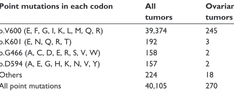
BRAF point mutations (Table 2). Mutated BRAF V600E
activates ERK1/2 without the need for extracellular signals.
Other relatively frequent BRAF missense mutations include
mutations at codons 466, 594, and 601.
BRAF point mutation has been reported in
approxi-mately 6% (270/4,386) of ovarian tumor samples tested for
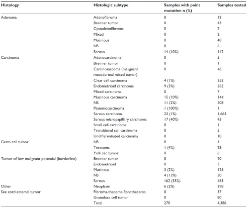
BRAF mutations in the COSMIC database (Tables 2 and 3).
The majority of these ovarian tumor samples (245/270)
have a mutation in codon 600, with p.V600E being the
pre-dominant missense mutation. BRAF gene amplification and
overexpression were found in approximately 12% and 7%,
respectively, of high-grade ovarian serous carcinomas using
the cBio Cancer Genomics Portal
5,6to interrogate the data
generated from The Cancer Genome Atlas study.
7The available data indicate that BRAF mutations mainly
occur in serous borderline tumors (SBTs),
8also known as
serous tumors of low malignant potential or atypical
pro-liferative serous tumors; SBTs also include micropapillary
serous carcinoma (MPSC),
9a minor morphologically
dis-tinct subgroup that was first described by Burks et al
10in
1996. The reported frequency of BRAF mutations in SBTs
ranges from 23% to 71%. We previously found that BRAF
mutation is rare in advanced-stage ovarian low-grade serous
carcinoma (LGSC).
11BRAF mutation is also rare in primary
clear cell ovarian carcinoma (1%),
12,13mucinous borderline
tumors (2%),
13–15and endometrioid carcinoma (3%),
16but
is relatively more frequent in mucinous carcinoma
4,17and
serous adenoma.
18Detection of mutated
BRAF
Most sequencing analyses of BRAF mutations in
ovar-ian cancer have used direct Sanger sequencing. Standard
polymerase chain reaction (PCR)-Sanger sequencing has a
mutation detection sensitivity of approximately 10%.
19Since
SBTs have a high component of stromal cells, the
sensitiv-ity for detecting the BRAF mutation may be compromised
if DNA is extracted from bulk tissue for direct sequencing
using regular PCR amplification of the targeted region
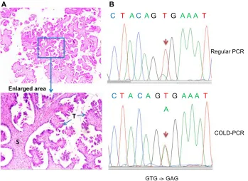
(Figure 1A). To increase the mutation detection sensitivity,
researchers can microdissect tumor cells from the paraffin
section, but this strategy can be quite tedious. Several other
methods, such as pyrosequencing,
20matrix-assisted laser
desorption/ionization time of flight mass spectrometry,
21and comparative allele-specific TaqMan PCR,
22can be used
to screen for BRAF mutations in samples with low tumor
cell purity. We have adopted the co-amplification at lower
denaturation temperature (COLD)-PCR approach, described
previously,
23to enrich low-level mutant BRAF alleles in the
DNA samples for detecting BRAF mutations by Sanger
sequencing (Figure 1B).
24As shown in Figure 1B,
COLD-PCR amplifies the mutated allele (A) to have the same signal
intensity as the wild-type allele (T).
Immunohistochemical staining for the BRAF V600E
mutation–specific monoclonal antibody VE1
25can also
be used to detect BRAF mutation. The sensitivity of VE1
immunostaining has been validated for detecting the BRAF
V600E mutation in formalin-fixed and paraffin-embedded
ovarian SBT tissues.
26Immunostaining was evaluable in
most cases with sufficient tumor cells, but rare cases with
scant cytoplasm and diffuse staining may be difficult to
interpret.
27The false-positive rate of VE1 immunostaining
can be as high as 30%, so initial VE1 immunostaining should
be validated with sequencing.
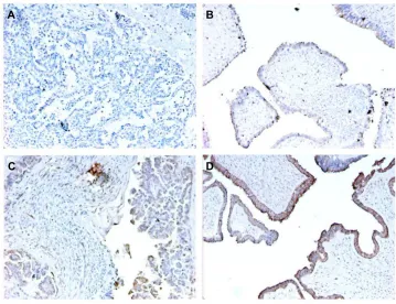
28Figure 2 is an illustration of
VE1 immunostaining on four ovarian tumor samples with
confirmed BRAF mutation status by DNA sequencing. One of
the SBT with wild-type BRAF gene (Figure 2B) (Figure 1A
Table 1 Frequency of BRAF point mutations in tumors of different primary organ sites, according to the Catalogue of Somatic Mutations in Cancer (COSMIC) database
Primary organ site Frequency of point mutation*
KRAS BRAF
Thyroid 1.8% (141/7,717) 41.5% (19,297/46,463) Skin 2.3% (86/3,729) 41.4% (8,134/19,667) Large intestine 34.5% (18,551/53,826) 12.5% (9,253/74,074)
eye 1.6% (4/258) 10.1% (84/828)
Bone 1.7% (11/643) 9.6% (53/552)
Hematopoietic and lymphoid
4.5% (532/11,956) 9.1% (786/8,636)
Pituitary 0% (0/315) 8.7% (20/230) Central nervous
system
0.9% (28/3,264) 7.0% (392/5,598)
Ovary 11.7% (660/5,653) 6.2% (270/4,386) Biliary tract 23.3% (631/2,707) 5.8% (50/865)
Note: *Includes only primary organ sites for which more than 5% of the tumors
had BRAF mutations.
Table 2 Frequency of common BRAF point mutations in all tumors and in ovarian tumors, according to the Catalogue of Somatic Mutations in Cancer (COSMIC) database
Point mutations in each codon All
tumors
Ovarian tumors
p.v600 (e, F, G, I, K, L, M, Q, R) 39,374 245
p.K601 (e, N, Q, R, T) 192 3
p.G466 (A, C, D, e, R, S, v, w) 158 2 p.D594 (A, e, G, H, K, N, v, Y) 157 2
Others 224 18
All point mutations 40,105 270
Pathology and Laboratory Medicine International downloaded from https://www.dovepress.com/ by 118.70.13.36 on 26-Aug-2020
Dovepress BRAF mutation in ovarian cancer
and 1B had wild-type BRAF; Figure 2C and 2D had mutated
BRAF V600E) had faint false positive staining, which could
be a result of an edge effect during staining and should be
interpreted cautiously.
Role of
BRAF
mutation in the
pathogenesis of ovarian cancer
There are four major histological subtypes of epithelial
ovar-ian cancer (EOC), which is thought to arise from the surface
epithelium of the ovaries but could also be from extra-ovarian
origins.
29An ovarian tumorigenesis model based on
morphol-ogy and molecular genetics has been proposed.
30,31EOCs
are classified as Type I or Type II. Type I tumors include
low-grade MPSC, mucinous, endometrioid, and clear cell
carcinomas and are characterized by high frequency of KRAS,
BRAF, PTEN, or beta-catenin mutations.
30–34Type II tumors
include high-grade serous carcinoma, malignant mixed
mesodermal tumors (carcinosarcomas) and undifferentiated
carcinomas and are characterized by high genetic
instabil-ity and high frequency of TP53 mutation.
31,32Separating
EOC into Type I and Type II based on genetic mutations
is controversial. High frequency of TP53 (57%, 8/14) and
KRAS (57%, 8/14) mutations has also been found in Type I
mucinous EOC.
35Similarly, Type I endometrioid cancer also
has a high frequency of TP53 mutation (63%, 17/27).
36Both
endometriosis-related cancers (endometrioid and clear cell
cancer) have high frequency of ARID1A mutations.
37,38SBT
has high frequency of BRAF and KRAS mutations while
LGSC has high frequency of KRAS mutation. Although the
progression of LGSC to high-grade serous carcinoma is very
Table 3 Frequency of BRAF point mutation detected in histologic subtypes of ovarian tumors, according to the Catalogue of Somatic Mutations in Cancer (COSMIC) database
Histology Histologic subtype Samples with point
mutation n (%)
Samples tested
Adenoma Adenofibroma 0 12
Brenner tumor 0 43
Cystadenofibroma 0 2
Mixed 0 2
Mucinous 0 40
NS 0 6
Serous 14 (10%) 142
Carcinoma Adenocarcinoma 0 5
Brenner tumor 0 1
Carcinosarcoma (malignant mesodermal mixed tumor)
0 46
Clear cell carcinoma 4 (1%) 352
endometrioid carcinoma 9 (3%) 262
Mixed carcinoma 0 7
Mucinous carcinoma 15 (10%) 144
NS 11 (2%) 508
Psammocarcinoma 1 (100%) 1
Serous carcinoma 23 (1%) 1,663
Serous micropapillary carcinoma 17 (40%) 43
Small cell carcinoma 0 1
Transitional cell carcinoma 0 5
Undifferentiated carcinoma 0 10
Germ cell tumor NS 0 1
Teratoma 1 (4%) 28
Yolk sac tumor 0 6
Tumor of low malignant potential (borderline) Brenner tumor 0 20
endometrioid 0 3
Mucinous 3 (2%) 125
NS 4 (13%) 30
Serous 162 (35%) 463
Other Neoplasm 6 (2%) 298
Sex cord-stromal tumor Fibroma-thecoma-fibrothecoma 0 37
Granulosa cell tumor 0 80
Total 270 4,386
Abbreviation: NS, not specified.
Pathology and Laboratory Medicine International downloaded from https://www.dovepress.com/ by 118.70.13.36 on 26-Aug-2020
Dovepress
wong et al
rare, several studies have reported the recurrence of
high-grade serous carcinoma from SBT or LGSC.
34,39,40Thus, each
ovarian carcinoma subtype should be treated as a different
disease as suggested previously for future biomarker studies
and clinical trials.
41No germline mutation of BRAF has been found, and thus
the presence of BRAF mutation is probably not a genetic
pre-disposition for the development of SBTs.
16Although a high
percentage of stage II–IV SBTs with noninvasive implants have
progressed to invasive LGSCs when patients were observed
for 5 years or longer,
42BRAF mutations are rare in LGSCs as
well as in invasive implants associated with SBTs.
43,44BRAF
mutations are also rare in high-grade serous carcinomas. BRAF
mutation is mainly found in ovarian serous adenoma, SBTs,
invasive MPSC, and mucinous carcinomas.
4,17,18,45,46It is generally believed that adenoma progresses to SBT
and then to LGSC. The pathologic difference between SBTs
and LGSC is the destructive stromal invasion in LGSC.
One study proposed that MPSC is a step in the progression
from SBTs to LGSC.
47Destructive invasion of the ovarian
stroma is rare in MPSC, but those with invasion are called
invasive MPSC or are considered LGSC. MPSC represents
approximately 6.5%–33% of all SBTs.
10,16,48–51However, there
is a consensus that both typical SBTs and MPSC should be
classified as LGSC when areas of stromal invasion are greater
than 5 mm. Patients with SBT with or without MPSC features
have no difference in recurrence or disease-related mortality,
although noninvasive SBTs with a micropapillary pattern
may have invasive peritoneal implants more often than those
without the micropapillary pattern.
50–54Using sequencing analysis, Singer et al
45found that
ovarian SBTs and low-grade invasive MPSC shared similar
frequencies of both KRAS and BRAF mutations, which are
involved in the progression of SBTs to LGSC.
45Another
study found the same mutations in ovarian SBTs and the
cystadenoma epithelium bordering the SBTs in six (86%) of
seven samples.
18However, other studies suggest that ovarian
SBTs with BRAF mutations may be less likely to spread or
progress to LGSC. Most studies of ovarian LGSC reported
a low frequency of BRAF mutations (Table 4).
11,27,45,55–59In
one study of the implants that accompany ovarian SBTs, only
six (13%) of 45 patients had BRAF mutations in SBTs with
noninvasive peritoneal implants, and none of the patients had
BRAF mutations in SBTs with invasive implants.
43Heublein
et al
44detected BRAF mutations in only noninvasive implants
associated with ovarian SBTs. Similarly, in our analysis of
36 patients with advanced-stage ovarian SBTs, we found that
BRAF mutation was mainly associated with SBTs that did
A
B
C
T
A
C
A
G
T
G
A A A
T
Regular PCR
COLD-PCR
GTG - GAG
C
T
A
C
A
T
A
T
A
A A
G
G
Enlarged area
>
Figure 1 Co-amplification at lower denaturation temperature polymerase chain reaction (COLD-PCR) increases ability to detect BRAF mutations in ovarian serous borderline tumors (SBTs).
Notes: (A) Hematoxylin and eosin staining of a representative SBT showing the presence of a large component of stromal cells (S) in comparison to the tumor cells (T). The
blue arrows indicate the tumor cells on the outside of the fibrous stroma. (B) Sequencing chromatogram showing the amplification of the mutant BRAF allele by COLD-PCR in comparison to regular PCR. The brown arrows indicate the mutated allele.
Pathology and Laboratory Medicine International downloaded from https://www.dovepress.com/ by 118.70.13.36 on 26-Aug-2020
Dovepress BRAF mutation in ovarian cancer
not progress to low-grade invasive tumors.
23BRAF
muta-tion is likely involved in the initiamuta-tion and progression
of ovarian adenoma to SBTs, but may not be involved in
the progression of SBTs to carcinoma. By comparing the
gene expression profiles between ovarian SBTs with
wild-type BRAF and those with mutated BRAF V600E, we
previ-ously found that SBTs with BRAF V600E express high levels
of genes associated with cell growth-inhibitory functions,
including CDC20B, PMEPA1, PAEP, FOXC1, and SFN.
11Using immunohistochemical analysis, Heublein et al
44also
observed that ovarian SBTs with BRAF-mutated implants
tended to express high levels of p16. High expression of
p16 may attenuate the mutated BRAF-induced MAPK
sig-nals that affect cell-cycle progression and thus may prevent
further tumor progression.
On the other hand, the total reported percentage of BRAF
mutations in mucinous carcinoma (20%) is much higher than
that of mucinous borderline tumor (5%) (Table 5).
4,13,15,17,46,60–62Unlike the serous adenoma with similar BRAF mutations as
SBTs, none of the mucinous adenomas (n
=
40) had a
detect-able BRAF mutation.
15Since it is believed that mucinous
adenoma can progress as mucinous borderline tumor and then
Table 5 BRAF mutation frequency in mucinous borderline tumors (mBT) and mucinous carcinomas (MOC)
mBT MOC Reference
0/15 (0%) 1/10 (10%) Mayr et al (2006)13
NA 3/20 (15%) Nakayama et al (2008)4
0/17 (0%) 3/21 (14%) Steffensen et al (2011)17
0/3 (0%) 5/12 (42%) Ryland et al (2013)46
NA 2/10 (20%) Rahman et al (2013)60
2/38 (5%) NA Anglesio et al (2008)61
3/22 (14%) NA Hunter et al (2012)15
NA 1/2 (50%) Gorringe et al (2008)62
Summary: 5% (5/95) 20% (15/75)
Abbreviation: NA, not applicable.
A
B
D
C
Figure 2 examples of immunostains for BRAF v600e mutant protein with ve1 monoclonal antibody in ovarian tumor samples with BRAF mutation status confirmed by DNA sequencing.
Notes: (A) Low-grade serous carcinoma with wild-type BRAF; (B) serous borderline tumor with wild-type BRAF; (C) serous borderline tumor with BRAF v600e mutation; (D) serous borderline tumor with BRAF v600e mutation.
Table 4 BRAF mutation frequency in serous borderline tumors (SBTs) and low-grade serous carcinomas (LGSCs)
SBT LGSC Reference
14/51 (28%) 7/22 (32%) Singer et al (2003)45
NA 2/20 (10%) Cho et al (2009)55
9/30 (30%) 1/39 (3%) wong et al (2010)11
10/43 (23%) 0/17 (0%) vereczkey et al (2011)56
NA 0/11 (0%) Sundov et al (2013)57
25/56 (45%) 1/19 (5%) Grisham et al (2013)58
NA 2/34 (6%) Farley et al (2013)59
22/31 (71%) 1/7 (14%) Bosmuller et al (2013)27
Abbreviation: NA, not applicable.
Pathology and Laboratory Medicine International downloaded from https://www.dovepress.com/ by 118.70.13.36 on 26-Aug-2020
Dovepress
wong et al
mucinous carcinoma, a higher frequency of BRAF mutations
in mucinous carcinoma may suggest a driver role of BRAF
mutation in the pathogenesis of some mucinous carcinomas
in contrast with that of SBTs.
Mutated
BRAF
as a potential
prognostic marker for ovarian
SBT or LGSC
Several studies have reported the association of BRAF
mutation with a better clinical outcome for patients with
ovarian cancer. Preusser et al
28reported that patients with
invasive ovarian carcinomas that stained positive for the
BRAF V600E monoclonal antibody had a strong trend
toward better survival. Similarly, in a study by Grisham
et al,
58BRAF V600E mutations were identified in 35% of 75
patients with ovarian SBTs or LGSC, and the BRAF V600E
mutation in these tumors was associated with early-stage
disease and improved prognosis. Grisham et al also
con-cluded that patients with ovarian SBTs or LGSC who need
systemic therapy are unlikely to have BRAF mutant tumors.
55Pathologically, ovarian SBTs with BRAF mutation are
associ-ated with cellular features indicative of senescence, such as
the expression of senescence-associated beta-galactosidase
activity and abundant eosinophilic cells.
63These data suggest
that BRAF mutation is a biomarker of favorable prognosis and
may prevent the progression of ovarian SBTs or early-stage
LGSC to more aggressive disease, despite the fact that BRAF
mutation is predictive of poor prognosis in other malignancies
such as thyroid cancer and melanoma.
Future perspectives
SBT appears to be a unique disease with high frequency of
BRAF mutations. Validation of BRAF mutations as a potential
prognostic marker in a large cohort of patients with ovarian
SBTs or LGSC is necessary to determine the clinical
signifi-cance of BRAF status in the management of patients with this
disease. Once the BRAF mutation has been confirmed as a
protective factor against the progression of ovarian SBTs and
early-stage LGSC into more aggressive disease,
11,23,43,58clini-cians will be able to use the BRAF status of surgically resected
ovarian SBTs to predict the risk of recurrence. Moreover,
for progressive SBT/LGSC with BRAF V600E mutation,
treatment with BRAF V600E specific inhibitor could be an
alternative regimen.
64,65Furthermore, investigation of ovarian
LGSC and mucinous carcinoma with BRAF mutation may
unveil why BRAF mutations are associated with poor
clini-cal outcomes in other cancers such as microsatellite-stable
colon cancer,
66melanoma,
67and thyroid cancer.
68In summary,
further study of BRAF mutation status will have a significant
impact on the management of ovarian cancer.
Disclosure
The authors report no conflicts of interest in this work.
References
1. Davies H, Bignell GR, Cox C, et al. Mutations of the BRAF gene in human cancer. Nature. 2002;417(6892):949–954.
2. Forbes SA, Bindal N, Bamford S, et al. COSMIC: mining complete cancer genomes in the Catalogue of Somatic Mutations in Cancer.
Nucleic Acids Res. 2011;39(Database issue):D945–D950.
3. Yachida S, Mudali S, Martin SA, Montgomery EA, Iacobuzio-Donahue CA. Beta-catenin nuclear labeling is a common feature of sessile serrated adenomas and correlates with early neoplastic progression after BRAF activation. Am J Surg Pathol. 2009;33(12):1823–1832.
4. Nakayama N, Nakayama K, Yeasmin S, et al. KRAS or BRAF mutation status is a useful predictor of sensitivity to MEK inhibition in ovarian cancer. British Journal of Cancer. 2008;99(12):2020–2028.
5. Cerami E, Gao J, Dogrusoz U, et al. The cBio cancer genomics portal: an open platform for exploring multidimensional cancer genomics data.
Cancer Discov. 2012;2(5):401–404.
6. Gao J, Aksoy BA, Dogrusoz U, et al. Integrative analysis of complex cancer genomics and clinical profiles using the cBioPortal. Sci Signal. 2013;6(269):pl1.
7. Cancer Genome Atlas Research Network. Integrated genomic analyses of ovarian carcinoma. Nature. 2011;474(7353):609–615.
8. Sieben NL, Macropoulos P, Roemen GM, et al. In ovarian neoplasms, BRAF, but not KRAS, mutations are restricted to low-grade serous tumours. J Pathol. 2004;202(3):336–340.
9. Seidman JD, Kurman RJ. Ovarian serous borderline tumors: a critical review of the literature with emphasis on prognostic indicators. Hum
Pathol. 2000;31(5):539–557.
10. Burks RT, Sherman ME, Kurman RJ. Micropapillary serous carcinoma of the ovary. A distinctive low-grade carcinoma related to serous bor-derline tumors. Am J Surg Pathol. 1996;20(11):1319–1330.
11. Wong KK, Tsang YT, Deavers MT, et al. BRAF mutation is rare in advanced-stage low-grade ovarian serous carcinomas. Am J Pathol. 2010;177(4):1611–1617.
12. Zannoni GF, Improta G, Chiarello G, et al. Mutational status of KRAS, NRAS, and BRAF in primary clear cell ovarian carcinoma. Virchows
Arch. 2014;465(2):193–198.
13. Mayr D, Hirschmann A, Lohrs U, Diebold J. KRAS and BRAF muta-tions in ovarian tumors: a comprehensive study of invasive carcino-mas, borderline tumors and extraovarian implants. Gynecol Oncol. 2006;103(3):883–887.
14. Gemignani ML, Schlaerth AC, Bogomolniy F, et al. Role of KRAS and BRAF gene mutations in mucinous ovarian carcinoma. Gynecol
Oncol. 2003;90(2):378–381.
15. Hunter SM, Gorringe KL, Christie M, et al. Pre-invasive ovarian mucinous tumors are characterized by CDKN2A and RAS pathway aberrations. Clin Cancer Res. 2012;18(19):5267–5277.
16. Ueda M, Toji E, Noda S. Germ line and somatic mutations of BRAF V599E in ovarian carcinoma. Int J Gynecol Cancer. 2007;17(4): 794–797.
17. Steffensen KD, Waldstrom M, Grove A, et al. Improved classification of epithelial ovarian cancer: results of 3 danish cohorts. Int J Gynecol
Cancer. 2011;21(9):1592–1600.
18. Ho CL, Kurman RJ, Dehari R, Wang TL, Shih IeM. Mutations of BRAF and KRAS precede the development of ovarian serous borderline tumors. Cancer Res. 2004;64(19):6915–6918.
19. Thomas RK, Nickerson E, Simons JF, et al. Sensitive mutation detec-tion in heterogeneous cancer specimens by massively parallel picoliter reactor sequencing. Nat Med. 2006;12(7):852–855.
Pathology and Laboratory Medicine International downloaded from https://www.dovepress.com/ by 118.70.13.36 on 26-Aug-2020
Dovepress BRAF mutation in ovarian cancer
20. Tan YH, Liu Y, Eu KW, et al. Detection of BRAF V600E mutation by pyrosequencing. Pathology. 2008;40(3):295–298.
21. Greaves WO, Verma S, Patel KP, et al. Frequency and spectrum of BRAF mutations in a retrospective, single-institution study of 1112 cases of melanoma. J Mol Diagn. 2013;15(2):220–226. 22. Richter A, Grieu F, Carrello A, et al. A multisite blinded study for the
detection of BRAF mutations in formalin-fixed, paraffin-embedded malignant melanoma. Sci Rep. 2013;3:1659.
23. Tsang YT, Deavers MT, Sun CC, et al. KRAS (but not BRAF) mutations in ovarian serous borderline tumour are associated with recurrent low-grade serous carcinoma. J Pathol. 2013;231(4): 449–456.
24. Milbury CA, Li J, Makrigiorgos GM. COLD-PCR-enhanced high-resolution melting enables rapid and selective identification of low-level unknown mutations. Clin Chem. 2009;55(12):2130–2143.
25. Capper D, Preusser M, Habel A, et al. Assessment of BRAF V600E mutation status by immunohistochemistry with a mutation-specific monoclonal antibody. Acta Neuropathol. 2011;122(1):11–19. 26. Hayashi Y, Sasaki H, Takeshita S, et al. Usefulness of
immunohis-tochemistry for the detection of the BRAF V600E mutation in ovarian serous borderline tumors. Oncol Rep. 2014;32(5):1815–1819. 27. Bosmuller H, Fischer A, Pham DL, et al. Detection of the BRAF
V600E mutation in serous ovarian tumors: a comparative analysis of immunohistochemistry with a mutation-specific monoclonal antibody and allele-specific PCR. Hum Pathol. 2013;44(3):329–335.
28. Preusser M, Capper D, Berghoff AS, et al. Expression of BRAF V600E mutant protein in epithelial ovarian tumors. Appl Immunohistochem
Mol Morphol. 2013;21(2):159–164.
29. Kurman RJ, Shih IeM. Molecular pathogenesis and extraovarian ori-gin of epithelial ovarian cancer – shifting the paradigm. Hum Pathol. 2011;42(7):918–931.
30. Shih IeM, Kurman RJ. Ovarian tumorigenesis: a proposed model based on morphological and molecular genetic analysis. Am J Pathol. 2004;164(5):1511–1518.
31. Kurman RJ, Shih IeM. The origin and pathogenesis of epithelial ovar-ian cancer: a proposed unifying theory. Am J Surg Pathol. 2010;34(3): 433–443.
32. Cho KR, Shih IeM. Ovarian cancer. Annu Rev Pathol. 2009;4: 287–313.
33. Kurman RJ, Shih IeM. Pathogenesis of ovarian cancer: lessons from morphology and molecular biology and their clinical implications. Int
J Gynecol Pathol. 2008;27(2):151–160.
34. Vang R, Shih IeM, Kurman RJ. Ovarian low-grade and high-grade serous carcinoma: pathogenesis, clinicopathologic and molecular biologic features, and diagnostic problems. Adv Anat Pathol. 2009; 16(5):267–282.
35. Rechsteiner M, Zimmermann AK, Wild PJ, et al. TP53 mutations are common in all subtypes of epithelial ovarian cancer and occur concomitantly with KRAS mutations in the mucinous type. Exp Mol
Pathol. 2013;95(2):235–241.
36. Okuda T, Otsuka J, Sekizawa A, et al. p53 mutations and overexpres-sion affect prognosis of ovarian endometrioid cancer but not clear cell cancer. Gynecol Oncol. 2003;88(3):318–325.
37. Wiegand KC, Shah SP, Al-Agha OM, et al. ARID1A mutations in endometriosis-associated ovarian carcinomas. N Engl J Med. 2010; 363(16):1532–1543.
38. Jones S, Wang TL, Shih IeM, et al. Frequent mutations of chromatin remodeling gene ARID1A in ovarian clear cell carcinoma. Science. 2010;330(6001):228–231.
39. Dehari R, Kurman RJ, Logani S, Shih IeM. The development of high-grade serous carcinoma from atypical proliferative (borderline) serous tumors and low-grade micropapillary serous carcinoma: a morphologic and molecular genetic analysis. Am J Surg Pathol. 2007; 31(7):1007–1012.
40. Parker RL, Clement PB, Chercover DJ, Sornarajah T, Gilks CB. Early recurrence of ovarian serous borderline tumor as high-grade carcinoma: a report of two cases. Int J Gynecol Pathol. 2004;23(3):265–272.
41. Kobel M, Kalloger SE, Boyd N, et al. Ovarian carcinoma subtypes are different diseases: implications for biomarker studies. PLoS Med. 2008;5(12):e232.
42. Silva EG, Gershenson DM, Malpica A, Deavers M. The recurrence and the overall survival rates of ovarian serous borderline neoplasms with noninvasive implants is time dependent. Am J Surg Pathol. 2006;30(11): 1367–1371.
43. Ardighieri L, Zeppernick F, Hannibal CG, et al. Mutational analysis of BRAF and KRAS in ovarian serous borderline (atypical proliferative) tumours and associated peritoneal implants. J Pathol. 2014;232(1): 16–22.
44. Heublein S, Grasse K, Hessel H, et al. KRAS, BRAF genotyping reveals genetic heterogeneity of ovarian borderline tumors and associated implants. BMC Cancer. 2013;13:483.
45. Singer G, Oldt R 3rd, Cohen Y, et al. Mutations in BRAF and KRAS characterize the development of low-grade ovarian serous carcinoma.
J Natl Cancer Inst. 2003;95(6):484–486.
46. Ryland GL, Hunter SM, Doyle MA, et al. RNF43 is a tumour suppressor gene mutated in mucinous tumours of the ovary. J Pathol. 2013;229(3): 469–476.
47. Gershenson DM. Is micropapillary serous carcinoma for real? Cancer. 2002;95(4):677–680.
48. Uzan C, Nikpayam M, Ribassin-Majed L, et al. Influence of histologi-cal subtypes on the risk of an invasive recurrence in a large series of stage I borderline ovarian tumor including 191 conservative treatments.
Ann Oncol. 2014;25(7):1312–1319.
49. Lazarou A, Fotopoulou C, Coumbos A, et al. Long-term follow-up of borderline ovarian tumors clinical outcome and prognostic factors.
Anticancer Res. 2014;34(11):6725–6730.
50. Fauvet R, Demblocque E, Morice P, Querleu D, Darai E. Behavior of serous borderline ovarian tumors with and without micropapil-lary patterns: results of a French multicenter study. Ann Surg Oncol. 2012;19(3):941–947.
51. Uzan C, Kane A, Rey A, et al. Prognosis and prognostic factors of the micropapillary pattern in patients treated for stage II and III serous borderline tumors of the ovary. Oncologist. 2011;16(2):189–196. 52. Deavers MT, Gershenson DM, Tortolero-Luna G, et al. Micropapillary and
cribriform patterns in ovarian serous tumors of low malignant poten-tial: a study of 99 advanced stage cases. Am J Surg Pathol. 2002; 26(9):1129–1141.
53. Longacre TA, McKenney JK, Tazelaar HD, Kempson RL, Hendrickson MR. Ovarian serous tumors of low malignant potential (borderline tumors): outcome-based study of 276 patients with long-term(5-year) follow-up. Am J Surg Pathol. 2005;29(6):707–723.
54. Prat J, De Nictolis M. Serous borderline tumors of the ovary: a long-term follow-up study of 137 cases, including 18 with a micropapil-lary pattern and 20 with microinvasion. Am J Surg Pathol. 2002; 26(9):1111–1128.
55. Cho YH, Kim DY, Kim JH, et al. Mutational analysis of KRAS, BRAF, and TP53 genes of ovarian serous carcinomas in Korean women. Yonsei
medical journal. 2009;50(2):266–272.
56. Vereczkey I, Serester O, Dobos J, et al. Molecular characterization of 103 ovarian serous and mucinous tumors. Pathology oncology research:
POR. 2011;17(3):551–559.
57. Sundov D, Caric A, Mrklic I, et al. P53, MAPK, topoisomerase II alpha and Ki67 immunohistochemical expression and KRAS/BRAF mutation in ovarian serous carcinomas. Diagnostic pathology. 2013;8:21. 58. Grisham RN, Iyer G, Garg K, et al. BRAF mutation is associated with
early stage disease and improved outcome in patients with low-grade serous ovarian cancer. Cancer. 2013;119(3):548–554.
59. Farley J, Brady WE, Vathipadiekal V, et al. Selumetinib in women with recurrent low-grade serous carcinoma of the ovary or peritoneum: an open-label, single-arm, phase 2 study. The Lancet. Oncology. 2013;14(2):134–140.
60. Rahman M, Nakayama K, Rahman MT, et al. PPP2R1A mutation is a rare event in ovarian carcinoma across histological subtypes. Anticancer
research. 2013;33(1):113–118.
Pathology and Laboratory Medicine International downloaded from https://www.dovepress.com/ by 118.70.13.36 on 26-Aug-2020
Pathology and Laboratory Medicine International
Publish your work in this journal
Submit your manuscript here: http://www.dovepress.com/pathology-and-laboratory-medicine-international-journal Pathology and Laboratory Medicine International is a peer-reviewed,
open access journal focusing on innovative basic research and translational research related to pathology or human disease. The journal includes original research, updates, case reports, reviews and commentaries on current controversies. The Academic Sponsor
of this journal is the Chinese American Pathology Association (CAPA). The manuscript management system is completely online and includes a very quick and fair peer-review system. Visit http://www.dovepress.com/testimonials.php to read real quotes from published authors.
Dovepress
Dove
press
wong et al
61. Anglesio MS, Arnold JM, George J, et al. Mutation of ERBB2 provides a novel alternative mechanism for the ubiquitous activation of RAS-MAPK in ovarian serous low malignant potential tumors. Molecular
cancer research: MCR. 2008;6(11):1678–1690.
62. Gorringe KL, Choong DY, Williams LH, et al. Mutation and methylation analysis of the chromodomain-helicase-DNA binding 5 gene in ovarian cancer. Neoplasia. 2008;10(11):1253–1258.
63. Zeppernick F, Ardighieri L, Hannibal CG, et al. BRAF mutation is associated with a specific cell type with features suggestive of senes-cence in ovarian serous borderline (atypical proliferative) tumors. The
American journal of surgical pathology. 2014;38(12):1603–1611.
64. Flaherty KT, Puzanov I, Kim KB, et al. Inhibition of mutated, activated BRAF in metastatic melanoma. N Engl J Med. 2010;363(9):809–819.
65. Robinson GW, Orr BA, Gajjar A. Complete clinical regression of a BRAF V600E-mutant pediatric glioblastoma multiforme after BRAF inhibitor therapy. BMC cancer. 2014;14:258.
66. Samowitz WS, Sweeney C, Herrick J, et al. Poor survival associated with the BRAF V600E mutation in microsatellite-stable colon cancers.
Cancer research. 2005;65(14):6063–6069.
67. Chapman PB, Hauschild A, Robert C, et al. Improved survival with vemurafenib in melanoma with BRAF V600E mutation. N Engl J Med. 2011;364(26):2507–2516.
68. Kebebew E, Weng J, Bauer J, et al. The prevalence and prognostic value of BRAF mutation in thyroid cancer. Ann Surg. 2007;246(3):466–470; discussion 470–461.
Pathology and Laboratory Medicine International downloaded from https://www.dovepress.com/ by 118.70.13.36 on 26-Aug-2020