Original Article
Trend of the incidence of cervical
spondylosis: decrease with aging in the elderly
and increase with aging in the young and the adults
Zhiyou Cai1, Nan Zhang2, Niya Ma2, Gaolei Dong2, Shengyuan Wang2, Yu Zhao2
1Department of Neurology, Renmin Hospital, Hubei University of Medicine, Shiyan Renmin Hospital, Shiyan
442000, Hubei Province, People’s Republic of China; 2Department of Neurology, The Fourth Affiliated Hospital of
Harbin Medical University, Harbin 150000, Heilongjiang Province, People’s Republic of China
Received February 3, 2016; Accepted June 8, 2016; Epub July 15, 2016; Published July 30, 2016
Abstract: Background and purpose: Numerous clinical studies have indicated that the incidence of cervical spon-dylosis increases with age. However, the relationship between age and the incidence of cervical sponspon-dylosis is still unclear. It is essential to address the relationship between age the incidence of cervical spondylosis via more clini-cal data. Methods: A retrospective analysis of 4271 cases of cerviclini-cal spondylosis has been done in this study. A chi-square test was used to analyze the associations between different variables.A One-Way Analysis of Variance was used to test the equality of three or more means at one time by using variances for risk factors. Results: The most prominent features of cervical spondylosis were bulge or herniation at C3-4, C4-5 and C5-6. The incidence of cervi-cal spondylosis increased with age before 50 years old and decreased with age after 59. The age risk factors have no relationship with the incidence of cervical spondylosis. There was an increase with age before 50 years old and a decrease with age after 59 for the occurrence of imaging features for cervical spondylosis patients. Conclusion: The incidence of cervical spondylosis decreases with aging in the elderly and increases with aging in the young and the adults. This analysis suggests that aging is not a contributor to the occurrence of cervical spondylosis in the elderly.
Keywords: Cervical spondylosis, incidence, age
Introduction
Cervical spondylosis is a chronic ‘wear and tear’ degenerative process of the cervical spine that affects the vertebral bodies and interver-tebral disks in the neck, and can develop into disk ruptures and herniation, osteophyte, com-pression of the spinal cord, or cervical spondy-lotic myelopathy [1, 2]. Cervical spondylosis can be identified in the majority of people older than 40 years. Symptoms of cervical spondylo-sis include one, or a combination, of the follow-ing: numbness, weakness and tingling in the neck and/or arms, pain in the neck and/or arms, neck stiffness, headaches, symptomatic compression of the spinal cord (myelopathy), nerve roots (radiculopathy), or problems with bladder function from cervical myelopathy [3, 4]. Prolonged cord compression from cervical
spondylosis can result into irreversible histo-logical spinal cord changes such as intradural fibrosis, ischemia, destruction of the blood-spi-nal cord barrier, demyelination, and neuroblood-spi-nal apoptosis within the spinal cord [5]. Decom- pressive surgery may rescue these changes and halt or even reverse the deterioration in myelopathy patients and contribute to an improvement in functional and neurological status [6].
spondylosis are increasing. However, the rela-tionship between age the incidence of cervical spondylosis remains obscure. It is essential to clarify relationship between age the incidence
Table 1. The distribution of different age range and gender
Gender Age distribution (year) N
≤29 30-39 40-49 50-59 60-69 70-79 80-89 ≥90
Total 21 186 837 1731 956 415 124 1 4271
Male 7 93 387 685 375 173 72 1 1793
[image:2.612.92.355.188.294.2]Female 14 93 450 1046 581 242 52 0 2478
Table 2. Percentage of risk factors
Risk factors Cases Percentage (%)
Hypertension 1413 33.1
Diabetes 573 13.4
Cardiovascular diseases 818 19.2
Cerebral infarct 2673 62.6
Smoking 667 15.6
Drinking 486 11.4
Hyperlipidemia 1546 36.2
of cervical spondylosis under more clinical data. This study has exam-ined the clinical data of cervical spondylosis from December 2014 to January 2014 in our hospital in Northern China. The present clini-cal investigation has demonstrat-ed that the trend of the incidence of cervical spondylosis decreases with aging in the elderly and increases with aging in the young and the adults.
Clinical data and methods
4271 cases of cervical spondylosis were collected in the Fourth Affiliated Hospital of Harbin Me- dical University in the Northern China. All data from January 2014 to December 2015 was obtained from the all departments of the Fourth Affiliated Hospital of Harbin Medical University. This study was approved by the research ethics Table 3. Clinical symptoms and signs
Symptoms or signs Cases Percentage (%)
Headache 84 2.0
Dizziness 390 9.1
Fatigue 72 1.7
Nausea and vomiting 197 4.6
Weakness and tingling in the neck and/or arms 848 19.9 Pain in the back, neck and/or arms 901 21.1
Vertigo and instability of walking 234 5.5
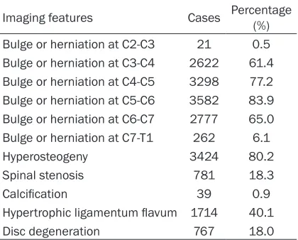
Table 4. Imaging features
Imaging features Cases Percentage (%) Bulge or herniation at C2-C3 21 0.5 Bulge or herniation at C3-C4 2622 61.4 Bulge or herniation at C4-C5 3298 77.2 Bulge or herniation at C5-C6 3582 83.9 Bulge or herniation at C6-C7 2777 65.0 Bulge or herniation at C7-T1 262 6.1
Hyperosteogeny 3424 80.2
Spinal stenosis 781 18.3
Calcification 39 0.9
Hypertrophic ligamentum flavum 1714 40.1
Disc degeneration 767 18.0
board of Harbin Medical University. Diagnosis was determined in accordance with 2012 ICD-9-CM Diagnosis Code 721 (721.0 Cervical spondylosis without myelopathy, 721.1 Cervical spondylosis with myelopathy) and the diagnos-tic criteria of diagnosis and treatment for cervical spondylosis issued by China’s Reha- bilitation Medicine association.
[image:2.612.92.357.333.447.2] [image:2.612.91.301.484.653.2]bles on sociodemographic characteristics, age-related risk factors, and physical examina-tion findings.
The clinical manifestation represented the fol-lowing: ① no symptoms of cervical spondylo-sis: cervical degenerative changes without clinical manifestations; ② mild stenosis of intervertebral space and less osteophyte for-mation with neck stiffness, stiff neck pain, shoulder pain and stiff symptoms and signs; ③
cervical radiculopathy: root distribution (num- bness or/and pain) and signs; ④ cervical spondylotic myelopathy: canal stenosis with the clinical manifestations of cervical cord compression such as numbness, gait instabili-ty, difficulty walking; ⑤ sympathetic cervical spondylosis: clinical manifestations of sympa-thetic nervous system such as dizziness, head-ache, poor sleep, loss of memory, tinnitus,
nau-sea, diarrhea, palpitations, or sweating; ⑥ the vertebral artery type of cervical spon- dylosis: cataplexy attack, ver- tigo.
Retrospective analysis of 4271-case cervical spondylo-sis has been conducted. The data was edited and entered into a computer to be ana-lyzed using SPSS, Windows version 13.0 (IBM Corpo- ration, Armonk, NY, USA). A chi-square test was intro-duced to analyze the associa-tions between two different variables. A One-Way Analysis of Variance was used to test the equality of three or more means at one time by using variances for risk factors. A P-value of less than 0.005 was taken to indicate statisti-cal significance.
Results
The general clinical data analysis
Demographic items included age and gender, clinical symptoms and signs, risk factors, and imaging fea- tures. The distribution of different age range and risk factors were detailed in Tables 1 and 2. 4271 cases included 1793 male and 2478 female.
[image:3.612.91.371.76.418.2]The general clinical data demonstrated that the main symptoms and signs of cervical spondylo-sis involved pain in the back, neck and/or arms, weakness and tingling in the neck and/or arms, dizziness, headache, vertigo and instability of walking, nausea and vomiting, and neck stiff-ness. The most prevalence symptom was pain in the back, neck or/and arms (Table 3). The imaging examination with computed tomogra-phy or/and magnetic resonance imaging pre-sented the most prominent characteristic fea-tures of cervical spondylosis: bulge or hernia-tion at C3-4, C4-5 and C5-6, spinal stenosis, vertebral hyperostosis, and hypertrophic liga-mentum flavum (Table 4).
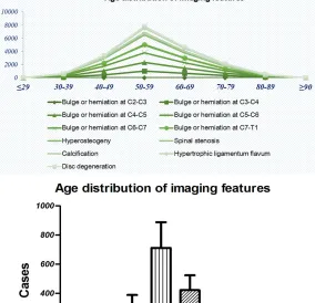
The analysis of age distribution for cervical spondylosis
The total data showed that the incidence of cer-vical spondylosis increased with age before 50 years old and decreased with age after 59. The same results were found in both the male and the female subjects (Figure 1 and Table 1). The imaging findings (Figure 2 and Table 5) are consistent with the previous results that the incidence of cervical spondylosis increased with age before 50 years old and decreased with age after 59. There was an increase with age before 50 years old and decrease with age after 59 for the occurrence of bulge or
tion at C3-C4, bulge or tion at C4-C5, bulge or tion at C5-C6, bulge or tion at C6-C7, bulge or hernia-tion at C7-T1, hyperosteoge- ny, spinal stenosis, hypertro- phic ligamentum flavum and disc degeneration. The same founding was observed in the bulge or herniation at C2-C3 and calcification although there was no obvious change in the number of cases among the different age group. The relationship between risk factors and the incidence of cervical spondylosis
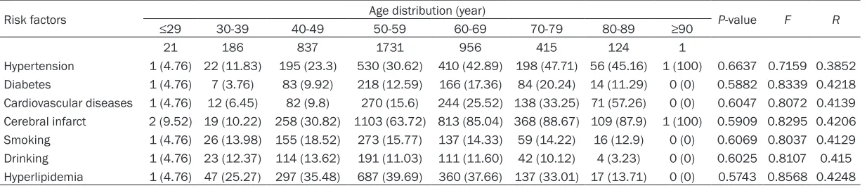
The association between the clinical risk factors and cervi-cal spondylosis on the basis of age had been demonstrat-ed in Table 6. A One-Way Analysis of Variance showed that the age-related risk factors (hypertension, hyper-lipidemia, diabetes, cerebral infarct, cardiovascular diseas-es, smoking and drinking) have no relationship with the incidence of cervical spon- dylosis.
Discussion
Cervical spondylosis is a com-mon pathological entity that can result in various clinical manifestations in the adult population. It can present in a variety of ways but the most common presentations are weak-ness and tingling in the neck and/or arms, pain in the neck and/or arms, functional deficits of the upper extremity, particularly the hand [10, 11]. In this study, the one-hospital based clini-cal data was retrospectively analyzed, which was collected from hospitalized patients with a diagnosis of cervical spondylosis. The one-hos-pital based clinical investigation identified that the most prevalence symptom of cervical spon-dylosis is pain in the back, neck and/or arms, and the most prominent characteristic features is bulge at C3-4, C4-5 and C5-6. There was no relation between the onset of cervical
[image:4.612.91.375.82.356.2]losis and the age-related risk factors (hyperten-sion, besides hyperlipidemia, diabetes, cere-bral infarct, cardiovascular diseases, smoking and drinking). The great significance of this study has found that the occurrence of cervical spondylosis increases with age before 50 years old, and decreases with age after 59, in spite of difference with the past reports that the occur-rence of cervical spondylosis increases with aging. This investigation implicates that aging is not a contributor to the occurrence of cervi-cal spondylosis in the elderly although the inci-dence of cervical spondylosis is proportional to the progress of age.
It is well-known that cervical spondylosis is an age-related ‘wear and tear’ degeneration of vertebrae and discs in the neck. Abundant clini-cal and pathologiclini-cal data evidences that cervi-cal spondylosis is a common occurrence in the elderly [9, 12]. With the progress of cervical spondylotic changes, symptoms often slowly come into emergence over time and get worse with aging, such as headache, neck stiffness, pain in the back, neck and/or arms, weakness and tingling in the neck and/or arms, dizziness, and so on [10, 13]. It is well accepted that age is the major risk factor of cervical spondylosis. However, our previous studies demonstrated that the incidence of cervical spondylosis decreases with age in the elderly population, especially after 60 years old, although increased with age before 50 years old. Our further investigation subsequently provided that there is no relationship between the inci-dence of cervical spondylosis in the elderly and the age-related risk factors.
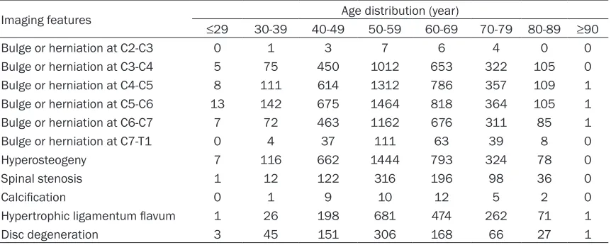
[image:5.612.92.523.85.258.2]The present clinical analysis has been execut-ed to further confirm the conclusion on the basis of cervical spondylosis 4271 cases from another hospital. The present results are con-sistent with the previous ones that the trend of age-related distribution for cervical spondylosis is an increase with age before 50 years old and a decrease with age after 59. These results also showed that the age-related risk factors (hypertension, hyperlipidemia, diabetes, cere-bral infarct, cardiovascular diseases, smoking and drinking) have no relationship with the trend of age-related distribution for cervical spondylosis. Therefore, this conclusion is more confirmed that the incidence of cervical spon-dylosis decreases with age in the elderly and increases with age in the young and adults. Accordingly, we inferred the pathogenesis of cervical spondylosis that the volume and inflammation of the nucleus gets lesser in the elderly since chronic age-related degeneration of cervical spondylosis will enhance the atrophy of the nucleus with the progress of age. On the contrary, with the advance of aging in the young and adults, discs gradually their strength and resiliency, and easily induce the occurrence of bulge or/and herniation. Therefore, the clinical presentation will become pronounced. Thus, the pressure from the nucleus in the elderly will become gradually less because of age-related atrophy, with the result in the lower incidence of annulus injury and occurrence of cervical spon-dylosis, and the less clinical manifestations, especially after 60 years old. In view of the dif-ferent age range, the inflammatory effect of the nucleus in the young and the adults before 50 Table 5. Age distribution of imaging features for cervical spondylosis patients
Imaging features Age distribution (year)
≤29 30-39 40-49 50-59 60-69 70-79 80-89 ≥90
Bulge or herniation at C2-C3 0 1 3 7 6 4 0 0
Bulge or herniation at C3-C4 5 75 450 1012 653 322 105 0
Bulge or herniation at C4-C5 8 111 614 1312 786 357 109 1
Bulge or herniation at C5-C6 13 142 675 1464 818 364 105 1
Bulge or herniation at C6-C7 7 72 463 1162 676 311 85 1
Bulge or herniation at C7-T1 0 4 37 111 63 39 8 0
Hyperosteogeny 7 116 662 1444 793 324 78 0
Spinal stenosis 1 12 122 316 196 98 36 0
Calcification 0 1 9 10 12 5 2 0
Hypertrophic ligamentum flavum 1 26 198 681 474 262 71 1
Table 6. The relationship between risk factors and the incidence of cervical spondylosis on the basis of age
Risk factors Age distribution (year) P-value F R
≤29 30-39 40-49 50-59 60-69 70-79 80-89 ≥90
21 186 837 1731 956 415 124 1
is stronger than the degenerative process, and the highest incidence of cervical spondylosis corresponds to the age range (around 50). Inversely, the inflammatory effect of the nucle-us is weaker than the degeneration in the elder-ly, the incidence of cervical spondylosis will decrease with the advance of age.
Age may play a different role in the pathogene-sis of cervical spondylopathogene-sis in the different age range. The young and adults will contribute to the occurrence of cervical spondylosis by “wear and tear” while the elderly will decrease the occurrence of cervical spondylosis by “atro-phy”. Thus, cervical spondylosis will have a dif-ferent clinical feature in difdif-ferent age range. In consideration that the previous finding that the incidence of cervical spondylosis increases with age in the young and adults, and decreas-es with age in the elderly, thdecreas-ese rdecreas-esults impli-cate that surgery is not the preferred treatment measures in the elderly patients with cervical spondylosis [12, 14]. If it isn’t an emergency suffering from cervical spondylosis, nonopera-tive therapeutic interventions will be designed for the elderly patients with cervical spondylo-sis as the first adoption, such as physical thera-py and pain medications. With the advance of age, the volume of nucleus becomes smaller because of its atrophy, and the pressure from the nucleus is less. Hence, the first-selected treatment for the elderly patients with cervical spondylosis is nonoperative interventions in a rehabilitation center if they have no acute symptoms and signs.
Acknowledgements
This work was supported by grants from the Youth Provincial Nature Science Foundation of Heilongjiang grant (QC2011C053) to Dr Yu Zhao, and the Natural Science Foundation of Hubei Province (2015CFB260), and the Hubei Province Health and Family Planning Scientific Research Project (WJ2015MB219) and the Shiyan Natural Science of Renmin Hospital, Hubei University of Medicine to Dr Zhiyou Cai. Disclosure of conflict of interest
None.
Address correspondence to: Yu Zhao, Department of Neurology, The Fourth Affiliated Hospital of Harbin Medical University, No. 37 Yiyuan Street, Harbin
150000, Heilongjiang Province, People’s Republic of China. Tel: +86-451-82576888; Fax: +86- 451-82576888; E-mail: [email protected]; Zhiyou Cai, Department of Neurology, Renmin Hospital, Hubei University of Medicine, Shiyan Renmin Hospital, No. 39 Chaoyang Middle Road, Shiyan 442000, Hubei Province, People’s Republic of China. Tel: 8637909; Fax: +86-719-8637909; E-mail: [email protected]
References
[1] Matz PG, Anderson PA, Holly LT, Groff MW, Heary RF, Kaiser MG, Mummaneni PV, Ryken TC, Choudhri TF, Vresilovic EJ, Resnick DK; Joint Section on Disorders of the Spine and Peripheral Nerves of the American Association of Neurological Surgeons and Congress of Neurological Surgeons. The natural history of cervical spondylotic myelopathy. J Neurosurg Spine 2009; 11: 104-111.
[2] Kotil K, Bilge T. Prospective study of anterior cervical microforaminotomy for cervical radicu-lopathy. J Clin Neurosci 2008; 15: 749-756. [3] Aurich M, Hofmann GO, Gras FM. [Cervical
my-elopathy after low grade distortion of the cervi-cal spine: Possible association with pre-exist-ing spondylosis of the cervical spine]. Unfallchirurg 2015; 118: 372-5.
[4] Kalb S, Zaidi HA, Ribas-Nijkerk JC, Sindhwani MK, Clark JC, Martirosyan NL, Theodore N. Persistent Outpatient Hypertension Is Inde- pendently Associated with Spinal Cord Dysfunction and Imaging Characteristics of Spinal Cord Damage among Patients with Cervical Spondylosis. World Neurosurg 2015; 84: 351-357.
[5] Tetreault L, Goldstein CL, Arnold P, Harrop J, Hilibrand A, Nouri A, Fehlings MG. Degene- rative Cervical Myelopathy: A Spectrum of Related Disorders Affecting the Aging Spine. Neurosurgery 2015; 77 Suppl 4: S51-67. [6] Lebl DR, Bono CM. Update on the Diagnosis
and Management of Cervical Spondylotic Myelopathy. J Am Acad Orthop Surg 2015; 23: 648-660.
[7] Iwanami A, Toyama Y. [Cervical spondylosis]. Nihon Rinsho 2014; 72: 1755-1760.
[8] Mamata H, Jolesz FA, Maier SE. Apparent diffu-sion coefficient and fractional anisotropy in spinal cord: age and cervical spondylosis-relat-ed changes. J Magn Reson Imaging 2005; 22: 38-43.
[10] Ando T. [Diagnosis and management of cervi-cal spondylosis]. Rinsho Shinkeigaku 2012; 52: 469-479.
[11] Zhang L, Zeitoun D, Rangel A, Lazennec JY, Catonne Y, Pascal-Moussellard H. Preoperative evaluation of the cervical spondylotic myelopa-thy with flexion-extension magnetic resonance imaging: about a prospective study of fifty pa-tients. Spine (Phila Pa 1976) 2011; 36: E1134-1139.
[12] Tauchi R, Imagama S, Inoh H, Yukawa Y, Kanemura T, Sato K, Matsubara Y, Harada A, Hachiya Y, Kamiya M, Yoshihara H, Ito Z, Ando K, Ishiguro N. Risk factors for a poor outcome following surgical treatment of cervical spon-dylotic amyotrophy: a multicenter study. Eur Spine J 2013; 22: 156-161.
[13] Mihara H, Ohnari K, Hachiya M, Kondo S, Yamada K. Cervical myelopathy caused by C3-C4 spondylosis in elderly patients: a radio-graphic analysis of pathogenesis. Spine (Phila Pa 1976) 2000; 25: 796-800.