STUDY ON SILVER NANOPARTICLE ENCAPSULATED CURCUMIN
FOR ANTICANCER ACTIVITY
P. Hemalatha1* and A. Premnath2
1,2
Department of Physics, Government Arts College, Coimbatore, Tamil Nadu, India.
ABSTRACT
Curcumin is one of the poly phenols, which is known for its medicinal
use since long time. Curcumin shows poor solubility and low
absorption and therefore, its use as nanoparticles is beneficial due to
their greater solubility and absorption. In this work curcumin was
extracted from curcuma longa by Soxhlet method. The particle size of
the curcumin was reduced by Ultrasonication method. Silver
nanoparticle was synthesized by chemical reduction method. These
two solutions were agitated about 30 minutes using magnetic stirrer to
form silver encapsulated curcumin. Particle size of the curcumin (36.6
nm), silver nano particle (50.6 nm) and silver encapsulated curcumin
(102.1 nm) was studied by using Particle size analyzer. The Rf value was calculated from
TLC analysis. Purity and percentage of the curcumin content was found by HPLC analysis.
Anticancer activity was assayed out for curcumin and silver nanoparticle encapsulated
curcumin. The study summarizes the challenges to develop curcumin delivery platforms and
up-to-date solutions for improving curcumin bioavailability and anticancer potential for
therapy.
KEY WORDS: Anti cancer activity, Silver nano particles, Curcumin, HPLC, TLC.
1. INTRODUCTION
From ancient times silver and turmeric are used for medicinal purposes. Silver has good
anti-oxidant properties. Turmeric has a compound called curcumin. Curcumin is responsible for
the medicinal properties and colour of turmeric. The bio availability of curcumin is very less.
It is hydrophobic and hence it is not absorbed by blood. Curcumin is all-in-one medicine for
Volume 5, Issue 10, 958-973. Research Article ISSN 2277– 7105
*Corresponding Author
Dr. P. Hemalatha
Department of Physics,
Government Arts College,
Coimbatore, Tamil Nadu,
India.
Article Received on 15 Aug. 2016,
Revised on 05 Sept. 2016, Accepted on 25 Sept. 2016
Nano medicines hold very good promise for target oriented reach of the drugs. The medicines
which are generally available react in the same way to all the cells. They can not differentiate
diseased cells and normal cells. So the available medicines kill the normal cells along with
the diseased cells thereby damaging the body in the process of curing it. The approaches in
nano medicine range from the medical use of nano materials, nano electronic biosensors and
even possible future applications of molecular nanotechnology. Current problems for nano
medicine involve understanding the issues related to toxicity and environmental impact of
nano scale materials. Nano medicine seeks to deliver a valuable set of research tools and
clinically helpful devices in the near future.
Topical drug delivery system for burn wound healing using SNPs prepared by the new
method has been successfully developed.[1] Biosynthesized silver nanoparticles (b-Ag NPs)
shows enhanced antibacterial activity compared to chemically synthesized silver
nanoparticles. Silver nano particle is one of the plausible mechanisms for the anticancer and
antibacterial activity.[2] Silver based nano particles are used in the field of infectious diseases
treatment.[3] Turmeric is safe and non-toxic for most patients, it has been shown to have
diverse biological effects in humans and animals. Turmeric/curcumin is a potent
anti-inflammatory and antioxidant. The evidence suggests that it can suppress tumor genesis,
tumor promotion, and metastasis and, therefore, has enormous potential as an anticancer
agent. Further study is needed to determine whether it, like other antioxidants, should be
avoided during chemotherapy.[4]
Pure curcumin and the crude ethanol extract have great potential in the prevention and cure of
cancer.[5] Extraction of curcuminoids from turmeric powder with the help of methanol as
solvent was carried out and curcuminoids were isolated in the form of crystals with 95%
purity and 75% yield. The isolated crystals show very good anti-inflammatory activity
against inflammation induced edema.[6] For curcumin extraction dichloromethane is the best
solvent for extraction. Cost-effective, simple and rapid identification method for
authentication of C. Longa rhizome have been developed.[7] The ethanol extract of curcuma
rhizome exhibited significant antibacterial activity. Ethanol extract is effective than aqueous
extract against bacteria and fungi.[8] Isolated turmeric extracts and pure curcumin showed
very weak activity against the studied myco bacteria but showed very good antioxidant
activity.[9] It is one of the best medicines to treat various life threatening diseases like
1.1 Curcumin
Curcumin differentiates between the normal cells and the cancer cells. It will not kill the
normal cells when it destroys the cancer cells. Such a medicine is hard to come by. But the
problem is that the curcumin has very less bio availability. It is not soluble in water. So it is
not present in the viable form in blood for long time after consumption to be effective for the
treatment. The bio availability of nano curcumin is more than that of the conventional type.
Attempts to improve the bioavailability of this novel compound curcumin are underway at
various places as we speak. Adjuvants which absorb, retain curcumin and then release it in
the blood stream after some time are being discovered. Nano particles of curcumin are being
studied. Liposomes and phospholipid complexes are used to improve the delivery of
curcumin.
1.2 Silver nanoparticle
Silver nanoparticles are nanoparticles of silver between 1 nm and 100 nm in size. Numerous
shapes of nanoparticles can be constructed depending on the application at hand. Their
extremely large surface area permits the coordination of a vast number of ligands. The
properties of silver nanoparticles applicable to human treatments are under investigation in
laboratory and animal studies, assessing potential efficacy, toxicity and costs.
Introduction of silver into bacterial cells induces a high degree of structural and
morphological changes, which can lead to cell death. As the silver nanoparticles come in
contact with the bacteria, they adhere to the cell wall and cell membrane. Silver nanoparticles
inhibit replication and are sufficient to cause the death of the cell. When silver dissolves in
cytosol, it ionizes to produce nanoparticles that increases its bactericidal activity. When
particle size decreases, reactivity increases because of increase in surface area to volume
ratio.
Recently, silver nanoparticles have been extensively used in electronics, engineering, textiles,
paints, food industry, cosmetics, bio-sensing, Chronic ulcers, and in many areas in medicine
and biology. Therefore, design and development of simple, one-step, reliable, low-cost, non
toxic and eco-friendly method for the fabrication of multifunctional silver nanoparticles is of
2. MATERIALS AND METHODS
2.1 Extraction of curcumin
Curcuma longa (turmeric) were collected from an organic farm which is located at Erode,
Tamilnadu. Fresh rhizomes were cleaned using deionized water, then rhizomes were
scrapped. It was dried in pollution free atmosphere at room temperature. After few days it
was powdered using mixer grinder.20 g of the turmeric powder was taken into a thimble and
placed in a Soxhlet apparatus, with ethanol as a solvent. Finally dark yellow extraction was
obtained and filtered. The filtrate was kept in Ultrasonicator at 20 KHz frequency for about
20 minutes to reduce the particle size of the curcumin.
2.2 Synthesis of silver nanoparticle
A multitude of chemical reduction methods have been used to synthesize silver nanoparticles
from silver salts. The reactions considered here were limited to those using silver nitrate as
the starting material. They vary in the choice of reducing agent, the relative quantities and
concentrations of reagents, temperature, duration of reaction, as well as the diameters of the
nanoparticles produced. In nearly all of them the colloidal silver products are described as
turbid and greenish-yellow or brown.[11–13]
A large excess of sodium borohydride is needed both to reduce the ionic silver and to
stabilize the silver nanoparticles that form. A 10-mL volume of 1.0 mM silver nitrate was
added drop wise (about 1 drop per second) to 30 mL of 2.0 M sodium borohydride solution
that has been chilled in an ice bath. The reaction mixture was stirred vigorously on a
magnetic stirrer plate. The solution turned light yellow after the addition of 2 mL of silver
nitrate and a brighter yellow, when all of the silver nitrate has been added. The entire addition
took about three minutes, after which the stirring was stopped and the stir bar removed.
Nanoparticles in colloidal sols can also be stabilized by adsorption to polyvinylpyrrolid
(PVP).[14,15] Only a concentration of PVP is needed to prevent the aggregation. The procedure
is repeated with diluted PVP until aggregation is observed upon addition of salt. The
minimum concentration of PVP required to stabilize the sol synthesized according to the
2.3 Silver nanoparticle encapsulated curcumin
Silver nanoparticle (brown colour) and curcumin (dark yellow colour) was taken in the ratio
of 2:3 and the sample was stirred using magnetic stirrer for about 30 minutes. By this process
finally dark brownish yellow colour solution was obtained.
3. RESULTS AND DISCUSSION
3.1 Particle size
Particle size analyzer method was used for measuring the attenuation ultrasound at the set of
frequencies in the MHz range. This attenuation spectrum is the raw data used for calculating
the particle size distribution.
Fig.1 Particle size measurement for curcumin
Fig.3 particle size measurement for silver nanoparticle encapsulated curcumin
In the present work, Nano Partica SZ-100 analyzer, Horiba Scientific make was employed to
measure the particle size of the samples. The obtained results are shown in figures (1-3).
From particle size analysis the size of curcumin, silver nanoparticle and the mixed solution
are found to be 36.6 nm, 50.6 nm and 102.1 nm respectively. The increase in particle size is
due to the encapsulation of silver nano particles with curcumin.
3.2 Thin layer chromatography
Thin layer chromatography of three solutions has been performed on TLC sheet using
dichloromethane: methanol in the ratio 19:1 as a mobile phase. These conditions gave
maximum resolution of spots. The Retention Factor was calculated using the formula,
Rf = Distance travelled by the solute/ Distance travelled by the solvent. Three major bands
were observed in extraction which corresponds to curcumin (C) (Rf = 0.885),
demethoxycurcumin (DCM) (Rf = 0.704) and bisdemethoxycurcumin (BDMC) (Rf = 0.557).
The chromatography of curcuminoids, silver nanoparticle and silver nanoparticle
encapsulated curcumin are shown in fig. 4, 5 and 6 respectively.
Fig.6 silver nanoparticle Encapsulated curcumin
3.3 HPLC analysis
HPLC method was used for qualitative and quantitative estimation of curcuminoids. Samples
containing different proportions of turmeric was accurately weighed and dissolved in
10 ml of distilled water. Samples were stirred and filtered through 0.45-mm filter membrane
to get clear solution. Then the samples were injected into the column and analyzed under
chromatographic condition.
Curcuminoids have immense biological properties in which curcumin (C) is reported for so
many medicinal properties. Recently the analogs of curcumin were reported for biological
activities. Demethoxycurcumin (DMC) were the best for inhibition of MCF -7 cells.[16]
Bisdemethoxycurcumin (BDMC) is active for modulation of MDR-1 gene expression.[17]
Compounds DMC and BDMC are not commercially available. Therefore to study biological
properties of individual curcuminoids we need to isolate compounds at high purity.
Ethanol is the suitable solvent for extraction of curcuminoids. Ethanol extracted
curcuminoids were quantified using HPLC. The chromatograms of 36.6 nm curcuminoids are
shown in figure7. From the figure it is clear that C, DMC, BDMC of 50.6 nm curcuminoids
in ethanol extract show single peaks at retention times of 10.3, 11.3, 12.3 min respectively.
Hence ethanol is a good substance for the isolation of individual curcumino ids.
The identity of each peak was confirmed by determination of retention times and by spiking
with standards. These purified compounds were further studied for biological activities and
Fig.7 HPLC for curcuminoids
3.4 Anticancer activity
3.4.1 Methodology
Cell line
Human breast adenocarcinoma cell lines (MCF7) obtained from National Centre for Cell
Science (NCCS), Pune and grown in Eagles Minimum Essential Medium containing 10%
Fetal bovine serum (FBS). The cells were maintained at 370C, 5% CO2, 95% air and 100%
relative humidity. Maintenance cultures were passed over weekly and the culture medium
was changed twice a week.
3.4.2 Cell treatment procedure
The monolayer cells were detached with trypsin-ethylenediaminetetraacetic acid (EDTA) to
make single cell suspensions and viable cells were counted using a hemocytometer and
diluted with medium containing 5% FBS to give final density of 1x10⁵ cells/ml. One hundred
microlitres per well of cell suspension were seeded into 96-well plates at plating density of
10,000 cells/well and incubated to allow for cell attachment at 370C, 5% CO2, 95% air and
100% relative humidity. After 24hr the cells were treated with serial concentrations of the test
samples. They were initially dispersed in phosphate buffered saline by sonication and an
aliquot of the sample solution was diluted to twice the desired final maximum test
concentration with serum free medium. Additional four serial dilutions were made to provide
a total of five sample concentrations. Aliquots of 100 µl of these different sample dilutions
were added to the appropriate wells already containing 100 µl of medium, resulting in the
required final sample concentrations. Following sample addition, the plates were incubated
for an additional 48 h at 370C, 5% CO2, 95% air and 100% relative humidity. The medium
3.4.3 Anticancer assay
Pure curcumin and silver nanoparticle encapsulated curcumin were chosen for anticancer
assay. In brief, the cells were seeded into 4 wells of a 96-well micro titer plate at 2 × 10⁴
cells per well with 100 μl growth medium and then incubated for 24 h at 37°C under 5% CO2.
Later, the medium was removed while fresh growth medium containing silver nanoparticle
encapsulated curcumin (or pure curcumin) at 100, 50, 25, 2.5, 0.25μg/ml were added. After 3
days of incubation at 37°C under 5% CO2, the medium was removed while 0.1 mg/ml MTT
[3-(4, 5-dimethyl thiazole-2yl) - 2, 5-diphenyl tetrazolium bromide] reagent was then added.
After incubation for 5 h at 37°C, the MTT [3-(4,5-dimethyl thiazole-2yl)-2,5-diphenyl
tetrazolium bromide] reagent was removed before adding 100 μl DMSO to each well and
gently shaken. The absorbance was then determined by microplate reader at 570 nm. Control
wells received only the media without the test samples. The conventional anticancer drug,
cisplatin, was used as a positive control in this study. The inhibition of cell growth by silver
nanoparticle encapsulated curcumin or pure curcumin were calculated as a percent anticancer
activity using the following formula: percent anticancer activity (Ac – As/Ac) × 100% where
Ac and As referred to the absorbance of control and the sample, respectively.
Curcumin
Fig.8e: 100µg
Silver nanoparticle encapsulated curcumin
Fig.9a: 0.25 µg Fig.9b: 2.5 µg
Fig.9e: 100 µg
The anticancer activity of the curcumin and silver nanoparticle encapsulated curcumin were
analyzed by anticancer assay method. Figure (8a-8e) shows the percentage inhibition of
curcumin in 0.25, 2.5, 25, 50 and 100μg/ml concentrations are increasing with increasing
concentration. The concentration of curcumin at 50 and 100μg/ml show good inhibition
value. Figure (9a-9e) shows the percentage inhibition of silver nanoparticle encapsulated
curcumin. The silver nanoparticle encapsulated curcumin shows good inhibition value from
25μg/ml and have 100% inhibition at100μg/ml. It shows that silver nano particle
encapsulated curcumin has high percentage inhibition than curcumin. Table 1 and 2 show the
percentage of inhibition for curcumin and silver nano particle encapsulated curcumin at
[image:11.596.166.433.502.603.2]various concentrations.
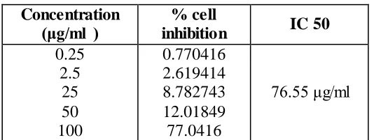
Table 1: Percentage inhibition of curcumin
Table 2: Percentage inhibition of silver nanoparticle encapsulated curcumin Concentration
(μg/ml )
% cell
inhibition IC 50
0.25 2.5 25 50 100 0.770416 2.619414 8.782743 12.01849 77.0416 76.55 μg/ml Concentration
(μg/ml) inhibition %cell IC 50
[image:11.596.179.417.638.738.2]Table 1 and 2 shows percentage inhibition of silver nano particle encapsulated curcumin
(10.27μg/ml) has good toxicity compared with curcumin (76.55μg/ml).
3.4.4 MTT assay
3-[4,5-dimethylthiazol-2-yl]2,5-diphenyltetrazolium bromide (MTT) is a yellow water
soluble tetrazolium salt. A mitochondrial enzyme in living cells, succinate-dehydrogenase,
cleaves the tetrazolium ring, converting the MTT to an insoluble purple formazan. Therefore,
the amount of formazan produced is directly proportional to the number of viable cells.
After 48 h of incubation, 15µl of MTT (5mg/ml) in phosphate buffered saline (PBS) was
added to each well and incubated at 370C for 4h. The medium with MTT was then flicked off
and the formed crystals were dissolved in 100µl of DMSO and then absorbance was
measured at 570 nm using micro plate reader. The % cell inhibition was determined using the
following formula.
% Cell Inhibition = 100- Abs (sample)/Abs (control) x100.
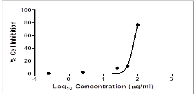
Nonlinear regression graph was plotted between % Cell inhibition and Log concentration and
[image:12.596.132.462.437.597.2]IC50 was determined using Graph Pad Prism software.
Fig. 12 MTT assay of Silver nano particle encapsulated curcumin (R²= 0.9966)
Two releases of curcumin and silver were fitted to these kinetic models to determine the
release kinetics and mechanisms from nanoparticles. The values of these kinetic rates, K and
R2, are presented in fig 11 and 12. In general, the release behavior for all nanoparticles did
[image:13.596.66.474.372.660.2]not obey zero order and first order kinetics based on the low R2 values obtained.
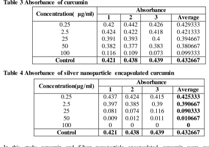
Table 3 Absorbance of curcumin
Table 4 Absorbance of silver nanoparticle encapsulated curcumin
In this study curcumin and Silver nanoparticle encapsulated curcumin were evaluated for
preliminary estimation of the in vitro tumor inhibition activities against cell line of human
breast carcinoma. The results revealed that curcumin and Silver nanoparticle encapsulated
curcumin extract show some correlation between antitumor activity and the structures.
Concentration( μg/ml) Absorbance
1 2 3 Average
0.25 2.5 25 50 100 0.42 0.424 0.391 0.382 0.116 0.442 0.422 0.393 0.377 0.109 0.426 0.418 0.4 0.383 0.073 0.429333 0.421333 0.394667 0.380667 0.099333
Control 0.421 0.438 0.439 0.432667
Concentration(μg/ml) Absorbance
1 2 3 Average
0.25 2.5 25 50 100 0.437 0.397 0.081 0.009 0 0.424 0.385 0.074 0.012 0 0.415 0.39 0.116 0.011 0 0.425333 0.390667 0.090333 0.010667 0
The concentrations that induce 50% inhibition of cell growth (IC50) in μg/ml are reported in
Table 1 and 2. Compounds were classified by their activity as highly active (IC50 < 1 μg/ml),
moderately active (1 μg/ml < IC50 < 10 μg/ml), or inactive (10 μg/ ml > IC50). The great
majority of the curcumin and ethanol extract samples were strongly cytotoxic against all cell
lines of human breast adenocarcinoma cell lines (MCF7) with IC50 below 1μg/ml, when
compared to cisplatin, a fact that supports their anticancer activity.
4. CONCLUSION
Curcumin was extracted from curcuma longa by Soxhlet method. Silver nano particles were
obtained by chemical reduction method. These two solutions were stirred to form silver nano
particle encapsulated curcumin. The particle size of the solutions was found by using particle
size analyzer. From the result it is concluded that the particle size is high for silver
encapsulated curcumin. TLC analysis shows the presence of curcuminoids. From HPLC the
purity and quantity of the curcumin content was found. Anticancer assay shows that silver
nanoparticle encapsulated curcumin is effective in inhibiting cancer growth than curcumin.
Pure curcumin and silver nanoparticle encapsulated curcumin have great potential in the
prevention and cure of cancer. From the results obtained from this research work, it is now
known that the Ag encapsulated curcumin has high potential to destroy cancer cells. The
research in nanoparticle encapsulated curcumin should be strengthened to improve
bioavailability and therapeutic efficacy in the treatment of various disorders.
Funding Agency
The authors are thankful to University Grants Commission (UGC) for funding to do this
research work under minor research project.
REFERENCES
1. Sonali Arjunrao Bhagat, Meera Chandradatt Singh, (Development and evaluation of
silver nanoparticles and its applications in topical drug delivery systems). Asian journal
of pharmaceutics, 2016; 1(10): 16-21.
2. Sudip Mukherjee, Debabrata Chowdhury, Rajesh Kotcherlakota, Sujata Patra,
Vinothkumar B, Manika Pal Bhadra, Bojja Sreedhar, Chitta Ranjan Patra (Potential
Theranostics Application of Bio-Synthesized Silver Nanoparticles [4-in-1 System]).
3. Quang Huy Tran, Van Quy Nguyenand, Anh-Tuan Le, (Silver nanoparticles: synthesis,
properties, toxicology, applications and perspectives). Advances In Natural Sciences:
Nanoscience And Nanotechnology, 2013; 4(3): 1-20.
4. Aggarwal BB, Kumar A, Bharti AC, (Anticancer Potential of curcumin: Preclinical and
clinical studies). Anticancer Res, 2003; 23(1): 363-398.
5. Patil. M.B, Taralkar. S.V, Sakpal. V.S, Shewale. S.P, Sakpal. R.S, (Extraction, Isolation
and Evaluation of Anti inflammatory activity of curcuminoids from curcuma longa).
International journal of chemical sciences and applications, 2011; 2(3): 172-174.
6. Hamdan abood aday, (Bioactivity of curcumin extract against of some pathogenic
strains). Eng & Tech. Journal, 2011; 29(10): 111-119.
7. Verma. S.C, Jain. C.L, Rani. R, Pant. P, Singh. R, Padhi. M.M, Devalla. R.B, (Simple and
rapid method for identification of curcuma longa rhizomes by physicochemical and
HPTLC fingerprint analysis). Chemical science transactions, 2012; 3(1): 709-715.
8. Jha Harit, Anand Barapatre, Mithlesh Prajapati, Keshaw Ram Aadil, Sunil Senapati,
(Antimicrobial activity of rhizome of selected curcuma variety). Int. J, Life Sc. Bt &
Pharm. Res, 2013; 2(3): 183-189.
9. Simay Cikrikci, Erkan Mozioglu, Hasibe Yilmaz, (Biological activity of curcuminoids
isolated from curcuma longa). Records of Natural Products, 2008; 2(1): 19-24.
10.Yogesh Panditrao Palve, Nayak. P.L, (Curcumin: A wonder anticancer drug). Int J Pharm
Biomed Sci, 2012; 2(3): 60-69.
11.Lee PC, Meisel D, (Absorption and surface-enhanced raman of dyes on silver and gold
sols). J. Phys. Chem, 1982; 86(17): 3391–3395.
12.Kamat PV, Flumiani M, Hartland GV, (Picosecond dynamics of silver nanoclusters.
Photoejection of electrons and fragmentation). J. Phys. Chem. B, 1998; 102(17):
3123–3128.
13.Nair AS, Pradeep T, (Halocarbon mineralization and catalytic destruction by metal
nanoparticles). Current Science, 2003; 84(12): 1560-1564.
14.Huang, HH, Ni, XP, Loy GL, Chew HW, Tan KL, Loh FC, Deng JF, Xu GQ,
(Photochemical formation of silver nanoparticles in poly (N-vinylpyrrolidone). Langmuir,
1996; 12(4): 909–912.
15.Yugang S, Gates B, Mayers B, Xia Y, (Crystalline Silver Nano wires by Soft Solution
16.Tonnesen HH, (Solubility, chemical and photochemical stability of curcumin in surfactant
solutions, Studies of curcumin and curcuminoids, XXVIII). Pharmazie, 2002; 57(12):
820–824.
17.Revathy S, Elumalai S, (In vitro evaluation of anticancer activity of curcuminoids from
turmeric (curcuma longa L) against multidrug resistant tumor cell lines). Int. J. Adv. Res,