organic papers
o136
Nakamura and Watanabe C18H38O2 DOI: 101107/S1600536801001052 Acta Cryst.(2001). E57, o136±o138 Acta Crystallographica Section EStructure Reports Online
ISSN 1600-5368
Octadecane-1,18-diol
Naotake Nakamura* and Ryoji Watanabe
Department of Applied Chemistry, Faculty of Science and Engineering, Ritsumeikan University, 1-1-1, Nojihigashi, Kusatsu, Shiga 525-8577, Japan
Correspondence e-mail: [email protected]
Key indicators Single-crystal X-ray study
T= 298 K
Mean(C±C) = 0.003 AÊ
Rfactor = 0.045
wRfactor = 0.065
Data-to-parameter ratio = 10.9
For details of how these key indicators were automatically derived from the article, see http://journals.iucr.org/e.
#2001 International Union of Crystallography Printed in Great Britain ± all rights reserved
The skeleton of the title molecule, C18H38O2, is all-transand
the molecules aggregate to form a layer structure along thec
axis as in a smectic C liquid crystal. The inclination angle of the long axis of the molecule to the layer plane is the same in each layer, but the direction of the long axis is opposite in alternate layers. These features are very similar to those of the homologues with an even number of C atoms, but different from those with an odd number.
Comment
Crystal structures of long-chain compounds such asn-alkanes (MuÈller, 1928) andn-higher primary alcohols (e.g.Watanabe, 1961; Seto, 1962), have been studied by many researchers from the viewpoint of basic polymer science. According to those results, the compounds have a simple straight hydrocarbon chain as a skeleton and the molecular shape can be regarded as rod-like, which is one of the typical features of liquid crystal molecules. In addition, some long-chain compounds construct a layer structure in the crystal state, which is similar to that of a smectic phase of liquid crystals. Therefore, these compounds have been studied from a structural point of view as models for smectic liquid crystals.
We have already reported molecular and crystal structures of ,!-alkanediols containing from 10 through 17 and 21 C atoms; these were investigated by Nakamura and co-workers: 1,10-decanediol (Nakamura & Sato, 1999a), 1,11-undecane-diol (Nakamuraet al., 1999), 1,12-dodecanediol (Nakamura & Setodoi, 1997), 1,13-tridecanediol (Nakamura et al., 1997), 1,14-tetradecanediol (Nakamura & Sato, 1999b), 1,15-penta-decanediol (Nakamura et al., 2000), 1,16-hexadecanediol (Nakamura & Yamamoto, 1994), 1,17-heptadecanediol (Nakamuraet al., 2001) and 1,21-henicosanediol (Nakamuraet al., 2000). The results showed a clear distinction in structure between the compounds with an even number of C atoms and those with an odd number of C atoms. In the,!-alkanediols with even number of C atoms, the hydroxyl groups located at both ends of a hydrocarbon skeleton showed an all-trans
conformation with respect to the skeleton. These molecules were arranged making layers in a herring-bone fashion,
just like the chiral smectic C liquid crystals. On the other hand, in the,!-alkanediols with an odd number of C atoms, one hydroxyl group hadgaucheconformation with respect to the hydrocarbon skeleton, whereas another hydroxyl group had
trans conformation. In this case, molecules made a layer structure which was very similar to that of the smectic A liquid
crystals. In addition, phase transition of,!-alkanediols from C13 through C24 was studied by means of a powder X-ray diffraction method and a linear relation of the longest axis of lattice constant versus number of C atoms was reported (Ogawa & Nakamura, 1999).
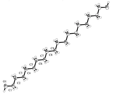
In the crystal structure of 1,18-octadecanediol, (I), shown in Fig. 1, an all-transconformation was observed not only in the hydrocarbon skeleton but also in both terminal hydroxyl groups. The molecule is centrosymmetric and forms a layer structure stacked along thecaxis. The inclination angle of the long axis of the molecule to the layer plane is equal in each layer, but the direction of the long axis of the molecule is opposite in alternate layers because of the existence of a twofold screw axis parallel tob. Such herring-bone structures can be regarded as a model structure of chiral smectic C liquid crystals. These features are very similar to those of 1,10-decanediol, 1,12-do1,10-decanediol, 1,14-tetradecanediol and 1,16-hexadecanediol. The average value of inclination angles of the above compounds was about 56, as found in the present
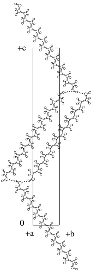
structure. These molecules form interlayer hydrogen bonds, as shown in Fig. 2, with the interlayer hydrogen-bonding distance O1 O1i= 2.843 (2) AÊ [symmetry code: (i) 3ÿx,1
2+y,12ÿz].
This value is in good agreement with those of other ,! -alkanediols with an even number of C atoms.
Experimental
According to the conventional method, the title compound, (I), was synthesized from commercially available octadecanedioic acid (Tokyo Kasei Kogyo Co. Ltd) by esteri®cation and reduction with LiAlH4. The single crystal used for analysis was grown by very slow
evaporation from a solution in a mixed solvent system comprising methanol, ethyl acetate and 1,3-dimethylbenzene (1:1:3 ratio). Crystal data
C18H38O2 Mr= 286.50
Monoclinic,P21/c a= 4.998 (2) AÊ
b= 5.220 (2) AÊ
c= 34.853 (2) AÊ
= 92.04 (2)
V= 908.8 (5) AÊ3 Z= 2
Dx= 1.047 Mg mÿ3
CuKradiation Cell parameters from 24
re¯ections
= 9.3±20.1
= 0.50 mmÿ1 T= 298 K Plate, colorless 0.400.200.02 mm
Data collection
Rigaku AFC-5Rdiffractometer
!±2scans
Absorption correction: scans (Northet al., 1968)
Tmin= 0.929,Tmax= 1.000
2843 measured re¯ections 1802 independent re¯ections 991 re¯ections withI> 2(I)
Rint= 0.025
max= 70.5 h=ÿ2!6
k= 0!5
l=ÿ42!42 3 standard re¯ections
every 150 re¯ections intensity decay: 2.6%
Re®nement
Re®nement onF R= 0.045
wR= 0.065
S= 1.94 991 re¯ections 91 parameters
H-atom parameters not re®ned
w= 1/[2(F
o) + 0.00053|Fo|2]
(/)max< 0.001
max= 0.12 e AÊÿ3
min=ÿ0.11 e AÊÿ3
Acta Cryst.(2001). E57, o136±o138 Nakamura and Watanabe C18H38O2
o137
organic papers
Figure 1
AnORTEPII (Johnson, 1976) view of the title molecule showing the crystallographic numbering scheme. Displacement ellipsoids are shown at the 50% probability level.
Figure 2
organic papers
o138
Nakamura and Watanabe C18H38O2 Acta Cryst.(2001). E57, o136±o138All H atoms, including hydroxyl H atoms, were ®xed in idealized positions.
Data collection and cell re®nement: MSC/AFC Diffractometer Control Software (Molecular Structure Corporation, 1992); data reduction: TEXSAN (Molecular Structure Corporation, 1995); program(s) used to solve structure:SAPI91 (Fan, 1991); program(s) used to re®ne structure:TEXSAN; software used to prepare material for publication:TEXSAN.
References
Fan, H.-F. (1991).SAPI91. Rigaku Corporation, Tokyo, Japan.
Johnson, C. K. (1976).ORTEPII. Report ORNL-5138. Oak Ridge National Laboratory, Tennessee, USA.
Molecular Structure Corporation (1992).MSC/AFC Diffractometer Control Software. MSC, 3200 Research Forest Drive, The Woodlands, TX 77381, USA.
Molecular Structure Corporation (1995).TEXSAN.Version 1.7. MSC, 3200 Research Forest Drive, The Woodlands, TX 77381, USA.
MuÈller, A. (1928).Proc. R.Soc. London Ser. A,120, 437±459. Nakamura, N. & Sato, T. (1999a).Acta Cryst.C55, 1685±1687. Nakamura, N. & Sato, T. (1999b).Acta Cryst.C55, 1687±1689. Nakamura, N. & Setodoi, S. (1997).Acta Cryst.C53, 1883±1885. Nakamura, N., Setodoi, S. & Ikeya, T. (1999).Acta Cryst.C55, 789±791. Nakamura, N., Tanihara, Y. & Takayama, T. (1997).Acta Cryst. C53, 253±
255.
Nakamura, N., Uno, K. & Ogawa, Y. (2000) Acta Cryst. C56, 1389± 1370.
Nakamura, N., Uno, K. & Ogawa, Y. (2001).Acta Cryst.C57. In preparation. Nakamura, N., Uno, K., Watanabe, R., Ikeya, T. & Ogawa, Y. (2000).Acta
Cryst.C56, 903±904.
Nakamura, N. & Yamamoto, T. (1994).Acta Cryst.C50, 946±948.
North, A. C. T., Phillips, D. C. & Mathews, F. S. (1968).Acta Cryst.A24, 351± 359.
supporting information
sup-1
Acta Cryst. (2001). E57, o136–o138supporting information
Acta Cryst. (2001). E57, o136–o138 [doi:10.1107/S1600536801001052]
Octadecane-1,18-diol
Naotake Nakamura and Ryoji Watanabe
S1. Comment
Crystal structures of long-chain compounds such as n-alkanes (Müller, 1928) and n-higher primary alcohols (e.g.
Watanabe, 1961; Seto, 1962), have been studied by many researchers from the viewpoint of basic polymer science.
According to those results, the compounds have a simple straight hydrocarbon chain as a skeleton and the molecular
shape can be regarded as a rod-like, which is one of the typical features of liquid crystal molecules. In addition, some
long-chain compounds construct a layer structure in the crystal state, which is similar to that of a smectic phase of liquid
crystals. Therefore, these compounds have been studied from a structural point of view as models for smectic liquid
crystals.
We have already reported molecular and crystal structure of α,ω-alkanediols containing from 10 through 17 and 21 C
atoms, these were investigated by Nakamura and co-workers: 1,10-decanediol (Nakamura & Sato, 1999a),
1,11-undecanediol (Nakamura et al., 1999), 1,12-dodecanediol (Nakamura & Setodoi, 1997), 1,13-tridecanediol (Nakamura et
al., 1997), 1,14-tetradecanediol (Nakamura & Sato, 1999b), 1,15-pentadecanediol (Nakamura et al., 2000),
1,16-hexa-decanediol (Nakamura & Yamamoto, 1994), 1,17-hepta1,16-hexa-decanediol (Nakamura et al., 2001) and 1,21-henicosanediol
(Nakamura et al., 2000a). The results showed a clear distinction in the structures between the compounds with an even
number of C atoms and those with an odd number of C atoms. In the α,ω-alkanediols with even number of C atoms, the
hydroxyl groups located at both ends of a hydrocarbon skeleton showed an all-trans conformation with respect to the
skeleton. These molecules were arranged making layers in a herring-bone fashion, just like the chiral smectic C liquid
crystals. On the other hand, in the α,ω-alkanediols with an odd number of C atoms, one hydroxyl group had gauche
conformation with respect to the hydrocarbon skeleton, whereas another hydroxyl group had trans conformation. In this
case, molecules made a layer structure which was very similar to that of the smectic A liquid crystals. In addition, phase
transition of α,ω-alkanediols from C13 through C24 was studied by means of a powder X-ray diffraction method and a
linear relation of the longest axis of lattice constant versus number of C atoms was reported (Ogawa & Nakamura, 1999).
In the crystal structure of 1,18-octadecanediol, (I), shown in Fig. 1, an all-trans conformation was observed not only in
the hydrocarbon skeleton but also in both terminal hydroxyl groups. The molecule is centrosymmetric and forms a layer
structure stacked along c axis. The inclination angle of the long axis of the molecule to the layer plane is equal in each
layer, but the direction of the long axis of the molecule is opposite in alternate layers because of an existence of a twofold
screw axis parallel to b. Such herring-bone structures can be regarded as a model structure of chiral smectic C liquid
crystals. These features are very similar to those of 1,10-decanediol, 1,12-dodecanediol, 1,14-tetradecanediol and
1,16-hexadecanediol. Average value of inclination angles of above compounds was about 56° as found in the present structure.
These molecules form interlayer hydrogen bonds, as shown in Fig. 2, with the interlayer hydrogen-bonding distance
O1···O1i = 2.843 (2) Å [symmetry code: (i) 3 - x, 0.5 + y, 0.5 - z]. This value is in good agreement these of other α,ω
supporting information
sup-2
Acta Cryst. (2001). E57, o136–o138S2. Experimental
According to the conventional method, the title compound, (I), was synthesized from commercially available
octadecane-dioic acid (Tokyo Kasei Kogyo Co. Ltd) by esterification and reduction with LiAlH4. The single-crystal used for analysis
was grown by very slow evaporation from a solution in a mixed solvent system comprising methanol, ethyl acetate and
dimetylbenzene (1:1:3 ratio).
S3. Refinement
[image:5.610.119.493.189.500.2]All H atoms, including hydroxyl H atoms, were fixed in idealized positions.
Figure 1
An ORTEPII (Johnson, 1976) view of the title molecule showing the crystallographic numbering scheme. Displacement
supporting information
[image:6.610.229.381.68.514.2]sup-3
Acta Cryst. (2001). E57, o136–o138Figure 2
The bc-projection of the crystal structure; dotted lines indicate the hydrogen bonds.
(I)
Crystal data
C18H38O2
Mr = 286.50 Monoclinic, P21/c
a = 4.998 (2) Å
b = 5.220 (2) Å
c = 34.853 (2) Å
β = 92.04 (2)°
V = 908.8 (5) Å3
Z = 2
F(000) = 324
Dx = 1.047 Mg m−3
Cu Kα radiation, λ = 1.5418 Å Cell parameters from 24 reflections
θ = 9.3–20.1°
µ = 0.50 mm−1
supporting information
sup-4
Acta Cryst. (2001). E57, o136–o138Data collection
Rigaku AFC-5R diffractometer
Radiation source: Rigaku rotating anode Graphite monochromator
ω–2θ scans
Absorption correction: ψ scans (North et al., 1968)
Tmin = 0.929, Tmax = 1 2843 measured reflections
1802 independent reflections 991 reflections with I > 2σ(I)
Rint = 0.025
θmax = 70.5°, θmin = 2.5°
h = −2→6
k = 0→5
l = −42→42
3 standard reflections every 150 reflections intensity decay: 2.6%
Refinement
Refinement on F
Least-squares matrix: full
R[F2 > 2σ(F2)] = 0.045
wR(F2) = 0.065
S = 1.67 991 reflections 91 parameters
0 restraints 0 constraints
H-atom parameters not refined
w = 1/[σ2(F
o) + 0.00053|Fo|2] (Δ/σ)max < 0.001
Δρmax = 0.12 e Å−3 Δρmin = −0.11 e Å−3
Special details
Experimental. none Geometry. none Refinement. None
Fractional atomic coordinates and isotropic or equivalent isotropic displacement parameters (Å2)
x y z Uiso*/Ueq
O1 1.4119 (3) −0.4953 (3) 0.23943 (4) 0.0802 (5)
C1 1.2271 (4) −0.5358 (4) 0.20834 (5) 0.0599 (6)
C2 1.1288 (4) −0.2830 (4) 0.19336 (5) 0.0540 (5)
C3 0.9330 (4) −0.3089 (4) 0.15912 (5) 0.0517 (5)
C4 0.8304 (4) −0.0540 (4) 0.14358 (5) 0.0514 (5)
C5 0.6396 (4) −0.0809 (4) 0.10891 (5) 0.0513 (5)
C6 0.5365 (4) 0.1724 (4) 0.09318 (5) 0.0500 (5)
C7 0.3465 (4) 0.1468 (4) 0.05827 (5) 0.0500 (5)
C8 0.2426 (4) 0.4007 (4) 0.04263 (5) 0.0501 (5)
C9 0.0519 (4) 0.3726 (4) 0.00781 (5) 0.0496 (6)
H0 1.4782 −0.6526 0.2493 0.0916*
H1a 1.3082 −0.6345 0.1882 0.0799*
H1b 1.0770 −0.6387 0.2167 0.0799*
H2a 1.2796 −0.1823 0.1854 0.0655*
H2b 1.0437 −0.1900 0.2132 0.0655*
H3a 1.0188 −0.3972 0.1390 0.0649*
H3b 0.7832 −0.4075 0.1667 0.0649*
H4a 0.9802 0.0478 0.1364 0.0598*
H4b 0.7398 0.0337 0.1633 0.0598*
H5a 0.7313 −0.1706 0.0889 0.0631*
H5b 0.4911 −0.1860 0.1158 0.0631*
supporting information
sup-5
Acta Cryst. (2001). E57, o136–o138H6b 0.4449 0.2630 0.1130 0.0601*
H7a 0.4376 0.0570 0.0384 0.0621*
H7b 0.1970 0.0410 0.0652 0.0621*
H8a 0.3905 0.5056 0.0355 0.0607*
H8b 0.1502 0.4899 0.0624 0.0607*
H9a 0.1427 0.2828 −0.0120 0.0594*
H9b −0.0979 0.2686 0.0149 0.0594*
Atomic displacement parameters (Å2)
U11 U22 U33 U12 U13 U23
O1 0.096 (1) 0.0647 (10) 0.0759 (9) −0.0011 (8) −0.0557 (8) 0.0046 (8)
C1 0.068 (1) 0.056 (1) 0.0534 (10) −0.0013 (10) −0.0282 (9) 0.0024 (9)
C2 0.059 (1) 0.056 (1) 0.0466 (9) 0.0017 (9) −0.0176 (8) −0.0008 (8)
C3 0.054 (1) 0.052 (1) 0.0479 (9) −0.0045 (9) −0.0158 (8) 0.0034 (8)
C4 0.0525 (10) 0.051 (1) 0.0490 (9) −0.0041 (9) −0.0152 (8) 0.0031 (8)
C5 0.054 (1) 0.051 (1) 0.0473 (9) −0.0007 (9) −0.0168 (8) 0.0010 (8)
C6 0.054 (1) 0.048 (1) 0.0471 (10) −0.0008 (8) −0.0163 (8) 0.0002 (8)
C7 0.054 (1) 0.049 (1) 0.0462 (9) −0.0031 (9) −0.0148 (8) 0.0011 (8)
C8 0.053 (1) 0.049 (1) 0.0475 (10) −0.0032 (8) −0.0155 (8) 0.0020 (7)
C9 0.055 (1) 0.049 (1) 0.0442 (10) −0.0019 (8) −0.0159 (8) 0.0026 (7)
Geometric parameters (Å, º)
O1—C1 1.414 (2) C5—C6 1.515 (2)
C1—C2 1.496 (3) C6—C7 1.523 (2)
C2—C3 1.522 (2) C7—C8 1.518 (3)
C3—C4 1.519 (2) C8—C9 1.523 (2)
C4—C5 1.519 (2) C9—C9i 1.521 (4)
O1···O1ii 2.843 (2) O1···C1ii 3.477 (2)
O1···O1iii 2.843 (2) O1···C2iii 3.554 (2)
O1—C1—C2 109.4 (1) C5—C6—C7 114.0 (1)
C1—C2—C3 112.9 (2) C6—C7—C8 114.0 (2)
C2—C3—C4 113.7 (2) C7—C8—C9 113.5 (1)
C3—C4—C5 113.4 (1) C8—C9—C9i 113.4 (2)
C4—C5—C6 113.8 (2)