USE OF MTA AS APICAL PLUG IN TEETH WITH OPEN APEX
1
Amitoj Kaur Walia,
2,*Harshita,
6
Harsimranjit Kaur and
1
Tutor, Department of Conservative Dentistry and Endodontics, Genesis Institute of Dental Sciences and
2
Post Graduate Student, Department of Conservative Dentistry and Endodontics, Genesis Institute of D
Sciences and Research, Ferozepur, Punjab, India
3
Post Graduate Student, Department of Conservative Dentistry and Endodontics, Genesis Institute of Dental
Sciences and Research, Ferozepur, Punjab, India
4
Senior Lecturer, Department of Conservative Den
and Research, Ferozepur, Punjab, India
5
Post Graduate Student, Department of Conservative Dentistry and Endodontics, Genesis Institute of Dental
Sciences and Research, Ferozepur, Punjab, India
6
Post Graduate Student, Department of Conservative Dentistry and Endodontics, Genesis Institute of Dental
Sciences and Research, Ferozepur, Punjab, India
7
Post Graduate Student, Master of Business Administration, Thapar Institute of Engineering and Tec
ARTICLE INFO ABSTRACT
Aim:
the aim of this case series is to use MTA plug in teeth with open apex. Summary:
for open apices, as it requires reasonably less time with predictable results and material to obtain periradicular healing in teeth with open apic
Copyright©2017, Amitoj Kaur Walia et al. This is an open access article distributed under the Creative Commons Att
use, distribution, and reproduction in any medium, provided the original work is properly cited.
INTRODUCTION
The primary objective of endodontic therapy is the complete obturation of the root canal space to prevent re
teeth with incomplete root development caused by trauma, caries or other pulpal pathosis, the absence of the natural constriction at the end of the root canal presents a challenge and by making control of obturating materials difficult. For success in these particular cases, the aim of the dentist is to adequately seal a sizeable communication between the root
*Corresponding author: Harshita,
Post Graduate Student, Department of Conservative Dentistry and Endodontics, Genesis Institute of Dental Sciences and Research, Ferozepur, Punjab, India
ISSN: 0975-833X
Article History:
Received 23rd July, 2017
Received in revised form
27th August, 2017
Accepted 18th September, 2017
Published online 31st October, 2017
Citation: Amitoj Kaur Walia, Harshita, Monika Vashisht, Fatinderjeet Singh, Pratima Sharma, Harsimranjit Kaur and Gajinder Krishan “Use of MTA as apical plug in teeth with open apex
Key words:
MTA, Necrotic pulp, Open apex, Apical plug.
RESEARCH ARTICLE
USE OF MTA AS APICAL PLUG IN TEETH WITH OPEN APEX - A REPORT OF 2 CASES
Harshita,
3Monika Vashisht,
4Fatinderjeet Singh,
Harsimranjit Kaur and
7Gajinder Krishan
Tutor, Department of Conservative Dentistry and Endodontics, Genesis Institute of Dental Sciences and
Research, Ferozepur, Punjab, India
Post Graduate Student, Department of Conservative Dentistry and Endodontics, Genesis Institute of D
Sciences and Research, Ferozepur, Punjab, India
Post Graduate Student, Department of Conservative Dentistry and Endodontics, Genesis Institute of Dental
Sciences and Research, Ferozepur, Punjab, India
Senior Lecturer, Department of Conservative Dentistry and Endodontics, Genesis Institute of Dental Sciences
and Research, Ferozepur, Punjab, India
Post Graduate Student, Department of Conservative Dentistry and Endodontics, Genesis Institute of Dental
Sciences and Research, Ferozepur, Punjab, India
ost Graduate Student, Department of Conservative Dentistry and Endodontics, Genesis Institute of Dental
Sciences and Research, Ferozepur, Punjab, India
Post Graduate Student, Master of Business Administration, Thapar Institute of Engineering and Tec
Patiala, Punjab, India
ABSTRACT
As management of non-vital teeth with open apices is a challenge to dental practitioners. So, the aim of this case series is to use MTA plug in teeth with open apex.
Summary: Mineral trioxide aggregate (MTA) apical plug method is an alternative treatment option for open apices, as it requires reasonably less time with predictable results and
material to obtain periradicular healing in teeth with open apices and necrotic pulps.
is an open access article distributed under the Creative Commons Attribution License, which use, distribution, and reproduction in any medium, provided the original work is properly cited.
The primary objective of endodontic therapy is the complete obturation of the root canal space to prevent re-infection. In teeth with incomplete root development caused by trauma, caries or other pulpal pathosis, the absence of the natural the end of the root canal presents a challenge and by making control of obturating materials difficult. For success in these particular cases, the aim of the dentist is to adequately seal a sizeable communication between the root
Post Graduate Student, Department of Conservative Dentistry and Endodontics, Genesis Institute of Dental Sciences and Research,
canal system and the periradicular tissue and provide a barrier against which obturation material can be compacted (Aggarwal, 2012). Recently, synthetic apical barriers with a variety of materials have been proposed as alternatives to the traditional apexification treatment method using calcium hydroxide. Mineral trioxide aggregate (MTA) is the most popular material for this purpose. It has been advocated for use as an apical barrier because of its sealing capabilities, ability to get hardened in the presence of blood, and biocompatibility. it can be used to create an apical plug at the root
to prevent the extrusion of the obturating materials.
case series presents the clinical applications of MTA as apical plug in teeth with open apex.
International Journal of Current Research
Vol. 9, Issue, 10, pp.59938-59942, October, 2017
INTERNATIONAL
OF CURRENT RESEARCH
Amitoj Kaur Walia, Harshita, Monika Vashisht, Fatinderjeet Singh, Pratima Sharma, Harsimranjit Kaur and Gajinder Krishan
apical plug in teeth with open apex - a report of 2 cases”, International Journal of Current Research
A REPORT OF 2 CASES
Fatinderjeet Singh,
5Pratima Sharma,
Tutor, Department of Conservative Dentistry and Endodontics, Genesis Institute of Dental Sciences and
Post Graduate Student, Department of Conservative Dentistry and Endodontics, Genesis Institute of Dental
Post Graduate Student, Department of Conservative Dentistry and Endodontics, Genesis Institute of Dental
tistry and Endodontics, Genesis Institute of Dental Sciences
Post Graduate Student, Department of Conservative Dentistry and Endodontics, Genesis Institute of Dental
ost Graduate Student, Department of Conservative Dentistry and Endodontics, Genesis Institute of Dental
Post Graduate Student, Master of Business Administration, Thapar Institute of Engineering and Technology,
vital teeth with open apices is a challenge to dental practitioners. So, the aim of this case series is to use MTA plug in teeth with open apex.
Mineral trioxide aggregate (MTA) apical plug method is an alternative treatment option for open apices, as it requires reasonably less time with predictable results and appears to be a valid
es and necrotic pulps.
ribution License, which permits unrestricted
canal system and the periradicular tissue and provide a barrier against which obturation material can be compacted Recently, synthetic apical barriers with a variety of materials have been proposed as alternatives to the fication treatment method using calcium hydroxide. Mineral trioxide aggregate (MTA) is the most popular material for this purpose. It has been advocated for use as an apical barrier because of its sealing capabilities, ability to
ce of blood, and biocompatibility.So
it can be used to create an apical plug at the root-end that helps to prevent the extrusion of the obturating materials. These two case series presents the clinical applications of MTA as apical
INTERNATIONAL JOURNAL OF CURRENT RESEARCH
Amitoj Kaur Walia, Harshita, Monika Vashisht, Fatinderjeet Singh, Pratima Sharma, Harsimranjit Kaur and Gajinder Krishan. 2017.
MATERIALS AND METHODS
Case Report 1
A 23 year old male patient reported Department of Conservative Dentistry and Endodontics, Genesis Institute of Dental Sciences and Research, Ferozepur with chief complaint of fractured and discolored upper right front tooth. Patient gave history of trauma at the age of 7 or 8 years and gave no history of pain or swelling. Clinical examination revealed Ellis class I fracture and discolored tooth in relation to 11. Tooth showed tenderness on percussion but no response to vitality tests.
Medical history was non contributory. Radiographic
[image:2.595.361.508.134.325.2]examination revealed fractured tooth with open apex and radiolucent lesion in proximity to apex of tooth 11 (Figure 1).
[image:2.595.91.233.242.428.2]Figure 1. Ellis class I fracture with open apex and periapical radiolucency in relation to 11relation to 11
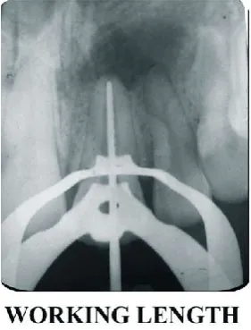
Figure 2. Working length determined in relation to 11
So, according to ‘AAE Consensus Conference Recommended Diagnostic Terminology’ the final diagnosis of Pulpal necrosis with asymptomatic apical periodontitis with open apex with respect to 11 was made. After the application of the rubber dam, access cavity preparation was done, the working length was determined (Figure 2). The canal was then cleaned biomechanically by using intracanal instruments. Irrigation was done using saline and 5 ml of 3% NaoCl (Shiva Products, Waliv , Palghar, Maharashtra, India) as an irrigant after each instrumentation for final flush.
Then, the canal was dried with sterile paper points and Metapex (Meta Dental Corp. Ltd., Elmburst, NY), a
commercially available product of composed Ca(OH)2,
[image:2.595.93.234.471.656.2]silicone oil, and iodoform was then placed in the canal and temporary restoration was done using Cavit (3M ESPE, St. Paul, MN, USA) (Figure3). Patient was recalled after 2 weeks.
Figure 3. Metapex placed in relation to 11
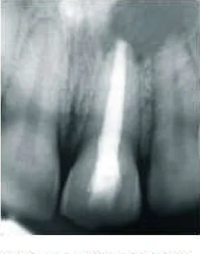
At follow up visit tooth was isolated using rubber dam and metapex was removed using H files and repeated rinsing with 3% NaOCl followed by rinsing with distilled water ( Figure4) . The canal was dried with sterile paper points and the MTA (Dental Tulsa Dentsply, Johnson City, TN, USA) was mixed according to manufacturer’s instructions and placed in canal with help of Dovgan carrier in the apical portion of the canal (Figure 5). and compacted into the coronal section of the canal using Schilder plugger [Dentsply Caulk, Milford, USA] to form a tight apical plug of 4mm. To check the correct position of the MTA placement, an IOPA radiograph was taken. A wet cotton pellet with distilled was then placed in the pulp chamber and the access cavity was closed with temporary filling material Cavit (3M ESPE, St. Paul, MN, USA).
Figure 4. Metapex removed in relation to 11
[image:2.595.358.510.521.721.2]Canal Sealer EWT Kerr, West Collins, CA) (Figure 7)
followed by placement of composite resin (Kulzer Charisma,
[image:3.595.92.235.101.294.2]Mumbai) as permanent restoration.
Figure 5. MTA placement in relation to 11
[image:3.595.315.552.156.351.2]Figure 6
Figure 7.
Case Report 2
A 25 year old male presented in the Department of Conservative Dentistry and Endodontics, Genesis Dental
College, Ferozepur with the chief complaint of discoloration in his right upper front tooth region. He gave history of trauma to this tooth at age of 9 and swelling and pus discharge from the right upper front since 1 month. The clinical examination revealed slight discoloration in relation to 12 and tooth did not showed any response to vitality tests. Radiographic examination (Figure 8) of the tooth revealed a wide open apex and prescence of periapical lesion.
So, according to ‘AAE Consensus Conference Recommended Diagnostic Terminology’ the final diagnosis of Pulpal necrosis with asymptomatic apical periodontitis with open apex in relation to 12 was made. The protocol for the creation of an apical plug with MTA mixture was implemented as in case 1 (Figure 9a, b, c and d).
RESULTS
In Case Report 1, patient was recalled after 6 months and 12 months for follow up. The radiographic follow up at 6 months revealed a decrease of the periapical rarefaction, regeneration of the periradicular tissue and hard tissue deposition indicating healing of lesion (Figure 8) and at 1 year significant healing was observed clearly (Figure 9).
[image:3.595.362.505.589.770.2]Figure 9
Case Report 2
In Case Report 2, patient was recalled after 6 months (9e) and 1 year (9f) for follow up. Complete resolution of lesion was seen on 1 year follow up.
DISCUSSION
During tooth development, the inner and outer dental epithelia fuse and form the cervical loop, which results in Hertwig’s epithelial root sheath, a structure responsible for root formation (Hargreaves, 2002). The presence of healthy pulp is essential for root development and apical closure. When the pulp is vital and the apex is not fully formed, it is imperative to maintain the pulp vitality for dentine formation. Dental caries and trauma are the most common challenges to the integrity of a tooth as it matures. Both insults can render the pulp non-vital. If this occurs prior to complete root formation and apical closure, normal root development is halted (Farhad, 2005). Clinically, there are several difficulties associated with treating non-vital teeth that have a widened or open apical foramen. Firstly, the apical diameter of the canal is often larger than the coronal diameter, so debridement is difficult. In addition, the lack of an apical stop makes the control of obturation materials very difficult. Finally, the thin walls of the root canal are prone to fracture, so that surgical treatment is generally not a viable
option (Cohen, 2006). To avoid these complications,
apexification prior to root canal filling should be attempted.
The use of calcium hydroxide for apical closure was first introduced in 1964 by Kaiser, when he proposed that using it after mixing with camphorated parachlorophenol (CMCP) would induce the formation of a calcified barrier across the apex (Dogra, 2005). The larger the apical opening, the longer is the time necessary to induce apical closure. Induction of apical healing takes at least 3–4 months and requires multiple appointments. Patient compliance with this regimen may be poor and many fail to return for scheduled visits. The temporary seal may fail resulting in reinfection and prolongation or failure of treatment. For these reasons one-visit apexification has been suggested. A number of materials have been proposed for this purpose including tricalcium phosphate, calcium hydroxide, freeze dried bone, and freeze dried dentine. Placement of MTA has been considered in these cases as it is effective as an apical barrier and its application results in predictable apical closing, reduced treatment time
and a reduced number of exposures to radiographs (Giuliani et
al., 2002).
It is a biomaterial that has been investigated for endodontic applications since the early 1990s. It was first described in the dental scientific literature in 1993 and was given approval for endodontic use by the U.S. Food and Drug Administration in
1998 (Howard et al., 2008).It is a powder that consists of fine
hydrophilic particles that set in the presence of moisture. The setting time in moisture is 2 hours 45 minutes (Hargreaves, 2002). Two commercial forms of MTA are available: ProRoot MTA as the grey and white MTA (Tulsa Dental Products, Tulsa, OK, USA) and MTA-Angelus (Angelus, Londrina, PR,
Brazil) is without tetracalcium alumioferrite.MTA is a mixture
of a refined Portland cement and bismuth oxide, and contains
trace amounts of SiO2, CaO, MgO. Investigations have found
that lower amounts of iron, aluminum, and magnesium are
present in white MTA than in grey MTA. Hydrated MTA
products have an initial pH of 10.2, which rises to 12.5 three hours after mixing. The setting process is described as a
hydration reaction of tricalcium silicate (3CaO·SiO2) and
dicalcium silicate (2CaO·SiO2), which the latter is said to be
responsible for the development of material strength (Kumari, 2011). It not only fulfills the ideal requirement of being bacteriostatic, but it might have potential bactericidal properties. The release of hydroxyl ions, a sustained high pH for extended periods, and the formation of a mineralized interstitial layer might provide a challenging environment for bacterial survival. These antibacterial properties can be a potent inhibitor of bacterial growth against species such as Entercoccocus faecalis, a microorganism prevalent in root canal failures. Moreover, Candida albicans, commonly present in refractory endodontic disease, is susceptible to the antifungal activity of freshly mixed MTA. These factors are important when considering nonsurgical treatments for patients with large periapical lesions associated with initial root canal
treatment (Bogen, 2009).MTA has also presented promising
outcomes when used for the repair of lateral and furcation perforations. Formation of cementum surrounding MTA was
observed, even after extrusion of MTA into a furcation (Pitt et
al., 1995).
On the basis of these findings, MTA may be an appropriate material for apical sealing of mature root canals with open apices, which may impose technical challenges in obtaining adequate obturation because of apical perforation, over-instrumentation, resorption, or former surgical treatment. Successful prognosis from conservative treatment with MTA for such difficult cases without surgical treatment is a great
benefit for patients (Hayashi et al., 2004). Furthermore, MTA
provides scaffolding for the formation of hard tissue and the
potential of a better biological seal (Steinig et al., 2003).It also
facilitates formation of normal periradicular architecture by inducing hard tissue barriers. The rationale is to establish an apical stop that would enable the root canal to be filled immediately. There is no attempt at root end closure. Rather an artificial apical stop is created (Mohammadi, 2011). The application of MTA mixture was preceded by a temporary calcium hydroxide dressing in order to limit bacterial infection in the tooth (Arzu, 2008). Obturation in this case was completed with thermoplasticized gutta-percha, since it can be placed without applying any compaction forces on thin dentinal walls in contrast to lateral condensation method.
Conclusion
In conclusion, MTA appeared to be a valid option for apexification with the added advantage of speed of completion of therapy.
Acknowledgement
Authors would like to thank Genesis Institute of Dental Sciences and Research, Ferozepur, Punjab, India for its support.
REFERENCES
Aggarwal, A 2012. Single visit apexification with MTA: A
report of two cases. Clinical Dentistry Mumbai, 30(9),
20-5.
Arzu P, Erdem E 2008 Mineral trioxide aggregate for obturation of maxillary central incisors with necrotic pulp
and open apices. Dental Traumatology, 24 (8), 38–41.
Bogen G, Kutter S 2009. Mineral Trioxide Aggregate
Obturation: A Review and Case Series. Journal of
Endodontics 35(6), 777- 90.
Cohen S, Hargreaves KM 2006. Pathways of the Pulp. St Louis
Mosby Inc 30(2), 610–49.
Dogra S, KS Mukunda, Arun A 2005. Apexification. Journal
of Dental sciences and Research 3(1), 41-4.
Farhad A, Mohammadi Z 2005. Calcium hydroxide: a review.
Int Dent J. 55(2), 293–301.
Giuliani V, Baccetti T, Pace R, Pagavino G 2002. The use of
MTA in teeth with necrotic pulps and open apices. Dent
Traumatol 18(5), 217-21.
Güneş B, Aydinbelge H 2012. Mineral trioxide aggregate apical plug method for the treatment of nonvital immature
permanent maxillary incisors: Three case reports. Journal
of Conservative Dentistry 15(1), 73-6.
Hargreaves KM, Goodis HE 2002. Seltzer and Bender’s Dental
Pulp. Quintessence Publishing Co. Inc 38(5), 13–40.
Hayashi M, Shimizu A, Ebisu A 2004. MTA for Obturation of Mandibular Central Incisors with Open Apices: Case
Report. Journal of Endodontics 30(2), 120-2.
Howard WR, Jeffrey M, David W, David G (2008) Mineral trioxide aggregate material use in endodontic treatment: A
review of the literature. Charlton dental materials 23(5),
149–64.
Kumari M, Takkar H, Nigam N 2011. Mineral Tri Oxide Aggregate Used As Apical Plug In Open Apex Cases - A
Review And Case Report. Indian Journal of Dental
Sciences 3(4): 15-7.
Matt, GD, Thorpe JR, Scott B 2004. Comparative Study of White and Gray Mineral Trioxide Aggregate (MTA) Simulating a One- or Two-Step Apical Barrier Technique.
Journal of Endodontics 30(12), 876-80.
Mohammadi Z 2011. Strategies to manage permanent non-vital
teeth with open apices: a clinical update. International
Dental Journal 61(5), 25–30.
Morse DR, O’Larnic J, Yesilsoy C 1990. Apexification:
review of the literature. Quintessence Int 21(7), 589–98.
Pitt Ford TR, Torabinejad M, McKendry JD, Hong CU, Kariyawasam SP 1995. Use of mineral trioxide aggregate
for repair of furcal perforations. Oral Surg Oral Med Oral
Pathol Oral Radiol Endod 7(9), 756–62.
Steinig TH, Regan JD, Gutmann JL 2003. The use and predictable placement of Mineral Trioxide Aggregate in
one-visit apexification cases. Aust Endod J 29(3), 34–42.