HPLC-MS/MS METHOD DEVELOPMENT AND VALIDATION FOR DETERMING
STABILITY OF ALECTINIB IN HUMAN PLASMA SAMPLES
*
Srika
Department of Pharmaceutical Sciences, University College of Pharmaceutical Sciences,
Acharya Nagarjuna University,
ARTICLE INFO ABSTRACT
The validated protein precipitation method was applied plasma with ATD8 as an internal standard (ISTD) by using
chromatographic separation was achieved with 0.1% formic acid in combination with methanol (25:75 v/v) using the
The total analysis time was 3 of AT
shows correlation coefficient (r using the linear regression model.
Copyright©2017, Srikanth and Prameela Rani.This
unrestricted use, distribution, and reproduction in any medium, provided the original work is properly cited.
INTRODUCTION
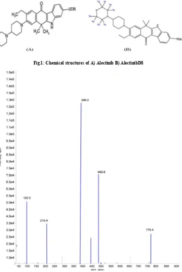
Alectinib (AT) is 9-ethyl-6,6-dimethyl-ylpiperidin-1-yl)-11-oxo-5H-benzo[b]carbazole
with chemical formula C30H34N4O2 (Fig.1) and its molecular
weight is 482.61656. Alectinib was used for the treatment of anaplastic lymphoma kinase (ALK) inhibitor (
2016; Dobbelstein et al., 2014; Prideaux
2010; Bennet et al., 2013). The literature survey reveals that, a variety of methods were reported on the pharmacokinetics of Alectinib in human plasma (Kim, 2008; Gode
2011) for quantification of Alectinib by using HPLC
From the literature review it was concluded that the reported methods used highly expensive extraction process (SPE), long run time and lack of deuterated internal standard by using HPLC-ESI-M/MS methods. There is no method reported for estimation of Alectinib using deuterated internal standard in biological samples. The main goal of the present study is to develop and validate the novel simple, sensitive, selective, rapid, rugged and reproducible analytical method for quantitative determination of AT in human plasma by HPLC ESI-MS/MS with a small amount of sample volume.
Corresponding author: Srikanth, I.
Department of Pharmaceutical Sciences, University College of Pharmaceutical Sciences, Acharya Nagarjuna University, Guntur 522510.
ISSN: 0975-833X
Article History:
Received 27th February, 2017
Received in revised form
29th March, 2017
Accepted 07th April, 2017
Published online 31st May,2017
Citation: Srikanth, I. and Prameela Rani, A. 2017.
samples”, International Journal of Current Research, 9, (0
Key words: Alectinib, Human Plasma, HPLC-ESI-MS/MS Bioanalysis.
RESEARCH ARTICLE
MS/MS METHOD DEVELOPMENT AND VALIDATION FOR DETERMING
STABILITY OF ALECTINIB IN HUMAN PLASMA SAMPLES
Srikanth, I. and Prameela Rani, A.
Department of Pharmaceutical Sciences, University College of Pharmaceutical Sciences,
Acharya Nagarjuna University, Guntur-522510
ABSTRACT
The validated protein precipitation method was applied for estimation of AT plasma with ATD8 as an internal standard (ISTD) by using
chromatographic separation was achieved with 0.1% formic acid in combination with methanol (25:75 v/v) using the C18 columnAscentis Express (50 mm × 4.6 mm, 2.7 µm).
The total analysis time was 3 min and flow rate was set to 0.6 ml/min. The mass transitions of AT, ATD8 obtained were m/z 482.6396.0 and 490.6
shows correlation coefficient (r2) greater than 0.9983 with a range of 5.00 using the linear regression model.
This is an open access article distributed under the Creative Commons Att use, distribution, and reproduction in any medium, provided the original work is properly cited.
-8-(4-morpholin-4-benzo[b]carbazole-3-carbonitrile
(Fig.1) and its molecular weight is 482.61656. Alectinib was used for the treatment of bitor (Hiroaki et al., Prideaux, 2012; Sugiura, The literature survey reveals that, a variety of methods were reported on the pharmacokinetics of Gode, 2013; Seeley, for quantification of Alectinib by using HPLC-MS/MS. From the literature review it was concluded that the reported methods used highly expensive extraction process (SPE), long run time and lack of deuterated internal standard by using There is no method reported for estimation of Alectinib using deuterated internal standard in The main goal of the present study is to develop and validate the novel simple, sensitive, selective, nd reproducible analytical method for quantitative determination of AT in human plasma by
HPLC-MS/MS with a small amount of sample volume.
Department of Pharmaceutical Sciences, University College of Pharmaceutical Sciences, Acharya Nagarjuna University,
Guntur-MATERIALS AND METHODS
Materials
Chemical Resources
Alectinib and AlectinibD8 (VARDA Biotech, Mumbai, India), methanol and acetonitrile (J.T Baker, USA), formic acid (Merck, Mumbai, India), Ultra pure
Millipore, Bedford, MA, USA), human plasma (Doctors pathological labs, hyderabad, India). The chemicals and solvents were used in this study a
Instrument Resources
An API 4000 HPLC-ESI
Biosystems), 1200 Series HPLC system (Agilent Technologies, Waldbronn, Germany), data acquisition and processing were accomplished using Analyst® Software 1.4.1.
Methods
Chromatographic conditions
The chromatographic separation was achieved with 0.1% formic acid in combination with methanol (25:75 v/v), gave the best peak shape and low baseline noise was observed using the Ascentis Express C18 (50 mm × 4.6 mm, 2.
International Journal of Current Research
Vol. 9, Issue, 05, pp.51506-51511, May, 2017
2017. Hplc-ms/ms method development and validation for determing stability of alectinib in human p
, 9, (05), 51506-51511.
MS/MS METHOD DEVELOPMENT AND VALIDATION FOR DETERMING
STABILITY OF ALECTINIB IN HUMAN PLASMA SAMPLES
Department of Pharmaceutical Sciences, University College of Pharmaceutical Sciences,
for estimation of AT in human plasma with ATD8 as an internal standard (ISTD) by using HPLC-ESI-MS/MS. The
chromatographic separation was achieved with 0.1% formic acid in combination with Express (50 mm × 4.6 mm, 2.7 µm). 0.6 ml/min. The mass transitions 490.6396.0. The standard curve ) greater than 0.9983 with a range of 5.00-10000.00 pg/ml
is an open access article distributed under the Creative Commons Attribution License, which permits
MATERIALS AND METHODS
VARDA Biotech, Mumbai, India), methanol and acetonitrile (J.T Baker, USA), formic acid Ultra pure water (Milli-Q system, Millipore, Bedford, MA, USA), human plasma (Doctors pathological labs, hyderabad, India). The chemicals and solvents were used in this study analytical and HPLC grade.
ESI-MS/MS system (Applied Biosystems), 1200 Series HPLC system (Agilent Technologies, Waldbronn, Germany), data acquisition and processing were accomplished using Analyst® Software 1.4.1.
The chromatographic separation was achieved with 0.1% formic acid in combination with methanol (25:75 v/v), gave the best peak shape and low baseline noise was observed using
(50 mm × 4.6 mm, 2.7 µm).
INTERNATIONAL JOURNAL OF CURRENT RESEARCH
Fig.1: Chemical structures of A) Alectinib B) AlectinibD8
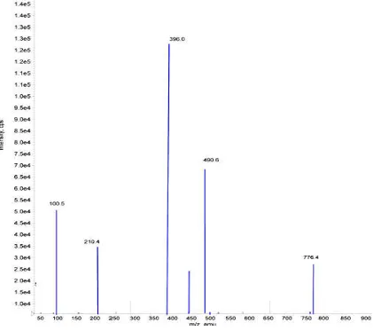
Fig. 2. Mass fragmentation pattren of Alectinib (AT)
Table. 1 - Calibration curve details
Spiked plasma Concentration (pg/ml)
Concentration measured (pg/ml) (Mean±S.D)
%CV (n=5) %Accuracy
5.00 4.99±0.01 1.4 99.9
10.00 10.24±0.02 3.6 101.7
50.00 49.89±0.15 2.7 101.3
100.00 100.24±0.22 2.5 100.1
500.00 501.6±0.27 3.8 100.1
1000.00 1004.22±0.21 2.6 101.7
2000.00 1999.18±1.02 3.1 99.4
4000.00 4001.35±1.10 3.4 101.7
6000.00 6003.76±1.11 1.7 102.6
8000.00 8001.12±1.96 3.8 101.5
10000.00 10000.07±1.23 2.5 100.5
[image:2.595.61.539.662.790.2]The total analysis time was 3 min and flow rate was set to ml/min. The temperature was set to 40°C for the column oven. The sample volume for the injection into mass spectrometry was adjusted to 10 µl for better ionization and chromatography.
Detection
The pure drug of AT and ATD8 were prepared in methanol
(10.00 ng/mL) and injected with a flow rate of 5 µL/min positive ion mode mass spectrometer for optimization of mass parameters like source temperature, IS, heater gas, nebulizer gas, curtain gas, CAD gas (all gas channels were purged with ultra high pure nitrogen gas), EP, DP, CE, FP and CXP were optimized. Analysis was performed using MRM positive ion mode with mass transitions of m/z (amu)
490.6396.0 for AT and ATD8.The mass fragmentation
pattern of parent and product ions mass spectras in Figure 2.
Standard calibration and quality control samples preparation
Stock solutions of AT (1000.00 µg/ml) and
µg/ml) were prepared in methanol. The internal standard (ATD8) spiking solution (500.00 ng/ml) was prepared in 75 methanol from ATD8 stock solution. Stock solutions of AT ATD8 and intermediate spiking solutions were
[image:3.595.90.511.297.670.2]refrigerated conditions (2-8°C) until analysis.
Fig. 3. Mass fragmentation pattren of flow rate was set to 0.6
ml/min. The temperature was set to 40°C for the column oven. The sample volume for the injection into mass spectrometry was adjusted to 10 µl for better ionization and
were prepared in methanol and injected with a flow rate of 5 µL/min into for optimization of mass parameters like source temperature, IS, heater gas, nebulizer ls were purged with ultra high pure nitrogen gas), EP, DP, CE, FP and CXP were optimized. Analysis was performed using MRM positive ion 482.6396.0 and The mass fragmentation arent and product ions mass spectras were depicted
Standard calibration and quality control samples
and ATD8 (1000.00 . The internal standard was prepared in 75% from ATD8 stock solution. Stock solutions of AT, and intermediate spiking solutions were stored in
C) until analysis.
Calibration standards (5.00, 10.00, 50.00, 100.00, 500.00, 1000.00, 2000.00, 4000.00, 6000.00, 8000.00 and 10000.00 pg/ml), quality control samples of
mid QC, high QC (5.00, 15.00, 3000.00, 7000.00 pg/ml) used by spiking the appropriate
the drug free plasma and stored at
Sample extraction
The protein precipitation method was applied to extract and ATD8. To each labelled polypropylene tube 50 µl of ATD8 (500.00 ng/ml) was mixed with the 100 µl plasma sample, then 0.25 ml of acetonitrile were added, vortexed for 5 min and centrifuged at 4000 rpm for 10 min at 20°C. The organic phase was transferred to auto sampler vials containing 100 µl of 0.1% formic acid and in
MS/MS for analysis.
Method validation
The developed method was validated over a linear concentration range of 5.0–10000.0 pg/ml.
The validation parameters include selectivity and specificity, LOQ, Linearity, precision and accuracy, matrix effect, recovery, stability (freeze–thaw, auto sampler, bench top, long term) was evaluated under validation section.
Selectivity and Specificity
Ten lots of blank plasma samples w
six lots free from interference were selected for Mass fragmentation pattren of Alectinib D8 (ATD8)
5.00, 10.00, 50.00, 100.00, 500.00, 1000.00, 2000.00, 4000.00, 6000.00, 8000.00 and 10000.00 ), quality control samples of lower limit QC, low QC, 5.00, 15.00, 3000.00, 7000.00 pg/ml) were used by spiking the appropriate amount of standard solution in the drug free plasma and stored at -30 °C till analysis.
The protein precipitation method was applied to extract AT . To each labelled polypropylene tube 50 µl of /ml) was mixed with the 100 µl plasma sample, then 0.25 ml of acetonitrile were added, vortexed for 5 min and centrifuged at 4000 rpm for 10 min at 20°C. The organic phase was transferred to auto sampler vials containing 100 µl of 0.1% formic acid and injected into the
HPLC-ESI-The developed method was validated over a linear 10000.0 pg/ml.
The validation parameters include selectivity and specificity, LOQ, Linearity, precision and accuracy, matrix effect, thaw, auto sampler, bench top, long term) was evaluated under validation section.
the selectivity and specificity. The endogenous/potential interfering peak areas for blank samples must be less than 20% of the LLOQ peak area of AT retention time and less than 5% for ATD8 retention time.
Limit of Quantification (LOQ)
Six LLOQ standards were prepared in screened plasma lot along with IS (500.00 ng/ml) and signal to noise ratio (S/N) was calculated using analyst software.
Linearity
Calibration standards were prepared to obtain linearity range of 5.00, 10.00, 50.00, 100.00, 500.00, 1000.00, 2000.00, 4000.00, 6000.00, 8000.00 and 10000.00pg/ml and assayed in five replicates on five different days.
Precision & Accuracy
One set of calibration standards and one set contains four different concentrations of quality control standards of Lower limit QC (5.00 pg/ml), Low QC (15.00 pg/ml), Mid QC (3000.00 pg/ml) and High QC (7000.00 pg/ml) concentrations were prepared in screened plasma and analyzed each quality control (QC) standards in six replicates on the same day (Intra day) and five different days (Inter day).
Matrix Effect
Six extracted blank plasma samples in three replicates were spiked with the un-extracted concentration of mid QC (3000.00 pg/ml) and compared with un-extracted standards of the same concentration.
Recovery
The recovery of samples was performed by protein
precipitation method.The extraction recovery was determined
in sextuplicate by comparing the extracted QC standards with un-extracted QC standards at three different concentrations of low (15.00 pg/ml), medium (3000.00 pg/ml), high (7000.00 pg/ml).
Stability studies
Bench top Stability (Room Temperature Stability, 24 h)
Six replicates of spiked low and high concentrations (BT stability samples) were set aside at ambient temperature up to 24 h. Samples were processed and compared with newly prepared low and high concentrations (comparison samples).
Freeze and thaw stability (after 3rd cycle at -30°C)
Six replicates of low and high concentrations (FT stability samples) were frozen at -30°C and subjected to three freeze-thaw cycles of 24, 36 and 48 h (-30°C to room temperature) and compared with newly prepared low and high concentrations (comparison samples).
Autosampler stability (2-8°C, 65 h)
Six replicates of low and high concentrations (AS stability samples) were stored in auto-sampler up to 65 h at 2-8°C.
Stability samples were compared with newly prepared low and high concentrations (comparison samples).
Long-term Stability (-30°C, 45 Days)
After completion of the stability period stored at -30 °C (45 days) six replicates of low and high concentrations (LT stability samples) were compared with newly prepared low and high concentrations (comparison samples).
RESULTS AND DISCUSSION
Method development
On the way to develop a simple and easy applicable method for determination of ATin human plasma, HPLC-MS/MS was selected as the method of choice. During method development process chromatographic (mobile phase composition, column, flow rate, injection volume, sample volume), mass spectrometric, sample extraction and internal standard parameters were optimized in logical and sequential manner to achieve the best results. Separation of the AT was performed with different branded RP-HPLC C18 columns. Initial
separation was performed with isocratic elution of 10mM ammonium formate and acetonitrile was selected as a mobile phase in varying combinations were tried, but a low response was observed. A mobile phase consisting of 0.1% acetic acid: acetonitrile (20:80 v/v) and 0.1%acetic acid: methanol (20:80 v/v) gave the best response, but poor peak shape was observed. After a series of trials a mobile phase consisting of 0.1% formic acid in combination with methanol and acetonitrile in varying combinations were tried.
Using a mobile phase containing 0.1% formic acid in combination with methanol (25:75 v/v), gave the best signal along with a marked improvement in the peak shape and low baseline noise was observed using the Ascentis Express C18 (50
mm × 4.6 mm, 2.7 µm) analytical column with a flow rate of 0.6 ml/min and reduced runtime to 3 min. The column oven temperature was kept at a constant temperature of about 40 °C and temperature of auto sampler was maintained at 4°C. Injection volume of 10 µl sample was adjusted for better ionization and chromatography. For selection of internal standard, Afatinib Dimaleate, Imatinib Mesylate and Lenvatinib Mesylate were tried with optimized mobile phase and column conditions. Finally AlectinibD8 (VLD9) was
selected as internal standard in terms of better chromatography and extractability.
The retention times of analyte (AT) and internal standard (ATD8) were eluted at 1.42 ± 0.2 min and 1.44 ± 0.2 min respectively with 3 min total runtime. Different procedures like PPT (Protein precipitation), SPE (solid phase extraction) and LLE (liquid-liquid extraction) methods were optimized. Out of all, it was observed that the PPT was suitable due to simple extraction, high recovery and the less ion suppression effect on drug and internal standard. Electro spray ionization (ESI) provided a maximum response over atmospheric pressure chemical ionization (APCI) mode, and was chosen for this method. The instrument was optimized to obtain sensitivity and signal stability during infusion of the analyte in the continuous flow of mobile phase to electrospray ion source operated at a flow rate of 20 μl/min. Alectinib gave more response in positive ion mode as compare to the negative ion mode.
To get high intense productions source dependent parameters were optimized like nebulizer gas flow 30 psi, CAD gas and curtain gas flow 25 psi, ion spray voltage 5500 V, and temperature 500°C. The compound dependent parameters such as the declustering potential (DP), focusing potential (FP), entrance potential (EP), collision energy (CE), cell exit potential (CXP) were optimized during tuning as 35, 25, 10, 20, 12 eV for Alectinib and AlectinibD8, respectively. The collision activated dissociation (CAD) gas was set at 4 psi using nitrogen gas. Quadrupole-1 and quadrupole-3 were both maintained at a unit resolution and dwell time was set at 200 ms for Alectinib and AlectinibD8. The predominant peaks in the primary ESI spectra of AT and ATD8 correspond to the MH+ ions at m/z 482.6 and 490.6 respectively.
Productions of AT and ATD8 scanned in quadrupole-3 after a collision with nitrogen in quadrupole-2 had a m/z of 396.0 for both respectively. The parent and productions mass spectrums of AT and ATD8 were shown in Figure 2 & 3.
Method validation
Selectivity and Specificity, Limit of Quantification (LOQ)
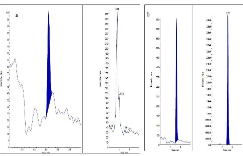
No significant response was observed at retention times of AT and ATD8 in blank plasma as compared to LLOQ and blank with IS samples. The limit of quantification for this method was proven as the lowest concentration of the calibration curve
which was proven as 5.0 ng/ml. Represent chromatograms
[image:5.595.53.538.56.367.2]were shown in Figure 4.
[image:5.595.46.548.442.504.2]Fig.4. Representative chromatograms of Alectinib in plasma a) Blank plasma chromatogram for interference free AT and ATD8 b) Chromatogram of LLOQ sample (AT with ATD8)
Table 2. Precision and accuracy (Analysis with spiked samples at three different concentrations)
Spiked Plasma Concentration (pg/ml)
Within-run (Intra-day) Between-run (Inter-Day)
Concentration measured (n=6;pg/ml;mean±S.D)
%CV %Accuracy Concentration measured
(n=6;pg/ml;mean±S.D)
%CV %Accuracy
15.00 14.8±0.07 5.10 94.78 14.9±0.08 3.2 91.66
3000.00 3002.34±1.23 2.36 96.00 2999.78±2.56 1.6 99.34
7000.00 6999.47±2.45 3.35 94.17 7004.33±3.61 2.4 97.55
Table. 3 - Stability studies of Alectinib in plasma
Spiked Plasma concentration (pg/ml)
Room temperature Stability
Processed sample Stability Long term stability Freeze and thaw stability
24h 65h 45 days Cycle (48h)
Concentration measured (n=6;pg/ml; mean±S.D)
%CV (n=6)
Concentration measured (n=6;pg/ml; mean±S.D)
%CV (n=6)
Concentration measured (n=6;pg/ml; mean±S.D)
%CV (n=6)
Concentration measured (n=6;pg/ml; mean±S.D)
%CV (n=6)
15.00 14.9±0.12 7.8 15.5±2.16 5.3 15.2±1.54 8.8 14.8±0.12 5.8
[image:5.595.40.551.536.625.2]Linearity
Linearity was plotted as a peak area ratio (AT peak area / ATD8 peak area) on the y-axis against AT concentration (pg/ml) on the x-axis. Calibration curves were found to be consistently accurate and precise for AT over a linearity range of 5 to 10000.00 pg/ml. The correlation coefficient was greater than 0.9980 for AT. The %CV was less than 15% and mean %accuracy was ranged between 99.40 -102.67%. Results were presented in Table 1.
Precision & Accuracy
Intra and inter batch %accuracy for AT was ranged between 94.17-96.00 and 91.66 to 99.34. %CV is 2.36 to 5.10 and
1.64% - 3.24%. Results are presented in Table 2.
Recovery
The mean %recovery for LQC, MQC, HQC samples of AT were 99.85%, 95.30% and 93.54% respectively. The overall mean %recovery and %CV of AT across QC levels is 96.23% and 5.95%. For the ATD8 (internal standard) the mean % recovery and %CV is 91.68% and 7.18%.
Matrix Effect
No significant matrix effect found in different sources of rat plasma tested for AT, ATD8. The %CV was found to be 3.71.
Stability (freeze–thaw, auto sampler, bench top, long term)
Quantification of the AT in plasma subjected to three freeze– thaw cycles (−30°C to room temperature), autosampler (processed), room temperature (Benchtop), long-term stability details were shown in Table 3.
Conclusion
The method described in this manuscript has been developed and validated over the concentration range of 5.0–10000.0 pg/ml in human plasma. The intra and inter-batch precision (%CV) was less than 6.0% and %accuracy ranged from 98.9%–102.4%. The overall %recovery for AT, ATD8 was greater than 90%. The selectivity, sensitivity, precision and accuracy obtained with this method make it suitable for the purpose of the present study. In conclusion, the method used in the present study is easy and fast to perform; it is also characterized with an adequate accuracy, precision, selectivity and stability. The simplicity of the method, and using rapid protein precipitation extraction with less run time of 3.0 min per sample, make it an attractive procedure in high-throughput bioanalysis of Alectinib.
Conflict of interest
Authors declare that, there is no conflict of interest.
REFERENCES
Bennet, R. V., Gamage, C. M. & Fernandez, F. M. 2013.
Imaging of biological tissues by desorption electrospray ionization mass spectrometry Journal of Visualized
Experimants 2013; e50575.
Dobbelstein, M. and Moll, U. 2014. Targeting tumour-supportive cellular machineries in anticancer drug development. Nat Rev Drug Discov ; 13:179–196.
Gode, D. & Volmer, D. A. 2013. Lipid imaging by mass spectrometry - a review. Analyst; 138:1289–1315.
Guidance for industry, March 2003. Bio availability and fed bio equivalence studies for orally administered drug products-general considerations: U.S. Department of Health and Human Services food and drug administration centre for drug evaluation and research (CDER).
Guidance for industry. December 2002. Food-effect bio availability and fed bio equivalence studies: U.S. Department of Health and Human Services food and drug administration centre for drug evaluation and research (CDER).
Guidance for industry. May 2001. Bioanalytical method validation, U.S. Department of Health and Human Services, food and drug administration, center for drug evaluation and research (CDER), Center for biologics evaluation and research (CBER).
Hiroaki Aikawa, Mitsuhiro Hayashi, Shoraku Ryu, Makiko Yamashita, Naoto Ohtsuka, Masanobu Nishidate, Yasuhiro Fujiwara and Akinobu Hamada. 2016. Visualizing spatial distribution of alectinib in murine brain using quantitative mass spectrometry imaging. Scientific reports, 6:23749.
Kim, J. H. et al. 2008. Label-free calcium imaging in ischemic retinal tissue by TOF-SIMS. Biophys Journal, 94:4095–4102.
Prideaux, B. & Stoeckli, M. 2012. Mass spectrometry imaging for drug distribution studies. Journal of Proteomics; 75:4999–5013.
Seeley, E. H., Schwamborn, K. & Caprioli, R. M. 2011. Imaging of intact tissue sections: moving beyond the microscope. J Biol Chem., 286:25459–25466.
Sugiura, Y. & Setou, M. 2010. Imaging mass spectrometry for visualization of drug and endogenous metabolite distribution: towardin-situ pharmacometabolomes.
Journal of Neuroimmune Pharmacol 2010; 5:31–43.