Mutations in multiple components of the
nuclear pore complex cause nephrotic
syndrome
Daniela A. Braun, … , Mustafa K. Khokha, Friedhelm
Hildebrandt
J Clin Invest.
2018;
128(10)
:4313-4328.
https://doi.org/10.1172/JCI98688
.
Steroid-resistant nephrotic syndrome (SRNS) almost invariably progresses to end-stage
renal disease. Although more than 50 monogenic causes of SRNS have been described, a
large proportion of SRNS remains unexplained. Recently, it was discovered that mutations
of
NUP93
and
NUP205
, encoding 2 proteins of the inner ring subunit of the nuclear pore
complex (NPC), cause SRNS. Here, we describe mutations in genes encoding 4
components of the outer rings of the NPC, namely
NUP107
,
NUP85
,
NUP133
, and
NUP160
, in 13 families with SRNS. Using coimmunoprecipitation experiments, we showed
that certain pathogenic alleles weakened the interaction between neighboring NPC
subunits. We demonstrated that morpholino knockdown of
nup107
,
nup85
, or
nup133
in
Xenopus
disrupted glomerulogenesis. Re-expression of WT mRNA, but not of mRNA
reflecting mutations from SRNS patients, mitigated this phenotype. We furthermore found
that CRISPR/Cas9 knockout of
NUP107
,
NUP85
, or
NUP133
in podocytes activated
Cdc42, an important effector of SRNS pathogenesis. CRISPR/Cas9 knockout of
nup107
or
nup85
in zebrafish caused developmental anomalies and early lethality. In contrast, an
in-frame mutation of
nup107
did not affect survival, thus mimicking the allelic effects seen in
humans. In conclusion, we discovered here that mutations in 4 genes encoding components
of the outer ring subunits of the NPC cause SRNS and thereby provide further evidence that
specific hypomorphic mutations in these essential genes cause a distinct, […]
Research Article
Genetics
Nephrology
Introduction
Steroid-resistant nephrotic syndrome (SRNS), a disease of the renal glomerular filter, is characterized by proteinuria, edema, and hypoalbuminemia. In contrast to other forms of nephrotic syndrome, SRNS does not respond to drug treatment and
Steroid-resistant nephrotic syndrome (SRNS) almost invariably progresses to end-stage renal disease. Although more than 50 monogenic causes of SRNS have been described, a large proportion of SRNS remains unexplained. Recently, it was
discovered that mutations of NUP93 and NUP205, encoding 2 proteins of the inner ring subunit of the nuclear pore complex
(NPC), cause SRNS. Here, we describe mutations in genes encoding 4 components of the outer rings of the NPC, namely NUP107, NUP85, NUP133, and NUP160, in 13 families with SRNS. Using coimmunoprecipitation experiments, we showed that certain pathogenic alleles weakened the interaction between neighboring NPC subunits. We demonstrated that morpholino
knockdown of nup107, nup85, or nup133 in Xenopus disrupted glomerulogenesis. Re-expression of WT mRNA, but not
of mRNA reflecting mutations from SRNS patients, mitigated this phenotype. We furthermore found that CRISPR/Cas9
knockout of NUP107, NUP85, or NUP133 in podocytes activated Cdc42, an important effector of SRNS pathogenesis. CRISPR/
Cas9 knockout of nup107 or nup85 in zebrafish caused developmental anomalies and early lethality. In contrast, an in-frame
mutation of nup107 did not affect survival, thus mimicking the allelic effects seen in humans. In conclusion, we discovered
here that mutations in 4 genes encoding components of the outer ring subunits of the NPC cause SRNS and thereby provide further evidence that specific hypomorphic mutations in these essential genes cause a distinct, organ-specific phenotype.
Mutations in multiple components of the nuclear pore
complex cause nephrotic syndrome
Daniela A. Braun,1,2 Svjetlana Lovric,1 David Schapiro,1 Ronen Schneider,1 Jonathan Marquez,3 Maria Asif,4,5,6
Muhammad Sajid Hussain,4,5,7 Ankana Daga,1 Eugen Widmeier,1 Jia Rao8,9 Shazia Ashraf,1 Weizhen Tan,1 C. Patrick Lusk,10
Amy Kolb,1 Tilman Jobst-Schwan,1 Johanna Magdalena Schmidt,1 Charlotte A. Hoogstraten,1 Kaitlyn Eddy,1 Thomas M. Kitzler,1
Shirlee Shril,1 Abubakar Moawia,4,5,6 Kathrin Schrage,5 Arwa Ishaq A. Khayyat,5,11 Jennifer A. Lawson,1 Heon Yung Gee,1
Jillian K. Warejko,1 Tobias Hermle,1 Amar J. Majmundar,1 Hannah Hugo,1 Birgit Budde,4 Susanne Motameny,4 Janine Altmüller,4,7,12
Angelika Anna Noegel,5,7,13 Hanan M. Fathy,14 Daniel P. Gale,15 Syeda Seema Waseem,4,5,6 Ayaz Khan,6 Larissa Kerecuk,16
Seema Hashmi,17 Nilufar Mohebbi,18 Robert Ettenger,19 Erkin Serdaroğlu,20 Khalid A. Alhasan,21 Mais Hashem,22,23,24
Sara Goncalves,25,26 Gema Ariceta,27 Mercedes Ubetagoyena,28 Wolfram Antonin,29 Shahid Mahmood Baig,6 Fowzan S. Alkuraya,22,23,24
Qian Shen,8,9 Hong Xu,8,9 Corinne Antignac,25,26,30 Richard P. Lifton,31,32 Shrikant Mane,31 Peter Nürnberg,4,7,13 Mustafa K. Khokha,3
and Friedhelm Hildebrandt1
1Department of Medicine, Boston Children’s Hospital, Harvard Medical School, Boston, Massachusetts, USA. 2Department of Internal Medicine D, University Hospital of Münster, Münster, Germany. 3Pediatric
Genomics Discovery Program, Department of Pediatrics and Genetics, Yale University School of Medicine, New Haven, Connecticut, USA. 4Cologne Center for Genomics, University of Cologne, Cologne, Germany.
5Institute of Biochemistry I, Medical Faculty, University of Cologne, Cologne, Germany. 6Human Molecular Genetics Laboratory, Health Biotechnology Division, National Institute for Biotechnology and Genetic
Engineering, Pakistan Institute of Engineering and Applied Sciences, Faisalabad, Pakistan. 7Center for Molecular Medicine Cologne, University of Cologne, Cologne, Germany. 8Department of Nephrology, Children’s
Hospital of Fudan University, Shanghai, China. 9Shanghai Kidney Development and Pediatric Kidney Disease Research Center, Shanghai, China. 10Department of Cell Biology, Yale University School of Medicine,
New Haven, Connecticut, USA. 11Biochemistry Department, King Saud University, Riyadh, Saudi Arabia. 12Institute of Human Genetics, University of Cologne, Cologne, Germany. 13Cologne Excellence Cluster on
Cellular Stress Responses in Aging-Associated Diseases (CECAD), University of Cologne, Cologne, Germany. 14Pediatric Nephrology Unit, Alexandria Faculty of Medicine, University of Alexandria, Alexandria, Egypt.
15Centre for Nephrology, University College London, Royal Free Hospital, London, United Kingdom. 16Birmingham Children’s Hospital NHS Foundation Trust, Birmingham, United Kingdom. 17Department of Pediatric
Nephrology, Sindh Institute of Urology and Transplantation, Karachi, Pakistan. 18Division of Nephrology, University Hospital Zurich, Zurich, Switzerland. 19Department of Pediatrics, University of California, Los
Angeles, California. 20Department of Pediatric Nephrology, Dr. Behçet Uz Children’s Hospital, Izmir, Turkey. 21Pediatric Department, College of Medicine, King Saud University and King Khalid University Hospital,
Riyadh, Saudi Arabia. 22Department of Genetics, King Faisal Specialist Hospital and Research Center, Riyadh, Saudi Arabia. 23Department of Anatomy and Cell Biology, College of Medicine, Alfaisal University,
Riyadh, Saudi Arabia. 24Saudi Human Genome Program, King Abdulaziz City for Science and Technology, Riyadh, Saudi Arabia. 25Laboratory of Hereditary Kidney Diseases, INSERM UMR1163, Imagine Institute,
Paris, France. 26Université Paris Descartes–Sorbonne Paris Cité, Imagine Institute, Paris, France. 27Universitat Autonoma de Barcelona, Hospital Universitari Vall d’Hebron, Pediatric Nephrology, Barcelona, Spain.
28Hospital Universitario Donostia, Pediatric Nephrology, Donostia–San Sebastian, Spain. 29Institute of Biochemistry and Molecular Cell Biology, Medical School, RWTH Aachen University, 52074 Aachen, Germany.
30Department of Genetics, Necker Hospital, Assistance Publique–Hôpitaux de Paris, Paris, France. 31Department of Genetics, Yale University School of Medicine, New Haven, Connecticut, USA. 32Laboratory of
Human Genetics and Genomics, The Rockefeller University, New York, New York, USA.
Conflict of interest: FH is a cofounder of Goldfinch Bio Inc. and receives royalties from Claritas Genomics.
Submitted: November 16, 2017; Accepted: July 24, 2018.
The Journal of Clinical Investigation
R E S E A R C H A R T I C L E
NUP85, NUP133, and NUP160, in 29 individuals of 13 families with SRNS. Furthermore, we detected a homozygous mutation in
NUP37 in a family with primary microcephaly, a phenotype that was also present in some families with NUP107 mutations. We use different in vivo and in vitro models to elucidate parts of the related pathogenesis.
Results
Hypomorphic mutations in 4 different genes encoding components of theouter ring subunitsof theNPC cause nephrotic syndrome. To identify additional genes that cause SRNS if mutated, we per-formed whole exome sequencing (25) in 160 families with SRNS. After excluding mutations in known SRNS genes, we used homo-zygosity mapping (26) in consanguineous families to identify potentially novel candidate loci for SRNS. This approach yielded a homozygous missense mutation (c.303G>A, p.Met101Ile) in the gene NUP107 (NM_020401.3) (Table 1 and Figure 1, B–D) in consanguineous family A4649 with 3 affected siblings with SRNS and microcephaly. In 4 additional consanguineous families with a similar phenotype (B1426, A802, FA, PN-1), the NUP107 locus was positioned within a homozygous peak region, and we identi-fied the same mutation in NUP107 (Figure 1, B–D, Supplemental Table 1, Supplemental Figure 1A, and Supplemental Figures 2 and 3; supplemental material available online with this article; https:// doi.org/10.1172/JCI98688DS1). The p.Met101Ile mutation rep-resents a South Asian founder allele that was reported previ-ously in patients with the combined phenotype of SRNS and pri-mary microcephaly (23, 27). Prior studies demonstrated that the p.Met101Ile mutation of NUP107 results in aberrant splicing and causes a reduction in the NUP107 protein level (23). NUP107 is an essential protein for NPC assembly (28, 29). In consanguineous family F797 with nonsyndromic SRNS, we found a homozygous missense mutation (c.2922T>G, p.Ser974Arg) in the gene NUP133
(NM_018230.2), which was located within a homozygous peak region on chromosome 1 (Figure 1, H–J).
Because NUP133 and NUP107 encode 2 interacting compo-nents of the outer ring subunits of the NPC, we hypothesized that alterations in other nuclear pore proteins may also cause monogenic SRNS. We therefore performed targeted sequencing of all exons of 19 genes that encode other NUPs in a worldwide cohort of 2,164 families with SRNS using a multiplex PCR–based high-throughput sequencing strategy (7, 30, 31) or using whole exome sequencing. As a result, we detected a homozygous mis-sense mutation of NUP107 (c.2666A>G, p.Tyr889Cys) in family A3825 and 2 compound heterozygous alleles (c.1021dup, p.Glu341Glyfs*3, and c.2129_2131delAAG, p.Glu710del) of
NUP107 in family A1830. None of these alleles were reported previously (Table 1 and Figure 1, A–D). All 5 families who carried the p.Met101Ile allele and family A1830 displayed the combined phenotype of SRNS with microcephaly and intellectual disabil-ity. In contrast, family A3825 and 9 previously reported families with NUP107 alleles other than p.Met101Ile did not have a neu-rodevelopmental phenotype (22, 24). We speculate that this allelism may be explained by the fact that only the homozygous p.Met101Ile allele resulted in a protein-truncating mutation (23), while all other families carried at least 1 recessive missense allele. This observation suggests that, as seen in other monogenic dis-inevitably progresses to end-stage renal disease (ESRD), thus
requiring dialysis or renal transplantation for survival (1). It constitutes the second most frequent cause of ESRD in the first 3 decades of life (2). In SRNS, renal histology reveals focal segmental glomerulosclerosis or diffuse mesangial sclerosis, which indicate irreversible damage to the glomerulus. Mutations in over 50 genes have been discovered to cause monogenic SRNS (3, 4). These genetic findings have revealed podocytes, specialized epithelial cells of the glomerular filter, as the critical site of injury in SRNS (5, 6). Furthermore, identification of disease genes has implicated multiple signaling pathways in the molecular pathogenesis of SRNS (3). Our group and others have recently demonstrated that in about 30% of patients with SRNS a causative single-gene mutation can be identified (7–9).
The nuclear pore complex (NPC) is a large (100 MDa) macro-molecular assembly that spans the nuclear envelope and forms a selective barrier between the cytoplasm and the nucleoplasm (10). The central channel of the NPC is filled with intrinsically disor-dered proteins, which are rich in phenylalanine-glycine (FG) amino acid residues. These FG-rich nucleoporins (NUPs) establish a size-selective diffusion barrier to macromolecules greater than approximately 40 kDa while providing binding sites for nuclear transport receptors (karyopherins, importins, and exportins) that ferry signal-bearing cargo across the NPC (10). The central chan-nel is supported by a structural scaffold built from the inner and outer ring complexes, which are formed by repeating modular units of the NUP93 subcomplex and the NUP107-160 complex, or “Y complex,” respectively (11, 12). The Y complex is the major subunit of the outer rings of the NPC. The atomic structures of the major domains of all 7 members of the Y complex (either alone or in complex), including NUPs 43, 85, 96, 107, 133, and 160, have been solved and modeled into electron cryomicroscopic maps of the human and Xenopus NPC (10).
Interestingly, while NUPs are best understood in their roles at the NPC, they also participate in additional functions in other subcellular locations (13–15). Studies in vertebrate model organisms additionally suggest that NUPs may have important regulatory functions during development and for tissue-specific progenitor cell maturation (16, 17). Genetic depletion of NUP transcripts caused severe developmental defects in zebra-fish (18), mice (16), and frogs (15). Interestingly, some NUP- encoding genes show differential expression in different tissues and in different developmental stages (15, 19). NUPs studied in this article (i.e., nup107, nup85, nup133, and nup160) were shown to be expressed in both the rostral portion of the embryo (relevant to CNS development) and the intermediate mesoderm (important for pronephric development) (20).
Recently, we identified mutations in NUP93 and NUP205, encoding 2 components of the inner ring subunit of the NPC, in patients with nonsyndromic SRNS (21). Intriguingly, patients with mutations in NUP107, encoding a component of the Y complex, also developed SRNS (22–24). These observations suggest that a specific pathogenic link between SRNS development and alterations in different NUPs exists.
Table 1. Phenot ypes in patients with SRNS and mutations in NUP10 7 , NUP85 , NUP133 , and NUP160 and in 1 f amily with a mutation in NUP3 7 and microc ephaly Family -individual Nucleo tide change
Amino acid change
Ex on ( zygosit y, se gr eg ation)
PPH2 score
SIFT MT Amino acid conser vation t o species gnomAD Sex
Ethnic origin
Par ent al consanguinit y Age o f
onset (yr)
Extr ar enal manif es tations Biopsy Renal pheno type and ther
apy (ESRD at
yr) NUP10 7 A464 9 –2 1 –2 2 –2 3 c.303G>A p.Met101Ile
4 (Hom, segreg
ates ) 0. 02 Del DC D. r erio 0/ 1/2 45,5 44
M F F
Pakist
ani
Y
6.5 12 14
MC, ID , S S MC, ID , S S MC, ID , S S
ND FSGS FSGS
Pr
oteinuria
SRNS
, ESRD (
13) Pr oteinuria A18 30_21 c.10 21dupG c.2129_213 1delA AG p. Glu34 1Gly fs*3 p. Glu710del
12 (het, m) 24 (het, p
) -- X. tr opic alis NP NP F Eur ope an N 4 MC, ID , DD , S S,FD , micr osc
opic hematuria, DCM, HTN
Glomeruloscler osis ESRD ( 5) A3825-21 c.2666A>G p.T yr88 9C ys
27 (Hom, segreg
ates ) 1 Del DC D. melanogas ter NP M Turkish Y 4. 5
Cleft lip, cleft palate
DMS
SRNS
, ESRD (
5) NUP8 5 A5 195-22 A c.1430C>T p. Ala477V al 15 (Hom) 0. 81 1 To l DC C. in tes tinalis NP F Eg yptian Y 8 SS , micr osc opic hematuria ND SRNS
, ESRD (
10 ) A325 9-21 c.1933C>T p. Ar g645T rp
19 (Hom, segreg
ates ) 0. 425 Del DC X. tr opic alis B 0/ 10 /2 77 ,22 4 M Ar abic N 11 Micr osc opic hematuria FSGS SRNS
, ESRD (
12)
NCR NCR322
7
NCR33
10
c.405+1G>A c.174
1G>C Donor splic e site p. Ala58 1Pr o
5 (het, p
)
17 (het, m)
0.99 Tol DC D. r erio NP NP F M
Eur
ope
an
N
7 4 ID ID , micr osc opic hematuria, shor t st ature , par tial GH deficienc y FSGS FSGS SRNS
, ESRD (
7)
SRNS
NUP133 A21
74 –2 1 –2 2 c.6 91C>G c.3 164T>C p. Ar g23 1Gly p.L eu1055Ser
6 (het, p
)
23 (het, m)
0.89 7 0.94 9 Tol Del DC DC D. r erio D. melanogas ter NP 0/ 17 /268 ,030
M F
Turkish N 9 10 None ND FSGS SRNS
, ESRD (
13) ESRD ( 20 ) F7 97 -21 c.2922T>G p.Ser9 74Ar g 21 (Hom) 0. 876 Del DC D. r erio NP F Eur ope an Y 3.9 None FSGS SRNS
, ESRD (
6)
NUP160 17F004
94 –2 1 –2 2 c.2 40 7G>A c.2 728C>T p. Glu803L ys p. Ar g910*
19 (het, p
)
22 (het, m)
0.05 Del NA D. r erio C 0/ 4/2 46,0 20 0/2/2 45, 806
M F
Asian
N
16 7 None None FSGS ND SRNS Pr oteinuria NUP3 7 PN-2 V-1 V-2 V-3 c.916C>T p. Ar g306*
10 (Hom, segreg
ates ) NA NA NA NA NP
M M M
Pakist ani Y MC, ID , CD MC, ID , CD MC, ID , CD
ND ND ND ND ND ND
The f
ollowing NCBI GenBank annotations wer
e used: NUP85 (NM_0 24844 .4); NUP10 7 (NM_0 20401.3); NUP133 (NM_0182 30.2); NUP160 (NM_0152 31. 1); NUP3 7 (NM_0 2405 7.3).
AOlder sister had similar clinical
phenot
ype, but w
as dec
eased so that no DNA sample c
ould be obtained.
BChemically c
onser
ved (
Arg or L
ys ) t o Dr osophila melanogast er .
CChemically c
onser
ved (
Glu or Gln) t
o Ciona int estinalis . CD , clinodact yly; C. int estinalis
, Ciona int
estinalis
; DC, dise
ase causing; DCM, dilated cardiomyopathy; DD
, de
velopmental delay; Del, deleterious
; D . melanogast er , Dr osophila melanogast er ; D . r erio
, Danio r
erio
; DMS, diffuse
mesangial sclerosis
; ESRD
, end-stage r
enal dise
ase; F
, f
emale; FD
, f
acial dysmorphism; FSGS, f
ocal segmental glomerulosclerosis
; GH, grow
th hormone; gnomAD
, Genome A
ggr
egation Database (http://
gnomad.broadinstitute.
org
); het, heterozygous
; Hom, homozygous
; HTN, ar
terial hyper
tension; ID
, intellectual disabilit
y; M, male; MC, microc
ephaly; MT
, Mutation T
aster (pr
ediction soft
w
ar
e); N, no; NA
, not
applicable; ND
, no data; NP
, not pr
esent; PPH2,
PolyPhen-2 (pr ediction soft w ar e); SIFT , Sor ting Int oler
ant From T
oler
ant (pr
ediction soft
w
ar
e); SRNS, steroid-r
esistant nephrotic syndrome; SS, shor
t statur e; TOL , t oler ant; V SD
, ventricular septum def
The Journal of Clinical Investigation
nese family, we discovered 2 compound heterozygous mutations (c.2407G>A, p.Glu803Lys, and c.2728C>T, p.Arg910*) in the gene NUP160 (NM_015231.1; Table 1 and Supplemental Figure 4). NUP160 encodes a direct interaction partner of NUP85 within the Y complex (Figure 2A). The older sibling presented at age 16 years with nephrotic syndrome that was resistant to therapy with steroids or other immunosuppressive drugs. His biopsy showed focal segmental glomerulosclerosis (FSGS), and his renal function was impaired (chronic kidney disease stage 3). The younger sister had proteinuria that first presented at age 7 years. Neither of the siblings displayed extrarenal symptoms (Table 1).
Twenty-five individuals of 13 families with recessive mutations of NUP107, NUP85, NUP133, or NUP160 had SRNS or proteinuria that manifested in childhood or adolescence (Table 1 and Supplemental Table 1). In 13 of these 25 patients, the disease progressed to ESRD before age 25 years. A kidney biopsy was performed in 15 patients. In all cases, light microscopy showed sclerosis of renal glomeruli, manifesting as FSGS or diffuse mesangial sclerosis (Table 1, Supplemental Table 1, Figure 1, and Supplemental Figure 5). Electron microscopy furthermore revealed partial podocyte foot process effacement, one of the hallmark symptoms of nephrotic syndrome (Figure 1 and Supplemental Figure 5). We termed these previously unrecognized monogenic causes of SRNS as NPHS17 (NUP85), NPHS18 (NUP133), and NPHS19 (NUP160).
In a consanguineous Pakistani family with 3 affected chil-dren with congenital microcephaly, we discovered a homozygous nonsense mutation (c.916C>T, p.Arg306*) in the gene NUP37
(NM_024057.3) (Table 1 and Supplemental Figure 6). NUP37
encodes an essential component of the Y complex of the NPC. Segregation analysis was performed when possible, and was com-patible with a recessive mode of inheritance (Supplemental Fig-ure 6). All 3 patients showed congenital microcephaly (–5 to –8 standard deviations [SD]), mild intellectual disability, cerebellar vermis hypoplasia, and clinodactyly of the fifth finger (Table 1). Proteinuria was not present in these patients, and renal ultrasound examination did not reveal any abnormalities (Table 1).
Mutations from SRNS patients impair intermolecular interactions between NPC components. NUP107, NUP85, NUP133, and NUP160 are components of the Y complex, whose molecular structure has recently been modeled into the outer ring scaffold of the NPC (11) (Figure 2A). To investigate the pathogenicity of alleles that we discovered in SRNS patients, we gleaned from available structural data (Protein Data Bank, pdb: 5A9Q) (11) that 1 mutation of
NUP133, p.Ser974Arg, and 1 mutation of NUP107, p.Tyr889Cys, were located in the interacting α-helix between the 2 proteins (Figure 2A). NUP85 mutations were positioned within a region for which no molecular structure of the human Y complex is available yet (Figure 2A).
We used cDNA constructs reflecting the mutations identified in patients with SRNS (Table 1) and performed half-endogenous coimmunoprecipitation (coIP) experiments in HEK293T cells. These experiments demonstrated that, as predicted based on the structural data, the alleles p.Tyr889Cys of NUP107 and p.Ser974Arg of NUP133 weakened the interaction between NUP107 and its direct binding partner NUP133 (Figure 2, B and C). Other missense alleles of NUP107 or NUP133 did not eases (32), severe mutations in NUP genes cause syndromic
phenotypes, while milder mutations give rise to distinct organ-specific phenotypes. Besides microcephaly, 2 families (B1426 and A3825) showed additional skeletal/facial phenotypes (in particular arachnodactyly, high arched palate, and cleft palate/ lip), and 1 family (A802) had congenital heart disease (Table 1 and Supplemental Table 1). The targeted screening additionally identified 2 segregating, compound heterozygous mutations of
NUP133 (c.691C>G, p.Arg231Gly, and c.3164T>C, p.Leu1055Ser, NM_018230.2) in family A2174 with nonsyndromic SRNS (Figure 1, I and K; Supplemental Figure 1C; and Table 1).
High-throughput exon sequencing furthermore yielded 3 mutant alleles of NUP85 (NM_024844.4), encoding a different com-ponent of the outer rings of the NPC, in 4 individuals of 3 unrelated families with SRNS (families A5195, A3259, and NCR3227/3310) (Figure 1, E–G; Supplemental Figure 1B; and Table 1). Two mutations were homozygous missense mutations (c.1430C>T, p.Ala477Val, and c.1933C>T, p.Arg645Trp). One family (NCR3227/3310) carried 2 compound heterozygous alleles (c.405+1G>A and c.1741G>C, p.Ala581Pro), which segregated from the maternal and the paternal side, respectively (Table 1). All families with NUP85 mutations had SRNS and microscopic hematuria, a finding that can be associated with SRNS. Family NCR3227/3310 additionally displayed intellec-tual disability, but showed no structural brain defects. Interestingly, in several families with NUP107 or NUP85 mutations, short stat-ure was noted as a clinical featstat-ure, and family NCR3227/3310 had partial growth hormone deficiency.
The Journal of Clinical Investigation
[image:7.585.42.521.46.694.2]R E S E A R C H A R T I C L E
interfere with the interaction between the 2 proteins. We fur-thermore show that the missense mutations p.Ala581Pro and p.Arg645Trp of NUP85 weakened the interaction with its binding partner NUP160 (Figure 2D). The obligatory splice site mutation c.405+1G>A of NUP85 is predicted to cause skipping of exon 5, thus resulting in a frameshift with premature truncating of the encoded protein. We modeled this mutation in a cDNA construct and found that it completely abrogated the interaction between NUP85 and NUP160 (Figure 2D). Confirmatory experiments for all coIPs using differently tagged fusion proteins are shown in Supplemental Figure 7. Next, we aimed to investigate the impact of an isolated depletion of SRNS-relevant NUPs on other compo-nents of the NPC. To address this question, we generated immor-talized human podocyte cell lines with doxycycline-inducible CRISPR/Cas9 mediated knockout of NUP107, NUP85, or NUP133
(Supplemental Figures 8–10 demonstrate successful depletion of targeted proteins). We used an inducible system because sustained knockout of any of the 3 investigated NUP genes had strong effects on cell viability, which we feared would mask any specific effects resulting from the knockout. Using the induc-ible CRISPR/Cas9 system, we could instead initiate depletion of the specific gene of interest shortly before the experiment and thereby study its direct effect. In this setting, we did not observe significant cell death. Interestingly, we found that short-term depletion of NUP85 or NUP133 had very little effect on the protein levels of other components of the NPC (Supplemental Figure 11).
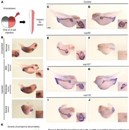
Morpholino knockdown of Xenopus nup107, nup85, or nup133 causes defects in glomerulogenesis. At the 2-cell stage, Xenopus
embryos were injected with a morpholino oligonucleotide targeting
nup85, nup107,or nup133. In Xenopus, cell divisions are holoblastic (complete); therefore, we can do lateralized injections of 1 cell at the 2-cell stage such that only half of the animal is affected during development. In this setting, the uninjected side can act as an inter-nal control. In contrast, the initial cleavages of zebrafish embryos are mesoblastic (incomplete); as a consequence, injected mRNAs cannot be localized to just 1 cell. For this reason, Xenopus is an ideal system in which to test rescue especially of lateralized structures such as the kidney (Figure 3A). At stage 35–37, we detected the pronephros using atp1a1 as a marker in whole-mount in situ hybridization experiments. At this stage, the proximal portion of the pronephros develops a convoluted shape that subsequently straightens out as it extends to the posterior portion of the embryo.
We categorized defects in pronephros morphology based on their severity (Figure 3B). Compared with controls (Figure 3, C and D), morpholino knockdown resulted in abnormal pronephros morphology in 65% of nup85 morphants (Figure 3, E vs. F, and K), 38% of nup107 morphants (Figure 3, G vs. H, and K), and 86% of
nup133 morphants (Figure 3, I vs. J, and K). Morpholino knockdown of pax8, which encodes an essential transcription factor in renal development, was used as a positive control to validate this approach (Supplemental Figure 12A). We used the gene NUP155
as a negative control, because the phenotype caused by NUP155
mutations, monogenic atrial fibrillation (33), does not involve the kidney. As expected, knockdown of nup155 did not result in a renal phenotype in the Xenopus model (Supplemental Figure 12B). These findings suggest that the nucleoporins Nup85, Nup107, and Nup133 are required for renal development in Xenopus.
In order to test the specificity of the knockdown experiment and to assess the pathogenicity of human mutations, we used rescue experiments (Supplemental Figure 13). We first injected
nup85, nup107, or nup133 morpholinos at the 1-cell stage to deplete these proteins throughout the embryo. At the 2-cell stage, we then injected 1 cell of these morphants with WT or mutant human mRNA of NUP85, NUP107, or NUP133, respectively (Supplemental Figure 13A). While WT mRNA rescued the abnormal kidney morphology seen in morphant kidneys, mutant mRNA reflecting the human SRNS mutations resulted in an impaired restoration of renal morphology (Supplemental Figure 13, B–D). Only the p.Ala477Val variant of NUP85 did not show a significantly reduced rescue efficiency. Based on this result, we would predict that this allele may have some residual function, and that it may represent a relatively mild allele as compared with the others. Interestingly, the phenotype in those patients was not notably different.
CRISPR/Cas9 knockout of NUP107, NUP85, or NUP133 increases the level of active Cdc42. Dysregulation of the Rho-like small GTPases RhoA, Rac1, and Cdc42, resulting in impaired actin dynamics, plays an important role in the pathogenesis of monogenic SRNS (34–39). Interestingly, we observed that CRISPR/Cas9–mediated knockout of NUP107, NUP85, or NUP133 increased the formation of filopodia in immortalized human podocytes (Figure 4, A and B), suggesting that loss of function of these genes alters the podocyte’s cytoskeleton. Because Cdc42 is known to induce filopodia formation, if active (40), we performed the colorimetric Cdc42 G-LISA Activation Assay to determine the cellular level of active Cdc42. We found that in comparison with control cells (Cas9 expression but no guide RNA [gRNA]), CRISPR/Cas9 knockout of any of the 3 genes significantly increased Cdc42 activity (Figure 4C). However, when using the IncuCyte system to assess the migration rate of podocytes, we did not find a significant difference between control and knockout cells (Supplemental Figure 14).
Truncating mutations of nup107 or nup85 in zebrafish induce early lethality, while an in-frame mutation of nup107 does not affect survival. We designed gRNAs targeting exon 2 of nup107, the zebrafish ortholog (NM_001030167.1). Using CRISPR/Cas9 technology, we then generated 2 different stable zebrafish lines, one with a protein-truncating “null” allele (c.50_56del7, p.Thr81Argfs*74) and one with an in-frame mutation, most likely acting as a hypomorphic allele (c.137_139del3, p.Ala46delAla). After het × het in-crossing, we monitored survival twice daily and generated Kaplan-Meier survival curves. Zebrafish with a homozygous truncating mutation of nup107 showed early lethality at 5 days postfertilization (dpf) (Figure 5A) and demonstrated severe developmental malformations, including small eyes, ventral body axis curvature, periorbital edema, and total body edema (Figure 5, C–G, and Table 2). These phenotypes developed at 4 dpf before death on 5 dpf. Zebrafish were born at Mendelian ratios. None of the 29 larvae homozygous for the null allele p.Thr81Argfs*74 were phenotypically normal. Conversely, none of the 42 heterozygous larvae and none of the 34 WT larvae developed any overt phenotype (Table 2). In contrast to the truncating allele, the hypomorphic mutation of
nup107 did not affect survival of zebrafish larvae (Figure 5B) and did not result in any overt phenotype (data not shown).
The Journal of Clinical Investigation
[image:9.585.59.507.54.505.2]R E S E A R C H A R T I C L E
To test for renal anomalies and for a kidney-specific pheno-type, we used transmission electron microscopy (TEM) to inves-tigate the ultrastructure of the renal glomerulus and used H&E staining to study renal histology. For the 2 null alleles of nup107
and nup85, we investigated 2 homozygous versus 2 heterozygous fish by TEM and 8 homozygous versus 8 heterozygous fish by renal histology for each gene. We detected significant tissue decay within the glomerular region, but also globally throughout the zebra fish line carrying the truncating mutation p.Arg107Cysfs*15
[image:10.585.47.542.59.469.2]of nup85, the zebrafish ortholog of human NUP85. As observed with nup107 knockout, homozygous nup85-knockout fish showed early lethality and died at day 8 dpf (Supplemental Figure 15A). The phenotypic features of nup85-knockout fish resembled those of nup107-knockout fish, including small eyes, body axis curvature, and edema (Supplemental Figure 15, B–E, and Supplemental Table 2).
Figure 4. CRISPR/Cas9–mediated knockout of NUP107, NUP85, or NUP133 induces filopodia formation and increases active Cdc42 in human podocytes.
The Journal of Clinical Investigation
R E S E A R C H A R T I C L E
(LCL) from a patient with the p.Met101Ile mutation of NUP107
(Supplemental Figure 16, A–C). These findings suggest that, as previously described (28, 29), proteins of the Y complex depend on each other for protein stability. Interestingly, in both cell lines residual amounts of the mutant protein were present.
When investigating the consequences of reduced NUP37 amounts for NPC density, we found a reduced intensity of the mAb414 antibody signal, suggesting a lower number of nuclear pores in mutant fibroblasts as compared with control cells (Figure 6, E and F). Using antibodies detecting heterochromatin (anti-HP1β; Figure 6G) and nucleoli (anti-fibrillarin; Figure 6, H and I), we demonstrated an alteration of chromatin organization and nucleo-lar morphology in NUP37 mutant fibroblasts. TEM confirmed this observation (Figure 6, J–M, arrowheads). Additionally, we found larva. We are therefore hesitant to interpret these findings as a
specific podocyte phenotype, but rather assume that the loss of an essential protein induces injury in all cell types.
Primary patient cell lines carrying protein-truncating mutations of NUP37 and NUP107 display changes in NPC composition and nuclear morphology. We obtained primary cell lines from 2 patients with microcephaly and with mutations of NUP37 and NUP107, respectively. In dermal fibroblasts from a patient carrying the p.Arg306* allele of NUP37, we demonstrated that at the mRNA (Supplemental Figure 6E) and protein level (Figure 6, A and D) NUP37 was reduced in comparison with control cells. Reduced NUP37 protein levels resulted in codepletion of other components of the Y complex, such as NUP107 and NUP160 (Figure 6, A–D). A similar effect was observed in a lymphoblastoid cell line
Figure 5. A truncating mutation but not a hypo-morphic mutation of nup107 causes early lethality and developmental defects in zebrafish. Zebrafish lines with mutations of nup107 were generated using CRISPR/Cas9 technology. Lethality following het × het in-crossing was monitored twice daily over the indicated periods. Genotyping was performed in all fish and was compatible with Mendelian ratios. (A) Kaplan-Meier survival curves of 86 larvae demonstrate that homozygous (hom) larvae carrying the frameshift mutation p.Thr81Argfs*74 of nup107 died before 5 dpf, contrary to heterozygous (het) and wild-type (WT) controls (n = 26 hom, 39 het, 21 WT). (B) Kaplan-Meier survival curves of a zebrafish line carrying a hypo-morphic mutation of nup107 (p.Ala46delAla). Note that, contrary to the truncating allele, this in-frame deletion of nup107 does not impair survival of homo-zygous larvae compared with WT fish or heterozy-gous clutch mates (n = 14 hom, 27 het, 15 WT). (C–G) Phenotypes of homozygous nup107-knockout larvae (p.Thr81Argfs*74) on day 4 dpf. Specifically, the pheno-type included small eyes, ventral body axis curvature, and peripheral as well as periorbital edema. (C and D) Yellow circumferences drawn around the pigmented area of the eyes of knockout fish (C) versus heterozy-gous clutch mates (D) assess eye size using ImageJ. (E) Quantification of eye size measurements (see C and D) demonstrates significantly smaller eyes in homozy-gous fish compared with heterozyhomozy-gous or WT clutch mates. One-way ANOVA with a standard confidence interval of 95% results in F(2, 84) = 84.72; P < 0.0001. Two-tailed P values (Šidák’s multiple-comparisons test) are shown in the figure (****P < 0.001). (F) Rep-resentative image showing ventral body axis curvature in a homozygous knockout fish. (G) Representative image displaying body and periorbital edema in a homozygous knockout fish. For quantification of F and
[image:11.585.37.339.53.547.2]The Rho-like small GTPases RhoA, Rac1, and Cdc42 are regu-lators of the actin cytoskeleton (43). Podocytes depend on a highly dynamic and tightly regulated actin cytoskeleton to generate and maintain their actin-based foot processes and the slit membrane of the renal glomerular filter (39). Dysregulation of these Rho-like small GTPases has been observed in several monogenic forms (34–37, 41) and mouse models of SRNS (38, 44). We therefore propose that the observed increase in the level of active Cdc42 upon knockout of
NUP85, NUP107, or NUP133 may contribute to the pathogenesis of SRNS in patients with mutations in these genes. Mechanistically, fur-ther studies will be needed to elucidate the molecular link between NPC disruption and dysregulation of Cdc42 activity. However, based on currently available data, it seems likely that alterations in NUPs are linked to the activity state of Cdc42 by an indirect effect. This functional link may be mediated, e.g., by nuclear import of specific regulatory elements or by nuclear export of distinct RNA species.
Prominent phenotypic features of nup107- and nup85-knockout zebrafish larvae included edema, ventral body axis curvature, and diminished eye size. This phenotype recapitulated a previously published transcription activator–like effector nuclease–mediated (TALEN-mediated) nup107-knockout zebrafish line (18). Before death, knockout zebrafish showed profound decay in all tissues, including the glomerular area. However, because of its global char-acter, we are hesitant to interpret these findings as a tissue-specific phenotype. We assume that knockout larvae die prematurely as a result of the complete lack of Nup85 and Nup107, two essential proteins, before a specific renal phenotype may develop.
Summarizing all findings, we suggest the following explanation for the observed genotype-phenotype correlation in patients with NUP107 mutations: While missense mutations likely have little impact on protein stability and cellular protein level, the p.Met101Ile allele of NUP107 resulted in a reduced amount of NUP107 protein with consecutive changes in NPC composition and nuclear organization. In conclusion, we propose that 3 types of mutations in essential NUP-encoding genes exist: (a) complete-loss-of-function mutations that cause embryonic lethality and are therefore not observed in human patients; (b) partial-loss-of- function mutations that reduce the amount of the encoded proteins and cause syndromic, developmental phenotypes, but are compatible with survival; and (c) strictly hypomorphic missense mutations that impair only specific aspects of the protein’s functionality without affecting its overall function and therefore result in a distinct, organ-restricted phenotype.
Mutations in the genes NUP93, NUP205, and NUP107, encoding proteins of the NPC, were recently described as causing SRNS (21–24). It was vexing that mutations in these essential genes of high evolutionary conservation and universal relevance gave rise to such a distinct phenotype. By identifying 3 additional genes that encode scaffold proteins of the NPC and cause SRNS, if mutated, we provide further evidence that distinct, hypomorphic mutations in this group of fundamental genes may specifically impact podocytes and may manifest in a cell type–specific manner.
Methods
Study participants. We collected blood samples and pedigree information after informed consent from individuals with SRNS or their guardians.
the perinuclear space to be widened and irregular (Figure 6, L and M, arrows), and we detected bulbous invasions of the nuclear envelope (Figure 6M, star) in NUP37 mutant fibroblasts. Nota-bly, the cell proliferation rate of NUP37-mutant as compared with control fibroblasts was reduced (Supplemental Figure 6F). We admit that, as mutations in NUP genes predominantly manifest in postmitotic, epithelial tissues, proliferating fibroblasts have impor-tant limitations as a model system.
Discussion
In summary, we here discovered recessive mutations in the genes
NUP85, NUP133, and NUP160 as 3 monogenic causes of SRNS. Interestingly, all 3 genes and the gene NUP107, in which we found 3 pathogenic alleles, encode proteins that are components of a distinct subunit of the nuclear pore complex (NPC), the so-called Y complex or outer ring subunit of the NPC. In coIP experiments, we demonstrated that 5 human mutations altered protein-protein interaction between direct binding partners in the NPC. Using morpholino knockdown in Xenopus larvae, we provided evidence that these NUPs play a distinct role in renal development. The renal phenotype of Xenopus morphants could be rescued by WT, but not mutant, mRNA, thus demonstrating pathogenicity for the alleles that we identified in patients with SRNS. In contrast to all other tested alleles, the p.Ala477Val allele of NUP85 rescued the phenotype, thereby suggesting that in this case the function of the encoded protein was at least partially preserved. This result may be explained by (a) the mild chemical difference between WT and altered amino acid (alanine vs. valine) and (b) the fact that this residue was not conserved between the human and the frog protein. As there was no difference in the severity of the phenotype in affected patients, we assume that the function of the human protein may still be significantly altered by this mutation. In cell culture studies, we observed that knockout of NUP107,
NUP85, or NUP133 in immortalized human podocytes changed the activity state of the Rho-like small GTPase Cdc42, a pathway that is known to be relevant to monogenic forms of SRNS (34–37, 41). When generating stable, transgenic zebrafish lines using CRISPR/ Cas9 technology, we observed that null alleles of nup107 or nup85
caused severe developmental malformations and early lethality. In contrast, an in-frame mutation of nup107 was compatible with survival and did not cause apparent developmental anomalies, thus reflecting the allelism seen in patients with NUP107 mutations.
Accumulating evidence suggests that apart from their universal function in nucleocytoplasmic transport, certain NUP proteins may have additional, individual functions in cell differentiation and dur-ing development (16, 42). By performdur-ing morpholino knockdown experiments, we generated the first evidence to our knowledge that Nup107, Nup85, and Nup133 are required for renal development in
Xenopus. The observation that knockdown of nup155 did not result in abnormal pronephros morphology suggests that this function is specific to certain NUPs. With one exception, mutant mRNAs, reflecting alleles that we found mutated in SRNS patients, were less efficient than WT mRNA in restoring the pronephros morphology in Xenopus morphants. Based on these observations, we hypothe-size that SRNS development in patients with mutations of NUP107,
The Journal of Clinical Investigation
paired-end sequencing on an Illumina MiSeq instrument (Illumina) to sequence the coding regions of 19 genes that encode different NUPs in 2,164 individuals with nephrotic syndrome. Bioinformatic analysis was conducted using CLC Genomics Workbench software (version 6.5.2; CLC bio). Identified mutations were confirmed by Sanger sequencing. Segregation analysis was performed whenever possible (Table 1).
In silico modeling of NUP85, NUP107, and NUP133 mutations. The model of the human Y complex (outer ring subunit of the NPC) was based on published data available from the Protein Data Bank (pdb). Figure 2A is based on pdb structures 5A9Q (11) and 3CQC (55). We used the open-source program Jmol (an open-source Java viewer for chemical structures in 3D; http://www.jmol.org/) to generate the 3D projections that are shown in Figure 2.
cDNA cloning and stable CRISPR/Cas9 cell lines. The follow-ing human cDNA constructs were used for this study: NUP107 (NM_020401.3), NUP85 (NM_024844.4), and NUP133 (NM_018230.2). The following expression vectors were used for cell culture experi-ments: pRK5-N-Myc, pDEST69-N-Flag, and pCDNA6.2-N-GFP. The inducible CRISPR/Cas9 backbone (TLCV2) was a gift from Adam Karpf (Eppley Institute, University of Nebraska Medical Center, Oma-ha, Nebraska, USA) (Addgene plasmid 87360). This backbone is based on LentiCRISPR-v2 and expresses Cas9-P2A-GFP under the control of a doxycycline-inducible promoter (tight TRE promoter). Additionally, a single gRNA and a puromycin-resistance cassette are expressed con-stitutively. Stable cell lines were generated using lentiviral transduction and puromycin selection (4 μg/ml) for more than 7 days. Cas9 expres-sion was induced 72 hours before all experiments using doxycycline (1 μg/ml). Western blotting and immunofluorescence experiments con-firmed a reduction in the expression level of targeted proteins (Supple-mental Figures 8–10).
gRNA sequences are provided in Supplemental Table 3. Antibodies are listed in Supplemental Table 4.
Cell lines. Immortalized human podocytes were a gift from Moin Saleem (University of Bristol, Bristol, United Kingdom) and have been extensively characterized in previous publications (56). HEK293T cells were purchased from the ATCC biological resource center and were used for virus production and coIP experiments. Cells were tested monthly for mycoplasma contamination. Primary cell lines, i.e., human dermal fibroblasts or lymphoblastoid cell lines (LCLs) from patients with a homozygous mutation of NUP107 (PN-1) or NUP37 (PN-2), were generated as described previously (57, 58).
Coimmunoprecipitation. CoIP experiments were performed in HEK293T cells. Thirty-six hours after cDNA transfection, protein lysates were harvested, and 450–600 μg of protein was incubated for 4 hours with (a) EZview Red Anti-c-Myc Affinity Gel (Sigma-Aldrich), (b) EZview Red ANTI-FLAG M2 Affinity Gel (Sigma-Aldrich), or (c) Chromotek-GFP-Trap Agarose Beads (Allele Biotechnology) depending on the expressed fusion protein and as indicated in the figures. Before elution, beads were washed 5 times with IP lysis buffer (Thermo Fisher Scientific). The result of each coIP study was confirmed in 3 independent experiments and was repeated using differently tagged fusion proteins.
Phenotyping in Xenopus embryos. Antisense morpholino oligonu-cleotides (MOs) or mRNAs were injected either at the 1-cell stage or into 1 cell of the 2-cell embryo as previously described (59). Sequences of MOs are provided in Supplemental Table 3. We generated mRNA of WT and mutated human sequences using a T7 promoter template in the pcDNA6.2-N-GFP backbone. In vitro capped mRNA was gener-Homozygosity mapping. gener-Homozygosity mapping was performed
using the GeneChip Human Mapping 250k StyI Array, Affymetrix, and the programs GENEHUNTER 2.1 (45, 46) and ALLEGRO (47) as described previously (48). Alternatively, homozygosity mapping was generated based on whole exome sequencing data using the Genome Analysis Tool Kit (49) and the program Homozygosity Mapper (50). For families PN-1 and PN-2, the Illumina HumanCoreExome 24 v1.1 array (Illumina) was used for genotyping. Further analysis was performed as described previously (51). See Supplemental Methods for a more detailed description.
Whole exome sequencing. Whole exome sequencing was performed on genomic DNA isolated from blood lymphocytes or saliva and subjected to exome capture using Agilent SureSelect human exome capture arrays (Agilent Technologies) followed by next-generation sequencing on a HiSeq Illumina sequencing platform. Sequence reads were mapped to the human reference genome (NCBI build 37/hg19) using CLC Genomics Workbench (version 6.5.2; CLC bio). Variants with minor allele frequencies greater than 1% in the dbSNP database (version 142) were excluded. Remaining variants were evaluated and ranked based on established criteria (3, 4, 52) and pedigree informa-tion. For families PN-1 and PN-2, whole exome sequencing was per-formed using the Agilent version 6 enrichment kit and the Illumina HiSeq 4000 sequencing system (paired-end reads, 2 × 75 bp) as described previously (53). For filtration and prioritization of variants, we used the Cologne Center for Genomics’ VARBANK database and analysis tool kit (54).
High-throughput mutation analysis by multiplex PCR with subsequent next-generation sequencing. As previously described (30, 31), we used 48.48 Access Array microfluidic technology (Fluidigm) and 2 × 250 bp
Figure 6. Impact of a NUP37 mutation on the composition of the NPC and on nuclear structure. A primary fibroblast cell line from patient PN-2 with the homozygous truncating mutation p.Arg306* of NUP37 (mutant) was compared with control fibroblasts (WT). DAPI stains DNA (blue). (A–C) Confocal microscopy of immunostaining for NUP37, NUP107, and NUP160 in mutant versus control fibroblasts. (D) Immunoblotting of NUP37, NUP160, and NUP107 in mutant versus control fibroblasts. α-Tubulin serves as a loading control. Note that protein levels are reduced in mutant fibroblasts. (E) Immunostaining with an antibody against several FG-repeat nucleo-porins (mAb414) in control (left) versus mutant (right) fibroblasts. (F) Quantification of E demonstrates a significant reduction in the number of NPCs per square micrometer in mutant versus control cells. Data were obtained for 100 cells from 3 different experiments. Error bars denote SEM. P = 0.0048 (Student’s t test). (G) Immunostaining of HP1β (green), labeling heterochromatin, demonstrates an altered pattern in mutant versus control fibroblasts. (H) Fibrillarin (green) was used to stain nucleoli. Note that fibril-larin staining was more dispersed in mutant fibroblasts. (I) Quantification of 150 cells from 3 independent experiments demonstrates a significantly increased percentage of nuclei with abnormal nucleoli in mutant versus control cells. Error bars represent SEM. P = 0.0018 (Student’s t test). Scale bars in A–C, E, G, and H: 5 μm. (J–M) TEM images of control versus mutant fibroblasts. In control (WT) cells, a regular nuclear envelope (arrow outside the nucleus in J) and well-arranged heterochromatin in the proximity of the nuclear envelope (arrow inside the nucleus in J) can be seen. Note that the nuclear architecture of mutant fibroblasts is altered; specifically, (a) there is abnormal arrangement of heterochromatin and nucleoli (arrowheads, J vs.
K); (b) the perinuclear space is widened and irregular (arrows, L vs. M); and (c) bulbous invasions of the nuclear envelope were observed (star, M). Scale bars are defined in each image. Immunofluorescence experiments (A–C, E,
The Journal of Clinical Investigation
R E S E A R C H A R T I C L E
bly expressing the inducible Cas9-P2A-GFP system with empty vector (MOCK) or single gRNA targeting NUP107, NUP85, or NUP133. Fifty cells for each condition were analyzed. The GFP signal confirmed expression of Cas9-P2A-GFP. Phalloidin stained F-actin fibers and was used to identify filopodia. To score cells as “filopodia positive” or “filopodia negative,” we used the 2 following criteria: (a) cells that displayed at least 1 filamentous protrusion with a length of more than one-quarter of the cell diameter, or (b) cells that had 3 or more actin-based, spike-like protrusions. Cells that fulfilled one or both criteria were scored as filopodia-positive cells.
Images in Figure 6 and Supplemental Figure 16A were taken with a confocal laser scanning microscope (TCS SP8 gSTED, Leica Microsystems) or a confocal microscope (LSM TCS SP5, Leica Microsystems). Nuclear pore density was analyzed using the “particle analysis” tool of ImageJ (version 1.51z; NIH). Samples were prepared for transmission electron microscope as reported previously (60) and viewed with a transmission electron microscope (JEOL JEM2100PLUS). Quantitative PCR experiments were performed as described previously (53). The cell proliferation assay was performed as described previously (57).
Assessment of the cell migration rate of immortalized human podocytes with CRISPR/Cas9–mediated knockout of NUP85, NUP107, or NUP133. We used the IncuCyte video microscopy system (Essen Biosciences). Podocytes were seeded on a 96-well plate and grown to confluence, and a standardized scratch wound was made using the Woundmaker device according to protocol. Wound closure was recorded by live cell imaging every hour for 20–24 hours. Data analysis was performed using the IncuCyte 96-well Kinetic Cell Migration and Invasion Assay software module. Individual data points are presented as mean ± SD resulting from at least triplicate measurements. Experiments were repeated 3 times independently.
Generation and characterization of stable zebrafish lines with CRISPR/ Cas9–mediated introduction of hypomorphic or truncating mutations of nup107 or nup85. We designed single gRNA targeting exon 2 of nup107 and exon 4 of nup85 using the CHOPCHOP online tool (https:// chopchop.rc.fas.harvard.edu) (61). Single gRNA and recombinant Cas9 protein were injected at the 1-cell stage. Successful mutagenesis was determined by a T7 endonuclease assay as described previously (62). Positive clutches were raised to adulthood and outcrossed against WT fish. Founder lines were genotyped using Sanger sequencing. We chose 1 line with a truncating mutation of nup107 (c.50_56del7, p.Thr81Argfs*74), 1 line with an in-frame deletion mutation of nup107 (c.137_139del3, pAla46delAla), and 1 line carrying a null allele of nup85 (c.323_332delGAGCCTGTATinsGCT, p.Arg107Cysfs*15).
Phenotyping in zebrafish larvae. We generated Kaplan-Meier survival curves by comparing WT, heterozygous, and knockout larvae. During the experimental period, we monitored the phenotype twice daily. All dead larvae and all surviving fish were genotyped at the end of the experiment using Sanger sequencing.
Phenotyping was performed at 4 dpf for nup107 fish and 7 dpf for nup85 fish. Zebrafish with ventral body axis curvature were imaged from a lateral view. Therefore, other parameters were not assessed in those fish. Larvae with normal body axis were imaged from a dorsal view. To determine eye size, we measured the surface of the black pigmented epithelium in the retina using the program ImageJ. We defined body edema as the presence of a clear, fluid-filled space around the torso in dorsal view, and periorbital edema as the presence ated using the T7 mMessage machine kit (Ambion, Thermo Fisher
Sci-entific). Five nanograms of MOs were injected for knockdown experi-ments, 10 ng of MOs for rescue experiexperi-ments, and 200 pg of mRNA. To label the pronephros, we detected Xenopus atp1a1 expression by generating a digoxigenin-labeled antisense probe using the T7 High Yield RNA Synthesis kit (New England Biolabs, E2040S). Embryos were collected at stage 35–37. Whole mount in situ hybridization was done as previously described (59). We qualitatively assessed pro-nephric morphology based on atp1a1 expression. Abnormalities were designated as mild, moderate, or severe (Figure 3B). All experiments were performed a minimum of 2 times, and numbers stated in graphs are the composite of multiple experiments. Statistical significance of glomerular abnormalities and rescues was evaluated by Fisher’s exact tests using GraphPad Prism version 7.00 (GraphPad Software). In all figures, statistical significance was defined as P < 0.05. P values, as indicates by asterisks, are defined in the respective figure legends. See Supplemental Methods for a more detailed description.
G-Lisa for active Cdc42. Active Cdc42 was assessed in immortal-ized human podocytes stably expressing doxycycline-inducible Cas9-P2A-GFP and single gRNAs targeting NUP107, NUP85, or NUP133 or no gRNA (MOCK). The measurements were performed using the colori-metric G-LISA Cdc42 Activation Assay Kit (Cytoskeleton Inc.) follow-ing the manufacturer’s instructions. Results are given as absorbance value at 490 nm and are normalized to the control condition (MOCK). Individual data points in Figure 4 represent the mean of 2 technical replicates derived from 3 independent experiments and are displayed in different colors with mean and SD. Statistical significance was calcu-lated using 1-way ANOVA with a standard confidence interval of 95%. F values resulting from ANOVA and 2-tailed P values derived from Šidák multivariate analysis are indicated. Statistical analysis was performed using GraphPad Prism. P < 0.05 was considered statistically significant.
Immunofluorescence, confocal microscopy, and quantification of filo-podia. Confocal imaging in human podocytes was performed using the Leica SP5X system with an upright DM6000 microscope, and images were processed with the Leica AF software suite. Experiments for filopodia quantification were performed in human podocytes sta-Table 2. Quantification of phenotypes in homozygous nup107-knockout zebrafish larvae (p.Thr81Argfs*74)
Small
eyes curvatureVentral A edemaBody B Periorbital edemaB,C Normal
Homozygous 11 18 7 5 0
Heterozygous 0 0 0 0 42
WT 0 0 0 0 34
Definitions of assessed phenotypes: Small eyes: assessed by measurement of the surface area of the retinal pigmented epithelium in dorsal view images (see yellow drawn circumferences in Figure 5, C and D). Body edema: defined as a clear, fluid-filled space around the torso in dorsal view images (see Figure 5G). Periorbital edema: defined as a clear, fluid-filled space before and/or behind the retinal pigment epithelium of the eyes in the dorsal view (see Figure 5G). AEye size and body and periorbital edema
could not by assessed in ventrally curved fish (see Figure 5F; n = 18). BAll
fish with either body edema or periorbital edema had small eyes. CFive of
the CECAD Imaging Facility, University of Cologne, for the elec-tron and confocal laser microscopy data. We furthermore thank the Regional Computing Center of the University of Cologne (RRZK) for providing computing time on the DFG-funded High Perfor-mance Computing (HPC) system CHEOPS as well as support.
This research was supported by grants from the NIH to FH (DK076683) and to MKK and CPL (HL124402). JM was sup-ported by the Yale Medical Scientist Training Program (MSTP) (NIH grant T32 GM07205) and NIH grant T32 GM007223 from the Yale Predoctoral Program in Cellular and Molecular Biology. PN and AAN acknowledge support from the Center for Molecu-lar Medicine Cologne. MSH was supported by the Köln Fortune Program of the Faculty of Medicine, University of Cologne. TJS was supported by grant Jo 1324/1-1 from the Deutsche Forsc-hungsgemeinschaft (DFG). EW was supported by the German National Academy of Sciences Leopoldina (LPDS-2015-07). AJM was supported by the Harvard Stem Cell Institute, Kidney Group. WT was supported by the ASN (American Society of Nephrology) Foundation for Kidney Research. TH was supported by the Ger-man Research Foundation, DFG fellowship (HE 7456/1-1). CA was supported by grants from the European Union’s Seventh Frame-work Programme (FP7/2007-2013/no 305608, EURenOmics), the Fondation Recherche Medicale (DEQ20150331682), and the Investments for the Future program (ANR-10-IAHU-01). SG was supported by the MD-PhD program of Imagine Institute (ANR-10-IAHU-01 and Fondation Bettencourt-Schueller). TMK was supported by a Post-Doctoral Fellowship award from the KRES-CENT Program, a national kidney research training partnership of the Kidney Foundation of Canada, the Canadian Society of Nephrology, and the Canadian Institutes of Health Research. MA, AM, and SSW were supported by the Higher Education Commis-sion of Pakistan. AIAK is supported by King Saud University.
Address correspondence to: Friedhelm Hildebrandt, Boston Children’s Hospital, Enders 561, Harvard Medical School, 300 Longwood Avenue, Boston, Massachusetts 02115, USA. Phone: 617.355.6129; Email: friedhelm.hildebrandt@childrens. harvard.edu.
of a clear, fluid-filled space before and/or behind the retinal pigment epithelium of the eyes. Statistical analysis of eye size was performed using GraphPad Prism to calculate 1-way ANOVA test with multiple comparisons and a standard confidence interval of 95%. The resulting 2-tailed P values and F values are indicated. Zebrafish experiments were performed in Danio rerio, strain l-fabp: VDBP-GFP (AB). This line was a gift from Weibin Zhou (University of Michigan, Ann Arbor, Michigan, USA). For additional details see Supplemental Methods.
Study approval. Approval for human subjects research was obtained from Institutional Review Boards of the University of Michigan (Ann Arbor, Michigan, USA), Boston Children’s Hospital (Boston, Massachusetts, USA), and the National Institute for Biotechnology and Genetic Engineering, Faisalabad, Pakistan. All national and institutional guidelines for the care and use of laboratory animals were followed. Zebrafish experiments were approved by the Boston Children’s Hospital Institutional Animal Care and Use Committee (IACUC; protocol 15-12-3087R). Xenopus experiments were approved by the Yale IACUC (protocol 2015-11035).
Author contributions
SL, DS, AD, JR, WT, SA, HYG, SS, JAL, JKW, TH, EW, AJM, HH, AM, BB, S Motameny, JA, S Mane, MSH, PN, RPL, and FH performed whole exome evaluation and mutation analysis. DAB performed coimmunoprecipitation experiments and in vitro experiments in podocytes. WA confirmed the coimmunoprecipitation experiments. RS, A Kolb, TJS, JMS, CAH, TMK, and KE performed zebrafish exper-iments. JM, CPL, and MKK performed Xenopus experiments. MA, KS, AIAK, AAN, and MSH performed in vitroexperiments in primary patient cell lines. SL, DS, AD, DAB, JR, WT, HMF, DPG, LK, SH, KAA, MH, NM, RE, ES, FSA, QS, HX, A Khan, SSW, GA, MU, SMB, SG, CA, and FH recruited patients and gathered detailed clinical information for the study. All authors critically reviewed the paper. FH conceived of and directed the project. DAB and FH wrote the paper.
Acknowledgments
We are grateful to study participants and their families for their con-tribution. We thank the Yale Center for Mendelian Genomics for whole exome sequencing analysis (U54HG006504). For technical help, we thank Ramona Casper and Maria Stumpf. We also thank
1. Trautmann A, et al. Long-term outcome of steroid-resistant nephrotic syndrome in children. J Am Soc Nephrol. 2017;28(10):3055–3065. 2. Smith JM, Stablein DM, Munoz R, Hebert D,
McDonald RA. Contributions of the Transplant Registry: The 2006 Annual Report of the North American Pediatric Renal Trials and
Collabora-tive Studies (NAPRTCS). Pediatr Transplant.
2007;11(4):366–373.
3. Lovric S, Ashraf S, Tan W, Hildebrandt F. Genetic testing in steroid-resistant nephrotic syndrome:
when and how? Nephrol Dial Transplant.
2016;31(11):1802–1813.
4. Vivante A, Hildebrandt F. Exploring the genetic basis of early-onset chronic kidney disease. Nat Rev Nephrol. 2016;12(3):133–146. 5. Machuca E, Benoit G, Antignac C. Genetics of
nephrotic syndrome: connecting molecular
genetics to podocyte physiology. Hum Mol Genet.
2009;18(R2):R185–R194.
6. Wiggins RC. The spectrum of podocytopathies: a
unifying view of glomerular diseases. Kidney Int.
2007;71(12):1205–1214.
7. Sadowski CE, et al. A single-gene cause in 29.5% of cases of steroid-resistant nephrotic syndrome. J Am Soc Nephrol. 2015;26(6):1279–1289. 8. Bierzynska A, et al. Genomic and clinical profiling
of a national nephrotic syndrome cohort advo-cates a precision medicine approach to disease
management. Kidney Int. 2017;91(4):937–947.
9. Giglio S, et al. Heterogeneous genetic altera-tions in sporadic nephrotic syndrome associate
with resistance to immunosuppression. J Am Soc
Nephrol. 2015;26(1):230–236.
10. Beck M, Hurt E. The nuclear pore complex: understanding its function through structural
insight. Nat Rev Mol Cell Biol. 2017;18(2):73–89.
11. von Appen A, et al. In situ structural analysis
of the human nuclear pore complex. Nature.
2015;526(7571):140–143.
12. Kosinski J, et al. Molecular architecture of the inner ring scaffold of the human nuclear pore
complex. Science. 2016;352(6283):363–365.
13. Wozniak R, Burke B, Doye V. Nuclear transport and the mitotic apparatus: an evolving
relation-ship. Cell Mol Life Sci. 2010;67(13):2215–2230.
14. Chatel G, Fahrenkrog B. Nucleoporins: leaving the nuclear pore complex for a successful
mitosis. Cell Signal. 2011;23(10):1555–1562.
15. Del Viso F, et al. Congenital heart disease genet-ics uncovers context-dependent organization
and function of nucleoporins at cilia. Dev Cell.
2016;38(5):478–492.
16. Lupu F, Alves A, Anderson K, Doye V, Lacy E. Nuclear pore composition regulates neural stem/ progenitor cell differentiation in the mouse
embryo. Dev Cell. 2008;14(6):831–842.
17. Jacinto FV, Benner C, Hetzer MW. The nucleo-porin Nup153 regulates embryonic stem cell