Toxic Encephalopathy
Jacob Valk1
and f\1. S. van der Knaap
From the Departments of Diagnostic Radiology (JV) and Child Neurology (MSvdK), the Free University Hospital, Amsterdam, The Netherlands
There is growing awareness that chronic intox-ications by industrial, agricultural, iatrogenic, and environmental pollution may have teratogenic or oncogenic influence or may cause neurologic or psychiatric syndromes.
Toxic encephalopathy (TE) is the result of the interaction of a chemical compound with the brain. Disturbance of normal brain function is caused by:
1. depletion of oxidative energy;
2. nutritional deprivation affecting nerves and neurons;
3. exposure to foreign material which may be a. exogenous in origin,
b. generated within the central nervous system, or
c. generated within the body; 4. derangement of neurotransmission; 5. altered ion balance;
6. antigenic activity.
The list of examples of toxic encephalopathy is long and reflects the real difficulty in recogniz-ing that slow deterioration of neurologic functions indicates poisoning by a toxin. In many cases, religious, superstitious, or racial "explanations"
have been believed for a long time, before the true cause of the disorder was detected (1, 2).
Well known examples of toxic encephalopathy include:
1
Address reprint requests to Dr. Valk, Department of Diagnostic Radiology, Free University Hospital, P.O. Box 7057, 1007MB Amsterdam, The Netherlands.
Index terms: Brain, effects of toxic substances on; Brain, diseases; Pediatric neuroradiology
AJNR 13:747-760 Mar/Apr 1992 0195-6108/92/1302-0747 © American Society of Neuroradiology
747
1941-Lathyrus sativus peas, spastic paraparesis; the toxic agent was identified to be
1)-JY-methylamino-L-alanine (BMAA);
1953-Guamalian type of Parkinsonism, caused by the seeds of Cycas circinalis; the toxic agent was
identified as
1)-JY-oxalylmethylamino-L-alanine (BOMAA);
1948-hexachloraphene encephalopathy;
1950-monosodium glutamate in baby food; 1953-Minamata disease, mercury
encephalopathy;
1960-housepainters dementia, organic solvents;
1983-methylphenyltetrahydropyridine (MPTP), "synthetic heroin," causing striatal dopamine deficiency and Parkinsonism.
Clinically toxic encephalopathy presents with one of more of the following neurologic or psy-chiatric symptoms:
1. decreased concentration and consciousness;
2. excitability and convulsions; 3. motor and sensory disturbances;
4. extrapyramidal movement disorders; 5. disturbance of specific senses;
6. disturbance of coordination; and 7. behavioral and psychological changes
(Bonhoeffer types).
Most of the intoxications of the central nervous
be-748
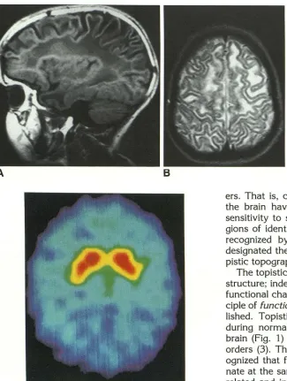
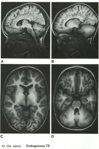
Fig. 1. Topistic area. T2-weighted im-ages at the level of the foramen of Monro (A) and of the midbrain (B) show the mor-phology of parts of the extrapyramidal sys-tem, as indicated by the deposition of iron in the pallidum, the substantia nigra, and the red nucleus.
Fig. 2. A 3-year-old boy nearly drowned in a swimming pool at home. CT scan (data not shown) demonstrated the so-called "white cerebellum" and bilateral hypodensity of the pallidum. Proton density axial MR (A) and T2-weighted axial MR (B) through the basal ganglia show nearly symmetrical in-volvement of the caudate nuclei, the putam-ina, the globi pallidi, the thalami, the genic-ulate bodies, the gray matter in the wall of the third ventricle, and the periaqueductal gray matter. Extensive cortical laminar ne-crosis is observed.
A
comes more important in cases of subacute or chronic toxicity with residual neurologic damage.
In such cases, magnetic resonance (MR) can
sometimes demonstrate striking pathologic changes associated with the encephalopathy, and can be of importance for further diagnosis.
However, even in cases with histologically proven organic cerebral damage, computed to-mography (CT) or MR is not always positive. For example, tardive dyskinesia is a severe move-ment disorder due to the chronic use of neurolep-tic drugs. The pathology is well known. Histology
AJNR: 13, March/April1992
B
shows a decrease in the number of ganglion cells in the substantia nigra. MR imaging shows no abnormalities. Similarly, in the acute malignant neuroleptic syndrome MR also shows no abnormalities.
Knowledge of the biomechanisms by which toxins cause encephalopathy helps us to under-stand the selectivity of the lesions seen in the imaging studies of toxic encephalopathy.
The characteristic MR images are the result of
[image:2.612.229.559.78.581.2]biochemi-AJNR: 13, March/ April 1992
A
BFig. 4. Topistic area. PET demonstrates the distribution of 18
fluorodopa and illustrates the concept of a topistic area related to the distribution of a particular neurotransmitter.
cal changes. These differing vulnerabilities reflect a number of factors, of which the most important are
1. regional cerebral blood flow/ oxygen demand;
2. distribution of neurotransmitters;
3. specific chemical affinity and vulnerability; and
4. developmental maturation of the patient at the time of intoxication.
Selective Vulnerability
As a general rule, specific groups of toxins tend to affect specific brain structures more than
oth-749
Fig. 3. Vacuolating myelinopathy. This type of myelin disorder is seen with a num-ber of conditions: hexachlorophene ence
ph-alopathy, triethyl tin encephalopathy, Can
-avan disease, and other organic acidopathies and aminoacidopathies.
A, Parasagittal Tl MR shows the char -acteristic swelling of the white matter in the
subcortical U fibers and the stretching of the cortex. The involved area has low signal intensity and reaches into the arcuate fibers. 8, T2-weighted transverse MR through the supraventricular parietal region shows extensive white matter involvement includ-ing the U fibers. The gyri stand out against the background of swollen white matter.
ers. That is, certain regions and systems within the brain have greater affinity for and greater sensitivity to specific types of toxins. These re-gions of identical affinity and vulnerability were recognized by German neuropathologists who designated them the "T opistische Bezirke" or to-pistic topographical areas.
The topistic areas often involve more than one structure; indeed, they often encompass a whole functional chain of neurons and tracts. The prin-ciple of functionally related systems is well estab-lished. Topistic areas can readily be identified during normal physiologic development of the brain (Fig. 1) and in systemic degenerative dis-orders (3). Thus, in 1920, Flechsig already rec-ognized that functionally related systems myeli-nate at the same time (4). Similarly, functionally related and interdependent nuclei appear to de-generate at the same time in multiple system atrophies such as Parkinson disease and progres-sive supranuclear palsy.
Other mechanisms of selective vulnerability are related to the similarity in particular characteris-tics that make the different geographic areas equally vulnerable to a particular noxious agent. Thus, apparently diverse areas may prove to have similar oxygen requirements, chemical composi-tions, and/or neurotransmitters.
[image:3.612.58.383.79.510.2]750
Fig. 5. Ecstasy encephalopathy. Selective involvement of the globus pallidus and the most dorsal part of the putamen in an 18-year-old girl, after drinking a love potion containing Ecstasy (3,4 methylenedioxymetamphetamine). Consequent behavioral changes required psychiatric hospitalization. Ecstasy influences particular neurotransmitter receptors.
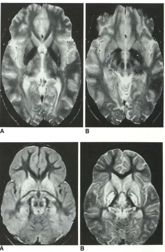
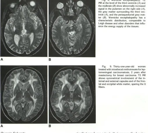
selective vulnerability of gray matter structures to energy depletion is also reflected in the pref-erential affliction of gray matter structures in carbon monoxide (CO) intoxication (especially the pallidum), and Leigh disease (the gray matter around the third ventricle, the periaqueductal gray matter, the tectum and tegmentum of the brain stem, and the dentate nuclei). Wernicke encephalopathy, a toxic encephalopathy caused by thiamine deficiency in alcoholics, shows the same pattern of selective involvement as Leigh disease, plus involvement of the mammillary bod-ies, presumably because thiamine deficiency also influences energy metabolism.
An example of selective vulnerability resulting from specific chemical composition is found in myelin. Myelin has an especially high lipid content and shows especially slow turnover. As a result, all the myelinated tracts are particularly vulnera-ble to the accumulation of lipophilic substances and to lipid peroxidation. One instance of such intoxication has become famous in medical liter-ature: hexachlorophene encephalopathy, a vac-uolating myelinopathy, was found in infants that were washed with antiseptic hexachlorophene solutions for dermal problems. The skin of pre-term neonates proved to be more permeable to these agents than the more mature skin, resulting
AJNR: 13, March/Apri11992
in increased absorption and unfortunate toxicity. In adults, vacuolating myelinopathy has been described after the use of hexachlorophene solu-tions in vaginal tampons and as an antiseptic agent on burned areas. Intoxication with triethyl tin has identical effects (Fig. 3).
T opistic areas related to the distribution of a particular neurotransmitter are best visualized by positron emission tomography (PETJ. PET, for example, shows the distribution of 1 fluorodopa in the basal ganglia (Fig. 4). Methylphenyltetra-hydropyridine (MPTP) interferes selectively with the dopamine neurotransmitters and leads to se-vere Parkinsonism. Tardive dyskinesia and malig-nant neuroleptic syndrome are other examples of toxic encephalopathy in a topistic area related to specific neurotransmitters. In this kind of involve-ment of a neurotransmitter system, imaging mo-dalities usually do not show abnormalities. How-ever, Figure 5 illustrates an example in which MR successfully depicts the topistic areas.
Selective vulnerability is also related to the level of activity during development. This con-cept was particularly stressed by Dobbing (5) and has broadened our insight into the origin of con-genital malformations of the central nervous sys-tem (6). The greatest impact of noxious agents is on those structures that grow and develop at the highest rate at the time of insult. Thus, migra-tional disorders result when toxic insults are suf-fered in the third to fifth month of gestation, the period in which neuronal migration occurs {Fig. 6). Similarly, disorders of myelination are ob-served when toxic insults occur during the last trimester of pregnancy and the first year of life, because myelination of the CNS occurs at a high rate in these periods. Since normal myelinatibn depends upon complex interactions between ax-ons, myelin-forming oligodendrocytes and the provision of substrates by the environment, the delicate interactive process is easily disturbed by adverse factors such as nutritional deficiencies and intoxications (7).
Classification
To facilitate our analysis of TE we have clas-sified the toxic encephalopathies by origin of the
[image:4.612.84.267.76.312.2]AJNR: 13, March/April 1992
A B
depends in part, on observer bias. Thus, Gram-negative endotoxin can be classified either as exogenous-external, because the Escherichia coli infection stems from outside the body, or as exogenous-internal because the endotoxin caus-ing the related encephalopathy is produced by the E. coli within the body. We prefer exogenous-internal, because the toxin itself is produced within-and in interaction with-the body.
The largest group of toxins are exogenous-external. Toxins within this group are usually categorized heterogeneously by chemical com-position, source, and effect. Dietary and chemical deficiencies may lead to abnormal biochemical processing that produces effects comparable to intoxications. They may show patterns of dam-age by virtue of selective vulnerability. These deficiency states are included among the exoge-nous-external toxic encephalopathies.
Exogenous-External TE
The group of exogenous-external TEs includes all intoxications from iatrogenic, agricultural, in-dustrial, environmental, and social (drugs, alco-hol, nicotine) sources that affect the CNS.
To this group belong Lathyrus sativus peas, the Guamanian type of Parkinsonism, organic mercury poisoning, organic lead poisoning, and toluene exposure or sniffing; so do other well known encephalopathies associated with ethanol abuse, such as the Marchiafava-Bignami syn-drome. Wernicke encephalopathy, and the Kor-sakoff syndrome, Iatrogenic exogenous-external TE includes all the TEs caused by the ingestion of prescribed drugs, such as anticancer agents, anticonvulsants, tranquilizers, and anesthetic
751
Fig. 6. Migration disorder; 14-year-old girl with psychomotor retardation and epi -lepsy. T2-weighted parasagittal and tran s-verse images show a combination of an occipital lissencephaly and frontotemporal pachygyria, with linear heterotopia of the gray matter in the frontal lobes. Irregularities around the occipital horns suggest an en ce-phaloclastic or toxic cause for the arrest of development.
gasses, or caused by medical treatments such as postdialysis progressive aluminum encephalop-athy. The fetal intoxication syndromes can also be considered in this category.
Organic Mercury Poisoning
Ingestion of fish caught in the poisoned bay of Minamata led to a neurologic disorder that was eventually identified as being caused by organic mercury (8). Mercury intoxication has also been reported after accidental ingestion of wheat that had been treated with organic mercury com
-pounds to prevent cropping. Neuropathology shows degeneration of the granular layer of the cerebellum and patchy loss of cells in the cerebral cortex, in particular the calcarine cortex.
MR images show the depositibn of mercury in cerebellar structures and under the occipital cor
-tex (Fig. 7).
Wernicke Encephalopathy
Wernicke encephalopathy (9-11) results from a deficiency of vitamin B 1 (thiamine), and, as such, is not confined to chronic alcoholics. Be
-cause vitamin B 1 is a cofactor of transketolase,
thiamine deficiency causes decreased activity of this enzyme. The precise relationship of reduced enzyme activity to damage in the characteristic
"topistic area" represented by Wernicke enceph-alopathy (Fig. 8) is conjectural. Korsakoff disease is now generally seen as a chronic stage of Wer
-nicke encephalopathy, with consistent atrophy of
the mammillary bodies and variable involvement
752 AJNR: 13, March/Apri11992
TABLE 1:
[TOXIC ENCEPHALOPATHY
j
_.---
~
EXOGENOUS
ENDOGENOUS
/
~INTERNAL
inborn errors of metabolismEXTERNAL
~
endotoxins
paraneoplastic toxins
uremia
ion balance disorders
TOXIC SUBSTANCES
classification
chemical composition source industrial
agricultural
effects
iatrogen on co gens
teratogens
DEFICIENCIES vitamins
nutritients
( para) sympathicomimetica
( para) sympathicolytica
Fig. 7. Mercury encephalopathy; 50-year-old man with a 2-year course of pro-gressive cerebellar and focal cerebral signs following use of a mercury-containing com-pound to prevent tulip bulbs from cropping. The midsagittal (A) and parasagittal (B) gra-dient recalled echo images show the mag-netic susceptibility effect of the mercury deposits in the cerebellum and beneath the occipital cortex, extending the histopatho-logic findings in this region.
Cytostatic Agents
A
Cytostatics, such as methotrexate and 5 fluo-rouracil can lead to severe changes in the white matter (Fig. 9) when they are used to treat extra-cranial tumors, or intraextra-cranial tumors (often in combination with radiotherapy).
Heroin Pyrolysite
During the 1980s, a group of chronic heroin addicts in Amsterdam was discovered to manifest
B
[image:6.612.120.561.64.595.2]AJNR: 13, March/ April 1992
A
BA
BOrganic Solvents
Chronic inhalation of organic solvents or other
preparations containing toluene is known to
cause multifocal neurologic and mental disorders,
cerebral, cerebellar and brain stem atrophy, and
diffuse focal white matter abnormalities detecta-ble by MR (Fig. 11) (14-17). Toluene is one example of a typical lipophilic substance that persists in the myelin for a long time, leading to severe functional disturbances.
Exogenous-Internal TE
The disorders caused by exogenous-internal toxins have in common a focal or generalized process in the body outside the blood-brain
bar-753
Fig. 8. Wernicke encephalopathy. T2
MR at the level of the third ventricle (A) and the midbrain (B) show abnormally increased signal in the putamen on the right side (A), the gray matter surrounding the third ven-tricle (A), and the periaqueductal gray
mat-ter (B). Wernicke encephalopathy has a
characteristic distribution, comparable to
Leigh disease and other disorders that influ-ence the energy supply of the tissues.
Fig. 9. Thirty-one-year-old woman
treated with intrathecal methotrexate for
lep-tomeningeal carcinomatosis, 2 years after
mastectomy for breast carcinoma. T2 MR shows symmetrical involvement of the in-ternal and exin-ternal capsules and of the fron-tal and occipifron-tal white matter, sparing the U fibers.
rier that produces a toxin that crosses the barrier, enters the brain, and causes encephalopathy. Paraneoplastic syndromes and parainfectious de-generations belong in this category. Malignant disease anywhere in the body may have a remote effect on the peripheral nerves, the spinal cord, and on the brain, causing peripheral neuropathy, subacute necrotizing myelopathy, and
encephal-omyeloradiculitis. The
encephalomyeloradiculi-tides group manifests in two different forms; brain stem "encephalitis," and so called limbic
"enceph-alitis" in which the changes are restricted to the limbic system. These differing manifestations presumably reflect the differing selective vulner-ability of brain structures to toxic agents. Parain
[image:7.612.54.562.84.541.2]754
Fig. 10. Vacuolating myelinopathy from unknown toxin; 26-year-old man who
sniffed heroin pyrolysite, polluted with a still unknown toxin. He and many others deve
l-oped severe neurologic motor and c oordi-nation problems, secondary to pathologi -cally confirmed vacuolating myelinopathy.
All the patients' lesions were restricted to the white matter, in a characteristic di stri-bution. A, T2 MR shows high signal intensity
of the posterior limb qf the internal capsule
and of the centromedian nucleus of the thal -amus. White matter lesions are present in the arcuate f(f>ers of the occipital lobes. 8,
Proton density MR shows involvement of
the cerebellar white matter.
Fig. 11. Industrial toxin; 58-year-old
man with progressive memory loss who
worked in the paint industry for more than
40 years. The T2 MR show diffuse focal
changes in the white matter and in the
cor-tex. There is loss of cortical tissue, with
widening of the subarachnoid spaces and
the Sylvian fissure.
A
A
E. coli, mycoplasma, and diphtheria infections.
Changes in the ion balance are held responsible for central pontine and extrapontine myelinolysis.
Porphyria, an inborn error of metabolism, may lead to a peripheral neuropathy and encephalop-athy with foci of myelin loss, myelin pallor, and ischemic changes in the gray matter that are probably secondary to vasospasm.
Hepatocerebral syndromes may be the result of an inborn error of metabolism such as Wilson
dis~ase. In Wilson disease, the encephalopathy is
caused by ceruloplasmin deficiency leading to excessive deposition of copper in tissues, espe-cially the globus pallidus. Hepatorenal syndromes may also result from cirrhosis or portocaval
shunt~ with c;~cquired hepatic failure, probably
AJNR: 13, March/Aprill992
B
B
mediated by hyperammonemia. This form will be discussed more extensively later, because it pro-duces special changes in signal intensity on MR.
Central Pontine Myelinolysis
[image:8.614.230.560.75.533.2]AJNR: 13, March/ April 1992
a too rapid correction of hyponatremia. The re-lation of CP M to alcoholism arises because alco-hol blocks the expression of antidiuretic hormone. Alcohol withdrawal leads to a rapid return of antidiuretic hormone function, causing hypona-tremia. Aggressive treatment with intravenous infusions of hypertonic solutions may then pre-cipitate the CPM (18).
The clinical manifestations of CPM vary from minimal symptoms to a complete locked-in syn-drome or coma, depending upon the extent of the lesions. During the last years it has become evident that the CPM is not invariably fatal; some patients survive and show a marked improve-ment of their neurologic disorders.
In CPM, there is usually a single, symmetrical lesion in the central part of the basis pontis. The myelinolysis can be extensive, occupying the whole basis pontis, with the exception of a small outer rim, or it may be quite limited and appear as a very small central lesion. Histologically, there is severe or complete loss of myelin in the lesion, with a concomitant loss of oligodendrocytes. The axons are mostly well preserved.
In CPM, MR shows the typical lesion in the basis pontis (Fig. 12). The MR abnormalities grad-ually disappear in cases that show clinical im-provement, but a lesion remains visible for a long time. Extrapontine myelinolysis preferentially in-volves the cerebellar white matter; the mammil-lary bodies; the tegmentum of the midbrain; the lateral geniculate bodies; the thalamus; the basal ganglia; the internal, external, and extreme cap-sules; the anterior commissure; the fornix; and the deep layer~ gf the cerebral cortex.
755
Fig. 12. Central pontine myelinosis; 56-year-old-man, admitted 3 weeks before the
MR for general malaise, vomiting, and desic-cation. A hyponatremia of 111.5 mmoi/L
was corrected in 2 days to 134 mmoi/L. Shortly thereafter, he developed severe neu -rologic symptoms. Midsagittal T1 MR (A)
and transverse T2 MR (B) demonstrate the characteristic features of central pontine
myelinolysis, typically sparing the outer rim of the pons. In this case, the clinical
condi-tion gradually improved and the MR
abnor-malities gradually regressed incompletely.
Hepatocerebral Syndromes
· Hepatocerebral syndromes manifest by atro-phy, by changes in signal intensity in the basal ganglia, and by T1 shortening of the white matter (particularly in infants and children). 1H MR spec-troscopy has shown a change in the glutamate/ glutamine complex and in myoinositol (19-24). Most investigators believe that ammonia plays a key role in hepatic encephalopathy. Ammonia appears to cause neurotoxicity by interacting with the glutamate/ glutamine metabolism. Apart from its other roles in metabolism, glutamate is the most important excitatory neurotransmitter.
Hy-i:>erammonia stimulates glutamine synthesis via
glutamine synthetase. It may also inhibit gluta-minase and inhibit glutamate re-uptake by the astrocytes. Thus, hyperammonemia acts to in-crease the concentration of glutamine. Pathways of glutamate include transamination, dehydro-genation, deamination, and decarboxylation to ')'-aminobutyric acid (GABA). GABA is the most important inhibitory neurotransmitter. However, the relation between these phenomena and the T1 shortening as seen on MR is not clear.
Other explanations for the T1 shortening of the basal ganglia have been suggested, such as accumulation of manganese or of lipid particles. McConnel et al (25) demonstrated intracytoplas-matic glial lipid accumulation in the caudate nu-cleus and putamen. According to Zeneroli et al (26) short-chain fatty acids accumulate in the blood and may play a role in the pathogenesis of hepatic encephalopathy.
[image:9.612.54.381.79.284.2]ipqi~t!ng11ish-756
Fig. 13. Hepatic encephalopathy; 3-year-old boy, status post multiple surgical procedures for Hirschsprung disease, with many postoperative complications, hepatic failure, and hepatic encephalopathy. Midsag-ittal (A) and parasagMidsag-ittal spin echo MR (B) and T1 inversion recovery MR ( C and D) show a high signal intensity in all cerebral and cerebellar white matter, especially the basal ganglia. The changes are most pro-nounced in the globus pallidus. The basis pontis is typically spared.
A
c
able from white matter (Fig. 13). At the same time, there is T1 shortening in the white matter, typically sparing the pons, but including the ar-cuate fibers. In older patients, this spread of T 1 shortening over the white matter is less clear.
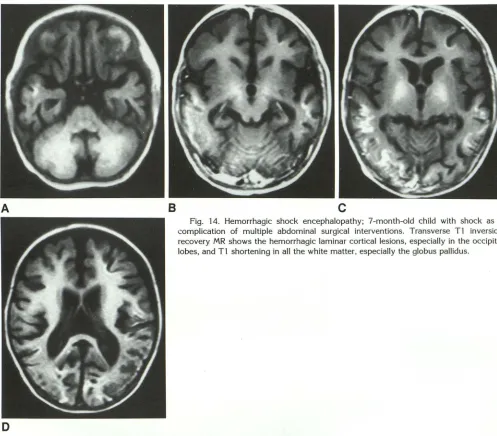
The T1 shortening of gray and white matter may be observed with a number of toxic-meta-bolic conditions, not just hepatic encephalopathy. Thus, we have seen this phenomenon in a child with a hemorrhagic shock encephalitis (Fig. 14). It may be that the glutamate/ glutamine complex is involved via another metabolic pathway, but we cannot identify any specific cause for the T1 shortening.
AJNR: 13, March/Apri11992
B
D
Endogenous TE
The endogenous forms of TE are its smallest group. A number of inborn errors of metabolism should be included here, because their effects are mediated by a toxic product. This is the case in phenylketonuria (27), maple syrup urine disease, and globoid cell leukodystrophy (Krabbe disease).
Krabbe Disease
[image:10.612.227.558.71.566.2]AJNR: 13, March/ April 1992 757
A
8
c
Fig. 14. Hemorrhagic shock encephalopathy; 7-month-old child with shock as a complication of multiple abdominal surgical interventions. Transverse Tl inversion recovery MR shows the hemorrhagic laminar cortical lesions, especially in the occipital lobes, and Tl shortening in all the white matter, especially the globus pallidus.
D
that has 2 functions: 1) it normally degrades
cerebroside into galactose and ceramide and 2) it normally hydrolyzes psychosine (galactosyl sphingosine). Psychosine is a toxic metabolite that is essentially nonexistent in normal brain.
Psychosine causes early, very rapid, almost
com-plete death of the oligodendroglia. The
oligoden-droglia normally make myelin. Cerebroside is
localized predominantly in myelin. Because
Krabbe patients cannot break down cerebroside,
one would normally expect an abnormally high
concentration of cerebroside within their white
matter. However, patients with Krabbe disease
always show less cerebroside in the white matter
than do normal individuals, because early death
of the oligodendroglia! cells terminates the
syn-thesis of cerebroside. Krabbe patients do show
an abnormally high ratio of cerebroside to
sulfa-tide, which is indicative of abnormal accumula
-tion of cerebroside, but this accumulation is in no
way comparable to the extreme storage of
sul-fatide seen in metachromatic leukodystrophy.
Fetal Syndromes
The toxic substance interfering with fetal de-velopment can be considered an
exogenous-ex-ternal intoxication, reaching the unborn child via
the mother. The fetal syndromes are unique,
because the effect of the toxin depends upon the
stage of development the child has reached, as
well as upon the nature of the toxin, the
concen-tration of the toxin, and the duration of the
[image:11.612.53.550.77.513.2]758
Fig. 15. Late-onset form of Krabbe dis-ease; 14-year-old girl with a progressive neurologic .syndrome. T1 MR (A) and T2 MR (B) show the general involvement of the white matter, with some atrophy. The deep infolding of the gray matter over the white is more conspicuous in B.
Fig. 16. Caudal regression syndrome;
4-month-old child of a diabetic mother.
A, Sagittal T1 MR. The abrupt cutoff and high position of the conus medullaris are characteristic of this syndrome.
B, Coronal Tl MR again shows the blunted termination of the conus. The sac-rum ends at S2.
A
A
Toxic or teratogenic substances have a great
impact on the developing fetus. The effects are
often widespread and involve multiple parts of
the body or the whole body, in addition to the
brain. The gamut of possible dysgeneses is
ex-tensive and ranges from lethal malformation with
early spontaneous abortion to mild alteration in
morphology and function. The sources of the
toxins include:
1. Iatrogenic-Syndromes can result from the
use of established drugs prescribed to the mother,
AJNR: 13, March/ April 1992
8
8
but taken during the pregnancy; most such cases are accidental. A few arise when drugs are given
knowingly in desperate cases (30, 31 ).
Antiepilep-tic drugs are known to cause developmental dam
-age, resulting for example in the fetal hydantoin
syndrome and the fetal valproate syndrome.
2. Alcohol and drug abuse-The most
impor-tant cause of fetal dysgenesis is the use of drugs
or alcohol during the pregnancy. The fetal alcohol
syndrome has been reported extensively in the
[image:12.615.229.561.74.590.2]re-AJNR: 13, March/ April 1992
tarded, have short palpebral fissures, a low nasal
bridge, epicanthus, a long convex upper lip, and mental retardation. The numerous CNS
anoma-lies of the fetal alcohol syndrome include:
mal-formation of neuronal and glial migration, micro-cephaly, hydromicro-cephaly, porenmicro-cephaly, agenesis
of the corpus callosum, meningomyelocele, and
Dandy Walker malformation.
3. Metabolic disorders of the mother-Fetal
development may suffer from a metabolic
disor-der of the mother. Diabetes mellitus may be one causative factor in the caudal regression
syn-drome, a complex abnormality of the caudal end
of the embryo with anal atresia, sacral dysgenesis
or agenesis, high position of the conus medullaris,
and diverse urologic, neurologic, and orthopedic disorders (Fig. 16).
4. Physical events-Hyperthermia, radiation,
amnionic bands, and mechanical trauma are all
potentially considered hazardous to the develop-ing fetus, possibly causdevelop-ing fetal dysgenesis. Strictly speaking, this category does not fall under
the heading of toxic encephalopathy.
5. Teratogens-Compounds with teratogenic action are now listed in a reference manual (33). Their discussion exceeds the scope of this communication.
In most of the fetal syndromes the direct role of neuroradiologic imaging methods are limited;
indirectly, of course, ultrasound, CT, and MR can
depict congenital malformations, dysgyria,
re-tarded myelination, and myelodysplasias.
Conclusion
Imaging modalities, in particular MR, play a
modest, but sometimes very important role, in
the diagnosis of toxic encephalopathy. The field
offers another example of the way the MR reader
should get involved in the pathophysiologic and pathomorphologic backgrounds of the observed lesions. The concepts of selective vulnerability and topistic (topographic) areas is helpful in understanding the patterned involvement of symmetrical areas in the brain with specific
char-acteristics. Recognition of these patterns may
lead to correct diagnosis.
Acknowledgments
We are grateful to Lenie de Vries, Els van Straten, Karin
Molleman, Marilene Winkelman, Ronald Prinsze, Ton
Schweigman, and Adrie Verburg for their assistance in
preparing the material for this article.
759
References
1. Vinken PJ, Bruyn GW. Intoxications of the central nervous system. In: Vinken PJ, Bruyn GW, eds. Handbook of clinical neurology. Vols 36 and 37. New York: North Holland, 1979
2. Spencer P, Hugen J, Ludolph A, et al. Discovery and partial
charac-terization of primate motor system toxins. Ciba Found Symp
1987;126:221-238
3. Drayer BP, Bird CR, Williams K, Keller P. Systemic metabolic disease and the globus pallidus: an MRI approach. AJNR 1989; 10:902-911 4. Flechsig P. Anatomie des menslichen Gehirns und Ruckenmarks auf
myelogentischer Grundlage. Leipzig, Germany: Georg Thieme, 1920
5. Dobbing J. Vulnerable periods in developing brain. In: Davison AN,
Dobbing J, eds. Applied neurochemistry. Oxford, England: Blackwell,
1968:287-316
6. Knaap vd MS, Valk J. Classification of congenital abnormalities of the CNS. AJNR 1988;9:315-326
7. Wiggins RC. Myelination: a critical stage in development. Neurotoxi
-cology 1986;7:103-120
8. Tokuomi H, Okajuma T, Kanai J, Tsimoda M. Minamata disease: an
unusual neurological disorder occurring in Minamata. Kurume Med J
1961;14:47-64
9. Galluci M, Bozzao A, Splendiani A, Masciocchi C, Passariello R. Wernicke encephalopathy: MR findings in five patients. AJNR
1990;11:887-892
10. Victor M. MR in the diagnosis of Wernicke-Korsakoff syndrome. AJNR 1990; 11 :895-896
11. Donnal JF, Ralph Heinze E, Burger PC. MR of reversible thalamic
lesions in Wernicke syndrome. AJNR 1990; 11:893-894
12. Wolters ECh, Wijngaarden van GK, Stam FC, et al. "Heroine"-l euko-encephalopathie: spongiforme leuko-myelo-encefalopathie na inhal-atie van het pyrolysaat van verontreinigde hero"ine. Ned Tijdschr
Geneeskd 1982; 126:508-514
13. Wolters ECh, Wijngaarden van GK, Stam FC, et al. Leukoencepha-lopathy after inhaling "heroin" pyrolysite. Lancet 1982; 1:1233-1236
14. Kelly CTW. Prolonged cerebellar dysfunction associated with paint-sniffing. Pediatrics 1975;56:605-606
15. Hermes JT, Filley CM, Rosenberg NL. Neurologic sequelae of chronic solvent vapor abuse. Neurology 1986;36:698-702
16. Escobar A, Aruffo C. Chronic thinner intoxication: clinicopathologic
report of a human case. J Neurol Neurosurg Psychiatry 1980 ;43:986-994
17. Ikeda M, Tsukagoshi H. Encephalopathy due to toluene sniffing:
report of a case with magnetic resonance imaging. Eur Neurol 1990;30:347-349
18. Valk J, van der Knaap MS. Central pontine and extrapontine
myeli-nolysis. In: Magnetic resonance of myelin, myelination and myelin disorders. Heidelberg, Germany: Springer-Verlag, 1989:253-262 19. Levy LM, Yang A, Hennigar R, Rothstein J, Bryan RN. The brain and
hepatic encephalopathy MR abnormalities. AJNR 1989; 10:900-905 20. Luyten PR, Hollander den JA, Bovee WMMJ, Ross BD, Bosman DK,
Chamuleau RAFM. 31
P and 'H NMR spectroscopy of the human brain
in chronic hepatic encephalopathy (abstr). In: Book of abstracts Society of Magnetic Resonance in Medicine. Vol 1. Berkeley, CA: Society of Magnetic Resonance in Medicine, 1989:375
21. Zeneroli ML, Cioni C, Vezzelli C, et al. Prevalence of brain atrophy in liver cirrhosis patients with chronic persistent encephalopathy. J
Hepatol1987;4:283-292
22. Kulisevsky J, Ruscalleda J, Grau JM. MR imaging of acquired
hepa-tocerebral degeneration. AJNR 1991;12:527-528
23. Ross BD. Biochemical considerations in 'H spectroscopy: glutamate and glutamine: myo-inositol and related metabolites. NMR Biomed 1991 ;4:59-63
24. Chamuleau RAFM, Bosman DK, Bovee WMMJ, Luyten PR, Hollander
den JA. What the clinician can learn from MR glutamine/glutamate
760
25. McConnell J, Castaldo P. Striatal hyperemia, transient liver failure and chorea after liver transplantation. J Hepatol1990; 1 O(suppl 1 ):
16-23
26. Zeneroli ML, Ciani G, Crisi G, Vezzelli C, Ventura E. Globus pallidus alterations and brain atrophy in liver cirrhosis patients with en ceph-alopathy: an MR imaging study. Magn Reson Imaging 1991; 9:295-302
27. Brismar J, Aqeel A, Gascon G, Ozand P. Malignant hyperphenylala-ninemia. AJNR 1990; 11:135-138
28. Valk J, van der Knaap MS. Globoid cell leukodystrophy. In: Magnetic resonance of myelin, myelination and myelin disorders. Heidelberg,
Germany: Springer-Verlag, 1990:77-82
AJNR: 13, March/April1992
29. Singer DB, Sung LJ, Wigglesworth RJS. Fetal growth and maturation.
In: Wigglesworth RJS, Singer DB, eds. Textbook of fetal and perinatal pathology. Oxford, England: Blackwell, 1991:11-47
30. Volpe JJ. Drugs and the developing nervous system. In: Volpe JJ, ed. Neurology of the newborn. Philadelphia: Saunders, 1987: 664-697
31. Vernadakis A, Parker KK. Drugs and the developing central nervous
system. Pharmacal Ther 1980; 11:593-647
32. Lemoine P, Herousseau H, Bortegru J. Children of alcoholic parents;
observed anomalies (127 cases). Quest Med 1968;21:476-482