VISUAL PROCESSING IN A PRIMATE TEMPORAL
ASSOCIATION CORTEX: INSENSITIVITY TO
SELF-INDUCED MOTION
Jari K. Hietanen
A Thesis Submitted for the Degree of PhD
at the
University of St Andrews
1993
Full metadata for this item is available in
St Andrews Research Repository
at:
http://research-repository.st-andrews.ac.uk/
Please use this identifier to cite or link to this item:
http://hdl.handle.net/10023/14576
A sso ciatio n C ortex: In se n sitiv ity to
Self-In d u ced M otion
Jari K. H ietanen
D epartm ent o f Psychology U niversity o f St. Andrews
All rights reserved
INFORMATION TO ALL USERS
The quality of this reproduction is d e p e n d e n t upon the quality of the copy subm itted.
In the unlikely e v en t that the author did not send a co m p le te m anuscript and there are missing p ag es, these will be noted. Also, if m aterial had to be rem oved,
a n o te will indicate the deletion.
uest
ProQuest 10166341
Published by ProQuest LLO (2017). Copyright of the Dissertation is held by the Author.
All rights reserved.
This work is protected against unauthorized copying under Title 17, United States C o d e Microform Edition © ProQuest LLO.
ProQuest LLO.
789 East Eisenhower Parkway P.Q. Box 1346
I, Jari Kaarlo Hietanen, hereby certify that this thesis has been composed by myself, that it is a record of my own work, and that it has not been accepted in partial or complete fulfilment of any other degree or professional qualification.
Signed: Date:
I was admitted to the Faculty of Science of the University of St. Andrews under Ordinance General No. 12 on 1st October 1988 and as a candidate for the degree in September 1989.
Signed: Date: v ^' I ' h
I hereby certify that the candidate has fulfilled the conditions of the Resolution and Regulations appropriate to the degree of Ph.D.
7'
Signature of Supervisor Date:
UNRESTRICTED
In submitting this thesis to the University of St. Andrews I understand that I am giving permission for it to be made available for use in accordance with the regulations of the University Library for the time being in force, subject to any copyright vested in the work not being affected thereby. I also understand that the title and abstract will be published, and that a copy of the work may be made and supplied to any bona fide library or research worker.
I am deeply grateful to my supervisor David Perrett for all the help and support I have received from him during the course of this study. It is not only that he guided me through the preparation of this thesis, but having had the opportunity to work alongside with him has given me much invaluable knowledge for my coming attempts to continue interesting and insightful neuroscience. I can just hope that if I am ever in the position of having students of my own, I would be as understanding towards them as he has been towards me. Thanks Dave.
Special thanks to Professor Tapio Nummenmaa, Ph.D., Assistant Professor Markku Ojanen, Ph.D., and Docent Ville Jântti M.D. at the University of Tampere, Finland, for their willingness to write referees’ reports for what must have seemed to be an endless row of grant applications.
I am grateful to the following foundations for the financial support during the course of this thesis: Pirkanmaan Kulttuurirahasto, Alfred Kordelinin Saatio, Tampereen Kaupungin Tiederahasto, Emil Aaltosen Saatid, and the Guarantors of
BRAIN.
Many thanks to my "friends in fight", Mike Oram and Phil Benson. Sharing an office with them has made an indelible impact on me, I fear. With Mike I collected most of the experimental data that will be documented here and his contribution of providing a multitude of user-friendly computer programmes for various kinds of data analyses is greatly appreciated. Likewise, Phil offered a helping hand, without hesitation, on a countless number of occasions.
Thanks also to following persons, Rhys Bevan, Derek Brook, Winand Dittrich, Mark Harries, Maarten Milders, Mary Latimer, Wendy Taylor, Sharon Thomas, Fiona Wallace, Pete Wilcox, and to all workshop technicians and departmental secretaries.
Chapter One: Introduction 1
Chapter Two: Processing of Self-Induced and Expected Stimulation 4 at the Neuronal Level
2.1. Introduction 4
2.2. Self-motion 5
2.3. Visual stability during self-induced eye movements 15 2.4. Processing of somatosensry stimuli as a result of 30
active exploratory movements
2.5. Attenuation of responses to self-vocalized sounds 33 2.6. Processing of expected externally-induced stimuli 34
Chapter Three: The Superior Temporal Polysensory Area in the 43 Primate Visual System
3.1. Introduction 43
3.2. Visual processing within the primate cortex 43
3.3. The anatomy and functional properties of superior 49 temporal polysensory area and adjacent visual areas
3.3.1. Architectonics 49
3.3.2. Connections 57
3.3.3. Functional properties 59
Chapter Four: Summary of the Literature Review and Rationale 76 for the Present Experiments
Chapter Five: General Methods for the Experiments 83
5.1. Subjects and training 83
5.2. Surgical procedures 85
5.3. Single-unit and eye movement recording 86
5.4. Data collection and general testing procedure 87
5.5. Perfusion and histology 88
5.6. Reconstruction of the cell location 90
Chapter Six: General Response Properties of Motion Sensitive Neurons 92 in Area STP
6.1. Introduction 92
6.2. Methods 94
6.3. Results 96
6.3.1. Cell classification 96
6.3.2. Discrimination between directions 101
6.3.3. Width of tuning of direction-selective cells 102
6.3.4. Average shape of direction tuning 104
6.3.5. Distribution of optimal directions 105
6.3.6. Temporal characteristics of cell responses 105
6.3.7. Location of cells 106
7.1. Introduction 7.2. Methods 7.3. Results
7.3.1. General response properties
7.3.2. Response selectivity for motion direction 7.3.3. Feature selectivity
7.3.4. Differential responses to the sight of object motion and motion of own hand
7.3.5. Responsivity to external stimuli during self-induced stimulation
7.3.6. Unexpected self-produced stimulation 7.3.7. Eye movements during self-produced and
external visual stimulation 7.3.8. Location of cells
7.4. Discussion 114 118 124 124 125 126 126 128 128 130 132 134
Chapter Eight: STP Cell Responses to Self-Induced Pattern Motion 141
8.1. Introduction 141
8.2. Methods 142
8.3. Results 147
8.3.1. General response properties 147
8.3.2. Discrimination between responses to externally- 148 induced and self-induced pattern motion
8.3.3. Cells responding to self- and externally-induced motion 154 8.3.4. Relative strength of responses in self- and externally- 156
induced stimulus conditions
8.3.5. Eye movements during self- and externally- 157 induced pattern motion
8.3.6. Location of cells 159
8.4. Discussion 159
Chapter Nine: STP Cell Responses to Visual Image Motion During 168 Object- and Self-Motion
9.1. Introduction 168
9.2. Methods 172
9.3. Results 176
9.3.1. General response properties 176
9.3.2. Responsivity during object motion and 176
self-motion
9.3.3. Observer-relative and object-relative motion 178 9.3.4. Eye movements during object-motion and self-motion 185
9.3.5. Location of cells 185
9.4. General Discussion 187
Andrews, St. Andrews, Fife KY16 9JU, SCOTLAND.
ABSTRACT
An animal’s own behaviour can give rise to sensory stimulation that is very similar to stimulation of completely external origin. Much of this self-induced stimulation has little informative value to the animal and may even interfere with the processing of externally-induced stimulation. A high-level association area in the temporal cortex of macaque (superior temporal polysensory area, STP) which has been shown to participate in the analysis of visud motion was targeted in a series of experiments in order to investigate whether this brain area discriminates externally- and self-induced stimulation in its visual motion processing. Earlier results in somatosensory processing within this same brain area provided grounds for this presumption
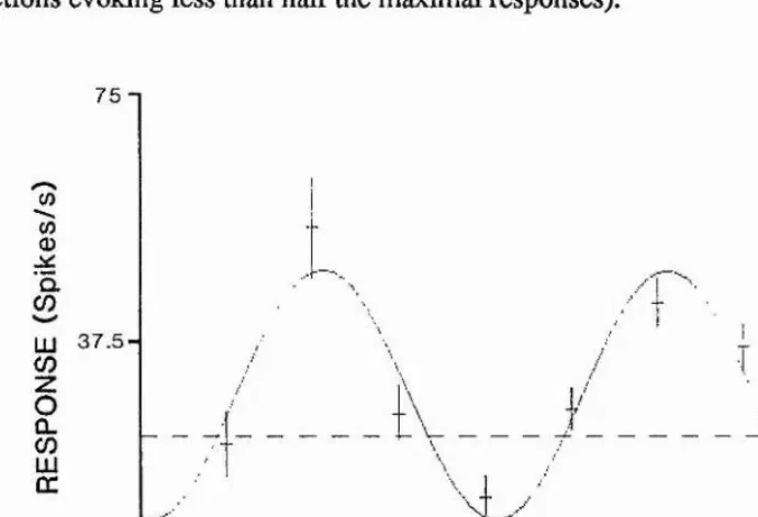
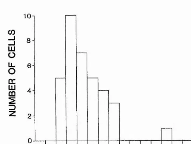
The cells studied in here were sensitive to the presence of motion but showed no selectivity for the form of the stimulus. 25% of all visually responsive cells in area STP were classified as belonging to this class of cells. This group of cells was further categorized into unidirectional (39%), bidirectional (4%) and pandirectional (57%) cells. Tuning to direction varied in sharpness. For most cells the angular change in direction required to reduce response to half maximal was between 45 and 70 degrees. The optimal directions of cells appeared clustered around cartesian axes, (up/down, left/right and towards/away). The response latency varied between 35.0-126.4 ms (mean 90.9 ms). On average cell responses showed a transient burst of activity followed by a tonic discharge maintained for the duration of stimulation. 83% of the motion sensitive cells lacking form selectivity responded to any stimuli moved by the experimenter, but gave no response to the sight of the animal’s own limb movements. The cells remained, however, responsive to external stimulation whüe the monkey’s own hand was moving in view. Responses to self-induced movements were recovered if the monkey introduced a novel object in its hand into view. That the response discrimination between externally- and self-induced stimulation was not caused by differences in the visual appearance of the stimuli was confirmed in the second experiment where the monkey was trained to rotate a handle connected to a patterned cylinder in order to generate visual motion stimulation over a fixation point. 61% of the tested cells discriminated between pattern motion generated by the monkey and by the experimenter. It was shown that the monkey’s motor activity as such (turning a handle without visible cylinder rotation) did not affect the cells’ spontaneous activity. Some indication was received to suggest that the discriminative mechanism is using not only (motor) corollary discharges but also proprioceptive input. These results also gave evidence of the plasticity of discriminative processing in STP for the animal’s life-time experiences. Finally, the cells were studied for their responsiveness for Image motion resulting from movements of external objects and movements of the animal’s body (self-motion). 84% of the cells responded only to visual object-motion and failed to respond to visual motion resulting from animal’s self-motion. The experiments also revealed that area STP processes visual motion mostly in observer- relative terms, i.e. in reference to the perceiver itself.
The results provide one explanation for the functional significance of the convergence of several modalities of sensory (and motor) input in the STP. It is suggested that area STP works as a "neural filter" to separate expected sensory consequences resulting from one’s own actions from those that originate from the actions of other animals or environmental events.
INTRODUCTION
Modem theories of perception emphasize the active nature of perception. Perception is not considered to be an involuntary automatic processing of all incoming sensory
signals but includes searching and extracting of relevant information for the ongoing behavioural tasks. However, because of the limited processing capacity at the certain high levels of the central nervous system, only a fraction of the flux of the sensory stimulation bombarding sense organs can be further processed.
Behaviour in the natural surroundings causes continuous stimulation upon sensory systems as an inevitable consequence of a mere action. The motor acts of a behaving subject produce basically two different sorts of sensory stimulation: proprioceptive and exteroceptive. Proprioceptive stimulation results from the excitation of peripheral sensory receptors, such as muscle spindles, tendon organs, and receptors in the joints, as a consequence of muscle contractions and movement of body parts. Motor acts can lead to innumerable kinds of exteroceptive stimulation affecting multiple sensory systems. Tapping your finger onto the surface of a table causes at the same time visual, auditory, and tactile stimulation in addition to proprioceptive changes.
on the individual’s own action and it "intrudes upon the course of action". The most familiar example is, perhaps, the perception of stable visual world during our eye movements. Even though the retinal image moves across the retina during saccades and tracking eye movements we do not experience a movement of the perceptual environment. The nervous system must, therefore, handle this visual stimulation differently from that when eyes are still and the environment moves. Similar discrimination between self-produced and non-self-produced stimulation can be found in other sensory systems in various animal species. Chapter 2 of this thesis concentrates on describing examples of sensory mechanisms which show discrimination in processing between self-induced signals and signals of completely external origin.
PROCESSING OF SELF-INDUCED AND
EXPECTED STIMULATION AT THE
NEURONAL LEVEL
2
.
1.
INTRODUCTIONfood (Bullock, 1988). In this context it should also be kept in mind that expectations do not have to be conscious states of mind, but can develop and extend their effects on sensory processing automatically.
2.2. SELF-MOTION
Present understanding regards image motion as a fundamental visual dimension with the same status as brightness, depth, size and colour. Movement as a visual quality provides undoubtedly very powerful signals for animals about events of interest or danger in their environment (Bruce & Green, 1990). Detecting the movements of other animals or objects, however, is only one of the several functions that image motion processing serves in vision (Nakayama, 1985). In addition to this most obvious use of motion sensitivity, Nakayama (1985) has listed six very diverse functions for motion processing. According to him image motion processing has a role in a) encoding of the third dimension, b) providing an estimate of the "time to collision", c) distinguishing "figure" from "ground", d) providing information about observer’s own movements in relation to environment, e) driving eye movements and f) assisting in pattern vision. The multitude of the functions suggests that there might exist several parallel motion processing systems.
capable in modifying the concomitant visual motion processing.
Obiect-motion vs. self-motion. It has long been recognized that the visual system must distinguish object-motion characteristics which help define object identity and actions of other animate objects from self-induced visual motion that instead help define the action of the observing organism (von Helmholtz, 1911; von Holst & Mittelstaedt, 1950; Gibson, 1966). In most cases the deformation of the retinal image due to ego- motion is qualitatively different from that caused by object motion. As an observer moves in the world the locomotion will be accompanied by flow in the optic array. The nature of optic flow patterns is specific to certain types of movement (Fig. 2.1). For example, approach is accompanied by centrifugal expanding of the texture elements in the world whereas retreat would be specified by inward streaming of optical texture elements towards the pole of the optic flow field (Bruce & Green,
1990).
Figure 2.1. An illustration of the optic flow field during retreat. The optical texture elements stream Inwards towards the pole of the optic flow field. (From Bruce & Green 1990).
stimulation for the sensation of self-motion becomes evident in artificial experimental situations (sometimes occurring in natural conditions as well) where appropriate visual flow stimulation leads to an experience of ego-motion of a stationary perceiver. Visual flow stimulation can override the vestibular, kinesthetic and somatosensory systems of a stationary perceiver and lead to an illusionary sensation of self-motion (Brandt et al., 1977; Dichgans & Brandt, 1978; Berthoz, 1981; Probst et al., 1985). Experiments with large optokinetic drums have revealed that particularly stimulation of the peripheral visual field induces vection (perception of self-movement), even when there is a conflict between the central visual field and the periphery (Brandt et al., 1973). It should be noted that illusionary self-motion can be evoked also by a somatosensory/kinesthetic stimulation in an objectively stationary observer and, like visual stimulation, this can predominate over the vestibular system (Brandt et al.,
1977; Bles, 1981; Probst et al., 1985).
The effects of visual flow stimulation have been shown to be powerful enough to be able to trigger compensatory reflex action for maintaining the postural balance (Lee & Aronson, 1974). In fact, the kind of reflexes triggered by typical visual flow patterns may be innate ("time to collision", Nakayama, 1985). Bower et al. (1970) and Ball and Tronick (1971) reported that young babies without experience about the effects of colliding objects exhibit defensive distress reactions (pulling the head backwards and raising arms to cover the face) to "looming" (optically expanding) display images. Similar results have been reported with infant rhesus monkeys (Schiff et al., 1962).
Under natural, environmental conditions the visual system, however, does not work in i isolation or alone, but the sensation of self-motion is induced by multimodal
interaction of the visual, the vestibular and the somatosensory systems (Probst et al., 1985). In fact, the role of vision in motion (and self-motion) perception is greatly modified when the observer is not just a passive stationary participant in a laboratory experiment but locomotes actively in the environment (Berthoz, 1981). Even in laboratory conditions the predominance of visual stimulation over vestibular stimulation is not absolute, but the perception of visually induced self-motion can be modulated by simultaneous vestibular stimulation in a passively moved subject (Probst et al., 1985).
Psychophysical experiments with humans have shown that thresholds for the detection of external object motion are elevated under conditions which mimic the stimulation received during natural locomotion and which induce the sensation of self-motion. Probst, Brandt and Degner (1986) studied the effects of induced self- motion on concurrent object-motion detection and found significantly raised thresholds for object-motion detection when self-motion perception was induced by either visual, vestibular or cervico-somatosensory stimulation (by having the subject’s head fixed stationary and rotating his trunk). Moreover, it was shown that these different sensory channels mediating self-motion perception had a cumulative inhibitory effect on object-motion perception.
Neurophysiolo^y o f self-motion. Self-motion signals from different sensory systems can be combined at various levels of the brain, i.e. vestibular nuclei, cerebellum, thalamus and neocortex. The vestibular nuclei are the focus of convergence of fibres from the visual system and firom cutaneous and neck proprioceptors (Berthoz, 1981). Single-units in the macaque vestibular nuclei which respond to stimulation of the horizontal semicircular canals have been shown to exhibit consistent frequency changes when stationary animals are exposed to moving visual fields or when they are
1977). At the thalamic level where vestibular nuclei send their projections (ventro- posterior nuclei, see Fig. 2.2) pure optokinetic visual stimulation and pure vestibular stimulation often results in comparable responses. By comparison to the vestibular nuclei visual input at the thalamic level produces stronger responses, with shorter latencies suggesting a greater visual contribution to visual-vestibular interaction at the higher levels of processing. A proportion of the thalamic units also respond to limb movements (Büttner & Henn, 1976; Büttner & Lang, 1979).
I»
PO V P to
YM . n
PO
B
c h air(d ark ) cylincler(Ught)
60
70 S 70 L'
^ ^®0)- 45 L
J nw.
0 2 Hz 0.2 Hz
5 sec
[image:18.614.77.492.299.519.2]At the cortical level the vestibular system has projections to the parietal cortex. Schwartz and Fredrickson (1971) and Büttner and Buettner (1978) have described a vestibular projection area in the anterior parts of the intraparietal sulcus (area 2v) which, contrary to other modality specific primary fields, was not strictly modahty specific but was shown to receive convergent visual and somatosensory input. Similarly to the thalamic level, optokinetic visual stimulation is able to produce comparable responses to the pure vestibular stimulation (Fig. 2.2). Joint movements or deep pressure on the skin influenced the same units which respond to vestibular stimulation. For some units the kinesthetic responses required the rotation of more than one joint in a way which occurs naturally during coordinated movements. This somatosensory-vestibular interaction which is already present at the level of vestibular nuclei and thalamus probably plays a central role in mediating the illusionary self-motion evoked by somatosensory/kinesthetic stimulation (Brandt et al., 1977; Bles, 1981; Probst et al., 1985). The thalamic ventro-posterior nucleus also projects to area 3a in the bottom of the central sulcus (Büttner & Lang, 1979). A further area receiving vestibular input and having response properties similar to area 2v has been identified in the upper bank of the lateral sulcus, area PIVC (parieto- insular vestibular cortex, Grüsser et al., 1990a,b). More posteriorly in areas 7a and 7b of the parietal cortex vestibular input has been shown to converge with visual or visuo-motor input (Pause & Schreiter, 1980; Kawano et al., 1984). Some of the units recorded in these studies might have been localized in area MST in the superior temporal sulcus (Fig. 1. in Kawano et al., 1984). In fact, Thier and Erickson (1992) have shown that cells in the lateral part of area MST (MSTl) do receive a head- movement related non-visual input which probably originates from the vestibular organs.
from object-motion. Neurophysiological studies from the motion processing systems have revealed neurons which are activated preferentially by whole field movements and neurons which need the motion to be restricted to a part of the cell’s receptive field. Neurons in the optic tectum of birds (Frost et al., 1990), suprasylvian area of cats (von Grünau & Frost, 1983), the superior colliculus (Bender & Davidson, 1966) and areas MT and MSTv (Allman et al., 1985; Saito et al., 1986; Tanaka et al., 1986; Sugita et al., 1990) of monkeys have a receptive field organization which consists of two (figure-background) directionally specific mechanisms having opposite directional preferences (see Fig. 2.3). The cells exhibit responses to a local stimulus movement in
o_o_d ■çMj* t_o_ec'‘c°oVo'’o‘’ '
vnjWL eg [J__
Figure 2.3. Response properties of a MT neuron. The cell responds to o bar movement against a stationary dot pattern (A), Response to the bar movement is suppressed when the dots move in the same direction with the bar (B), but is facilitated when the dot pattern moves in the opposite direction (C). Filled arrows: direction of the bar movement; open arrows; direction of the dot pattern movement. (From Tanaka et aL 1986).
example, by looking at one’s objectively stationary hand against a background at different distances while moving the head sideways. The hand and the background seem to move in opposite directions. In fact, one easily experiences an illusionary movement of the hand despite the somatosensory system signals that this is not the case).
rotation. The directions of these axes coincide with the best-response axes of the semicircular canals showing thus that the visual motion detection system at this level shares the same set of co-ordinate axes (Simpson et al., 1988). The response properties of primate AOS neurons seem to differ of those observed in rabbit, cat and birds in that the responses are also evoked by small single objects (Westheimer & Blair, 1974, Hoffmann & Distler, 1989). Hoffman and Distler (1989) have suggested that this is due to the strong projections from cortical areas specialized in motion analysis to the AOS.
Visual Cortex
Superior Colliculus
pontine /
nuclei ' > system
Vestibular R eceptors Cerebellum
! 0 D C .CO Flocc,
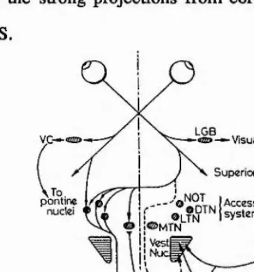
Figure 2.4. Visual pathways to the accessory optic system (AOS) in brainstem. From the retinae visual information Is distributed directly to LGB, to pontine nuclei, to superior colllculi and to AOS. The pontine nuclei link with the vestibular system (not indicated). Abbreviations: LGB, lateral geniculate body; NOT, nucleus of the optic track; DTN, dorsal terminal nucleus; MTN, medial terminal nucleus; LTN, lateral terminal nucleus; NRTP, nucleus reticularis tegmenti ponti. (From Berthoz, 1981).
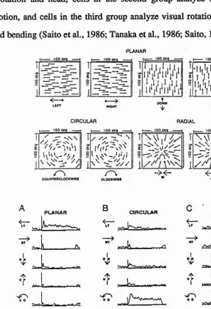
[image:22.612.185.367.238.433.2]2.5). Like neurons in the AOS, a large fraction of the MSTd cells within each category prefers the movement of a wide field to movement of a small object. It has been suggested that the cells in MSTd are involved, therefore, in the analysis of visual field flow caused by self-movement of the animal. The cells belonging to the first group analyze straight and parallel visual flow caused mainly by eye-movement or head rotation and head, cells in the second group analyze radial flow caused by locomotion, and cells in the third group analyze visual rotational flow accompanied by head bending (Saito et al., 1986; Tanaka et al., 1986; Saito, 1992).
PLANAR
T ■ lOOQfQ — 1k—— 100 d^Q .■■.■■■I
T ^
i U 1 ^ 1^ j 1 }H— *00 dfd — H'I (, If 1,1 If ^1 I,s s S I ' t ' , V i'
1 o
li l l ^ li l 2
1 1 11*1*111*1* 1*
i
I I I ' l MBOWH
CIRCULAR too iwg ---1 I--- 100 a«a
RADIAL
-couMttflCtocKwise
T
W ' f .
(xocxwne
\ T
OUT
PLANAR B CIRCULAR RADIAL
V
“Î
\ A.
U _____^
“Î
4—
K— —^
?V >. A T I. I l f f —
[image:23.613.117.424.209.658.2]Roy and Wurtz (1990) have found that MST neurons that are responsive to motion in the fronto-parallel plane code the direction of movement as well as the depth of motion from disparity cues. When stimulated with random dot displays the neurons responded, for example, when the “foreground" (i.e. in front of the plane of fixation) moved to the left or when the "background" (a stimulus behind the plane of fixation) moved to the right, or both of these movements. Roy and Wurtz argue that even more effective in signalling the direction of self-motion were cells (40% of those tested) which preferred one direction of motion with visual stimuli of one sign of disparity and the opposite direction of motion for stimuli of the opposite sign of disparity. Such a neuron could respond, for example, when the foreground moves to the left or when the background moves to the right or both. This arrangement of optic flow occurs in the situations where the line of sight and the direction of motion are not parallel, for example, during lateral displacement of the head. The proposition that these neurons contribute a signal about the direction of self-motion was further supported by the findings that a predominance of the disparity-dependent direction- selective neurons preferred horizontal motion as opposed to oblique or vertical motion. Macaque monkeys are primarily terrestrial animals and their locomotion occurs mainly in the horizontal plane.
2.3. VISUAL STABILITY DURING SELF-INDUCED EYE MOVEMENTS
however, does not evoke sensation of visual movement (or Illusionary self movement).
Theories accounting for experienced perceptions. When the eye is voluntarily moved and the optical image slips across the retina, we do not experience any movement and the world appears stationary. Instead, a gentle pressure onto the eye-ball leading to a slight lateral displacement of the eye evokes an apparent motion of the visual world (see Fig. 2.6). Experiments with visual afterimages have shown that afterimages (i.e. images with fixed retinal location) appear to move during normal eye movements or with attempts to move a paralyzed eye, but remain still during movements imposed externally onto the eye ball (for a review see Grüsser, 1986). These results have
0 0 S- 0 0
Gaze directed aheod Ooze voluntarily shifted Imposed mov’t shifts to left gaze to left Eye stationary
No commond Tor get appeors
stationary
Eye moves Command given Target appears
stationary
Eye moves No command Target appears to
move to right
+
-Voluntary attempt to look to left — eye muscles paralysed Eye stationary Commond given Target oppeors to
move to left
Figure 2.6. Diagrams showing actual positions of the eye, viewed target and its retinal image. Explanations underneath each picture describe the details. Plus (+) and minus
(r) signs in the pictures refer only to the ogonlst/ontagonist eye muscle activation. (From McCloskey, 1981).
[image:25.612.98.449.307.525.2]cancel the visual motion signal and this leads to the sensation of visual motion in the opposite direction. Correspondingly, voluntary eye-movement does not result in the retinal displacement of an afterimage and as there is no afferent motion signal present to be cancelled, the subject experiences an illusionary sensation of movement of the afterimage.
Sperry (1950) and von Holst and Mittelstaedt (1950) have provided two well known theories of the neural basis of the visual stability during voluntary eye movements. Sperry (1950) introduced a notion that the motor command centers generate two types of signals: a motor command to oculomotor centers for moving the eyes and a corollary discharge into the visual centers to compensate for the "expected" retinal displacement. Von Holst and Mittelstaedt (1950) presented a slightly different theory in which they proposed the motor command to be accompanied by an ejference copy to the sensory systems. An efference copy is sent to sensory structures simultaneously with a command sent from the oculomotor stmctures to move the eyes. This efference copy can be supposed to be a positive (+) signal whereas the sensory signal ("re-afference") caused by eye movement is a negative (-) signal. When these two signals are summed at some level in the central nervous system, no signal of movement arises and the visual surround is perceived as stable (Fig. 2.7).
movements would not contribute any information about the environment itself and, therefore, would not modify the representation. In this model, there would be no question of suppressing these reafferent sensory signals as they would be used for
R O T A T IO N E YE
Figure 2.7. The efference copy - re-cafference principle. An efferent command Is sent from a motor center (Ci) to move the eye to the right. An efference copy of this signal marked with a plus (+) sign Is also sent to a sensory center. This sensory center receives an afferent sensory signal from the retina which Indicates that the point x on the retina has moved from 1 to 2. This afference (re-afference) has a minus (-) sign and it is cancelled by the efference copy (+ signal). Higher centers (Cn) receive no message of motion. (From Hyvdnnen, 1982).
providing sensory feedback for the guidance of the locomotor activity. The function of corollary discharges is to provide information (or set criteria) to the central mechanisms to evaluate whether incoming afferent sensory signals require a readjustment of the internal representation of the environment.
Sherrington, 1918). It has been shown that normal subjects with completely paralyzed
eye muscles by drugs do not experience displacement of the visual world on attempting to move their eyes (Siebeck, 1954; Brindley et al., 1976). Brindley et al. (1976) also studied the effects of drug induced paralysis of one eye on movements of retinal afterimages. Afterimages formed in the completely paralyzed eye did not move during attempts to move the eyes, whereas this illusion was perceived in the other, non-paralyzed eye. At least a part of these differences between the studies just described and the early studies may be explained by the fact that in the early studies the patients* eye muscle paralysis was not complete (Brindley et al., 1976; McCloskey, 1981). Referring to the possible role of afferent proprioceptive information from eye muscles in visual stabilization during eye movements, McCloskey (1981) points out that afferent input may be more effective during voluntary eye movements as compared to imposed rotations. "The mechanical state of the extraocular muscles, the background of contraction-induced proprioceptive reafference, and the central neural context into which afferent impulses flow could all be expected to be different in active and imposed eye movements." McCloskey considers the possibility that corollary discharge acts as a gate allowing relevant kinaesthetic input access to perceptual mechanisms only during self-induced, voluntary eye movements. Thus during normal eye movements "expected" self initiated retinal motion would be ignored by central mechanisms on the basis of incoming proprioceptive input and an efference copy of command signals.
by pressing the eye ball. For example, the peak velocity of a 20 degree saccadic eye movement reaches 900 degrees/s (Wurtz et al., 1980). Smooth pursuit eye movements, such as those in tracking a moving target or those generated by vestibular stimulation, are evoked reflexly and are much slower. Their function is to stabilize the retinal image of a target when either the target or the head moves. Saccadic eye movements differ from slow pursuit movements in several perceptual and physiological characteristics and they are generated by two different oculomotor sub systems (Leigh & Zee, 1983). It is possible that the lack of voluntary control over smooth pursuit movements is a naturally evolved mechanism to prevent an illusionary sensation of "world motion". Interestingly, if a slowly moving object is tracked with a smooth pursuit movement, subjects tend to experience a illusory motion of the background (i.e. the whole field). This is known as the Filehne illusion (Filehne, 1922; Mack & Hermann, 1973). In this case the motion sensation is, however, complicated by other motion cues present (i.e. relative movement between the target and background). A smooth pursuit in absence of a target would produce an optimal experimental situation for comparing slow velocity image movement caused by real object motion with that caused by eye motion. Unfortunately, as mentioned above, voluntary pursuit in the absence of a target is impossible.
of corollary signals. Corollary discharges provide the information about eye position in darkness, and they have a role especially in intersensory coordination of vision with other modalities in both structured and unstructured visual conditions.
Stevens et al. (1976) presents a theory for the existence of three independent systems in explaining visual stabilization which also accounts for some of the controversies in experimental results. They studied visual perceptions during drug- induced paralyses. One of the crucial findings was that attempts to make saccades during partial paralysis resulted not in the sensation of dynamic motion of the visual field (as earlier studies had described) but a sudden "static displacement" of the field, i.e. a jump of the field from one position to a second. According to their theory an eye position system receives input from sensory receptors located in the conjuctival sac
and extraocular muscles, and this system is responsible for the sensation of effort reported by their subjects during attempted eye movements. A second pattern visual system receives its input from the retina. When the whole retinal surface is activated by a large rapid image movement (e.g. during a saccade), pattern vision is suppressed, whereas an activation of a limited area of retina by an image movement leads to perception of movement. This way the pattern system may distinguish between self induced image movements and movements of objects in the external world. Finally, Stevens et al. (1976) propose a spatial system which is responsible for perceptions of spatial localization receiving an input from the retina and a corollary discharge from motor centres. This system makes it possible to differentiate between self-induced displacements and external displacements of visual world, producing a perceptually stable visual world.
the visual field, independently of the eye movements, and indeed, this type of neuron has been found in several visual areas.
Single cells in monkey’s superior colliculus have been found to respond selectively to real stimulus movement and to "ignore" comparable motion of stimuli across the retina produced by saccadic eye movements (Fig. 2.8; Robinson & Wurtz, 1976; Richmond & Wurtz, 1980). Moreover, these experiments showed that the
t
r
FIX A TIO N L IC H T
SA CCADE TA RG ET
STA TIO N A RY STIMULUS
R E C E P T IV E FIE L D FIX A TIO N LIG H T
a
H
V
A .250-j
[image:31.612.86.475.221.615.2]discrimination is caused by an extraretinal signal suppressing the visual responses in collicular cells (evident from the suppression of spontaneous activity in the dark during eye movements), and that the most likely source of this extraretinal input is a corollary discharge from some part of the oculomotor system. Similar results have been obtained from the pulvinar which receives a direct projection from the superior colliculus (Robinson & Petersen, 1992). Even though the existence of connections from oculomotor muscle spindles to the superior colliculus has been demonstrated (e.g. Cooper & FiUenz, 1955), the possibility of this suppressive input being of proprioceptive origin has been excluded. For example, the latency of the suppressive input acting on these collicular cells was too short to originate from oculomotor muscle spindles (Robinson & Wurtz, 1976), and the suppression persisted even after blocking of motor and proprioceptive nerve fibers entering and leaving the orbit (Richmond & Wurtz, 1980).
Evidence in favour of corollary discharges providing eye position information in monkeys has also been reported by Guthrie et al. (1983). They showed that monkeys with transections of the ophthalmic nerves (which eliminated extraocular muscle proprioception) were able to generate accurate compensatory saccades after perturbation of eye position produced by stimulation of the superior colliculus, to targets which were extinguished before the eye position perturbation.
effectiveness of the visual stabilization in a wide variety of conditions. The coroUary discharge mechanisms are effective over a wide range of light and contrast levels, but modify the responses of only a half of the collicular cells. The retinal mechanisms instead affect all cells but work reliably only in a patterned visual environment with high contrast levels present.
(
J
B
i
Figure 2.9. Response of a collicular cell to stimuli moving across the cell's receptive field (dashed circle). A) Response of the cell to a stimulus which moves across the suppressive surround of the cell before entering the receptive field. B) and C) The responses were Improved when the surrounding areas of the receptive field were shielded. The approximate time at which the stimulus crossed the receptive field is indicated by the arrow beneath each histogram. (From Wurtz et a!., 1980).
received neither proprioceptive or visual information. The activity of one type of the cells was correlated solely to the motor component during saccades, whereas the second type of cells was found to code eye position. The finding that the activity of both cell types began after eye movements indicated that these cells were not involved in creating eye movement commands, but were very suitable for producing corollary discharges to other brain areas. However, monkeys with ablations of the frontal eye fields still show suppressive activity in the superior colliculus, which makes it questionable that the frontal eye fields are the (only) source of corollary discharge (Richmond & Wurtz, 1980).
While the above results indicate that saccades affect the processing of visual information in the tectopulvinar visual system, the geniculocortical pathway at the level of lateral geniculate nucleus remains visually sensitive during saccades and no visual and oculomotor interaction has been observed in LGN cells (Biittner & Fuchs,
1973).
26
suppression might be evident perceptually as a pause in continuous vision. This is not our experience.
When comparisons have been made between responses to slow stimulus movements and to equivalent stimulus conditions during tracking eye movements, striate cells have also been observed to differentiate between these two conditions. Fischer et al. (1981) reported stronger responses to slow real stimulus movement in about 5% of the recorded striate neurons (from their Fig. 5). GaUetti et al. (1984) have reported 10% of the cells in VI to be sensitive only for "real motion" (i.e. stimulus motion across stationary retina).
In the prestriate visual areas the proportion of cells discriminating between self-produced and externally-produced motion across retina is considerably higher. The general rule seems to be that the proportion of such cells increases as one records from visual areas increasingly higher in the hierarchy of visual areas. In area V2, Galletti et al. (1988) reported about 14% of the studied neurons gave high responses to real moving stimuli, but weak responses to equivalent retinal image displacements due to eye movements. In area V3a the proportion of these ’real-motion* cells has increased to 48% (Galletti et al., 1990).
property increases within the STS along a posterior-to-anterior axis from MT to MST" (Erickson & Thier, 1991).
The difference in results from those of Galletti and his co-workers could be, not necessarily due to shortcomings in controlling experimental variables as suggested by Erickson and Thier (1991), but rather because of the backward projections from area MST to cortical areas at the earlier stages (see Fig. 3.2 in Chapter 3 and for a review of the cortical connectivity see, Felleman & Van Essen, 1991). If it is postulated that area MSTd presents the stage in the hierarchy of visual motion analysis which is responsible for processing self-induced retinal motion input differently from externally-induced signals, MSTd could return the discriminated information to lower levels. It is not surprising that Erickson and Thier (1991) did not find real-motion or ’passive-only* cells from area V4. This would be because there does not exist a projection from area MSTd back to area V4, whereas MSTd does project directly back to areas such as MT, V3A and V2, and real-motion cells in VI would in turn depend on influence of back projections from V2 to VI. When it comes to areas below V4, it is not clear exactly which areas Erickson and Thier (1991) recorded from, and in any case the number of the recorded cells was so small (as the total number of cells recorded from V1-V4 was reported to be 24) that it is hardly surprising that they did not find any passive-only (real-motion) cells from these areas. According to Galletti et al, (1984, 1988, 1990) the proportion of real-motion cells was
10%, 14% and 41% in areas VI, V2 and V3A, respectively.
alone (Fig. 2.10). This suggests that where the corollary discharge is acting its input selectively inhibits the visual input but does not induce a general shut down of responsiveness. In this respect there is an important difference in comparison to the processing in the superior colliculus. Collicular cells do show a general suppression of spontaneous activity connected to the saccadic eye movements themselves, instead of a selective inhibition of visual responses (Robinson & Wurtz, 1976; Richmond &
EYE MOVEMENT VISUAL
BACKGROUND STIMULUS MOVEMENT CONTROL
c
1
LIGHT 1
un>t O iM T
. . , A . . . . ^ lillluUiiUi ^1 1 iVt 1
<0 r
O - Q.
<0 1
DARK II il II i«#0M*#AI«««i i4J É É iI i i . i . i a t A É A j i U i u Ml II 1 1 limlilll III lill W W
1 0 0 m s e c /d iv is io n
§
w
LIGHT 1 im It III ill 1 III 1 I lilIlih U y i 1 ■ mIi Im 1 1 H ^
w <D a
DARK ” 1 ■■ 1 « lü ü h if I I 1 1 U É i i . 1 1
100 m s e c /d iv is io n
Figure 2.10. Responses of two "real-motion" ceils during stimulus movement (steady fixation), during tracking eye movement and during tracking eye movement without any visual stimulation on the receptive field (control). The cell above maintained Its real-motion behaviour also when tested with an illuminated stimulus in darkness. This indicated that the ceil received an extra-retinal Input. The cell below failed to show real motion behaviour in darkness which suggest that this ceil received a retinal signal. (From Galletti et al., 1990).
Wurtz, 1980). Cells in areas MT and MSTv, as described in the previous section, also contribute in generating the retinal stabilization mechanisms through the surround suppression of large field motion (Allman et al., 1985; Saito et al., 1986; Tanaka et al., 1986; Sugita et al., 1990).
MacKay (1973) which were described earlier. He proposed that corollary discharges are not used to eliminate the sensory changes due to voluntary eye movements but to provide information to the central mechanisms that the internal representations perceivers have build up and stored from their environment do not need readjustment. Recently, Duhamel et al. (1992) have shown that for some parietal neurons the location of the receptive field shifts transiently in accordance with the intended eye movement before the eye movement is actually executed. This shows that parietal neurons ’anticipate’ the retinal consequences of intended eye movements. Moreover, those parietal neurons which do not show predictive remapping do exhibit, however, responses that reflect a visual memory trace that has been remapped in conjunction with the eye movement. These neurons respond when the eye movement brings the stimulus into the receptive field, even though the stimulus has been distinguished earlier. Duhamel et al. (1992) suggest that these response properties reflect the effects of signals corollary to eye movements and that they cause the parietal representation of the visual world to undergo a shift that predicts the location of reafferent visual input.
between the two mechanisms could be that corollary discharges facilitate the detection of real movement in the environment over a wide range of light and contrast levels, and provide information about spatial attributes of the environment to other sensory systems, whereas processing of whole field motion and selective sensitivity to local relative motion provide highly accurate information of movement in the complex, illuminated visual world. Two parallel systems secure efficient function over a wide range of external conditions.
2,4. PROCESSING OF SOMATOSENSORY STIMULI AS A RESULT OF ACTIVE EXPLORATORY MOVEMENTS
As Gibson (1966) points out "the perceptual capacity of the hand goes unrecognized because we usually attend more to its motor capacity, and also because the visual input dominates the haptic in awareness". Indeed, it is interesting to compare exploratory hand movements and exploration of the visual surroundings with voluntary eye movements. A quick comparison would show that both "perceptual systems" consist of a movable organ having a mosaic of sensory receptors (skin/retina) attached to it.
subsystems. He remarked how sensory psychology had failed to emphasize the difference between active touch (touching) and passive touch (being touched). Active touch is purposive by its nature and it consists of two kinds of input information, objective and subjective. What is most important, the purpose of active, exploratory hand movements is to isolate and enhance objective components over the subjective ones.
Single-unit recordings from the somatosensory cortex (areas 1 and 2) in monkeys have revealed tactile neurons which are closely related to active exploratory hand movements (Iwamura et al., 1985). The cells exhibit stronger responses when the tactile stimulation results from active touch as opposed to otherwise similar, but passive tactile contact. Both excitatory and inhibitory activity changes are present. Some neurons signal the movement of skin relative to the object, and again for these cells active hand movement over the surface texture produces stronger responses as compared to object motion across a passive hand/skin. The receptive fields of the cells have very precise requirements and, for example, an activation may necessitate an active manipulation of an object with two particular fingers. What is especially interesting, is that the cell discharges do not precede the contact of finger with the objects (cf. the response properties of posterior parietal neurons which are described later).
these results; afferent suppression induced by displacement of the stimulated skin area, or inhibition by descending motor commands. Psychophysical experiments showed that the perceptual suppression started more than 100 ms before the onset of muscular activity, but similar perceptual attenuation was also observed before and
after a passive displacement of the stimulated skin area. These results were taken to
4
suggest that active or passive displacement of the stimulated skin area may causeinhibition of afferent somatosensory input transmission and induce perceptual ® masking. In this respect all these results resemble those obtained with saccadic eye
movements as described in the previous section. It was argued that saccadic suppression involves both motor (corollary discharge) and afferent (retinal displacement) mechanisms.
The results described above seem to contradict the notion that active movements of the hand enhance its capacities in perceptual tasks. One reason for this seeming discrepancy could be that the simple movements that the subjects were required to perform during the experiments were not of an exploratory character, controlled by tactile and proprioceptive feedback (Gordon, 1978). Another possibility is that observations of overall reduction of ascending activity as made in macroelectrode recordings at different neural levels may conceal a pattern of selective depression. For example, in exploratory movements the spatial acuity might be improved by increasing lateral inhibition in a relay nucleus, but a macroelectrode recording would only show a general attenuation of activity (Gordon, 1978).
said to become "expected". Active exploratory hand movements made out of sight by the animal itself were found to produce no tactile responses when the monkey contacted familiar, expected surfaces such as its own body or highly familiar items within reach. By contrast equivalent tactile stimulation as a result of encountering novel, unexpected surfaces produced vigorous neuronal responses. What kind of information is then used for forming expectations about one’s incoming tactile
sensations? The authors suggested that this requires knowledge of the tactile surface 1 properties of the objects in the environment, spatial memory for the normal location
of objects in the environment and information as to the current position and trajectory
of limb movements. In fact, visual information could have been added to the previous | list, but as it reflects the effects of intersensory information transfer, this case is
handled separately in section 2.7.
2.5. ATTENUATION OF RESPONSES TO SELF-VOCALIZED SOUNDS
Most vertebrates rely on vision in orienting their behaviour with respect to the environment, and it is the visual system which provides the information necessary for responding adequately to such environmental features as size, shape, distance, nature and movements of objects (Henson, 1965). However, some of these functions are shared by the auditory system. In humans it is easily forgotten that audition, besides its social-communicative role, can play an important role in assessing the nature and movement of animate and inanimate objects. Experiments with blind people have shown that the human ear is capable to make relatively fine size discrimination, simple shape discrimination, and target location in space based on echo-information received from self-emitted vocal sounds (Rice & Feinstein, 1965; Rice, 1967).
that within the midbrain most of the analyzed cells responded to self-produced vocalizations in a similar way as they did to the same vocalizations played back from an audiotape. Rather less than 10% of the cells showed no or only a weak response during the self-produced calls. By contrast, in the thalamus and in the auditory cortex a considerable number of neurons that reacted to playback calls did not respond to self-produced vocalizations. It seemed, thus, that higher stations of the auditory pathway display less activity during animal’s vocal activity than lower ones. Two different functions were proposed for this selective neural processing. First, it could have a monitoring function required for controlling and learning species specific vocal performance. Secondly, calls are not of communicative value to the producer itself, and hence, the increasing number of cells in the higher auditory stations not engaged in the processing of self-produced calls would be free for analyzing essential signals coming from the environment This would make these higher level cells appropriate for selective auditory attention to external sounds.
2.6. PROCESSING OF EXPECTED EXTERNALLY-INDUCED STIMULI
This section describes situations where an externally-induced stimulus has become, in a way or another, expected to the animal. These are probably the cases which most usually come to mind when examples of "expectation" are considered. As will be seen, there are several ways in which expectations can be created in experimental conditions but the focus here is, again, only in single cell studies.
delivery of a certain signal and an occurrence of another signal shortly after the first one (cf. second-order conditioning). If most of the time the following stimulus matches that which subjects were forewarned about, the subjective probability of the stimulus is high and the stimulus will be expected. If instead, a stimulus is presented contrary to the cues given beforehand, then the stimulus can be said to be unexpected to the animal.
Hocherman et al. (1981, 1990) trained rhesus monkeys to push a lever either to the left or right depending on whether they were presented an auditory signal of noise or tone, respectively. After the monkeys had mastered this task the behavioural paradigm was changed and additional light signals preceded the auditory ones. A flash of light on the left side signalled the occurrence of the noise signal (and a lever push to the left) and a light signal from the right preceded each tone stimulus. Now, it was possible for monkeys to predict the nature of the auditory stimulus based on the location of the visual signal. In a predetermined number of trials the auditory stimulus was preceded by the "wrong" visual signal. Responses of single units in the auditory cortex and the medial geniculate nucleus were recorded to tone and noise stimuli. Comparison between unit activity in true vs false conditioning trials revealed that on trials with correct behavioural responses half of the studied units were not affected by the predictability of the nature of the auditory stimulus. The other half was divided between neurons whose responses to the auditory stimuli were greater on true conditioning trials and neurons which responded more vigorously on false conditioning trials.
revealed that in the subgroup of units whose responses were greater in true conditioning trials than in false conditioning trials, the responses were actually facilitated in the former case and inhibited in the latter case. A different pattern was found for units whose responses in false conditioning trials were greater than in true conditioning trials. These units showed a base-line response activity in true conditioning trials, but a strong facilitation in false conditioning trials.
These observations led the authors to suggest the existence of two neural mechanisms (Hocherman et al., 1981). A signal-match detection mechanism involves units whose response to a correctly predicted stimulus is facilitated and whose response to a wrongly predicted stimulus is inhibited. An error-detection mechanism, on the contrary, includes units emitting a base- line activity to correctly predicted stimuli but showing strong facilitation to wrongly predicted (and hence unexpected) stimuli. These mechanisms are thus involved in the comparison of actual auditory signals with existing predictions about those signals. The authors proposed that such a comparison can be done if the realization of a particular prediction took the form of a specific spatial template of excitability through one or several levels of the auditory system. The observation that subcortical cells were affected by the effects of anticipation (Hocherman & Yirmiya, 1990) suggested that ’anticipation-related’ modulation of auditory information processing starts at the subcortical levels and that it is probably through descending corticothalamic projections that cortex controls the information processing in the lower levels of auditory system.
types were interleaved and the nature of the experimental condition was indicated to the monkey before the reaction signal with a light stimulus. In other words, responses to high tones were always rewarded, whereas responses to low tones were only rewarded in the condition where the pitch did not matter (signalled by the light cue). Single unit recordings from auditory cortex revealed that 25% of the sampled units showed definite alterations in responses to the same tonal stimuli under different experimental conditions. All these cells (exept one) showed increased activity to the same low tone stimulus in the unrewarded condition relative to the responses evoked in the rewarded condition. No response modulation was observed to the high frequency stimuli which always signalled reinforcement. When trying to provide an explanation for their results Beaton and Miller (1975) stated that the "data do not fit easily within an attention or alerting model", because the monkeys had to attend to the stimuli in both conditions. They concluded that the activity of these neurons was correlated with a specific behavioural state which was a result of experimental conditions that controlled the monkeys’ performance. Any other descriptions or definitions of this "behavioural state" were not offered.
had the property that they emit a baseline activity to correctly predicted stimuli but respond to unexpected stimuli.
Hypotheses about specific neural activity patterns created by expectation of particular sensory stimulation have gained experimental evidence in the studies by Freeman (1979). He found that the spatial pattern of EEG activity on the surface of the olfactory bulb in mice tended to be similar irrespective of the sensory input, whereas it changed to a new pattern when animals were expecting a particular odor. Freeman suggested that during learning, the pattern of neuronal activity which is induced by an odor provides the specification for a neural template in a form of strengthened connections between the neurons activated by that odor. After this learning phase the template could be activated by centrifugal connections in order to serve as a selective filter for expected odors.
The classical matching to sample paradigm can be seen as a task which requires that a neural template is formed from the sample stimulus and that the matching is based on comparing subsequent incoming sensory stimulation with this template. In this paradigm the subject is first shown a sample stimulus (a cue) and thereafter the subject is required to select a corresponding match stimulus from a number of possible stimuli. In other words the sample stimulus must be used to build up a kind of prediction about the subsequent matching stimuli, of which one is to fit to the prediction. Now, transferring this paradigm to neurophysiological experiments it is possible to study, for example, whether responses to matching stimuli depend on the preceding sample stimulus.
that it could not see). In homomodal trials the sample orientation was presented visually on the screen in front of the animal. The four matching stimuli were presented as a sequence of visual gratings and the animal was required to release a switch when it saw a grating whose orientation matched the sample orientation.
For over half of the neurons the visual responses to match stimuli were affected by the nature of preceding sample stimuli. The largest set of neurons was tested using the tactile-visual matching task. Two thirds of the neurons that were sensitive to both sample cue and match stimulus orientation showed a significant response interaction between cue and match stimulus orientation. About half of these neurons responded best to one of the four matching combinations in which both the cue and match stimulus orientations were the same (Fig. 2.11) and the other half responded best in a certain non-matching condition. No clear example was found of a neuron that responded well to each of the matching conditions but did not respond to non-matching conditions.
STIMULUS
M
C
u
# I :1
oJUJ
[uIl
■ ■ ■ JiALM III
Jliki
5 s / s e cThe results of visual-visual matching task were generally similar to those using the tactile-visual match. It is worth noticing that in visual-visual tasks the cue orientation was removed before the presentation of any stimuli. So, for example, cells were found whose responses showed sensitivity to a certain cue orientation, but to a different matching stimulus orientation, even though both were presented visually. Haenny et al. (1988) concluded that the cell tuning to cue orientation represented information specifically relevant to the matching task, rather than a basic sensory signal - either somatosensory or visual. Generally, the results provide support to the existence of "neural templates" created in the matching task in V4 and suggests that there are V4 units that are involved in comparing cue information to the actual stimulus information.
In the examples presented above, the expectations that the animals had, were formed because of a previous training in a certain type of behavioural task and, moreover, the expectations were likely to be present at a "conscious" level. However, it can be argued that natural every-day life is full of instances where a sensory experience is expected only at a "unconscious" level. Most environmental events are perceived and analysed by more than just one sensory channel, and this leads animals (and humans) to form intersensory associations. These associations can have a predictive function. For example, seeing an object approaching one’s body signals an impending tactile sensation.
relatively easy to see the functional importance for such mechanisms. As the authors suggest, when someone or something touches us from out of sight, there is a compelling sensation of being "touched" and this may indicate a need for interaction with another individual.
Anticipatorv activity before stimulation. Still another effect of expectation on neuronal responses can be found in cases where changes (reflecting the expectation) in neuronal activity precede the actual stimulation of the sensory system in question. Single neurons in the dorsolateral prefrontal cortex (Sakai, 1974; Niki & Watanabe, 1979; Joseph & Barone, 1987), cingulate cortex (Niki & Watanabe, 1979), premotor cortex (Mauritz & Wise, 1986), primary motor cortex (Lamour et al., 1980) and caudate nucleus (Rolls et al., 1983; Hikosaka et al., 1989) have been shown to exhibit "anticipatory" activity changes preceding a task relevant stimuli. Extensive experience in performing a certain behavioural task is likely to lead to predictions about events within the task and it has been suggested that these neurons have a role in predicting environmental changes and preparing the animal for appropriate motor responses.
CH APTERS
THE SUPERIOR TEMPORAL POLYSENSORY
AREA IN THE PRIMATE
VISUAL SYSTEM
3.1. INTRODUCTION
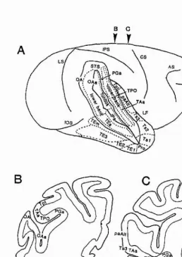
The principal object of this chapter is to describe the anatomy, connections and known physiological properties of the brain area that is the target of the neurophysiological experiments. The chapter starts with providing first a short overview of some of the anatomical and functional principles of visual processing within the primate cortex. This is particularly important because, as it wül be seen later, the investigated brain area seems to be a site of convergence of separate information processing pathways within the cortex and consideration of the functional properties within these pathways is relevant to the present study. After the general overview, the scope is narrowed. The order of presentation of individual brain areas proceeds from describing the target area to include some other both anatomically and functionally closely related brain areas. This kind of order (rather than following hierarchy of processing stages from the bottom up) is deliberately selected in the belief that it guides the reader to a better understanding of the reasons for the particular experiments that are carried out in this thesis.
3.2. VISUAL PROCESSING WITHIN THE PRIMATE CORTEX