University of Warwick institutional repository:http://go.warwick.ac.uk/wrap
A Thesis Submitted for the Degree of PhD at the University of Warwick
http://go.warwick.ac.uk/wrap/72926
This thesis is made available online and is protected by original copyright. Please scroll down to view the document itself.
User-Centred Design of a Task-Oriented
Upper-Limb Assessment System for Stroke
by
Christopher Golby
A thesis submitted in partial fulfilment of the requirements for the
degree of
Doctor of Philosophy in Engineering
WMG, University of Warwick
2 | P a g e
Contents
List of Figures……… ... 8
List of Tables……… ……..10
Acknowledgements……… .. ………..11
Declaration………...………...13
Abstract………..……….……...14
List of Abbreviations………..………15
Chapter 1: Introduction ... 17
1.1 - Context and Motivation ... 17
1.2 - The Need ... 21
1.3 - Aim of the Thesis ... 22
1.4 - Research Question ... 24
1.5 - Specific Objectives ... 24
1.6 - Contribution to Knowledge ... Error! Bookmark not defined. 1.7 - Organisation of the Thesis ... 25
Chapter 2: Literature Review ... 28
2.1 - Upper-Limb Motor Control Assessment of Stroke Patients in the NHS ... 28
2.2 - Motion Tracking as an Assessment Tool for Upper-Limb Motor Control. ... 33
2.2.1 - Examples of Marker-based Motion Tracking Systems ... 33
3 | P a g e
2.2.3 - Data Processing when Analysing Upper-Limb Motion ... 41
2.2.4 - Advantages and Disadvantages of Motion Tracking in Upper-Limb Motor Control Assessment ... 42
2.3 - The Use of the Microsoft Kinect as a Tool for Assessing Upper-Limb Motion. ... 43
2.4 - Alternative Technologies used in Stroke Patient Upper-Limb Motion Assessment (from which lessons may be learned). ... 50
2.4.1 - Virtual Reality ... 50
2.4.2 - Augmented Reality ... 51
2.4.3 - Robotics ... 53
2.4.4 - Haptic Technologies ... 56
2.4.5 - Brain-Computer Interfacing ... 58
2.5 - Summary ... 59
Chapter 3: Methods ... 62
3.1 - Requirements Elicitiation ... 62
3.1.1 - Elictiation Study Design ... 63
3.1.2 - Recruitment ... 64
3.1.3 - Sample ... 65
3.1.4 - Protocol ... 65
3.1.5 - Analysis ... 66
4 | P a g e
3.2 - System Design and Implementation Methodology ... 67
3.2.1 - Defining of Requirements ... 68
3.2.2 - Design and Development of Sytem ... 68
3.2.3 - Review of the Prototypes Technical Limitations ... 69
3.3 - System Verification ... 69
3.3.1 - Verification Study Design ... 70
3.3.2 - Recruitment ... 70
3.3.3 - Sample ... 70
3.3.4 - Protocol ... 71
3.3.5 - Analysis ... 71
3.3.6 - Ethics ... 72
3.3.7 - Hypothesis ... 73
3.4 - System Validation with Stroke Patients ... 73
3.4.1 - Validation Study Design ... 74
3.4.2 - Recruitment ... 74
3.4.3 - Sample ... 76
3.4.4 - Protocol ... 76
3.4.5 - Analysis ... 77
3.4.6 - Ethics ... 78
3.5 - Summary ... 80
5 | P a g e
4.1 - Findings from Non-Participant Observation ... 82
4.1.1 - Non-Participant Observation of Occupational Therapist with Patient 1 82 4.1.2 - Non-Participant Observation of Occupational Therapist with Patient 2 85 4.1.3 - Non-Participant Observation of Physiotherapist with Patient 3 ... 87
4.1.4 - Summary of Findings from Non-Participant Observation ... 90
4.2 - Findings from Semi-Structured Interviews ... 91
4.2.1 - Initial Patient Assessment ... 93
4.2.2 - Continuing Assessment ... 94
4.2.3 - Outcome Measures created by Individual Hospitals ... 97
4.2.4 - Issues ... 98
4.2.5 - Specific Outcome Measures ... 104
4.2.6 - Demonstration of outcomes to show progress in rehabilitation ... 106
4.2.7 - Technology ... 107
4.2.8 - Coping with Stroke ... 111
4.2.9 - Summary of Findings from Semi-Structured Interviews ... 113
4.3 - Discussion of findings from Requirements Elicitation ... 114
4.4 - Summary ... 117
Chapter 5: System Implementation ... 119
5.1 - A Critical Summary of the Issues from Requirements Elicitation. ... 119
5.2 - The derived Requirements of the System. ... 121
6 | P a g e
5.3.1 - Overview of the Developed System and its Aims ... 122
5.3.2 - The In-Therapy (data collection) Version of the System. ... 128
5.3.3 - The ADL (data collection) Version of the System. ... 134
5.3.4 - Presentation of Assessment Results in the User-Interface ... 139
5.3.5 - Video of the User Interface ... 143
5.4 - Technical Limitations ... 143
5.5 - Summary. ... 146
Chapter 6: System Evaluation ... 147
6.1 - System Verification ... 147
6.2 - System Validation ... 151
6.2.1 - Field Notes ... 151
6.2.2 - Post Assessment Interviews ... 155
6.3 - Summary ... 171
Chapter 7: Discussion ... 173
7.1 - Discussion of the Designed System (Requirements Elicitation and System Implementation) ... 173
7.2 - Comparison of the Implemented System against the System Requirements 175 7.3 - Discussion of System Verification ... 179
7.4 - Discussion of System Validation ... 181
7.4.1 - Advantages of the System ... 181
7 | P a g e
7.4.3 - Overall Disadvantages of the System ... 186
7.4.4 - Issues in Collected Results ... 187
7.5 - Generated Requirements for a New Version of the Current Prototype ... 190
7.6 - Generated User Needs for Future Systems and Components ... 193
7.7 - Limitations of this Research... 195
7.8 - Summary ... 197
Chapter 8: Conclusion ... 199
8.1 - Summary of Work and Results ... Error! Bookmark not defined. 8.2 - Conlcusions ... Error! Bookmark not defined. 8.3 - Contribution to Knowledge ... Error! Bookmark not defined. 8.4 - Further Study ... Error! Bookmark not defined. 8.4.1 - Extension of the Current Systems Evidence-BaseError! Bookmark not defined. 8.4.2 - Development of the Current Prototype .. Error! Bookmark not defined. 8.4.3 - User Needs for a Future System ... Error! Bookmark not defined. References ... 206
8 | P a g e
List of Figures
FIGURE 2-1:AGONIOMETER ... 32
FIGURE 2-2:USING THE PHILIPS RESEARCH STROKE REHABILITATION EXERCISER FOR ADL.THESE INCLUDE:A:SENSOR PLACEMENT AND GARMENTS.B:EXAMPLE OF DRINKING FROM A CUP.C AND D:EXAMPLES OF EATING WITH KNIFE AND FORK... 34
FIGURE 2-3:INTERACTIVE REHABILITATION AND EXERCISE SYSTEM (IREX) ... 39
FIGURE 2-4:BIOSTAGE PLATFORM FROM ORGANIC MOTION ... 40
FIGURE 2-5:MICROSOFT KINECT ... 44
FIGURE 2-6:MEAN± STANDARD ERROR MOTOR POWER SCORES (MAX 20) FOR PATIENTS USING THE MIT-MANUS ... 54
FIGURE 2-7:COMPARISON OF FEEDBACK TYPE INDIVIDUALS USING A COMPRATIVE FEEDBACK ROBOT. THE Y COLUMN SHOWS TIME SPENT PERFORMING REHABILITATION. ... 55
FIGURE 2-8:SENSABLE PHANTOM OMNI ... 56
FIGURE 4-1:TEMPLATE OF EMERGENT THEMES AND SUB-THEMES USED TO ANALYSE INTERVIEWS ... 92
FIGURE 5-1:THE PROTOTYPE SYSTEM PROVIDED TO THE NHS(INCLUDING LAPTOP,KINECT,EZ430 CHRONOS WATCH AND ADL OBJECTS ON PORTABLE TABLE) ... 123
FIGURE 5-2:MOVEMENTS (TOP:ABDUCTION/ADDUCTION,BOTTOM LEFT:PRONATION/SUPINATION, BOTTOM RIGHT:FLEXION/EXTENSION) ... 124
FIGURE 5-3:MICROSOFT KINECT AND THE JOINTS IT TRACKS ... 126
FIGURE 5-4:EZ430CHRONOS WATCH ... 127
FIGURE 5-5:SYSTEM LOGIN SCREEN ... 128
FIGURE 5-6:SKELETAL REPRESENTATION ON-SCREEN ... 129
FIGURE 5-7:‘ACTIVITY ZONE’, SHOWING THE AREA IN WHICH THE PATIENT MUST STAND, AS MEASURED BY THE SYSTEM. ... 131
FIGURE 5-8:OBJECTS USED IN ADL SECTION OF THE SYSTEM ... 134
FIGURE 5-9:USER INTERFACE DURING PATIENT-LED TASK STAGE ... 136
FIGURE 5-10:SYSTEM RESET SCREEN, AS DISPLAYED BETWEEN ADL. ... 137
9 | P a g e
FIGURE 5-12:USER INTERFACE DURING RESULTS PRESENTATION – ... 139
FIGURE 5-13:ELBOW ROMASSESSMENT DATA OVER TIME – ... 141
FIGURE 5-14:ADLACTIVITY BREAKDOWN ... 142
FIGURE 7-1:ELBOW ROMOVER TIME AS MEASURED BY THE SYSTEM IN PAT2 ... 188
FIGURE 7-2:PRONATION/SUPINATION ROMOVER TIME AS MEASURED BY THE SYSTEM IN PAT2 ... 189
10 | P a g e
List of Tables
TABLE 2-1:KINECT SPECIFICATIONS ... 47
TABLE 4-1:DIFFERENT OUTCOME MEASURES AND TIMES MENTIONED ... 105
TABLE 4-2:TECHNOLOGIES MENTIONED BY THERAPISTS IN THE INTERVIEWS ... 108
TABLE 6-1:SUMMARY OF T-TESTS. ... 149
TABLE 7-1:SUMMARY OF MAIN TECHNICAL ISSUES ... 184
TABLE 8-1:SUMMARY OF ADAPTATIONS REQUIRED TO CURRENT SYSTEM ... ERROR!BOOKMARK NOT DEFINED.
11 | P a g e
Acknowledgements
I would firstly like to thank all the supervisors involved in this work.
Professor Gillian Hundt’s sheer drive and determination, constant advice, support
and general care has been invaluable throughout. She was always there supporting
when times were tough towards the end, and I am genuinely grateful for this.
I am also highly grateful to Professor Theodoros Arvanitis, who stepped into this
work towards the latter half, without any hesitation. Without his guidance,
knowledge and constant support, this work would not have turned into what it is, and
I am extremely thankful for that.
I would also like to thank Professor Vinesh Raja, who steered the project from the
beginning. Without Vinesh’s ideas, trust and motivational talks, this work would not
have been possible.
The combination of skills and experience from having three supervisors throughout
has led to the rounded approach I have managed to adopt in this work, and I have
learned so many lessons from each of them.
Perhaps more formally, I am grateful to the Engineering and Physical Sciences
Research Council (EPSRC) and Economic and Social Research Council (ESRC) for
providing me with the funding to conduct this research and I hope it moves on to
have an impact in healthcare systems, helping the patients who are so in need of this
12 | P a g e Thanks must also be given to everybody I have met in WMG and the University of
Warwick who have allowed me to have such a great time, whether in work or
socially.
I would also like to take time to thank my Mother and Father for their support
throughout this PhD and for all their care up to this point. Without what they have
put up with and been through, none of this could have been possible, and I shall
always be grateful. I fully understand how much you have surrendered over the years
to achieve this, both in your own individual ways. Mom, I love our constant chats,
and always relish coming home to see and spend time with you. Dad, I’m always
excited by my trips away to see you and am hoping that this shall continue and the
amount of trips increase in the near future!
I also would like to thank Jo for her constant encouragement and kindness through
the past few years; I truly believe she is the most caring individual I have ever met.
She has changed my views of the world and I have no doubt that I would not have
completed this without her. I grow more and more excited every day about our future
together.
Another mention must be given to Jo’s family for all the times they have supported
me. I’m not sure they realise how welcome they have made me feel, and how they
have turned Leamington into my second home!
I would like to mention my Nan and Grandad, who have put up with my ramblings
for the last few years, yet have always been there to listen and fill me with
confidence! Much of what they taught me as a child has led to this; I always wanted
13 | P a g e Mentions must also be given to my two sisters, Corrina and Siobhan, and also to
Richard, who have always been there, without a moments doubt. I know I can
always rely on them.
Finally, I would like to thank all the participants who were involved in this research,
with particular thanks to the two NHS therapists who were involved in the
structuring of this research, I cannot mention names at this point for ethical reasons,
14 | P a g e
Declaration
This thesis is submitted to the University of Warwick in support of my application
for the degree of Doctor of Philosophy. It has been composed by myself and has not
been submitted in any previous application for any degree
The work presented (including data generated and data analysis) was carried out by
the author except in the cases outlined below:
Thematic coding of interviews - Transcripts were also read by Professor Gillian
Hundt (supervisor) in order to collaborate on themes to be used within the full
analysis.
Parts of this thesis have been published by the author:
Golby, C., Raja, V., Hundt, G. (2013). Physiotherapists' and Occupational
Therapists' Perceptions of the Assessment of Stroke Patients for Musculoskeletal
Rehabilitation in the UK National Health Service, Journal of Health 5(9)
pp.1478-1485.
Golby, C., Raja, V., Hundt, G., Badiyani, S. A Low Cost Activities of Daily Living
Assessment System for the Continual Assessment of Stroke Patients, from Acute
Rehabilitation through to Telerehabilitation. In: Successes and Failures in
15 | P a g e
Abstract
During rehabilitation from Stroke, patients require assessment of their upper-limb motor control. Outcome measures can often be subjective and objective data is required to supplement therapist/patient opinion on progress. This can be performed through goniometry; however, goniometry can be time-consuming, have
inaccuracies of ±23º, and is therefore, often not used.
Motion tracking technology is a possible answer to this problem, but can also be costly, time-consuming and not suitable for the clinical environment. This thesis aims to provide an objective, digital intervention method for assessing range of motion to supplement current outcome measures which is suitable for the clinical environment. This was performed by creating a low-cost technology through a user-centred design approach.
Requirements elicitation demonstrated that a motivational, portable, cost-effective, non-invasive, time saving system for assessing functional activities was needed. Therefore, a system which utilised a Microsoft Kinect and EZ430 chronos wrist watch to track patient’s movements during and/or outside of therapy sessions was created. Measurements can be taken in a matter of minutes and provide a high quantity of objective data regarding patient movement.
The system was verified, using healthy volunteers, by showing similar error rates in the system across 3 weeks in 10 able-bodied individuals, with error rates produced by a physiotherapist using goniometry. The system was also validated in the clinical setting with 6 stroke patients, over 15 weeks, as selected by 6 occupational therapists and 3 physiotherapists in 2 NHS stroke wards.
The approach which has been created in this thesis is objective, repeatable, low-cost, portable, and non-invasive; allowing it to be the first tool for the objective
16 | P a g e
List of Abbreviations
Abbreviation Definition
ADL Activities of Daily Living
API Application Programming Interface
BCI Brain Computer Interface
CAHAI Chedoke Arm and Hand Activity Inventory 7 point score
CE Conformité Européenne
CG Christopher Golby
CI Confidence Interval
EEG Electroencephalography
EMG Electromyography
EVREST Effectiveness of Virtual Reality Exercises in Stroke Rehabilitation
FAAST Flexible Action and Articulated Skeleton Toolkit
FIM-FAM Functional Independence Measure and Functional Assessment Measure
GH Gillian Hundt
GP General Practitioner
HMD Head Mounted Display
ICI Inferential Confidence Interval
IISU the Interface IS yoU
IREX Interactive Rehabilitation and Exercise System
M Mean
MIT-Manus Massachusetts Institute of Technology Manus
NIHSS National Institutes of Health Stroke Scale
NHS National Health Service
NICE National Institute for Health and Care Excellence
NRES National Research Ethics Service
OPEN NI Open Natural Interfaces
PC Personal Computer
REC Research Ethics Committee
RF Radio Frequency
ROM Range Of Motion
SD Standard Deviation
SDK Software Development Kit
T-TOAT Technology-supported Task-Oriented Arm Training
Ubi-Rehab Ubiquitous Rehabilitation System
UK United Kingdom
17 | P a g e
Chapter 1:
Introduction
This chapter will provide an introduction and overview of this thesis. A description
of the background to the thesis shall be given, placing it in context and assessing the
need for it. The specific aim, objectives and associated research questions shall then
be discussed, before a summary is given of the contribution to knowledge provided
by this thesis. Finally, an overview of the structure of the main body of the thesis
shall be presented.
1.1 -Context and Motivation
The World Health Organization defines stroke as:
“An acute neurologic dysfunction of vascular origin with sudden (within seconds) or
at least rapid (within hours) occurrence of symptoms and signs corresponding to the
involvement of focal areas in the brain.” (World Health Organization Task Force,
1989, p.1412).
In the ‘Atlas of Heart Disease and Stroke’ also published by the World Health
Organization, stroke is defined as follows:
“Strokes are caused by disruption of the blood supply to the brain. This may result
from either blockage (ischaemic stroke) or rupture of a blood vessel (haemorrhagic
18 | P a g e On average, fifteen million people worldwide suffer from a stroke every year. Five
million of these strokes will result in fatality, and another five million will result in
permanent disability (Mackay and Mensah, 2004).
Owing to the increase in risk factors for stroke, such as hypertension, ageing,
diabetes and obesity, there is an ever increasing incidence of stroke worldwide
(Feigin, 2005, World Health Organization, 2011).
In the United Kingdom, there are around 152,000 cases of Stroke each year, and
there are currently 1.1 million individuals living in the United Kingdom who have
survived a stroke (Townsend et al., 2012), with more than half of all survivors left
dependent on others (Adamson, Beswick and Ebrahim. 2004). The National Audit
Office’s 2010 report, entitled ‘Progress in Improving Stroke Care’ states:
“There are approximately 110,000 strokes and 20,000 TIAs (Transient Ischaemic
Attack) per year in England alone. Around 300,000 people are living with moderate
to severe disabilities as a result of stroke. We estimate that, in 2008-09, the direct
care cost of stroke was at least £3 billion annually, within a wider economic cost of
about £8 billion. Without preventative action, there is likely to be an increase in
strokes as the population ages” (National Audit Office, 2010, p.4).
Stroke results in a ‘neurological lesion’ in the brain, causing symptoms such as
Hemiplegia (paralysis of one side of the body), Hemiparesis (loss of strength in the
arm and leg) and Dysphasia (speech problems) (Anderson, 1992). Patients who
experience a stroke may need to undergo rehabilitation. Rehabilitation allows new
neural pathways to be formed away from the lesion caused by the stroke (Teasell,
19 | P a g e The United Kingdom (UK) National Health Service (NHS) provides rehabilitation to
help patients establish new neural pathways, in order to regain motor skills and
improve their quality of life (NICE, 2008).
During this time stroke patients will be seen by a variety of professionals, including
nurses; physiotherapists; occupational therapists; speech and language therapists; and
dieticians. Rehabilitation is split according to each area covered by these
professionals, and patients should receive a minimum of 45 minutes rehabilitative
care daily (Intercollegiate Stroke Working Party, 2012). This theses shall focus
specifically on occupational therapy and physiotherapy.
Occupational therapists carrying out stroke rehabilitation aim to help with
task-specific aspects of the patient’s life. The occupational therapist decides, after
collaborating with the patient, what the best form of treatment is and this will include
help with both mental and emotional issues, such as anxiety and depression,
cognitive impairment, attention and concentration and memory, as well as physical
functioning, such as splinting and stretching, task specific training, and Activities of
Daily Living (ADL) (Intercollegiate Stroke Working Party, 2012).
Occupational therapists ensure patients and carers have support structures in place
when they leave hospital, and, at regular intervals thereafter. Patients and carers
should see the occupational therapist at least every six months, for a formal
interview.
Physiotherapists in the NHS aim to support stroke patients by setting long and short
term goals. The patient is usually encouraged to stay as mobile as possible and to
20 | P a g e which treatments are most suitable for the patient and these may include fitness
training, arm re-education, functional electrical stimulation, mental practice,
positioning, robotic assisted movement therapy, splinting and stretching, strength
training and task specific training (Intercollegiate Stroke Working Party, 2012).
After discharge from attending outpatient physiotherapy, patients are encouraged to
remain active and, as with occupational therapy, have formal reviews, at least every
six months.
Rehabilitation for stroke is an essential part of the stroke care pathway, however,
there are issues with this process. The time that staff members have ‘face-to-face’
with patients is important as evidence suggests that more intensive rehabilitation
produces a better functional outcome (Kwakkel et al., 2004). Tyson and Turner
(1999) argued that the most frequently cited reason for deficiencies in care was the
lack of time that support staff members spent with patients. Patients can spend a lot
of their rehabilitation time in bed or in a hospital room, being inactive (De Wit et al.,
2005, 2007). The NHS have addressed this issue by releasing National Clinical
Guidelines stating that a minimum of 45 minutes per day should be spent with a
patient (Intercollegiate Stroke Working Party, 2012). However, there is still evidence
that this guideline is not being followed. Rudd et al. (2009) state that 75% of patients
receive less than an hour of treatment a day and 25% of patients received less than
half an hour a day of treatment.
Rudd et al. (2009) suggest that the NHS currently struggles with providing enough
time for rehabilitation, due to the limited number of staff available. There are on
21 | P a g e NHS. By asking staff what would be required to achieve a high level of care, Rudd et
al. calculated that an additional 1 occupational therapist and 1.3 physiotherapists per
10 beds were needed.
However, extra staff may be currently unachievable, due to rising costs in this area.
The National Audit Office evaluated current stroke care costs in the U.K. and stated
that the main burden of stroke was in the cost of rehabilitation and life after stroke.
There has now been an allocation of £30 million to support care at the post-hospital
stage (National Audit Office, 2010). There are currently issues with how productive
the NHS can be with the resources it has at its disposal in this area, particularly when
taking into account the increasing prevalence and risk factors of stroke and the
increasing amount of individuals who live with a long term disability as a result of
stroke, each of which long term rehabilitation.
1.2 -The Need
The NHS has an aim to provide a minimum of 45 minutes rehabilitation per stroke
patient, per day (Intercollegiate Stroke Working Party, 2012). However, it has been
noted that this may be difficult, as the prevalence of stroke and risk factors increase,
and the current system has limited resources available, in particular medical staff, to
provide the rehabilitation services to meet targets (Rudd et al., 2009).
There has been an increased amount of funding added to the system, in order to
provide rehabilitation to stroke patients (National Audit Office, 2010); however, staff
shortages are still a problem, and increased funding may not necessarily improve
22 | P a g e method to simply keep adding more staff to the system, using the current
methodologies. Therefore, an argument can be put forwards that a change to the
current methodologies may be required, by finding an approach which allows current
medical staff to optimise their processes, and create more time for rehabilitation. One
method for achieving this, may be through the use of technology.
One particular area which could be used to optimise rehabilitation, and has been
suggested in the literature, is motion tracking technology, particularly for assessing
motor control in the upper-limb. However, such motion tracking systems can be
characterised as being technologically driven, not suited to the clinical environment,
costly and difficult to use. While valuable lessons may be learned from current
technologies which have been developed (inside and outside the field of motion
tracking), a solution is required which is developed using a clinical perspective to
accompany current outcome measures, which is therefore suited to the clinical
environment. This will be discussed in more detail in Chapter 2: Literature Review.
1.3 -Aim of the Thesis
This aim of this research was to create a clinically driven digital intervention and
associated tool which could assist in the functional assessment of upper-limb motor
control in stroke patients using motion tracking technology.
In order to create a clinically driven technology, a user-centred design approach was
followed. A user-centred design approach aims to put the user at the centre of any
product designed, constantly referring to them for opinion throughout the design
23 | P a g e development is the lack of research conducted in the early design stages, and how
users are often not spoken to until technology driven design briefs have already been
created; yet it can cost up to ten times as much to alter a system after the design stage
to suit users’ needs (Johnson et al., 2005). User-centred design allows the users of
the system to be involved in the design process, and subsequently observed in their
natural environment, developing a rich data source not available through other data
collection methods (Sharp et al., 2002). Sharp et al. suggest five points, which would
allow therapists to be involved in the design process, allowing the tool to be
clinically driven:
1. Users’ tasks and goals are the driving force behind the development.
2. Users’ behaviour and context of use are studied and the system is designed to
support them.
3. Users’ characteristics are captured and designed for.
4. Users’ are consulted throughout development from earliest phases to the latest
and their input is seriously taken into account.
5. All design decisions are taken within the context of the users, their work, and
their environment.
This research placed the user at the centre of the design process, following the above
points through the use of observational studies (Johnson et al., 2005) and
semi-structured interviews with users (Martin et al., 2012).
This was performed by initially gathering user requirements through a requirements
24 | P a g e prototype. Designing a prototype allows for a trial of different design types, and for
the possibility to identify certain issues and solutions (Sommerville, 2011).
This tool was then evaluated by therapists to help derive user needs for a complete
system. This evaluation was performed through a rigorous verification and validation
process using the system. The process of evaluating a prototype can allow users to
see how the tool may support them, identify strengths and weaknesses and be able to
suggest ideas for improvements and additional needs (Sommerville, 2011). This
resulted in a set of requirements and user needs for future versions of a more
complete system in a potential subsequent development stage.
1.4 -Research Question
In line with the aforementioned aim for this research, the main research question in
this thesis was as follows:
What clinically-driven method (technology) can supplement current outcome
measures by providing an objective assessment of upper-limb motor control in
stroke patients?
1.5 -Specific Objectives
In order to answer the research question and achieve the thesis’ aim, specific
objectives were set as secondary research questions. These were as follows:
What are the current concerns of Occupational Therapists and
25 | P a g e control assessment and how do these therapists feel they can they be
alleviated through the use of technology?
What initial prototype can be developed to assist therapists in the assessment
of upper-limb motor control in stroke patients, based on current issues
identified in the literature and the outcomes of the previous question?
How does such a device perform at this initial prototype stage, in healthy
volunteers, and in the clinical setting?
What further system refinements and user needs can be derived from the
verification and validation of the developed prototype?
1.6 -Organisation of the Thesis
Initially, to put this work in context, a literature review was conducted (chapter 2);
this involved reviewing the current-state-of-the art and detailing the current problem
areas in motor control assessment of the upper-limb for stroke patients in the NHS.
This review included a critical discussion of how motion tracking technology has
been used to address the issues in this area, and where research currently stands.
Finally, a review of alternative technologies to motion tracking is presented, from
which lessons may be learned.
A methodology for the thesis then needed to be derived and this is presented in
chapter 3. This chapter details the three major stages in this research: requirements
elicitation, system development and system evaluation.
In order to establish initial requirements, a requirements elicitation study was
26 | P a g e control assessment in stroke rehabilitation were sought and this is presented in
chapter 4. This process utilised semi-structured interviews and non-participant
researcher-based observation within the clinical setting.
Following this stage of the work, design and implementation of the prototype was
undertaken which is shown in chapter 5. Critically derived elements from the
literature review of this thesis were intertwined with the initial requirements drawn
from the elicitation exercise to create this.
Chapter 6 presents an overview of the evaluation of the system. This details results
from the verification and validation steps followed, as part of this process.
The verification stage involved comparing any error rates made by the system, with
any errors made by a chartered Physiotherapist when measuring Range of Motion
(ROM). This was performed by comparing measurements taken by the therapist /
system across a three week period, in 10 participants, and comparing any
discrepancies between weeks.
The validation stage involved a feasibility study, which involved trialling the
prototype system in two clinical settings. The system was evaluated through a
user-centred design trial, in which the system was placed in the users ‘natural’ clinical
environment for up to 15 weeks, with therapists using the system to assess selected
stroke patients over time. Semi-structured interviews were conducted at the end of
this stage to evaluate use of the system from the perspective of the user.
Chapter 7 includes a discussion of the thesis as a whole, including both a critical
27 | P a g e system developed as part of the thesis’ proposed solution in motor control
assessment of the upper-limb for stroke patients, and the evaluation (verification and
validation). This discussion is followed by a summary of ideas for future system
developments, based on the advantages of the proposed solution and by taking care
of any current limitations observed during the evaluation of the work.
Chapter 8 provides a conclusion for the thesis, details of the contribution to
28 | P a g e
Chapter 2:
Literature Review
This chapter discusses the current state-of-the-art for upper-limb motor control
assessment for stroke patients in the NHS through the use of motion tracking
technology. The chapter begins by reviewing the literature which describes the
current problems with upper-limb motor control assessment for stroke patients in the
NHS. This is followed by a review which describes how this is currently being
addressed through the use of motion tracking technology, what stage this is at, and
what the current problems associated with using motion tracking technology to
assess upper-limb motor control in stroke patients are. This latter stage also contains
a discussion of the specific motion tracking tool used in this research, the Microsoft
Kinect (with a justification for this) specifically for this purpose. Finally, a review of
other technologies which have been utilised in this field is also presented; this is to
derive any lessons which can be learned from deploying different technologies in
similar circumstances.
2.1 -Upper-Limb Motor Control Assessment of Stroke Patients in the NHS
Part of stroke therapy is the rehabilitation of the upper-limb. 50-75% of stroke
patients suffer from a neurological problem in which they lose motor control in the
upper-limb, and suffer from reduced ROM (Olsen, 1990). This is an important issue,
as this functionality is needed for most ADL and this in turn can affect the patient’s
29 | P a g e predominantly lower-limb based tasks such as walking require upper-limb control
(Patten et al., 2006).
Upper-limb function can improve over time, as patients demonstrate neuroplasticity
(Young and Tolentino, 2011). However, faster improvements have been shown
through regular therapy and exercise aimed at increasing ROM. Evidence shows that
task-oriented, high repetition therapy is most successful in the re-learning of motor
skills (Jones et al., 2011).
Even though this is such a major aspect of stroke rehabilitation, previous reports
have shown that up to 50% of stroke survivors suffer from upper-limb impairment 6
months after their first stroke encounter, despite high investment of resources in this
area from the NHS (Kwakkel et al., 2004). The amount of time required to provide
such high intensity upper-limb rehabilitation is difficult in the current NHS care
pathway (an interactive tool demonstrating the National Institute for Health and Care
Excellence (NICE) care pathway can be found online (NICE, 2014)), as it is so
resource intensive already.
An important part of any rehabilitation process, and the particular focus of this
thesis, is the assessment of the individual throughout therapy. This is key for goal
setting, motivation of the patients and feedback to the therapists.
Assessment in this field needs to evaluate the control and coordination of the
upper-limb, including the ability to conduct ADL and the biomechanical parameters
involved in motor skills. However, National Clinical Guidelines for Stroke (linked to
the care pathway (NICE, 2014)) do not provide methodologies specifically for the
30 | P a g e on rehabilitative assessment of the upper-limb, which could potentially be linked to
technology, is instructions on the use of outcome measures:
“Measurement of function is central to rehabilitation. Many valid tools exist, and
although these guidelines do not specify which ones should be used, some
suggestions are made in the appropriate parts of the document… It is important staff
are trained in whichever scales are chosen to ensure consistency of their use within
the team and an understanding of their limitations and purposes. This section only
considers general principles” (Intercollegiate Stroke Working Party, 2012, p.30).
In current practice, occupational therapists and physiotherapists provide feedback to
patients through verbal communication of progress, or outcome measures. However,
non-formalised feedback has the potential to contain a degree of subjective and/or
inaccurate analysis (Talvitie, 2000). Therefore, therapists often use set outcome
measures to assess rehabilitative progress, which have been evaluated in the
literature (reliability, responsiveness, validity, etc.) (Quinn et al., 2009; National
Stroke Association, 2006). Outcome measures come in a number of forms for
assessing different areas. Scales that are particularly applicable to this research
evaluate ROM in the upper-limb and the patient’s ability to conduct ADL. Some
examples of these types of outcome measures include the Barthel Index, National
Institute of Health Stroke Scale (NIHSS), Rankin Scale, Glasgow Outcomes Scale,
Scandinavian Stroke Scale, The Timed Walk and the Frenchay Activities Index
(Quinn et al., 2009). A more thorough description of these outcome measures can be
31 | P a g e These outcome measures allow for a robust and standardised way of assessing
upper-limb motor control. However, as described above, the National Clinical Guidelines
for Stroke do not recommend any particular one for use in the NHS. There are a
large amount of these outcome measures and this can make it difficult to maintain a
structured care pathway. For example, there are over 200 ADL scales and research
has previously shown that some of these can potentially produce qualitative and
ambiguous data (Shah et al., 1989), with refinement sometimes needed to detect
clinically important differences (Gompertz et al., 1993).
Duncan et al. (2000) stated that there was no consistency in the selection of outcome
measures, or the timing of assessments. The National Audit Office (2010, p.8) have
stated that it is “difficult to assess the cost-effectiveness of long term care provision
because of a lack of outcome measures”. Despite developments over the last decade,
stroke patients are still not adequately assessed for rehabilitative purposes (Skinner
and Turner-Stokes, 2006).
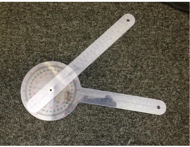
One pertinent type of outcome measure, which currently provides quantitative data,
is that of goniometry. Goniometry allows for the assessment of ROM, and can be
used to assess the upper-limb. In this type of assessment, a goniometer, as shown in
Figure 2-1, can be used to measure minimum and maximum ROM, manually, by a
therapist. However, although goniometry allows for a more objective measure of
upper-limb functionality, it can be time consuming and evidence has shown that
goniometry displays inaccuracy rates, shown to be anywhere from ±5 degrees up to
14-23 degrees (Clapper and Wolf, 1988, Garcia-Elias et al., 1989, Hayes et al.,
32 | P a g e
Figure 2-1: A Goniometer
The use of technology has been suggested as a way forward, and evidence for this
area shows improvement in outcomes. Research has shown that motion tracking
could potentially provide quantitative and objective assessment data. It has been
recommended that ways of providing highly technical biomechanical data to
therapists/patients, in a usable manner, could assist in motor skill re-learning.
There is a focus on cost saving, alongside improving care, within the NHS and a way
to achieve this could be through creating “more with the same, not more of the
same” (Appleby et al., 2010, p.1). It is possible that physiotherapy and occupational
therapy services could be streamlined through the automation and increased
quantification of rehabilitative assessment of the upper-limb in stroke patients, which
can sit alongside current outcome measures, to create a more holistic approach. If an
33 | P a g e data at first contact with a patient and, at each session thereafter, therapy could be
optimised and patients could receive more direct therapy time. One way to achieve
this would be to utilise motion tracking technologies to provide these biomechanical
data.
2.2 -Motion Tracking as an Assessment Tool for Upper-Limb Motor Control.
A method for assessing motor control in the upper-limb is through the use of motion
tracking technology. Motion tracking allows motion patterns and estimations of body
poses to be devised through computer analysis (Alexander et al., 2010). This can be
used to evaluate the movement of the upper-limb by evaluating aspects, such as
speed and smoothness of movement. Assessment may be performed by breaking
down the components of motor control or through using the technology to calculate
existing outcome measures. Motion may be detected through the placement of
markers on the individual and the recording of the movement of these markers; or
through special markerless motion tracking cameras, which can identify a human
shape and track various aspects of its movement. These two types of tracking and
their use within upper-limb motor control assessment in stroke patients shall be
discussed in the following sections of this thesis.
2.2.1 - Examples of Marker-based Motion Tracking Systems
One method for performing motion capture is through the placement of markers on
the patient. An example of this, when used in stroke upper-limb motor control
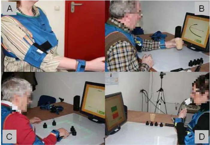
34 | P a g e utilised a Philips Research Stroke Rehabilitation Exerciser (a full rehabilitative suite
with a patient and therapist station), shown in Figure 2-2. The Philips Research
Stroke Rehabilitation Exerciser uses “wireless inertial sensors for measuring joint
kinematics, an active exercise board which is capable of interaction with real-world
interactive objects and a Personal Computer (PC) with touch screen via which
exercises are offered and feedback on performance is provided” (Timmermans et al.,
[image:35.595.142.493.387.629.2]2010, p.115).
Figure 2-2: Using the Philips Research Stroke Rehabilitation Exerciser for ADL. These include: A: Sensor placement and garments. B: Example of drinking from a cup. C and D: Examples of
eating with knife and fork.
35 | P a g e Timmermans et al. used a method they described as the ‘Technology-supported
task-oriented arm training’ (T-TOAT). This method involves the breaking down of ADL
into sub-categories. For example, drinking from a cup may be broken down into
reach out to cup; grasp cup; lift cup; bring cup to mouth; empty cup into mouth;
place cup on table and release cup. Timmermans et al. state that the reason for this
categorisation is that “exercise programs can be implemented in
technology-supported training” (Timmermans et al., 2010, p.117). The system is also adaptable
and is based on exercise physiology and motor learning. The system and software
were tested with nine participants. The participants demonstrated improvements after
eight weeks on the Fugl–Meyer, Action Research Arm Test, and Motor Activity
Log. A lot can be taken from this paper and applied to this research, particularly the
breaking down of tasks into sub-categories and the adaptability of the system.
However, the low numbers of participants in the research by Timmermans et al.
means that large scale conclusions cannot be drawn. Also, participants may have
improved on the tests specified without the system present.
Another example of a marker based research system which has been used to evaluate
the upper-limb, is the ‘Computer Assisted Rehabilitation Environment’ (CAREN)
(Subramanian, 2007). This system includes a Head Mounted Display (HMD), a
CyberGlove haptic device and an OptoTrak motion tracking system. The user is able
to work in a 3-dimensional virtual reality environment and conduct goal-directed
upper-limb exercises for the improvement of upper-limb motor function. This is
performed by asking the patient to move their arm towards set targets in the
environment, with the movement tracked through the marker based system. To test
36 | P a g e at the same distance as the ones in the virtual environment. The tests were performed
with 15 stroke patients and 8 age-matched non-disabled individuals. Results showed
good correlation with the non-disabled individuals showing a range of 257–356 mm
in the physical environment and 275–370 mm in the virtual environment and stroke
patients showing a range of 263–363mm in the physical environment and 275–379
mm in the virtual environment.
Another marker based system that can be potentially utilised in stroke rehabilitation
and upper-limb assessment is the Vicon motion capture system, a marker based
commercial motion tracking system (Hingtgen et al., 2006). Hingtgen et al. used a
Vicon workstation to evaluate upper-limb activity using a kinematic model,
developed specifically for this research. This is an important aspect of any motion
tracking system, as the initial data must be turned into clinically relevant data, and
kinematic analysis must be performed in order to achieve this. Hintgen et al. tested 8
adults who had suffered a stroke and analysed the accuracy of the system. They were
able to successfully quantify the movement and demonstrate greater velocity in
unaffected arms of the patient when compared to the affected arm.
One popular use of a motion tracking system is demonstrated by Nintendo’s Wii™.
The Wii is a low cost, commercially available gaming system (Deutsch et al., 2008)
and could potentially be used in stroke rehabilitation and upper-limb assessment. The
‘WiiMote’ (The Wii’s remote control) can be utilised as a handheld pointing device
and although its efficacy is unproven within a rehabilitation setting, studies have
37 | P a g e Mouawad et al. (2011) carried out an experiment using the Wii as a rehabilitation
tool. In this experiment seven patients utilised the Wii over a two week period. The
authors claim that clinically relevant improvements were made with mean
performance time decreased from 3.2 to 2.8 seconds and Fugl-Meyer score increases
from 42.3 to 47.3, however, the small cohort warrants further trials to validate the
results fully.
Celinder and Peoples (2012) also utilised the Wii in a similar manner. Nine stroke
patients used Wii Sports as a supplement to traditional therapy. Qualitative analysis
was drawn from this in the form of semi-structured interviews and field notes. The
results showed that use of the Wii helped increase variety and engagement, whilst
helping to overcome specific obstacles and challenges. These reported results
provide evidence for the additional benefits of using such tools in stroke
rehabilitation and assessment.
However, it should be noted that the use of the Wii as a tool for stroke rehabilitation
and assessment has some disadvantages. Due to the nature of the device, only hand
positions in space may be measured and not individual joint movements, limiting the
amount of assessment which can be performed.
There is currently a limited evidence base for the use of the Nintendo Wii within
stroke upper-limb assessment. Saposnik et al. (2010a), in their initial protocol, have
investigated the ‘Effectiveness of Virtual Reality Exercises in Stroke Rehabilitation’
(EVREST). The importance of this research is identified in their claim that despite
advancement in this area, this is the first clinical trial that used the Wii for stroke
38 | P a g e therapy group or a group which utilised the Wii in therapy. Results produced from
this study (Saposnik, 2010b) showed that patients in the Wii therapy group
demonstrated an improvement in mean motor function of 7 seconds (Wolf Motor
Function Test, 7.4 seconds; 95% CI (Confidence Interval), -14.5, -0.2) over patients
in standard rehabilitation.
Overall, marker-based motion tracking systems may be used for the assessment of
upper-limb, and the low-cost and portable nature of commercial devices such as the
Wii could provide a method for achieving this. However, limitations in terms of
evidence base, limited feedback (e.g. the Wii only returning the positions of the
controllers and nothing else) and time-consumption when setting up equipment (i.e.
placing the markers on an individual) provide limitations for this area when using the
technology in the clinical environment.
2.2.2 - Examples of Markerless Motion Tracking Systems
An alternative method for the motion tracking of the upper-limb is through
markerless tracking systems, which take away the need for markers to be placed on a
patient. Markerless tracking employs video capture techniques in order to achieve its
goal, allowing real time feedback without head mounted displays, gloves or markers
(Weiss et al., 2004).
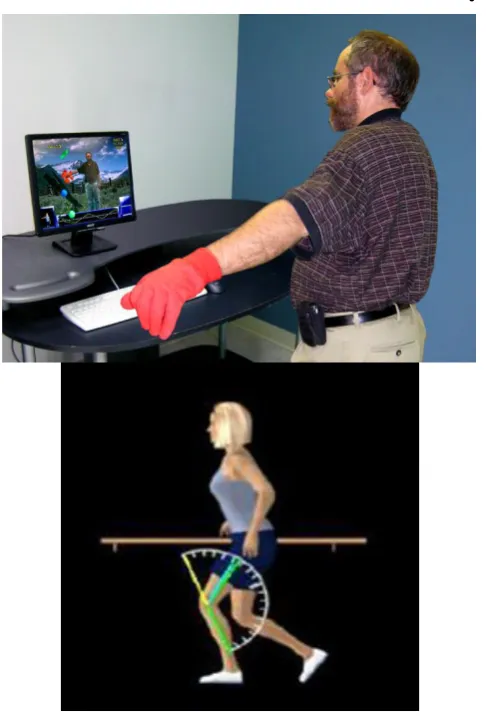
These systems are being adopted and becoming available commercially, for
example, a company called ‘Gesturetek’ have released a system specifically aimed at
the rehabilitation sector which could be used for the assessment of upper-limb motor
39 | P a g e
Figure 2-3: Interactive Rehabilitation and Exercise System (IREX)
(Source: GestureTek, 2007)
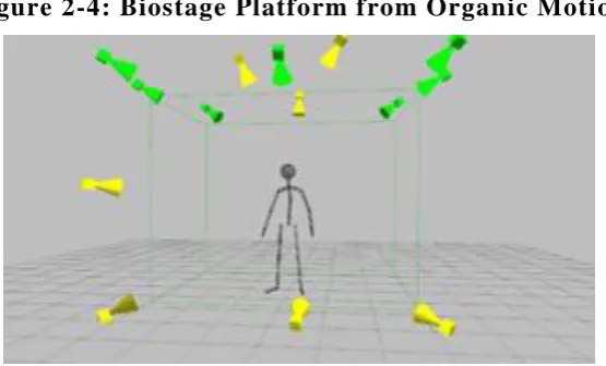
Figure 2-4 shows another commercially available markerless motion tracking
system, the Biostage platform from Organic Motion. This is a multi-camera
markerless motion tracking system, with cameras set-up around an individual to
40 | P a g e A further commercial system is the ‘EyeToy’ for the Sony PlayStation II (Huber et
al., 2008). The EyeToy is an off-the-shelf low cost gaming application, and
represents one of the first large scale commercial attempts to employ a markerless
[image:41.595.181.459.234.402.2]motion tracking system.
Figure 2-4: Biostage Platform from Organic Motion
(Source: Organic Motion, 2012)
Another system to be developed, which is low-cost, portable, and could be suitable
for the clinical environment, is the Microsoft Kinect system. This is a one-camera
system originally designed for use with Microsoft’s XBOX 360 console, and could
be applicable to stroke rehabilitation (Chang et al., 2012). Due to these attributes, the
Microsoft Kinect has been selected for use in this research. The reasoning behind
this, including further details of this system, will be discussed in more depth in
section 2.3 - The Use of the Microsoft Kinect as a Tool for Assessing Upper-Limb
41 | P a g e These systems could potentially be used for the assessment of upper-limb motor
control in stroke patients. Allin et al. (2010) actually created a specific algorithm for
tracking the arm of stroke patients using multiple cameras in markerless systems,
such as the biostage platform. The system divides the arm into three segments
(upper-arm, lower-arm and hand). It has been evaluated using two methods; the first
of these was to compare the system to the commercially available marker based
tracking system Vicon (Vicon, 2012); the second of these was to evaluate the system
with seven stroke patients using the upper extremity section of the Fugl–Meyer
Score. The system demonstrated positive results, confirming that the “average
absolute discrepancies between 15 infrared markers measured by a VICON and 15
corresponding virtual markers measured with a parts-based tracker to be 80 mm,
with a standard deviation of 5 mm” (Allin et al., 2010, p.8).
Various devices now exist, and are commercially available. However, as previously
described in this thesis, research must now be performed to identify how to make
these systems suitable for the clinical environment. This research will particularly
focus on the Microsoft Kinect technology and how it can be used within the clinical
environment, and a discussion of this will take place later in this chapter.
2.2.3 - Data Processing when Analysing Upper-Limb Motion
A difficult area which affects all motion tracking systems, and has to be taken into
account for assessing upper-limb motion control, is the way in which data is
processed in order to review the tracked motions. This processing requires human
42 | P a g e 2003) or other various kinds of kinematic models (Cheung et al., 2005). There are a
range of algorithms that have been used in order to achieve this processing, that
estimate human motion (Mündermann et al., 2006).
2.2.4 - Advantages and Disadvantages of Motion Tracking in Upper-Limb
Motor Control Assessment
When using marker based systems, there is a requirement for special cameras and
equipment (Tao and Hu, 2003). The markers that are placed on a suit or on the skin
restrict movement causing problems with realism (Zheng et al., 2005). There are also
problems with the accuracy of readings as soft tissue can move causing noisy data,
the marker itself can ‘wobble’ or actually move, and, creating standardised spots to
place the marker on the body is a problem due to diminishing accuracy levels (Zhou
and Hu, 2008). Marker based systems also take sections of limbs as solid entities and
employ estimation algorithms in order to achieve motion playback, causing problems
with accuracy (Mündermann et al., 2006). In line with these problems, many marker
based systems are too expensive for the clinical environment and simply not
designed for it, making this technology unsuitable for this purpose. A tool, such as
the Wii, may alleviate some of these issues as it provides off-the-shelf, affordable
availability, which could be suited to the clinical environment. Some promising
studies have been shown in this area; however, there are certain limitations to what
the Wii may show, as it effectively only demonstrates the position of 2 markers (the
controllers) in space and provides no analysis of movement in the body, so there is
43 | P a g e performing compensatory movements. The Wii may be suited to the rehabilitation
environment, when utilised for basic exercise and motivational activities. However,
it does not currently provide enough data to analyse the upper-limb successfully and
is therefore currently outside of the scope of this research.
Markerless motion tracking may be able to relieve some of the problems set out
above, but this may come with a trade-off in terms of accuracy levels. Also,
markerless based motion tracking still has problems with depth, occlusion,
appearance deformation, kinematics issues (Tao and Hu, 2003), line of sight and the
true animation of human motion (Zhou and Hu, 2008). Again, markerless motion
tracking systems may be expensive and unsuitable to the clinical environment.
However, a recent progression in terms of technology, has been in the development
of commercially available off-the-shelf devices, which allow for markerless motion
tracking. If it can be shown that these systems are capable of providing a high
enough accuracy level to be suited to the clinical environment, whilst still meeting
needs such as portability and cost, then they may be utilisable within a rehabilitation
setting. As previously stated, this research shall use the Microsoft Kinect technology
to learn whether such a system can be created which is adapted to the clinical
environment, deriving needs through a user-centred design approach.
2.3 -The Use of the Microsoft Kinect as a Tool for Assessing Upper-Limb
Motion.
A technology that has recently been developed is a markerless commercial motion
44 | P a g e originally designed for use with the Microsoft Xbox 360 console and can be used to
track up to 20 joints in the ‘skeleton’ of the player, which it displays on screen. The
Microsoft Kinect is available at a low price (below GBP£200), making it affordable
[image:45.595.138.501.235.397.2]in the clinical environment.
Figure 2-5: Microsoft Kinect
Chang et al. (2012) state that the Microsoft Kinect could be a valuable tool for stroke
rehabilitation and shows high promise for clinical settings and in remote
environments.
A system has actually already been developed which could be used for rehabilitative
purposes with the Microsoft Kinect, which could allow for upper-limb assessment.
The system allows for programs to be developed using the Microsoft Kinect at a
quicker rate through the use of a user interface and is called the ‘Flexible Action and
Articulated Skeleton Toolkit’ (FAAST) which allows for rapid game development
45 | P a g e “FAAST considers two broad categories of information from the sensor: actions and
articulated skeletons. Articulated skeletons consist of the positions and orientations
for each joint in a human figure, and are useful for virtual reality and video game
applications in allowing direct body-based control of a virtual avatar. FAAST
retrieves these skeleton joints from the OpenNI drivers and transmits them to the
end-user application” (Suma et al., 2011, p.245).
Suma et al. state that they developed their own methodology for streaming skeletal
data at each joint as a six-degree-of-freedom tracker. This could be a useful toolkit
within this research and could allow for rapid product development. The only issue,
at this current point in time, is that end users are asked to initially stand in a
calibration pose with their arms held in the air, a position that most stroke patients
would find difficult to achieve.
Current upper-limb rehabilitative assessment games have already been developed for
research purposes and include tasks such as ‘reaching for objects’ and ‘cognitive
challenges’ (Lange et al., 2011). 20 participants were recruited into this study (10 of
which had suffered a stroke), alongside 10 clinicians. A game was developed which
was goal-directed; it involved collecting gems, while driving through a mine and
placing them in a cart. The distance and angle of the gems was altered according to
the patient. This paper demonstrated the use of usability testing to gain initial
feedback on the system. Participants asked for more instructions when using the
game; better visual effects were identified as a need, as users struggled to see the
gems and 3 participants stated that they would like more of a ‘story’ within the
46 | P a g e must provide ample instructions as cognitive issues can be a problem within this
patient group. However, most of the patients stated that they found the system highly
motivating and challenging, complying with the principles of goal-driven tasks and
therapy. Any assessment tool could eventually be placed within a full rehabilitative
system. This could potentially involve the use of gamification to improve motivation
and help alleviate time pressures on clinical staff.
A study, which has been performed to demonstrate the effectiveness of the Kinect
within rehabilitation, utilises a methodology that could be used for upper-limb motor
control assessment (Chang et al., 2011). Chang et al. developed a ‘KineRehab’
system which was used by the participants whilst in wheelchairs. The participants
were asked to carry out certain movements and these were assessed using gesture
recognition. The participants were assessed using an ABAB system. The authors
describe this as four stages of an experiment, with A demonstrating a
non-intervention stage, followed by phase B in which an non-intervention phase is introduced,
followed by another repetition of each of these phases. It was stated that the amount
of movements, which were correct, were higher at B than at A. However, more
research is needed with more than the two utilised participants to prove this result. A
point, raised in this paper, is with regards to the motivational factor that the
Microsoft Kinect can provide, and how mundane tasks can be made increasingly
more interesting. However, this is performed with a group of students, and further
validation with patients would be required to prove this.
It was also stated that the therapist using the system believed it provided excellent
47 | P a g e Although, this would require further validation as the therapist had been involved in
the research and their opinion is open to bias.
Clark et al. (2012) state how useful the Microsoft Kinect could potentially be as a
tool for stroke rehabilitation, describing how the reference points provided by the
system are comparable to that of a standard commercial motion tracking system at a
much lower cost.
Table 2-1 shows the results of a number of functionality tests performed on the
Microsoft Kinect (Livingston et al. (2012) provide more details of these tests in their
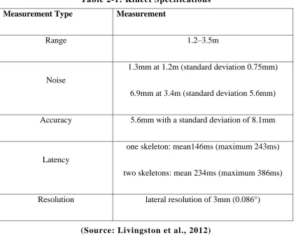
[image:48.595.109.533.386.732.2]paper ‘Performance measurements for the Microsoft Kinect skeleton’).
Table 2-1: Kinect Specifications
Measurement Type Measurement
Range 1.2–3.5m
Noise
1.3mm at 1.2m (standard deviation 0.75mm)
6.9mm at 3.4m (standard deviation 5.6mm)
Accuracy 5.6mm with a standard deviation of 8.1mm
Latency
one skeleton: mean146ms (maximum 243ms)
two skeletons: mean 234ms (maximum 386ms)
Resolution lateral resolution of 3mm (0.086°)
48 | P a g e Leyvand et al (2011) discuss how the optimum measurement range for the Microsoft
Kinect is 1-3 metres, with accuracy improving as the user moves closer to the 1m
range. As the user will predominantly be in the same gross body position during
assessment, the system will prompt the user to stand at a distance of between 1-2
metres from the Kinect camera, eliminating difficulties with detection range.
The accuracy of the Kinect is currently to within 5.6mm, as stated by Livingston et
al. (2012). A stroke patient, when using the system, may often make movements
which require a large ROM (above 65° at any one joint), making a 5.6mm
inaccuracy level of low significance. Fernandez-Baena et al. (2012) specify a mean
inaccuracy level, when measuring ROM on the Kinect, of 8.63 degrees.
To place this data in perspective, various authors have released papers stating that
the current method for measuring ROM, namely goniometry, also displays
inaccuracy rates, shown to be anywhere from ±5 degrees up to 14-23 degrees of
inaccuracy (Clapper and Wolf, 1988, Garcia-Elias et al., 1989, Hayes et al., 2001),
demonstrating a similar and possibly greater inaccuracy level to that of the Microsoft
Kinect. However, there are other additional issues with goniometry. Goniometry has
been shown to have a low inter-rater reliability (R=.53 (Petherick et al., 1988)),
whilst the Kinect is automated and eliminates the need for different testers. The
Kinect is also capable of taking a high amount of measurements (up to 30 Frames
Per Second of every joint) (Microsoft, 2012a), which is also without a therapist
49 | P a g e joints ROM (for which a separate maximum and minimum position must be
measured).
Researchers have also utilised the Microsoft Kinect in collaboration with other
sensors to create greater accuracy rates. Bó et al. (2011) state that the potential when
combining the Microsoft Kinect with other systems (e.g., accelerometers) is
encouraging.
Stroke patients predominately suffer from reduced movement rates (speed and
smoothness) (Rohrer et al., 2002). These reduced movement rates should limit the
effects of noise and latency in line with a speed-accuracy trade off. The inaccuracy
rate of only 5.6mm in large ROM activities demonstrates that noise and latency do
not cause an underlying issue when used for this purpose.
It must be taken into account that the Microsoft Kinect has a limit on the types of
movement it is capable of tracking. The Kinect is capable of measuring large
movements such as flexion/extension at the elbow and abduction/adduction at the
shoulder. However, it is not capable of detecting smaller movements, such as the
pronation/supination movement at the wrist joint that is essential to most ADL. This
may be solved through combination with other sensors if required.
In summary, initial studies have shown that the Microsoft Kinect may be suitable to
the assessment of the upper-limb in stroke rehabilitation, due to -its low-cost nature
and off-the-shelf availability. Initial studies also show that accuracy levels should be
high enough for use in the assessment of the upper-limb. However, research is now
required in how to best utilise the device for this purpose. Systems must be