Original Article
Effect of total glucosides of paeonia on TGF-β1 and
ICAM-1 expression in the kidney of diabetic rats
Feili Lin1, Jianqing Wang1, Yin Hu1, JinKang Jia1, Feng Zhang2
1Department of Nephrology, The Second Affiliated Hospital of Zhejiang University School of Medicine, Zhejiang, China; 2Department of Orthopedics, The Second Affiliated Hospital of Zhejiang University School of Medicine,
Zhejiang, China
Received May 26, 2015; Accepted June 29, 2015; Epub February 1, 2016; Published February 15, 2016
Abstract: Intercellular adhesion factor 1 (ICAM-1) and transforming growth factor (TGF-β1) expression enhanced following the diabetes extension. They may be involved in the pathogenesis of diabetic nephropathy. Total gluco-sides of paeonia (TGP) have anti-inflammation and immune regulation effect. We discussed TGP effect on TGF-β1 and ICAM-1 expression and its renal protection mechanism on diabetic rats. 7 weeks old healthy male SD rats were randomly divided into control group (C), diabetes group (B), and TGP group H, M, and L with 10 in each group. Fasting blood glucose (BG), creatinine (Scr), urea nitrogen (BUN), and 24 h urine trace albumin excretion rate (UAER) were measured and renal pathological morphology was observed. Immunohistochemistry was applied to measure TGF-β1 and ICAM-1 expression in the kidney. TUNEL assay was performed to observe renal cortical cell apoptosis. Compared with group C, BG, Scr, BUN, and UAER significantly increased in group B (P < 0.05). Their levels in TGP group H, M, L were higher than the group C but lower than group B (P < 0.05). Histological changes of the lesions were improved obviously in TGP group. TGF-β1 and ICAM-1 expression in the kidney presented similar trends. Cell apoptosis index increased markedly in group B compared with group C, it showed different degree of decrease in TGP group (P < 0.05).TGP can improve early renal damage in diabetic rats, which might be related to its reducing TGF-β1 and ICAM-1 expression in the kidney to inhibit cell apoptosis.
Keywords: Total glucosides of paeonia, diabetic nephropathy, ICAM-1, TGF-β1
Introduction
Diabetic nephropathy (DN) is a type of diabetes complication that is the leading cause of end-stage renal failure. Its incidence increased in recent years. However, its etiology and patho-genesis still have not been fully elucidated [1]. Studies have shown that [2, 3] diabetes is
associated with oxidative stress and inflamma
-tion, while intercellular adhesion factor-1 (ICAM-1) is the important reason for macrophages
and monocytes infiltration in renal tissue.
Animal experiments showed that [4, 5] ICAM-1 overexpressed in the kidney of the type 1 dia-betes model induced by streptozocin (STZ), which is consistent with glomerular structure changes. High glucose can increase the extra-cellular matrix hyperplasia by inducing
trans-forming growth factor β (TGF-β) gene and pro
-tein expression in glomerular mesangial cells.
Several TGF-β and related receptors overex
-pressed in diabetic rats renal tubular. Following
duration extension, TGF-β1 protein expression
gradually upregulated and its overexpression played an important role in diabetic kidney
inju-ry [6, 7]. Immune/inflammatoinju-ry inhibitors and
antioxidants can reduce renal tissue inflamma
-tion cells infiltra-tion and oxidative stress reac
-tion, reduce urinary albumin levels, and improve renal structure and function in diabetes models [8]. Total glucosides of paeonia (TGP) are
extracted from the dry root of paeonia lactiflora
Pall, which contains hydroxy paeoniflorin, pae
-oniflorin, and other active ingredients. Radix
paeoniae alba can protect blood and liver. Pharmacological studies showed that [9, 10]
TGP has anti-inflammatory, anti-stress and
immune regulation function, and it was an
anti-inflammatory immune regulatory drug in clinic.
In this paper, we aimed to investigate the pos-sible mechanism of TGP protection effect on diabetic rats by observing TGP impact on the
Materials and methods
Experimental animals and grouping
7 weeks old healthy male SD rats weighted 200~240 g were provided by the Chinese acad-emy of medical sciences animal experiment center (license SYXK-2013-0025) and main-tained in SPF laboratory. The rats were random-ly divided into control group (C), diabetes group (B), and TGP group H, M, L with 10 in each group.
Rats were used for all experiments, and all pro-cedures were approved by the Animal Ethics Committee of our hospital.
Drugs and reagents
TGP was purchased from Hunan KinglongBio Co., LTD. and suspended in 1% carboxymethyl cellulose. Streptozocin (STZ) was provided by Sigma and suspended in citrate buffer. Blood glucose, urea nitrogen, serum creatinine detec-tion kits were got from Nanjing Jiancheng
bio-logical technology co., LTD. TGF-β1 and ICAM-1
primary antibodies, DAB chromogenic agent were provided by Wuhan Boster Biological Co., LTD.
Modeling
STZ were used to establish diabetic rat model. After fasting for 12 h, the rats in the model group received 60 mg/kg 1% STZ solution intraperitoneal injection at one-time, while the control group received equal amount of citric acid buffer injection. Diabetes model was
eval-Measurement
Rat’s general information such as mental state, eating and drinking, stool and urine were observed daily. The weight was measured every 2 weeks. Blood glucose was tested once a month. At the end of 8 weeks’ test, urine was collected after 24 h fasting and urine protein level was detected. The rat abdominal venous blood was collected to inspect BG, BUN, and Scr levels. The left kidney was used for
weight-ing, while the right kidney was fixed in 10%
formaldehyde. Glucose oxidase method was applied to determine BG. Scr and BUN were measured using automatic biochemical analyz-er. Kidney weight index KI = left kidney weight/ body weight. Kidney pathological morphology was observed under optical microscope.
TGF-β1 and ICAM-1 expression were evaluated by
immunohistochemistry, and the positive result was evaluated by mean absorbance in the
posi-tive area. TGF-β1 mainly expressed in renal
tubular epithelial cells and part expressed in glomerular. ICAM-1 mainly expressed in glomer-ular endothelial cells and mesangial cells. They both located in the cytoplasm.
TUNEL assay
After stained with TUNEL, apoptotic cells were
observed under microscope. Five fields under
400× were enrolled for calculation. Apoptosis index (AI) = apoptotic cell number/total cell number × 100%.
Statistical analysis
All statistical analyses were performed using SPSS17.0 software (Chicago, IL). Numerical data were presented as means and standard deviation (± SD). Differences between multiple groups were analyzed using one-way ANOVA and LSD-t test. P < 0.05 was considered as
[image:2.629.100.294.81.250.2]sig-nificant difference.
Results
General information comparison
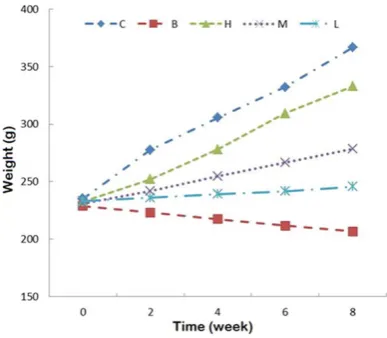
Rats in group C exhibited significant weight
gain, responsive, smooth and shining furs. Rats in group B appeared more eating and drinking, polyuria, with weight loss, unresponsive, and lackluster furs. Rats in H, M, and L groups pre-sented weight gain, good spirit. They were more
sensitive than in group B, but worse than group C (Figure 1).
TGP impact on BG and renal function
Compared with group C, BG, Scr, BUN, UAER,
and KI in group B significantly increased (P <
0.01). Their levels in TGP group H, M, L were higher than the group C but lower than group B (P < 0.01 or P < 0.05) (Figure 2).
Figure 2. TGP impact on diabetic rats’ BG and renal function indexes.
*P < 0.05, **P < 0.01, compared with
group C; #P < 0.05, ##P < 0.01,
com-pared with group B.
[image:3.629.100.531.72.619.2]TGP effect on diabetic rat pathological
chang-es
HE staining revealed that rat glomerular struc-ture in group C was clear with no obvious abnor-mality. Glomerular volume increased in group B. Glomerular mesangial cells appeared hyper-plasia and cystic wall adhesion. TGP groups
showed histological lesions improved signifi
-cantly compared with group B, and the improve-ment degree showed dose dependent (Figure 3).
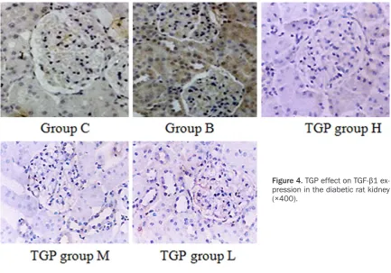
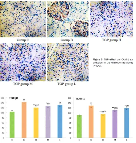
TGP effect on TGF-β1 and ICAM-1 expression
in the diabetic rat kidney
TGF-β1 and ICAM-1 expression were weak in
group C, and they obviously overexpressed in group B (P < 0.01). Their expression significant -ly reduced in TGP group H, M, L compared with group B (P < 0.05) (Figures4-6).
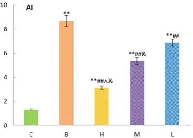
TGP impact on diabetic rat renal cortical cell
apoptosis
Cell apoptosis index increased markedly in group B compared with group C, it showed dif-ferent degree of decrease in TGP group (P < 0.05) (Figure 7).
Discussion
Inflammatory reaction runs through the whole process of diabetes. Inflammatory factor is
closely associated with diabetic nephropathy (DN). The pathological basis of DN is kidney
high filtration, high perfusion, glomerular
mesangial cell proliferation, basement mem-brane thickening, and extracellular matrix hyperplasia, following with nodular or diffuse sclerosis, eventually leading to hypertension, proteinuria and renal failure [12, 13].
Mononuclear macrophages infiltration existed
in the renal tissue in both type 1 and type 2 DN
at early stage. Infiltrated mononuclear macro
-phages may cause kidney damage through
secreting inflammatory factors and oxygen free
radicals, promote glomerular sclerosis. Kidney under pathological condition can produce
inflammatory cytokines such as TNF-α and NO itself. Secreted inflammatory cytokines can augment inflammation through paracrine or autocrine and trigger the inflammatory cascade reaction [14, 15]. Inflammation might be a
downstream of DN to cause renal damage.
Macrophages mediated kidney inflammation plays a significant role in the progression of DN. Inflammation/immune inhibitor application in
diabetes model can inhibit macrophage infiltra
[image:4.629.98.532.76.379.2]tion found in the kidney tissue, significantly
reduce UAER, and improve the renal
pathologi-cal tissue damage [16]. TGF-β1 and ICAM-1
overexpressed following the extension of diabe-tes. It was found that they can increase cell apoptosis and lead to renal function deteriora-tion [14-16]. TGP has multiple pharmacological
activities such as inflammation and
anti-stress. In vivo animal experiment results showed that [17] in adjuvant arthritis rats, TGP
can inhibit basic fibroblast growth factor and
vascular endothelial growth factor expression by downregulating macrophage mediated
TNF-α and IL-1 expression. This paper dis
-cussed TGP effect on TGF-β1 and ICAM-1
expression in the kidney of diabetic rats to investigate its protection mechanism to diabetes.
We used STZ to damage islet β cells and the
[image:5.629.99.540.78.542.2]rats in model group appeared polydipsia, poly-phagia, and polyuria, elevated BG, UAER, BUN,
Figure 5. TGP effect on ICAM-1 ex-pression in the diabetic rat kidney (×400).
Figure 6. TGP effect on ICAM-1 expression in the diabetic rat kidney (×400). *P < 0.05, **P < 0.01, compared with
group C; #P < 0.05, ##P < 0.01, compared with group B; &P < 0.05, compared with group L; ΔP < 0.05, compared with
Scr and KI, suggesting that diabetes caused renal damage. They decreased at different degree in TGP group H, M, L compared with group B, but still higher than in group C. Histological lesions improved obviously in TGP group, indicating that TGP can alleviate early renal damage, reduce proteinuria, and improv-ing renal function in diabetic rats.
Studies have shown that [18, 19] ICAM-1 can mediate WBC migration and adhesion. It mainly expressed in renal tubule-mesenchymal cells and vascular epithelial cells. ICAM-1 knockout diabetic rat model showed obvious alleviated
kidney macrophages infiltration, albumin excre
-tion rate, glomerular hypertrophy, and mesan-gial matrix expansion. Our results revealed that ICAM-1 overexpressed in group B, while it reduced in TGP group H, M, L with dose depen-dent, indicating that TGP can inhibit ICAM-1 protein expression in renal tissue, which may be related to inhibiting macrophages
infiltration.
Animal and clinical trials demonstrated that
[20, 21], TGF-β1 plays an important role on glo
-merular-interstitial damage in DN process. It was an important cytokine for cell growth and producing extracellular matrix. Its expression level increased in the kidney of diabetes
patients and animal models. TGF-β1 can induce
renal pathological changes by promoting kid-ney hypertrophy, inducing extracellular matrix hyperplasia, and inhibiting cell proliferation.
Our study showed that TGF-β1 expression
increased in group B, while it decreased in TGP group H, M, L with dose dependent, indicating
matory cytokines and adhesion factors may
damage glomerular filtration barrier and cause proteinuria. TGP treatment can decrease TGF-β
and ICAM-1 expression, suggesting that TGP can improve early renal damage and reduce
proteinuria through decreasing TGF-β and
ICAM-1 expression in the kidney of diabetic rats.
Above all, TGP can significantly improve early
renal damage in diabetic rats, which may be
related to its reducing TGF-β and ICAM-1
expression in kidney and inhibiting cell apoptosis.
Disclosure of conflict of interest
None.
Address correspondence to: Dr. Feng Zhang, Department of Orthopedics, The Second Affiliated Hospital of Zhejiang University School of Medicine, 88 Jiefang Road, Hangzhou 310009, Zhejiang, China. Tel: 0086-571-87783777; E-mail: [email protected]
References
[1] Graham ML, Schuurman HJ. Validity of animal models of type 1 diabetes, and strategies to enhance their utility in translational research. Eur J Pharmacol 2015; 759: 221-30.
[2] Nowotny K, Jung T, Hohn A, Weber D, Grune T. Advanced Glycation End Products and Oxidative Stress in Type 2 Diabetes Mellitus. Biomolecules 2015; 5: 194-222.
[3] Tykhomyrov AA, Shram SI, Grinenko TV. Role of angiostatins in diabetic complications. Biomed Khim 2015; 61: 41-56.
[4] Lu CY, Yang YC, Li CC, Liu KL, Lii CK, Chen HW. Andrographolide inhibits TNFalpha-induced ICAM-1 expression via suppression of NADPH oxidase activation and induction of HO-1 and GCLM expression through the PI3K/Akt/Nrf2 and PI3K/Akt/AP-1 pathways in human
endo-Figure 7. TGP impact on renal cortical cell apoptosis in diabetic rats. *P < 0.05, **P < 0.01, compared with
group C; #P < 0.05, ##P < 0.01, compared with group
B; &P < 0.05, compared with group L; ΔP < 0.05,
[image:6.629.99.299.81.224.2]thelial cells. Biochem Pharmacol 2014; 91: 40-50.
[5] Sozer V, Himmetoglu S, Korkmaz GG, Kaya S, Aydin S, Yumuk V, Hatemi H, Uzun H. Paraoxonase, oxidized low density lipoprotein, monocyte chemoattractant protein-1 and ad-hesion molecules are associated with macro-vascular complications in patients with type 2 diabetes mellitus. Minerva Med 2014; 105: 237-244.
[6] Park JT, Kato M, Lanting L, Castro N, Nam BY, Wang M, Kang SW, Natarajan R. Repression of let-7 by transforming growth factor-beta1-in-duced Lin28 upregulates collagen expression in glomerular mesangial cells under diabetic conditions. Am J Physiol Renal Physiol 2014; 307: F1390-1403.
[7] Castro NE, Kato M, Park JT, Natarajan R. Transforming growth factor beta1 (TGF-beta1) enhances expression of profibrotic genes through a novel signaling cascade and microR-NAs in renal mesangial cells. J Biol Chem 2014; 289: 29001-29013.
[8] Chang BC, Chen WD, Zhang Y, Yang P, Liu L, Wang J. Effect of total glucosides of paeony on Wnt/beta-catenin signal transduction pathway expression in kidney of diabetic rats. Zhongguo Zhong Yao Za Zhi 2014; 39: 3829-3835. [9] Zhang W, Zhao L, Su SQ, Xu XX, Wu YG. Total
glucosides of paeony attenuate renal tubuloin-terstitial injury in STZ-induced diabetic rats: role of Toll-like receptor 2. J Pharmacol Sci 2014; 125: 59-67.
[10] Julia C, Czernichow S, Charnaux N, Ahluwalia N, Andreeva V, Touvier M, Galan P, Fezeu L. Relationships between adipokines, biomark-ers of endothelial function and inflammation and risk of type 2 diabetes. Diabetes Res Clin Pract 2014; 105: 231-238.
[11] Fuchs D, Birklein F, Reeh PW, Sauer SK. Sensitized peripheral nociception in experi-mental diabetes of the rat. Pain 2010; 151: 496-505.
[12] Xu XX, Qi XM, Zhang W, Zhang CQ, Wu XX, Wu YG, Wang K, Shen JJ. Effects of total glucosides of paeony on immune regulatory toll-like recep-tors TLR2 and 4 in the kidney from diabetic rats. Phytomedicine 2014; 21: 815-823.
[13] Wang K, Wu YG, Su J, Zhang JJ, Zhang P, Qi XM. Total glucosides of paeony regulates JAK2/ STAT3 activation and macrophage proliferation in diabetic rat kidneys. Am J Chin Med 2012; 40: 521-536.
[14] Zhang P, Zhang JJ, Su J, Qi XM, Wu YG, Shen JJ. Effect of total glucosides of paeony on the ex-pression of nephrin in the kidneys from dia-betic rats. Am J Chin Med 2009; 37: 295-307. [15] Khan S, Jena G. Sodium butyrate, a HDAC
in-hibitor ameliorates eNOS, iNOS and TGF-beta1-induced fibrogenesis, apoptosis and DNA damage in the kidney of juvenile diabetic rats. Food Chem Toxicol 2014; 73: 127-139. [16] Su J, Zhang P, Zhang JJ, Qi XM, Wu YG, Shen JJ.
Effects of total glucosides of paeony on oxida-tive stress in the kidney from diabetic rats. Phytomedicine 2010; 17: 254-260.
[17] Wu Y, Ren K, Liang C, Yuan L, Qi X, Dong J, Shen J, Lin S. Renoprotective effect of total glu-cosides of paeony (TGP) and its mechanism in experimental diabetes. J Pharmacol Sci 2009; 109: 78-87.
[18] Correa RR, Pucci KR, Rocha LP, Pereira Junior CD, Helmo FR, Machado JR, Rocha LB, Rodrigues AR, Gloria MA, Guimaraes CS, Camara NO, Reis MA. Acute kidney injury and progression of renal failure after fetal program-ming in the offspring of diabetic rats. Pediatr Res 2015; 77: 440-446.
[19] Serban AI, Stanca L, Geicu OI, Munteanu MC, Costache M, Dinischiotu A. Extracellular matrix is modulated in advanced glycation end prod-ucts milieu via a RAGE receptor dependent pathway boosted by transforming growth fac-tor-beta1 RAGE. J Diabetes 2015; 7: 114-124. [20] Hsu YC, Lee PH, Lei CC, Ho C, Shih YH, Lin CL.
Nitric oxide donors rescue diabetic nephropa-thy through oxidative-stress-and nitrosative-stress-mediated Wnt signaling pathways. J Diabetes Investig 2015; 6: 24-34.