ISSN Online: 2164-5558 ISSN Print: 2164-5531
DOI: 10.4236/ojanes.2018.81003 Jan. 29, 2018 27 Open Journal of Anesthesiology
The Cardiac Function in the Beach Chair
Position under General Anesthesia
Kumiko Tanabe
1*, Yuko Yamada
1, Kiyoshi Nagase
1, Nobuo Terabayashi
2, Hiroki Iida
11Department of Anesthesiology and Pain Medicine, Gifu University Graduate School of Medicine, Gifu, Japan 2Department of Orthopaedic Surgery, Gifu University Graduate School of Medicine, Gifu, Japan
Abstract
Background: Shoulder surgery is performed in the beach chair position (BCP). The systemic arterial blood pressure (BP) must be increased to prevent cerebral hypoperfusion. However, it is not clear how the cardiac function is affected when BP increase to maintain cerebral perfusion pressure in anesthe-tized patients. Methods: An analysis was performed using the data from 13 patients. We prepared a parallel circuit using a FloTrac Sensor transducer and an arterial BP transducer. Following the transfer of the patient to the BCP under general anesthesia, the FloTrac Sensor transducer was placed at the lev-el of the fourth intercostal space, the arterial BP transducer was placed at the external auditory meatus level. We selected two points before surgery (120 s apart), during which the mean arterial BP (mABP) at the level of the brain was stable and at which the values in the supine position and the BCP were within 5 mmHg. Results: While the patients were in the supine position, the mean mABP at the mid-axillary level was 65.7 mmHg. In the BCP, the mean mABP was 66.5 mmHg at the external auditory meatus and 80.7 mmHg at the fourth intercostal space. The cardiac index changed from 2.2 (supine position) to 2.5 l/min/m2 (BCP). The stroke volume index was significantly increased from 35.8 to 42.3 ml/m2 (P = 0.003). The heart rate changed from 63.0 to 58.6 beats/min. The stroke volume variation was significantly decreased from 12.4% to 8.8% (P = 0.024). Conclusion: In order to ensure patient safety, close attention should be paid to the systemic cardiovascular changes that oc-cur when the BP is increased.
Keywords
Beach Chair Position, Cardiac Function, Cerebral Perfusion
1. Introduction
Arthroscopic or open shoulder surgeries are performed in the beach chair posi-How to cite this paper: Tanabe, K.,
Ya-mada, Y., Nagase, K., Terabayashi, N. and Iida, H. (2018) The Cardiac Function in the Beach Chair Position under General Anes-thesia. Open Journal of Anesthesiology, 8, 27-34.
https://doi.org/10.4236/ojanes.2018.81003
Received: December 12, 2017 Accepted: January 26, 2018 Published: January 29, 2018
Copyright © 2018 by authors and Scientific Research Publishing Inc. This work is licensed under the Creative Commons Attribution International License (CC BY 4.0).
DOI: 10.4236/ojanes.2018.81003 28 Open Journal of Anesthesiology tion (BCP) or the lateral decubitus position [1]. There is no evidence that one position is superior to the other, and both have advantages and disadvantages
[1]. Surgical procedures involving a combination of the lateral decubitus posi-tion and regional anesthesia are poorly tolerated, and regional anesthesia can be associated with inopportune patient movement during surgery [1]. Regional or general anesthesia may be utilized in conjunction with the BCP [1]; however, under general anesthesia, the BCP is associated with an increased risk of neuro-logical complications, including stroke, spinal cord ischemia, and transient loss of vision [2] [3]. The pathophysiology of these events has not been completely determined but it has been suggested to be related to cerebral or upper spinal cord hypoperfusion due to improper blood pressure (BP) management [2] [3]. Thus, anesthesiologists supply oxygenated blood to the brain through various interventions, including the regulation of the systemic arterial BP or end-tidal carbon dioxide [3] [4] [5].
In a conscious human, postural changes directly influence the cardiac preload and afterload [6]. Tilting the head up from the supine position has an immediate affect on the perfusion of the brain; the grater the difference between the pres-sure within the heart and the brain, the greater the impact on perfusion of the brain [6]. Thus, when correcting the BP to maintain an adequate cerebral perfu-sion pressure, one must account for the hydrostatic pressure gradient between the brain and the site at which the BP is measured [6]. However, under normal physiological conditions, the cerebral blood flow is kept at a constant level when the mean arterial BP (mABP) is between 50 and 150 mmHg [7]. In conscious in-dividuals, there is a significant increase in the mABP of the upper extremities af-ter a change in posture from the supine position to the BCP [8] [9], while the cerebral tissue oxygen saturation (SctO2), a noninvasive indicator of cerebral perfusion, is unaffected by postural change [9] [10]. Moving into an upright po-sition activates the sympathetic nervous system, resulting in an increase in the systemic vascular resistance (SVR) and the systemic BP and a reduction in the cardiac output (CO) [11]. In contrast, the upper extremity mABP and SctO2 values are significantly decreased when the position of an anesthetized patient is changed to the BCP [8] [9] [10] [12]. It has been suggested that the hydrostatic pressure gradient between the brain and the upper extremities plays a role in the decrease in the brain BP that is observed when anesthetized patients are placed into the BCP and that the SctO2 value decreases due to cerebral hypoperfusion
af-DOI: 10.4236/ojanes.2018.81003 29 Open Journal of Anesthesiology fected when the BP is increased to the level that is required to maintain cerebral perfusion pressure in anesthetized patients in the BCP. Accordingly, we investi-gated the changes in the cardiac index (CI), and stroke volume index (SVI) that occur when a patient receiving the standard, recommended BP support, is moved to the BCP from a supine position while under general anesthesia.
2. Materials and Methods
2.1. Anesthetic Management
The present study was approved by Gifu University Graduate School of Medi-cine Ethics Committee (Gifu, Japan). The study was registered in the University Hospital Medical Information Network in Japan (registration number: UMIN 000017158). Twenty-five consecutive patients enrolled in this study and pro-vided written informed consent. All of the patients refrained from any oral in-take or intravenous infusion for 10 h before the induction of anesthesia. General anesthesia was induced with thiopental and remifentanil and maintained with sevoflurane and remifentanil. The patients were mechanically ventilated at a rate and tidal volume that maintained normocapnia (as measured by end-tidal cap-nography). The depth of anesthesia was monitored with a Bispectral Index Mon-itor (BIS) (Medtronic Minimally Invasive Therapies, Minneapolis, MN, USA) and maintained within a BIS target range of 40 to 60. All of the patients were po-sitioned in a 60° head-up position (BCP). The patient’s BP was managed using standard clinical practices (intravenous ephedrine and phenylephrine, the ad-justment of the anesthetic concentration, and the alteration of fluid load), and was measured at the external auditory meatus level with the patient in the BCP.
2.2. The Measurement of Cardiac Function
We prepared a parallel circuit using a FloTrac Sensor transducer (Edwards Li-fesciences, Irvine, CA, USA) and an arterial BP transducer. Following the induc-tion of anesthesia, a 22-gauge cannula was placed into the non-surgical radial artery and connected to the combination circuit. The patient data were extracted from the FloTrac Sensor using an EV1000 monitor (software version 1.5) (Ed-wards Lifesciences, Irvine, CA, USA), which estimates the CO, CI, stroke volume (SV), SVI and stroke volume variation (SVV). Both transducers were placed at the mid-axillary level while the patient was in the supine position. When the pa-tient was shifted into the BCP, the FloTrac Sensor transducer was moved to the level of the heart (the fourth intercostal space) for the continuous measurement of the cardiac function and the mABP. The arterial BP transducer was placed at the level of the external auditory meatus for the continuous measurement of the mABP at the level of the brain.
2.3. Statistical Analysis
DOI: 10.4236/ojanes.2018.81003 30 Open Journal of Anesthesiology at the level of the brain was stable (within 5 mmHg) in both the supine position and the BCP. The paired Student’s t-test was used to compare the mABP values measured at the level of the heart, as well as CI, SVI, and SVV recorded at these two time points using Excel for Macintosh, version 14.6.0 (Microsoft Redmond, WA, USA). P values of <0.05 were considered to indicate statistical significance. We estimated that 25 patients would be needed because this study protocol was within-subject design, power analysis statistical power as 80%, and based on our previous anesthetic records in which BCP-related mABP elevation and standard deviation (SD) of mABP change was 10 and 5 mmHg, respectively.
We performed an interim analysis at the half sample size of protocol design using the Pocock method and set the p value to 0.029 (instead of 0.05) to avoid increasing the chance of a type 1 error [13]. The data were expressed as the mean ± SD.
3. Results
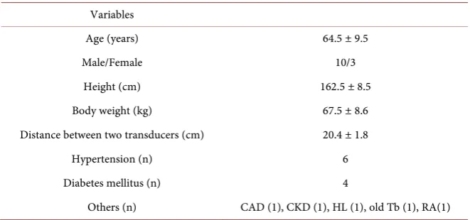
From May 2015 to March 2016, 22 consecutive patients undergoing shoulder surgery in the BCP at Gifu University Hospital were enrolled in the present study. Nine patients were excluded because of technical failures from the analy-sis due to the following factors: hemodynamic instability (n = 4), failure of radial artery cannulation (n = 2), transducer placement error (n = 2), and use of pro-pofol for anesthetic maintenance (n = 1). Thus, the data of 13 patients were in-cluded in the analysis. The characteristics of the patient are listed in Table 1. The mean distance between the fourth intercostal space and the external audito-ry meatus was 20.4 ± 1.8 cm.
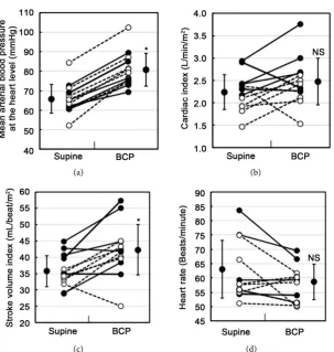
[image:4.595.209.540.521.676.2]Following the induction of anesthesia, while the patients were in the supine position, the mABP at the mid-axillary level was 65.7 ± 7.4 mmHg. After reposi-tioning the patients in the BCP, the mABP was 66.5 ± 8.1 mmHg at the external auditory meatus and 80.7 ± 8.4 mmHg at the fourth intercostal space (Figure 1(a)). The mABP values recorded at the level of the heart while the patients were
Table 1. The characteristics of the patients in the present study (n = 13).
Variables
Age (years) 64.5 ± 9.5
Male/Female 10/3
Height (cm) 162.5 ± 8.5
Body weight (kg) 67.5 ± 8.6
Distance between two transducers (cm) 20.4 ± 1.8
Hypertension (n) 6
Diabetes mellitus (n) 4
Others (n) CAD (1), CKD (1), HL (1), old Tb (1), RA(1)
DOI: 10.4236/ojanes.2018.81003 31 Open Journal of Anesthesiology
(a) (b)
[image:5.595.224.528.64.384.2]
(c) (d)
Figure 1. The hemodynamic values in the supine position and the beach chair position
(BCP) in patients receiving blood pressure support. In the BCP, the blood pressure was maintained with reference to the mean arterial blood pressure (mABP) at the level of the external auditory meatus. Two time points were selected (120 s apart) during which the mABP at the level of the brain was stable and within 5 mmHg in the supine position and the BCP before surgery. (a) The mABP at the level of the heart; (b) the cardiac index; (c) the stroke volume index; and (d) the heart rate. Error bar chart values are expressed as the mean ± standard deviation. *P < 0.029 in comparison to the value in the supine position. NS: not significant. White circles: non-hypertensive patients. Black circles: hypertensive patients.
in the BCP were significantly higher than those recorded while patients were in the supine position (P < 0.0001). The position-related changes in the CI, SVI and heart rate are shown in Figures 1(b)-(d), respectively. The CI changed from 2.2 ± 0.4 to 2.5 ± 0.5 l/min/m2 (P = 0.13, Figure 1(b)) and while this value increased in nine patients, it decreased in the other four. Overall, the SVI showed a signif-icant increase from 35.8 ± 4.8 to 42.3 ± 7.8 ml/m2 (P = 0.003, Figure 1(c)). The SVI decreased in three patients and these patients also experienced a decreased CI. The heart rate changed from 63.0 ± 10.0 to 58.6 ± 6.1 beats/min (P = 0.046,
Figure 1(d)). The SVV, which was 12.4% ± 3.5% in the supine position, was sig-nificantly decreased in the BCP (8.8% ± 2.3%, P = 0.024).
4. Discussion
DOI: 10.4236/ojanes.2018.81003 32 Open Journal of Anesthesiology the same in the supine position and the BCP, then the SVI increased leaving the CI unaffected. Previous studies have reported that postural changes in anesthe-tized patients result in cardiovascular changes as the patient’s head is elevated from a supine position. The elevation of the head was shown to result in a de-crease in the CI [14] [15] [16] and SVI [14] [16]. Many studies have reported that the mABP recorded at the level of the heart decreases as patients are shifted to the BCP [8] [10] [14] [15]. However, the changes that occur in these parame-ters after the postural change and the increase of the BP are unknown. Similarly to previous reports, it was hypothesized that the mABP recorded at the level of the heart, as well as the CI, and SVI would decrease in our patients following a shift to the BCP. It was hypothesized that both the CI and the SVI (especially the SVI) would increase after the BP was increased in the BCP in comparison to the values measured in the BCP when the BP was not increased. However, the changes in the CI, SVI and heart rate showed various patterns in the present study. The main cause of this discrepancy is the various methods that were used to increase the BP. Furthermore, various methods are used to increase the BP in the clinical setting and these methods exert varying degrees of influence on the systemic circulation, including the parameters that were reported in the present study. In any case, we need to pay attention to the changes in the cardiac func-tion and to manage patients according to their individual condifunc-tions.
The present study is associated with several limitations, namely the small sample size and the various methods that were applied to increase the BP. It is very difficult to keep the mABP at the level of the brain the same in both the su-pine position and the BCP. Thus, we could not collect a sufficient number of pa-tients and could not use a single method to increase the BP in all papa-tients. Our study protocol was designed to make comparisons within individuals. We calcu-lated that a study population of 25 would be needed. Because the statistical pow-er was 80%, a diffpow-erence in mABP of 10 mmHg was considpow-ered to be clinically relevant, and the SD in the change in the mABP (between before and after the change in position) was 5 mmHg. We performed an interim analysis using the Pocock method with a study population that was half the size of that in the pro-tocol and set the p value at 0.029 instead of 0.05 in order to avoid increasing the chance of a type 1 error [13]. The methods that we used to increase the BP have been utilized in previous studies [9] [17]. However, these methods have different effects on the cardiac function. Further studies should be performed to investi-gate the effects of each of these methods on the cardiac function in the BCP and methods for increasing the BP should be selected according to their effects on the cardiac function.
DOI: 10.4236/ojanes.2018.81003 33 Open Journal of Anesthesiology approximately 67 mmHg. It was calculated that the mABP at the level of the brain would decrease by 0.77 mmHg for each 1 cm in head elevation with a heart-level mABP of approximately 82 mmHg. This calculated value was the same as the value that was directly recorded in the radial artery in our study (approximately 81 mmHg). In a conscious, healthy individuals, the cerebral blood flow is maintained at a constant level of between 50 (recently 70 has been proposed) and 150 mmHg, despite changes in the cerebral perfusion pressure
[11] [18]. Reports indicate that cerebral autoregulation is unaffected by general anesthesia [18]. In our patient population, the mABP at the level of the brain was between 54 and 87 mmHg and safe mABP levels could be maintained.
5. Conclusion
In conclusion, it is important to maintain the cerebral blood flow when a patient is in the BCP. However, to ensure patient safety, close attention should be paid to the systemic cardiovascular changes that occur in each patient when the BP is increased. Further studies are needed to determine optimal method for main-taining the BP that avoids both cerebral desaturation and the over-loading to the heart in anesthetized patients in the BCP.
References
[1] Peruto, C.M., Ciccotti, M.G. and Cohen, S.B. (2009) Shoulder Arthroscopy Posi-tioning: Lateral Decubitus versus Beach Chair. Arthroscopy, 25, 891-896.
https://doi.org/10.1016/j.arthro.2008.10.003
[2] Bhatti, M.T. and Enneking, F.K. (2003) Visual Loss and Ophtalmoplegia after Shoulder Surgery. Anesthesia & Analgesia, 96, 899-902.
https://doi.org/10.1213/01.ANE.0000047272.31849.F9
[3] Pohl, A. and Cullen, D.J. (2005) Cerebral Ischemia during Shoulder Surgery in the Upright Position: A Case Series. Journal of Clinical Anesthesia, 17, 463-469. https://doi.org/10.1016/j.jclinane.2004.09.012
[4] The Official Journal of the Anesthesia Patient Safety Foundation. (2009) APSF Newsletter, 24, 45-48.
[5] Murphy, G.S., Szokol, J.W., Avram, M.J., Greenberg, S.B., Shear, T.D., Vender, J.S., Levin, S.D., Koh, J.L., Parikh, K.N. and Patel, S.S. (2014) Effect of Ventilation on Cerebral Oxygenation in Patients Undergoing Surgery in the Beach Chair Position: A Randomized Controlled Trial. British Journal of Anaesthsia, 113, 618-627. https://doi.org/10.1093/bja/aeu109
[6] Hinghofer-Szalkay, H. (2011) Gravity, the Hydrostatic Indifference Concept and the Cardiovascular System. European Journal of Applied Physiology, 111, 163-174. https://doi.org/10.1007/s00421-010-1646-9
[7] ter Laan, M., van Dijk, J.M., Elting, J.W., Staal, M.J. and Absalom, A.R. (2013) Sympathetic Regulation of Cerebral Blood Flow in Humans: A Review. British Journal of Anaesthesia, 111, 361-367. https://doi.org/10.1093/bja/aet122
DOI: 10.4236/ojanes.2018.81003 34 Open Journal of Anesthesiology [9] Meex, I., Vundelinckx, J., Buyse, K., Deburggraeve, F., De Naeyer, S., Desollvere, V., Anné, L., Truijen, J., Vander Laenen, M., Heylen, R., De Deyne, C. and Jans, F. (2016) Cerebral Tissue Oxygen Saturation Values in Volunteers and Patients in the Lateral Decubitus and Beach Chair Positions: A Prospective Observational Study. Canadian Journal Anesthesia, 63, 537-543.
https://doi.org/10.1007/s12630-016-0604-3
[10] Koh, J.L., Levin, S.D., Chehab, E.L. and Murohy, G.S. (2013) Neer Award 2012: Ce-rebral Oxygenation in the Beach Chair Position: A Prospective Study on the Effect of General Anesthesia Compared with Regional Anesthesia and Sedation. Journal of Shoulder and Elbow Surgery, 22, 1325-1331.
https://doi.org/10.1016/j.jse.2013.01.035
[11] Murphy, G.S. and Szokol, J.W. (2011) Blood Pressure Management during Beach Chair Position Shoulder Surgery: What Do We Know? Canadian Journal of Anes-thesia, 58, 977-982. https://doi.org/10.1007/s12630-011-9573-8
[12] Ozzeybek, D., Oztekin, S., Mavioğlu, O., Karaege, G., Ozkardeşler, S., Ozkan, M., Canyilmaz, M. and Elar, Z. (2003) Comparison of the Haemodynamic Effects of In-terscalene Block Combined with General Anaesthesia and InIn-terscalene Block alone for Shoulder Surgery. Journal of International Medical Research, 31, 428-433. https://doi.org/10.1177/147323000303100512
[13] Schulz, K.F. and Grimes, D.A. (2005) Multiplicity in Randomized Trials II: SUB-Group and Interim Analyses. Lancet, 365, 1657-1661.
https://doi.org/10.1016/S0140-6736(05)66516-6
[14] Buhre, W., Weyland, A., Buhre, K., Kazmaier, S., Mursch, K., Schmidt, M., Sydow, M. and Sonntag, H. (2000) Effects of the Sitting Position on the Distribution of Blood Volume in Patients Undergoing Neurosurgical Procedures. British Journal of Anaesthesia, 84, 354-357. https://doi.org/10.1093/oxfordjournals.bja.a013439 [15] Soeding, P.F., Hoy, S., Hoy, G., Evans, M. and Royse, C.F. (2013) Effect of
Pheny-lephrine on the Haemodynamic State and Cerebral Oxygen Saturation during Anaesthesia in the Upright Position. British Journal of Anaesthesia, 111, 229-234. https://doi.org/10.1093/bja/aet024
[16] Jo, Y.Y., Jung, W.S., Kim, H.S., Chang, Y.J. and Kwak, H.J. (2014) Prediction of Hypotension in the Beach Chair Position during Shoulder Arthroscopy Using Pre-Operative Hemodynamic Variables. Journal of Clinical Monitoring and Com-puting, 28, 173-178. https://doi.org/10.1007/s10877-013-9512-z
[17] Murphy, G.S., Szokol, J.W., Marymont, J.H., Greenberg, S.B., Avram, M.J., Vender, J.S., Vaughn, J. and Nisman, M. (2010) Cerebral Oxygen Desaturation Events As-sessed by Near-Infrared Spectroscopy during Shoulder Arthroscopy in the Beach Chair and Lateral Decubitus Positions. Anesthesia & Analgesia, 111, 496-505. https://doi.org/10.1213/ANE.0b013e3181e33bd9
[18] Dagal, A. and Lam, A.M. (2009) Cerebral Autoregulation and Anesthesia. Current Opinion in Anaesthesiology, 22, 547-552.