ISSN Print: 2158-7027
DOI: 10.4236/jbnb.2018.93013 Jun. 26, 2018 233 Journal of Biomaterials and Nanobiotechnology
Surface Modification of PEEK and Its
Osteoconductivity and
Anti-Inflammatory Properties
Kensuke Kuroda
1*, Kenta Igarashi
2, Hiroyasu Kanetaka
3, Masazumi Okido
11Institute of Materials and Systems for Sustainability, Nagoya University, Nagoya, Japan
2Department of Materials Science and Engineering, Graduate School of Engineering, Nagoya University, Nagoya, Japan 3Liaison Center for Innovative Dentistry, Graduate School of Dentistry, Tohoku University, Sendai, Japan
Abstract
Polyetheretherketone (PEEK) is known as one of the “super-engineering plas-tics” and is used as an intervertebral disk spacer in the body. PEEK has a hy-drophobic surface (water contact angle (WCA) > 80˚) and high chemical re-sistance, and it is thus difficult to perform any surface treatment, such as hy-drophilization. In this study, we aimed to form a hydrophilic surface on PEEK without coating layers by using hydroprocessing (aqueous solution processing), and we examined the osteoconductivity and anti-inflammatory properties of surface-treated PEEK in vivo compared with Ti implants. The WCA value of PEEK reached ~20˚ using a combination of immersion in a solution of >16.2 M H2SO4 and ultraviolet irradiation (172 nm). In in vivo testing, the
hydrophi-lization of PEEK by surface modification without a coating layer improved the osteoconductivity and anti-inflammatory properties. The relationship between the bone-implant contact ratio and the WCA values of the surface-modified PEEK agreed well with that of the surface-treated Ti.
Keywords
Polyetheretherketone, Hydrophilization, Protein Adsorption, Osteoconductivity, Inflammatory
1. Introduction
Polymer materials are used in the orthopedic and dental fields as important biomaterials alongside metallic and ceramic materials. In general, when bioma-terials are used in the body, it is known that the bioresponse depends on surface How to cite this paper: Kuroda, K.,
Iga-rashi, K., Kanetaka, H. and Okido, M. (2018) Surface Modification of PEEK and Its Osteoconductivity and Anti-Inflammatory Properties. Journal of Biomaterials and Nanobiotechnology, 9, 233-243.
https://doi.org/10.4236/jbnb.2018.93013
Received: April 8, 2018 Accepted: June 23, 2018 Published: June 26, 2018
Copyright © 2018 by authors and Scientific Research Publishing Inc. This work is licensed under the Creative Commons Attribution International License (CC BY 4.0).
DOI: 10.4236/jbnb.2018.93013 234 Journal of Biomaterials and Nanobiotechnology characteristics such as hydrophilicity [1] [2] [3] [4]. Our previous study using valve metals and their alloys showed that hydrophilic metallic surfaces have high osteoconductivity [5]. Many kinds of polymers have a high chemical stability as well as a hydrophobic surface. For example, polyetheretherketone (PEEK), which is used as an intervertebral disk spacer, has a hydrophobic surface (water contact angle (WCA) > 80˚); however, based on our previous studies with metal-lic materials, this feature does not indicate that PEEK has high biocompatibility. Indeed, postoperative infection caused by a PEEK disc spacer has been reported and solutions to such potential bio-incompatibility have been proposed [6]. PEEK is known as one of the “super-engineering plastics” that have a high chemical resistance; consequently, a surface treatment, such as hydrophilization, is very difficult to apply in the same manner as for polytetrafluoroethylene (PTFE). Initially, hydrophobic PEEK was coated with hydroxyapatite (HAp), which was believed to be a bioactive substance [7], but the adhesion of HAp on PEEK was poor. It is believed that the root of this problem is that PEEK shows very stable hydrophobicity.
Plasma irradiation is commonly used for the hydrophilization of various po-lymer materials [8]. However, the nearly line-of-sight nature of plasma irradia-tion and the normally relatively inhomogeneous irradiairradia-tion can produce surface unevenness, thereby limiting the usefulness of this technique because many im-plants have a complex shape. In this study, we aimed to form a hydrophilic sur-face on PEEK without coating layers by using hydroprocessing (aqueous solu-tion processing), and we examined the osteoconductivity and anti-inflammatory properties of the surface-treated PEEK compared with the surface-treated Ti im-plants in vivo.
2. Theory of Polymer Hydrophilization
It is well known that the copolymer of polylactic acid-glycolic acid (PLGA) de-grades in the body [9]. However, its degradability is not very fast and accelera-tion methods have been proposed [10]; for example, it was reported that the immersion of PLGA into alkaline aqueous solution was effective in accelerating the degradation. In our preliminary experiment, a PLGA sheet was immersed in several aqueous solutions, such as distilled water (DW), and ~5 mol L−1 NaOH
DOI: 10.4236/jbnb.2018.93013 235 Journal of Biomaterials and Nanobiotechnology
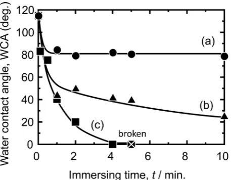
Figure 1. Water contact angle change with immersing time in (a) distilled water, (b) 0.1 M NaOH aqueous solution, and (c) 0.5 M NaOH aqueous solution.
trigger for the hydrophilization of the polymer, disconnection of the bonds (e.g., hydrolysis), and its acceleration. In addition, fixation of the hydrophilic group (e.g., -OH, -COOH) to the disconnected bond was an indispensable condition for hydrophilization.
3. Experimental
3.1. Surface Modification
We previously reported that the surface roughness of implants affected osteo-conductivity after implantation [11]. PEEK plates (φ6 mm) were polished by buffing using Al2O3 particles (particle size = 0.05 μm) and the surface roughness
(Ra) was set as Ra/μm < 0.1. Samples were immersed in ~18 M (=mol L−1)
H2SO4, ~12 M HCl, ~13 M HNO3, ~17 M CH3COOH, or ~30 vol.% H2O2 for up
DOI: 10.4236/jbnb.2018.93013 236 Journal of Biomaterials and Nanobiotechnology
3.2. Protein Adsorption Testing
In the protein adsorption testing, fibronectin and albumin were picked up as a protein that was either a cell-adhesive protein or not, respectively [12]. A fibro-nectin (~1.0 mg mL−1) or albumin (~50 mg mL−1) aqueous solution droplet (40
μL) was put on the surface of the WCA-controlled PEEK samples, respectively, and these were kept at 37˚C for up to 3 d. Then, they were rinsed out with ultra-sonic cleaning in the distilled water and dried naturally. The amount of adsorbed protein was determined using FTIR in attenuated total reflection (ATR) mode. The protein content was evaluated from the peptide binding (1650 cm−1) in the
protein in the FTIR spectra. The content of the different three positions was measured for each samples using calibration curve, and the mean value was cal-culated.
3.3.
In Vivo
Testing
All the animal studies were conducted in the laboratory at HAMRI Co., Ltd., Ja-pan, of the Association for Assessment and Accreditation of Laboratory Animal Care International (AAALAC).
1) Testing of osteoconductivity [13]
The sides of PEEK rods (2 mm in diameter, 5 mm in length) were polished by buffing using 0.05 μm Al2O3 particles. Hydrophilic PEEK rods (WCA = 85˚)
were prepared in a combination of immersion in 16.2 M H2SO4 for 5 s at 30˚C
and UV irradiation (172 nm) for 3 h. For the formation of the protein-adsorbed samples, hydrophilic PEEK samples were immersed in 1.0 mg mL−1 fibronectin
aqueous solution for 48 h at 37˚C. Next, they were rinsed with ultrasonic clean-ing in the distilled water and dried naturally. All the surface-treated rod samples (n = 5) before and after protein adsorption were implanted in rat tibia for 14 d for evaluation of osteoconductivity. The samples were longitudinally sliced and stained with toluidine blue. The interface between the implant and the cortical bone, as well as the cancerous bone, was observed by optical microscopy. The sum of the linear bone contact with the implant surface was measured and was expressed as a percentage over the entire implant length (the bone-implant con-tact ratio, BIC) in the cancerous bone and in the cortical bone parts. Significant differences in BIC were analyzed statistically using the Tukey-Kramer method [14].
2) Testing of anti-inflammatory properties
methacry-DOI: 10.4236/jbnb.2018.93013 237 Journal of Biomaterials and Nanobiotechnology late (MMA) polymer. The samples were sliced and stained with toluidine blue. Optical microscope observation of the interface between the implanted and the surrounding tissue revealed whether inflammatory cells such as lymphocytes and neutrophils were present, as well as a new generation of vascular, and for-mation of a fibrous capsule film. The capsule film thickness of the eight posi-tions was measured for each samples, and the mean value was expressed as an index of anti-inflammatory properties.
4. Results and Discussion
[image:5.595.211.539.419.673.2]4.1. Surface Modification
Figure 2 shows photographs taken after immersion in several aqueous solutions and washing in distilled water. The immersion in 12 M HCl, 13 M HNO3, 17 M
CH3COOH, and 30 vol.% H2O2 did not change the surface morphology of PEEK.
The WCA values were 81˚, 84˚, 80˚, and 83˚, respectively, and they did not in-crease or dein-crease dramatically from the as-polished PEEK (~85˚). Immersion in H2SO4 had a clear effect on the surface appearance. In particular, 18 M H2SO4
attacked the surface and the color turned white from beige, and the Ra value in-creased from 0.05 to 0.44 μm. For immersion in 16.8 M H2SO4, SEM observation
confirmed the attack, although discoloration and Ra change did not occur. For the immersion in less than 15 M, no changes were observed. Figure 3 shows FTIR spectra of samples after 18 M H2SO4 immersion (Figure 3(b)) and washing
in DW (Figure 3(c)). Figure 3 indicates that the immersion in H2SO4 broke the
Figure 2. Optical micrograph and surface morphology of PEEK samples processed by immersion in different aqueous solutions: ((a1), (a2)) 18 M H2SO4, ((b1), (b2)) 16.2 M
H2SO4, ((c1), (c2)) as-polished, (d) 12 M HCl, (e) 13 M HNO3, (f) 17M M CH3COOH,
DOI: 10.4236/jbnb.2018.93013 238 Journal of Biomaterials and Nanobiotechnology
Figure 3. FT-IR spectra of PEEK samples processed by following treatment: (a) as-polished, (b) immersion in 18 M H2SO4, (c) after rinse of (b) in distilled water, (d) after ultraviolet
(UV) irradiation (172 nm) to (c), and (e) atmospheric plasma irradiation to (c).
ether bond, the C–C single bond, and the C=C double bond, and increased the sulfo group. Because the subsequent washing process decreased the sulfo group, it was thought that the WCA value did not decrease after immersion in H2SO4,
which in any case satisfied the first step for polymer hydrophilization, that is, the disconnection of the bonds.
As a second step, UV (172 nm) irradiation (>10 mW cm−2, with 3 mm
dis-tance) and atmospheric plasma irradiation (500 W, 50 mm disdis-tance) were se-lected and applied on the solution-treated samples: immersion in 16.2 M H2SO4
for 5 s and rinsing in DW. With atmospheric plasma, the WCA value decreased rapidly and reached a constant WCA of ~10˚ in 10 min, regardless of the first step. FTIR showed that there was no change in the surface functional group by the plasma treatment (Figure 3(e)), and therefore it was thought that radicals might contribute to hydrophilization. However, the hydrophilicity formed by plasma irradiation, could not be maintained, and the WCA value rose rapidly in 1 d (Figure 4). In contrast, it took 1 - 2 h for the solution-treated samples to reach a constant WCA of ~20˚ under UV irradiation, and the Ra value was the same as after H2SO4 immersion (Ra = 0.05 μm). FTIR showed that the UV
irrad-iation created a carboxyl group. By examination of UV irradirrad-iation under differ-ent partial pressure environmdiffer-ents of CO2 (Ar or CO2 atmosphere), we
deter-mined that this carboxyl group was formed from CO2 in air. The UV treatment
DOI: 10.4236/jbnb.2018.93013 239 Journal of Biomaterials and Nanobiotechnology
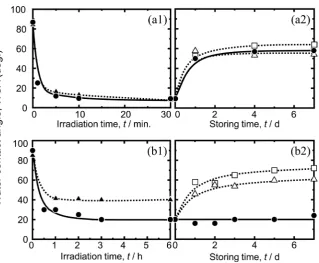
Figure 4. Water contact angle change with (1) irradiation time (●: 18 M H2SO4 immersed
sample and ▲: as-polished sample) and (2) storing time (●: five times concentrated phosphate buffered saline (x5 PBS(-)), □: air, and Δ: distilled water) of (a) atmospheric plasma irradiation and (b) ultraviolet (UV) irradiation (172 nm).
7 d by storing in ×5 PBS (−) in the same manner as hydrophilic Ti and its alloy samples [15]. It is thought that ions in ×5 PBS (−), such as Na+, were adsorbed
on the hydrophilic PEEK surface. Thus, in the following hydrophilization expe-riment, PEEK was immersed in 16.2 M H2SO4 for 5 s and rinsed in DW, and
then irradiated with UV (172 nm) for more than 3 h in air. To maintain hydro-philicity, the surface-treated PEEK was stored in ×5 PBS (−).
4.2. Evaluation of Osteoconductivity
DOI: 10.4236/jbnb.2018.93013 240 Journal of Biomaterials and Nanobiotechnology
Figure 5. Relationship between bone-implant contact ratio (BIC) and water contact angle (WCA). (1) as-polished, (2) ultraviolet (UV) irradiated to as-polished, ((3), (4)) ultravio-let (UV) irradiated to 16.2 M H2SO4 immersed, and (5) fibronectin adsorbed to sample
(3) and ○: surface treated Ti sample. *: p < 0.05.
Figure 6. Adsorbed value of protein ((a) fibronectin and (b) albumin) to the water con-tact angle controlled PEEK. ●: fibronectin, ▲: albumin, and ○, Δ: surface treated Ti sam-ple.
depending on the WCA value. This tendency agreed well with our results for surface-treated Ti [15]. The BIC value of the fibronectin-adsorbed samples (n = 5) on the hydrophilic samples (n = 3) was also high, and the same level of hy-drophilic sample, in spite that the fibronectin-adsorbed sample did not have a hydrophilic surface (WCA = 48˚). This also supports the notion that the osteo-conductivity was not directly affected by the WCA of implants.
4.3. Evaluation of Anti-Inflammatory Properties
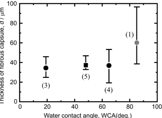
[image:8.595.240.508.304.516.2]DOI: 10.4236/jbnb.2018.93013 241 Journal of Biomaterials and Nanobiotechnology detected around the surrounding tissue of implants. A fibrous capsule film formed over the entire surface in all samples, and the inflammatory reaction oc-curred over the whole surface of all samples. The thickness of the capsule film was used as an index for the evaluation of anti-inflammatory properties. Figure 8 shows the thickness of the capsule film formed on the implants. On the
Figure 7. Photographs of the inside of the striped skin and the optical micrographs of surface treated PEEK samples implanted in the subcutaneous of back of rats for 14 d.
Figure 8. Relationship between the thickness of fibrous capsule and WCA. (1) as-polished, ((3), (4)) ultraviolet (UV) irradiated to 16.2 M H2SO4 immersed, and (5) fibronectin
[image:9.595.234.515.477.682.2]DOI: 10.4236/jbnb.2018.93013 242 Journal of Biomaterials and Nanobiotechnology as-polished sample, a thick capsule film was formed, suggesting that the inflam-mation was strong, as reported [17]. However, the inflaminflam-mation was weaker on the surface-treated PEEK, which was not related to the WCA value. In the im-plantation of the fibronectin-adsorbed PEEK samples on the hydrophilic surface in advance, the inflammation was not suppressed further. It is noteworthy that the surface treatment can suppress the inflammation without an anti-inflammatory drug, compared with as-polished PEEK.
5. Conclusion
The WCA value of PEEK reached ~20˚ using a combination of immersion in >16.2 M H2SO4 and UV irradiation (172 nm). The immersion of the
sur-face-treated PEEK in ×5 PBS (−) maintained the hydrophilic WCA. Although as-polished PEEK could not adsorb protein, hydrophilization gave rise to pro-tein adsorption. In vivo, the hydrophilization of PEEK by surface modification without a coating layer improved the osteoconductivity and anti-inflammatory properties. The relationship between BIC and WCA values of surface-modified PEEK agreed well with that of the surface-treated Ti.
Acknowledgements
This research was financial supported partially by JSPS KAKENHI (Grant-in-Aid for Scientific Research (A) 15H02310 and (B) 25289248), and Joint Research Project on Life Innovation Materials by MEXT.
Conflicts of Interest
The authors declare that there are no conflicts of interest regarding the publica-tion of this paper.
References
[1] Buser, D., Broggini, N., Wieland, M., Schenk, R.K., Denzer, A.J., Cochran, D.L., Hoffmann, B., Lussi, A. and Steinemann, S.G. (2004) Enhanced Bone Apposition to a Chemically Modified SLA Titanium Surface. Journal of Dental Research, 83, 529-533. https://doi.org/10.1177/154405910408300704
[2] Cochran, D.L., Buser, D., ten Bruggenkate, C.M., Weingart, D., Taylor, T.M., Ber-nald, J.P., Peters, F. and Simpson, J.P. (2002) The Use of Reduced Healing Times on ITI® Implants with a Sandblasted and Acid-Etched (SLA) Surface: Early Results from Clinical Trials on ITI® SLA Implants. Clinical Oral Implants Research, 13, 144-153. https://doi.org/10.1034/j.1600-0501.2002.130204.x
[3] Eriksson, C., Nygren, H. and Ohlson, K. (2004) Implantation of Hydrophilic and Hydrophobic Titanium Discs in Rat Tibia: Cellular Reactions on the Surfaces dur-ing the First 3 Weeks in Bone. Biomater, 25, 4759-4766.
https://doi.org/10.1016/j.biomaterials.2003.12.006
[4] Park, J.-W., Park, K.-B. and Suh, J.-Y. (2007) Effects of Calcium Ion Incorporation on Bone Healing of Ti6Al4V Alloy Implants in Rabbit Tibiae. Biomater, 28, 3306-3313. https://doi.org/10.1016/j.biomaterials.2007.04.007
DOI: 10.4236/jbnb.2018.93013 243 Journal of Biomaterials and Nanobiotechnology Surface of Pure Titanium by Hydrothermal Treatment. Journal of Biomaterials and Nanobiotechnology, 4, 284-290. https://doi.org/10.4236/jbnb.2013.43036
[6] Toth, J.M., Wang, M., Estes, B.T., Scifert, J.L., Seim, H.B. and Turmer, A.S. (2006) Polyetheretherketone as a Biomaterial for Spinal Applications. Biomater, 27, 324-334.
https://doi.org/10.1016/j.biomaterials.2005.07.011
[7] Suzuki, N., Umeda, T., Sumi, T., Horikoshi, S., Kuwahara, H., Toyama, T., Musha, Y. and Itatani, K. (2016) Rapid Formation of Hydroxyapatite Layer on Polyethere-therketone by Vacuum Ultraviolet Irradiation and Microwave Heating Techniques. Journal of the Ceramic Society of Japan, 124, 49-54.
https://doi.org/10.2109/jcersj2.15209
[8] Chvatalova, L., Cermak, R., Mracek, A., Grulich, O., Vesel, A., Ponizil, P., Minarik, A., Cvelbar, U., Benicek, L. and Sajdl, P. (2012) The Effect of Plasma Treatment on Structure and Properties of Poly(1-butene) Surface. European Polymer Journal, 48, 866-874. https://doi.org/10.1016/j.eurpolymj.2012.02.007
[9] Caffesse, R.G., Nasjleti, C.E., Morrison, E.C. and Sanchez, R. (1994) Guided Tissue Regeneration: Comparison of Bioabsorbable and Non-Bioabsorbable Membranes. Histologic and Histometric Study in Dogs. Journal of Periodontology, 65, 583-591.
https://doi.org/10.1902/jop.1994.65.6.583
[10] Tsuchiya, S., Ohmori, M., Hara, K., Fujio, M., Ikeno, M., Hibi, H. and Ueda, M. (2015) An Experimental Study on Guided Bone Regeneration Using a Polylac-tide-co-glycolide Membrane-Immobilized Conditioned Medium. The International Journal of Oral & Maxillofacial Implants, 30, 1175-1186.
https://doi.org/10.11607/jomi.3915
[11] Yamamoto, D., Kawai, I., Kuroda, K., Ichino, R., Okido, M. and Seki, A. (2011) Os-teoconductivity of Anodized Titanium with Controlled Micron-Level Surface Roughness. Materials Transactions, 52, 1650-1654.
https://doi.org/10.2320/matertrans.M2011049
[12] Porté-Durrieu, M.C., Guillemota, F., Pallu, S., Labrugère, C., Brouillaud, B., Bareille, R., Amédée, J., Barthe, N., Dard, M. and Baquey, Ch. (2004) Cyclo-(DfKRG) Pep-tide Grafting onto Ti-6Al-4V: Physical Characterization and Interest towards Hu-man Osteoprogenitor Cells Adhesion. Biomater, 25, 4837-4846.
https://doi.org/10.1016/j.biomaterials.2003.11.037
[13] Kuroda, K., Nakamoto, S., Miyashita, Y., Ichino, R. and Okido, M. (2006) Osteoin-ductivity of HAp Films with Different Surface Morphology Coated by the Thermal Substrate Method in Aqueous Solutions. Materials Transactions, 47, 1391-1394.
https://doi.org/10.2320/matertrans.47.1391
[14] Kramer, C.Y. (1956) Extension of Multiple Range Tests to Group Means with Un-equal Numbers of Replications. Biometrics, 12, 307-310.
https://doi.org/10.2307/3001469
[15] Zuldesmi, M., Waki, A., Kuroda, K. and Okido, M. (2014) Enhancement of Valve Metal Osteoconductivity by One-Step Hydrothermal Treatment. Materials Science and Engineering: C, 42, 405-411. https://doi.org/10.1016/j.msec.2014.05.049
[16] Kuroda, K. and Okido, M. (2015) A New Approach for Controlling Osteoconduc-tivity of Valve Metals Based on TiO2 Coatings on Ti Substrates. Materials
Technol-ogy, 30, B13-B20. https://doi.org/10.1179/1753555714Y.0000000221