Abstract— A study was conducted to investigate the effect of load on upper limb muscles and brain activities in light assembly task. The task was conducted at two levels of load (Low and high). Surface electromyography (EMG) was used to measure upper limb muscle activities of six subjects. The results found that the Mean Power Frequency (MPF) of the brachioradialis and upper trapezius activities were higher on the low-load task compared to high-load task. The EMG MPF also tended to decrease as time increases, that reflects muscle fatigue. Electroencephalography (EEG) was simultaneously recorded with EMG to record brain activities from Fz, Pz, O1 and O2 channels. Mean power of the EEG alpha bands from the channels were found to be higher on the low-load task compared to high-load task. The results indicated that high-load task require more brain activitie than the low-load task. It is also found that there was relation between upper limb muscles and brain activities while performing light assembly task. Thus these results can would be potentially useful in the management of fatigue, especially related to muscle and mental workload.
Index Terms— Upper limb muscle, brain activity, load, light assembly.
I. INTRODUCTION
Fatigue can occur because of either mental or muscle activity. The concept of mental fatigue earlier introduced by Grandjean clearly differentiated mental fatigue from muscle fatigue. He defined that muscle fatigue was concerned with reduced muscular system performance while mental fatigue deals with much reduced mental performance and the sense of weariness. Muscular fatigue contributes to impaired co-ordination and increased chances of errors and accidents [1].
When people become fatigued, besides muscle fatigue, they usually report difficulties in concentrating and focusing their attention on the tasks they are required to perform [2]. It is one indication of mental fatigue. Mental fatigue is believed to be a gradual and cumulative process and is thought to be associated with a disinclination for any effort, reduced efficiency and alertness and impaired mental performance [3]. Generally, there is no desire for physical or mental effort
Manuscript received December 30, 2009.
Hilma Raimona Zadry is a Ph.D student in Department of Engineering Design and Manufacture, Faculty of Engineering, University of Malaya, 50603 Kuala Lumpur, Malaysia. (Corresponding author: phone: +603 7967 5382; fax: +603 7967 5330. e-mail: [email protected]).
Siti Zawiah Dawal is an Associate Professor in Department of Engineering Design and Manufacture, Faculty of Engineering, University of Malaya, 50603 Kuala Lumpur, Malaysia. e-mail: [email protected].
Zahari Taha is a Professor in Department of Engineering Design and Manufacture, Faculty of Engineering, University of Malaya, 50603 Kuala Lumpur, Malaysia. E-mail: [email protected].
and there is an associated heavy, drowsy feeling. A number of mental fatigue tests have already been adopted, but it still hard to draw a generalized conclusion as to the method of selecting the most appropriate test battery for a given workload [4].
Based on an empirical review, there are many studies on muscle fatigue. Some of the studies analyzed muscle fatigue during repetitive tasks. They found that time pressure, lack of influence over one's work and constant involvement in repetitive tasks of short duration often characterize jobs associated with a high risk for muscular problems. In contrast to muscle fatigue, relatively few studies investigated on both muscle and mental fatigue. Most studies observed the impact of mental activities on muscle and mental fatigue [5].
Light-assembly work is a clear example of low intensity work with elevated risks of neck and shoulder disorders [6]. Although there were many studies investigating about muscle fatigue in assembly task, few research investigate the correlation between upper limb muscle and brain activities during light assembly task.
Many of the studies were carried out using laboratory experiments method, and only a few was carried out by survey. Electromyography (EMG) is the most popular tool for measuring muscle activation and fatigue. While blood pressure, blink rate, and heart rate were rarely used. Changes in the electromyographic activity (a decrease in the mean power frequency and/or an increase in the EMG amplitude) during standardized voluntary contractions have frequently been used as indicators of muscle fatigue [7, 8].
On the other hand, there were still a few studies measuring brain activity in light repetitive task using Electroencephalography (EEG) [9]. Most studies used EEG to measure drowsiness or fatigue on drivers [10, 11] and night work [12, 13]. In addition, subjective measurements were also carried out simultaneously.
This study investigates the effect of load on upper limb muscles (brachioradialis and upper trapezius) and brain activities (Fz, Pz, O1 and O2 channels) and the relationship between upper limb muscle and brain activities while doing light assembly tasks in different load.
II. METHODOLOGY
A.Subjects
Eight subjects consisted of five males and three females from the university population were recruited to participate in the experiment. The subjects were 18 to 30 year-old (22.80 ± 1.48 years). Potential participants were excluded if they had a history of any neurological, muscular or skeletal disease or disorder. Subjects were also excluded if they took any medication or substance that could affect motor and
Effect of Load on Upper Limb Muscle and
Brain Activity in Light Assembly Task
neurological performance.
B. Apparatus and Material
Noraxon Surface Electromyography (SEMG) and Telemyo “2400” Gen2 Telemetric Real Time 8 channel SEMG System completed with disposable surface electrodes Ag/AgCl/Solid Adhesive pregelled were used to record electrical activity of muscles. EEG BIOPAC MP150 System with AcqKnowledge 4.0 software and Electrode Cap (CAP100C) were used to record brain activity. Vertical electrooculogram (EOG) was also recorded, and later used to identify blink artefacts from the recorded EEG data. The EEG and EOG data was sampled at 1000 Hz.
C.Procedures
The subjects had to perform a simulated light assembly task at two levels of load (low load, LL and high load, HL). The task was to assemble and disassemble irons with different weight. Low load level includes assembling the iron with the weight of 300 g and high load level includes assembling the iron with the weight of 2 kg. The work task was terminated after two hours for each level. The cycle time was determined based on Methods Time Measurement (MTM).
Each tasks were performed in a random order on two consecutive days between 9.00 am until 12.00 pm. To become familiar with the experimental equipment and procedures, a training session was performed before the experiment. All session were performed in a laboratory at a normal temperature of 250 C.
The subjects were seated in an ergonomically chair with the back vertical and the feet in full contact with the floor or with a footrest. The desk was adjusted to elbow height so that the upper arm and forearm formed 900 angles when the hand was positioned at the middle of the desk and the upper arm was vertical.
At the start of each experimental day, three maximal voluntary contractions (MVC) of the right and left of brachoradialis and the right and left of upper trapezius were performed. The subjects also filled up the subjective measurement of fatigue (Borg’s CR-10 scale and mental fatigue scale) before and after the experiment. EMG and EEG were recorded concurrently whilst performing the tasks.
D.Data collection 1) Surface EMG
EMG signals were recorded from four muscles: brachioradialis and descending part of the upper trapezius on the right and left hands. Bipolar Ag/AgCl surface electrodes were placed with an inter electrode distance of 20 mm at the belly of the muscles. The electrode positions were located according to Hermens et al. [14]. A reference electrode was placed on the piciform bone. The electrode positions were marked with a waterproof pencil, in order to place the electrodes at the exact same position in both conditions.
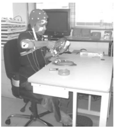
[image:2.595.329.524.46.264.2]Before the electrodes were applied, the skin was shaved, scrubbed and cleaned with alcohol. The recording was started after the inter-electrode resistance was less than 10 kΩ. Raw EMG signals were sampled during the test contraction with a sample frequency of 1500 Hz and band-pass filtered (20–400 Hz). Data was continuously recording using Telemyo 2400T G2 Telemetry EMG System.
Fig. 1. Subject was doing the task
The mean power frequency (MPF) was analysed using a fast Fourier transformation with a sliding window of 1000 samples. The MPF values were normalized to the measurement at the beginning of the working day. MPF values were calculated per window and averaged over one trial, resulting in one MPF value for each trial. Consequently, 24 MPF were obtained during the task. A simultaneous decrease of the MPF is generally considered indicative of fatigue [15].
2) Electroencephalography (EEG)
EEG recorded the brain activity simultaneously with the surface EMG during the experiments. It was recorded using an AgCl electrodes cap with electrodes placed at Fz, Pz, O1, and O2 of the International 10-20 electrodes placement system [16] and with an electronically earlobe reference. Data was continuously recorded for two hours with an MP150 system and analyzed with AcqKnowledge 4.0 software (BIOPAC Systems Inc.).
Electrodes were checked before each testing session to ensure that impedances were 5 kΩ or less. The bipolar recording technique was used to record the signals. The signals were band-pass filtered between 1.0 Hz and 100 Hz and recorded digitally (1000 Hz sample frequency). The EEG was checked off-line for artefacts. EEG alpha band was defined as the frequency between 8–13 Hz. For this measurement, an average power value over 5 minutes (24 epochs) periods were computed.
Eye movements were recorded by means of Electrooculogram (EOG). Right eye EOG was obtained with electrodes positioned above and below the eye with a ground on the masseter. The EOG signal was used to identify blink artefact in the EEG data as well as changes in blink types such as the small and slow blinks that characterize fatigue.
3) Subjective Measurement of Fatigue
The advantage of using subjective measurements such as rating scales is that they are easy to administer and no instrumentation or calibration is required. The process is generally non-invasive (although it may interrupt the task) and the data easy to interpret. In this study, the measurement used was a modified Borg-CR10 and mental fatigue scale. They were measured before and after the experimental task. The Borg-CR10 is a category scale with ratio properties that can yield ratios and levels and allow comparisons [18]. The scale ranges from 0 to 10 where 0 represents no fatigue and 10 represents maximum fatigue. The mental fatigue scale followed the measurement from a study by Huston [19]. The scale used is a five point bipolar scale consisting of four categories with descriptors. The categories are the subjects’ feelings of “fresh-weary”, “awake-sleepy”, “physically strong-physically weak” and the level of “interest-boredom”.
E. Data Analysis
The Mean Power Frequency (MPF) for EMG and mean power of the EEG alpha band were analyzed using SPSS 16.0. While for processing and filtering the signal, the software available together with the hardware was used. Shapiro-Wilk test was used to analysis the normality of the data. It was found that the data was normally distributed.
Independent-samples t-test was used to investigate the differences of EMG MPF and EEG alpha band power between genders. Paired-samples t-test was used to investigate the differences of EMG MPF and EEG alpha band power between LL and HL levels and the analysis of subjective measurements. Significance was accepted at p<0.05. Correlation analysis was carried out to examine the relationship between EMG MPF and EEG alpha band power.
III. RESULTS
A. EMG MPF
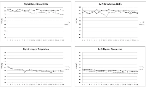
Fig. 2 shows the EMG MPF data for all subjects in two hours experiment. The data is divided into 24 intervals. The graph illustrates that EMG MPF are higher on the HL task than on the LL task for all muscles. It is also observed that the EMG MPF of the the muscles tends to deccrease with time for LL and HL task, especially on upper trapezius muscles.
B. EEG Alpha Band Power
Fig. 3 demonstrates the mean power of EEG alpha bands are rather similar between the HL and the LL task. The graphs also show that the mean power of EEG alpha bands on all channels, tend to increase as the time increase .
C.Comparison between Genders
The difference in muscle contraction between male and female subjects was investigated using independent-samples t-test. The analysis showed that the EMG MPF between male and female subjects was not significantly different for both LL and HL tasks. The gender differences were also not found on the EEG alpha band power for LL and HL tasks.
D.Comparison between Low and High Load Tasks
Analysis using paired-samples t-test showed there were no significant differences of EMG MPF between LL and HL tasks for all muscles. Similar to the EMG MPF, the EEG
alpha band power for all channels were also not different significantly between LL and HL tasks (Table 1).
Table1. Paired-samples t-test results for comparison of EMG and EEG MPF between low and high-load tasks
No Muscles Mean
Difference SD
Sig. (2-tailed)
1 Right Brachioradialis 0.700 7.642 0.803
2 Left Brachioradialis 2.705 8.068 0.375
3 Right Trapezius 4.953 13.510 0.334
4 Left Trapezius -1.735 6.842 0.497
5 Fz-Pz 3.035E-08 1.02E-07 0.429
6 O1-O2 -4.542E-08 5.27E-07 0.827
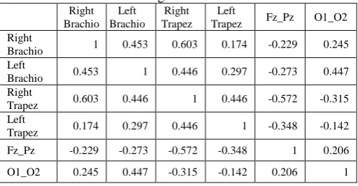
E. Correlation Analysis
[image:3.595.301.556.438.573.2]Table 2 and 3 summarize the correlation analysis between muscle and brain activities on the LL and HL tasks. The significant and high correlation between muscles and brain activities is found on the LL task between right brachioradialis muscle and O1-O2 channel. The results show that on the LL task, the p-value of correlation is less than 0.01 (R=0.882, p=0.009). Therefore there is a strong, positive, and significant correlation between right brachioradialis muscle and brain activity on O1-O2 channels. It indicates that if the right brachioradialis muscle activity increases, brain activity in the Fz and Pz channels will also increase. However, no significant correlation between muscle and brain activities was found for the HL task, because the p-value>0.5.
Table 2. Correlation analysis between EMG and EEG MPF for low-load task
Right Brachio
Left Brachio
Right Trapez
Left
Trapez Fz_Pz O1_O2 Right
Brachio 1 0.867** 0.107 0.118 -0.041 0.882** Left
Brachio 0.867** 1 0.242 -0.051 0.035 0.749 Right
Trapez 0.107 0.242 1 -0.429 0.583 0.041
Left
Trapez 0.118 -0.051 -0.429 1 -0.312 -0.183
Fz_Pz -0.041 0.035 0.583 -0.312 1 0.543
O1_O2 0.882** 0.749 0.041 -0.183 0.543 1
[image:3.595.301.557.616.749.2]**Correlation is significant at the 0.01 level (2-tailed)
Table 3. Correlation analysis between EMG and EEG MPF for high-load task
Right Brachio
Left Brachio
Right Trapez
Left
Trapez Fz_Pz O1_O2 Right
Brachio 1 0.453 0.603 0.174 -0.229 0.245 Left
Brachio 0.453 1 0.446 0.297 -0.273 0.447 Right
Trapez 0.603 0.446 1 0.446 -0.572 -0.315 Left
Trapez 0.174 0.297 0.446 1 -0.348 -0.142
Fz_Pz -0.229 -0.273 -0.572 -0.348 1 0.206
Fig. 2. Change of EMG MPF during the task (LL=Low load, HL= High load)
Fig. 3. Change of EEG MPF during the task (LL=Low load, HL= High load)
F. Subjective Measurement 1) Perceived Muscle Fatigue
The perceived muscle fatigue ratings using Borg CR-10 scale increased during the task for both levels. During the LL task, the perceived muscle fatigue of subjects for the whole body increases significantly (p=0.032) from 0.100 (no fatigue) before the experiment to 1.225 (weak) after the experiment. The significant increase of perceived muscle fatigue ratings were also found on the right and left shoulder and lower arm muscles (Table 4).
During the HL tasks, perceived muscle fatigue ratings increased significantly for the whole body (p=0.022) from 0.000 (no fatigue) to 1.425 (weak). Similar result was found on the right and left shoulder and lower arm. However, the
increase of perceived muscle fatigue ratings was not significant between LL and HL tasks.
Table 4. Perceived Muscle Fatigue Analysis
Body Part
LL HL
Before After Before After
Whole body 0.100 1.225** 0.000 1.425**
Right shoulder 0.100 1.688** 0.038 1.963**
Left shoulder 0.090 1.560** 0.350 1.940**
Right lower arm 0.063 0.913** 0.000 0.963**
[image:4.595.308.546.641.755.2]2) Perceived Mental Fatigue
The results of the subjective ratings for mental fatigue were statistically analyzed by the paired-samples t-test. This statistical technique tested the significance of the time and condition factors. The first subjective measurement analyzed was the “fresh-weary” measurement. The results showed there was an obvious increase in the mental fatigue ratings after the experiment for each tasks (Table 5). The results indicated the subjects would feel wearier in the later parts of experiment while feeling fresher in the earlier stages of experiment.
Table 5. Perceived Mental Fatigue Analysis Mental Fatigue
Scale
LL HL
Before After Before After
Fresh-weary 1.125 2.250** 1.125 2.500**
Awake-sleepy 1.125 2.500** 1.250 2.875**
Strong-weak 1.250 2.375** 1.125 2.375**
Interested-bored 1.250 2.625** 1.286 2.714** ** p<0.01
The next category pertained to the subjects’ drowsiness. This category was the “awake-sleepy” measurement. Once more, there was an obvious rising of the mean rating after the experiment. The differences of mean rating were significant between before and after the experiment for both levels. It was felt that the subjects would feel sleepier toward the end of the experiment as opposed to the beginning.
The “strong-weak” measurement analysis shows that the mean values of rating were significantly higher after the experiment for both levels. The final subjective category was the level of boredom. The ratings also increased after the experiment compared to before the experiment for both levels. However, the t-test analyzed that the load factor (LL and HL) was not significantly difference on all the scales.
IV. DISCUSSION
In the current study, the muscle and brain activities were investigated in the laboratory on subjects while performing a repetitive task in two load levels. The muscle activity was recording using surface EMG, while the brain activity was recording using EEG.
The study of muscle fatigue in the upper extremity muscle has been studied extensively during low force isometric contractions [20, 21]. However, the muscle fatigue study during dynamic and light assembly tasks has been reported only in a few studies [6, 22, 23]. Moreover, there were relatively few studies investigated on both muscle and mental fatigue. Most studies observed the impact of brain activities directing on muscle and mental fatigue. Those studies indicated that there is a relation between muscle and mental fatigue which can lead to mental stress and muscle tension. In addition, slow and fast conditions, which can be defined as time pressure, may lead also to muscle fatigue. However, there is a lack studies investigating the effect of load while doing repetitive assembly task on muscle and mental fatigue and their correlations.
Muscle fatigue can be characterized by a feeling of tightening in the muscle, a sustained cramp with a deep and intermittent pain, and a continuous pain with a desire to cease
the work or activity. Indications of muscle fatigue using EMG include an increase of the EMG amplitude and a decrease of the mean power frequency (MPF) [15]. EEG is sensitive to variety of states ranging from stress state, alertness to resting state, and sleep. During normal state of wakefulness with open eyes, beta waves are dominant. In relaxation or drowsiness alpha activity rises and if sleep appears power of lower frequency bands increase [24].
Results show that EMG MPF are lower on the LL task than on the HL task for all muscles. It indicates that the HL task needs more muscle activity that the LL task. Different with results from surface EMG, the results from EEG show that the mean power of EEG alpha bands are rather the same for the LL task and the HL task. The difference can be seen after 14th interval, the alpha bands power for O1-O2 channel were higher for the LL task than HL task. However, the differences between load factors (LL and HL) were not significant. It can be explained because the task is a light load task. The difference between the load is not too distant. Therefore to detect such differences; the longer time of experiment may be needed.
It is also observed that the EMG MPF of the the muscles tends to deccrease with time for LL and HL task, especially on upper trapezius muscles. It can be a sign of muscle fatigue. This study supported Bosch et al. study [23], that found a significant decrease in the mean power frequency (MPF), at two intensity levels while subjects doing light assembly work. Contradictory to EMG MPF, the graphs show that the mean power of EEG alpha bands on all channels tend to increase as the time increase. The increasing of EEG alpha band power specified that the subjects are in the drowsiness. The results were aligned with several EEG studies related to driving. Akerstedt and Thorsvall [25] and Akerstedt et al. [26] that EEG power of alpha and theta bands was increased as the alertness level of the driver decreased. Alpha activity reflects a relaxed wakefulness state, and decreases with concentration, stimulation or visual fixation. According to Akerstedt and Thorsvall [25] alpha activity was the most sensitive measure that could be used in detecting fatigue, followed by theta and delta activities.
The results of independent t-test using SPSS show there is no significant difference for the EMG MPF and EEG alpha band power between male and female on both levels. It can be clarified because the small numbers of experiment subjects. The results can be differed if there is the addition of the subjects.
the rating of perceived exertion for the workload selected by the subjects [28, 29].
Similar results are found for the perceived mental fatigue analysis. This finding indicates that the HL task is considered the most fatiguing treatment level. It means that subjects felt “wearier”, “sleepier”, “weaker”, and “more bored” on the HL task compared to the LL task. Furthermore, the results show that subjects felt “wearier”, “Sleepier”, “weaker”, and “more bored” after the experiment. In other words, the subjects perceive more fatigue towards the end of the experiment.
The subjective measurement results support the objective measurement (EMG and EEG) results. They show that the upper limb muscles and brain activities increased after the experiment. The longer the time and the heavier the load of the task, the subjects will be more fatigue physically and mentally.
V. CONCLUSION
Within the scope of this study, it can be concluded that: a. Muscle and brain activities are greater on the high-load
task than on low-load task. Thus the level of load affects muscular and brain activities.
b. EMG MPF tends decrease with time indicating a sign of muscle fatigue.
c. EEG alpha band power tends to increase with time indicating drowsiness or mental fatigue.
d. There is a relationship between muscle and brain activities while doing light assembly task without rest or break time.
It is important to design a job task that considers workers’ physical and mental capacity and capability in order to prevent muscle and mental fatigue. There are still lacking studies investigating muscle and mental fatigue in industries. Therefore in the future, it is proposed to develop a quantitative model for predicting time to muscle and mental fatigue, which would be potentially applicable to the management of fatigue.
REFERENCES
[1] Kroemer, K. H. E. and Grandjean, E., Fitting the Task to the Human, Fifth Edition-A Text Book of Occupational Ergonomics. Taylor and Francis Group, 2001.
[2] Boksem, M., Meijman, T. F., and Lorist, M. M. “Effects of mental fatigue on attention: An ERP study,” Cognitive Brain Research, 25: 107-116, 2005.
[3] Grandjean, E., 1979. Fatigue in Industry. Brit. J. Ind. Med. 36, 75-186. [4] Saito, K., “Measurement of Fatigue in Industries,” Industrial Health,
37: 134-142, 1999.
[5] Zadry, H. R., Dawal, S. Z., Taha, Z., 2007. Muscle and Mental Fatigue on Repetitive Tasks in Industry: A Mini Review. Proceedings of the International Conference on Ergonomics (ICE 2007), 3-5 December 2007, Kuala Lumpur, Malaysia.
[6] Mathiassen, S.E. and Winkel, J., “Physiological comparison of three interventions in light assembly work: reduced work pace, increased break allowance and shortened working days,” International Archives of Occupational and Environmental Health, 68, 94–108, 1996. [7] Bigland-Ritchie B, Woods JJ. Changes in muscle contractile
properties and neural control during human muscular fatigue. Muscle Nerve 1984;7(9):691–9.
[8] Merletti R, LoConte LR, Orizo C. Indices of muscle fatigue. J Electromyogr Kinesiol 1991;1:20–33.
[9] Zadry, H. R., Dawal, S. Z., 2008. Future Research on Muscle and Mental Fatigue in Industry: A Mini Review. IFMBE Proceedings, the 4th Kuala Lumpur International Conference on Biomedical
Engineering 2008 (BIOMED 2008), 25–28 June 2008, Kuala Lumpur, Malaysia,. ISSN: 1680-0737, Volume 21.
[10] Lal, S. K. L. and Craig, A. 2001. Review: A critical review of the psychophysiology of driver fatigue. Biological Psychology 55 (2001) 173–194.
[11] Eoh, H. J., Chung, M. K., Kim, S. H. 2005. Electroencephalographic study of drowsiness in simulated driving with sleep deprivation. International Journal of Industrial Ergonomics 35 (2005) 307–320. [12] Gillberg, M., Kecklund, G., Goransson, B. and Akerstedt, T. 2003.
Operator performance and signs of sleepiness during day and night workin a simulated thermal power plant. International Journal of Industrial Ergonomics 31 (2003) 101–109
[13] Åkerstedt, T., Kecklund, G. and Gillberg, M. 2007. Sleep and sleepiness in relation to stress and displaced work hours. Physiology & Behavior 92, 250–255.
[14] Hermens, H.J., Freriks, B., Disselhorst-Klug, C. and Rau, G., “Development of recommendations for SEMG sensors and sensor placement procedures,” J. Electromyogr. Kinesiol.10, pp. 361–374, 2000.
[15] Basmajian, J.V. and DeLuca, C.J., Muscles Alive: Their Functions Revealed by Electromyography, 5th ed. Baltimore: Lippincott, Williams and Wilkins, 1985.
[16] Andreassi, J.L., 2000. Psychophysiology: Human Behavior and Physiological Response, 4th ed. Lawrence Erlbaum NJ.
[17] Quiñones-Vientós, S., 2005. Quantifying Localized Muscle Fatigue of the Forearm during Simulations of High Pressure Cleaning Lance Tasks. Unpublished Master Thesis, Faculty of Virginia Polytechnic Institute and State University, Blacksburg, VA.
[18] Borg, G., 1998. Borg's Perceived Exertion and Pain Scales. Stockholm: Human Kinetics.
[19] Huston, T. R., 1985. An Investigation into Knowledge Worker Mental Fatigue and Restbreak Duration. PhD Thesis, Department of Mechanical and Industrial Engineering, College of Engineering, [20] Jorgensen, K., Fallentin, N., Krogh-Lund, C., Jensen, B., 1988.
Electromyography and fatigue during prolonged, low-level static contractions. Eur. J. Appl. Physiol. O. 57(3), 316–21.
[21] Madeleine, P., Farina, D., Merletti, R., Arendt-Nielsen, L., 2002. Upper trapezius muscle mechanomyographic and electromyographic activity in humans during low force fatiguing and non-fatiguing contractions. Eur. J. Appl. Physiol. 87, 327–36.
[22] Bosch, T., De Looze, M.P., Van Dieen J.H., 2007. Development of fatigue and discomfort in the upper trapezius muscle during light manual work. Ergonomics. 50(2), 161–177.
[23] Bosch, T., De Looze, M.P., Kingma, I., Visser, B., Van Dieen J.H., 2009. Electromyographical manifestations of muscle fatigue during different levels of simulated light manual assembly work. J. Electromyogr. Kinesiol. 19, 246-256.
[24] Teplan, M.M “Fundamentals of EEG Measurement,” Measurement Science Review, 2: 2, 2002.
[25] Akerstedt, T., Thorsvall, L., 1984. Continuous electrophysiological recordings. In: Cullen, J.J., Siegriest, J. (Eds.), Breakdown in Human Adaptation to Stress. Towards a Multidisciplinary Approach, Vol. I. Martinus Nijhoff, The Hague, 567–584.
[26] Akerstedt, T., Kecklund, G., Knuttsson, A., 1991. Manifest sleepiness and the EEG spectral content during night work. Sleep 14, 221–225. [27] Zadry, H. R., Dawal, S. Z., Taha, Z., 2009. Investigation of Upper limb
Muscle and Brain Activities on Light Assembly Tasks: A Pilot Study. Proceedings of the International Conference of Technical Postgraduates (TECHPOS) 2009. Kuala Lumpur, Malaysia, 14-15 December 2009. (Won the best paper award).
[28] Garg, A., Badger, D., 1986. Maximum acceptable weights and maximum voluntary isometric strength for asymmetric lifting. Ergonomics. 29(7), 879–892.