Original Article
Combination therapy of
hepatic arterial chemoembolization and
radiofrequency ablation for primary liver cancer
Feng Pan1, Hongfen Ni2, Yifeng Zheng1, Lidi Yao1, Xiaoyan Huang1, Lixin Ru3, Zhangyu Li1
1Department of Radiology, Huzhou Central Hospital, Huzhou City, Zhejiang Province, P. R. China; 2Department of Clinical Laboratory, Huzhou Third People’s Hospital, Huzhou City, Zhejiang Province, P. R. China; 3Department of Interventional Therapy, Huzhou Central Hospital, Huzhou City, Zhejiang Province, P. R. China
Received November 5, 2017; Accepted December 10, 2017; Epub January 15, 2018; Published January 30, 2018
Abstract: Objective: To explore the therapeutic effect of the combination therapy of hepatic arterial chemoemboliza-tion and radiofrequency ablachemoemboliza-tion (RFA) in the treatment of primary liver cancer (PLC). Methods: From January 2014 to May 2016, 80 patients with PLC undergoing interventional therapy in our hospital were recruited in the current study. They were subdivided into the experiment group (n=40) and the control group (n=40). The experiment group underwent the combination therapy of hepatic arterial chemoembolization and RFA, whereas the control group received hepatic arterial chemoembolization alone. The two groups were compared regarding the changes in the tumor recurrence-related markers (serum alpha fetoprotein (AFP) and carcinoembryonic antigen (CEA)) and tumor activity-related markers (E-calcium protein and vascular endothelial growth factor (VEGF)) at 1 month after surgery, the total response rate at month 3 after surgery as well as the rates of recurrence and survival at month 6.Results: The levels of serum AFP, CEA, E-calcium protein, and VEGF of the patients in the experiment group were all markedly lower at 1 month after surgery when compared with those in the control group, (All P<0.05); the total response rate was strikingly higher in the experiment group at month 3 (82.5% vs 60%; P=0.026); at month 6, the recurrence rate lowered substantially but the survival rate increased significantly in the experiment group (45% vs 20%; 75% vs 92.5%, respectively; both P<0.05). Conclusion: Compared with hepatic arterial chemoembolization alone, the combination therapy of radiofrequency ablation and hepatic arterial chemoembolization for treating PLC strikingly is associated with reduced tumor recurrence and activity-related markers, improved survival rate and lower recur-rence rate, which is worthy of clinically extensive use.
Keywords: Hepatic arterial chemoembolization, radiofrequency ablation, primary liver cancer, tumor markers
Introduction
Primary liver cancer (PLC) is a malignant tumor which occurs most frequently in the digestive system. It poses a serious threat to the life and health of the patients due to its high malignan-cy and mortality [1, 2]. Surgical treatment is the major treating method for PLC, but it gives rise to great trauma and high postoperative recur-rence rate [3]. Additionally, 80% of patients with advanced PLC are not candidates for sur-gery because of tumor metastasis, special lesion sites and other reasons [4, 5]. Inter- ventional therapy, such as hepatic arterial che-moembolization, is considered to be a pre-ferred technique for PLC patients, but it cannot enter into the tumor as it may cause damage to
controversial, and needs further comprehen-sive evaluation. In this context, this study was mainly designed to investigate the therapeutic effect of RFA in combination with hepatic arte-rial chemoembolization for the treatment of PLC, in hope of laying experimental basis for the treatment of the disease in the future.
Materials and methods
Clinical data
Each patient and their families provided written informed consent, and approval was obtained from the Hospital Ethics Committee. From January 2014 to May 2016, a total of 80 PLC patients undergoing interventional therapy in our hospital were enrolled as subjects in this study. Diagnosis of all the patients was con-firmed by CT, MRI, B ultrasound and other imag-ing examinations, laboratory diagnosis includ-ing serum alpha fetoprotein (AFP), and pa- thology of cytopuncture. Among the eligible patients, 55 were male and 25 were female, with a mean age of (57.2±6.4) years. Patients were included if they met the indications for hepatic arterial chemoembolization or RFA, but they could not undergo surgical resections due to high risk for anesthesia and other reasons or they clearly refused surgical treatment; no pre-vious systemic chemotherapy or radiotherapy; the Karnofsky Performance Status (KPS) score ≥70; the liver function Child-Pugh of Grade A or B; the number of tumor <3. Patients were not eligible for enrollment if they had contraindica-tions to hepatic arterial chemoembolization or RFA; tumor located in the sites near major blood vessels, the portal hepatis or the bowels; the presence of extrahepatic metastases of tumor, or with cachexia, jaundice and ascites; severe cardiovascular and cerebrovascular dis-ease; massive or diffuse hepatocellular carci-noma; abnormal blood coagulation, with plate-let counts of less than 50*109/L; incomplete
clinical data or reluctance to cooperate in fol-low-ups. The eligible 80 patients were assigned to the experiment group (n=40) and the control group (n=40) according to a random number table.
Methods
Hepatic arterial chemoembolization: The pa-tients in the control group underwent hepatic artery chemoembolization alone. They were re-quired to be anesthetized with an
intramuscu-lar injection of diazepam (10 mg) half an hour before chemoembolization after fasting for 4 h. With conventional disinfection draping and un-der local anesthesia, the patients had the fem-oral artery punctured by the Seldinger method and hepatic artery angiography performed. After the blood-supply artery for hepatic tumor was confirmed, an artery catheter was inserted selectively, into which 5 ml of nonionic contrast agent lipiodol, 2 g of 5-fluorouracil, and 200 mg of oxaliplatin were injected. Some gelatin sponge and polyvinyl alcohol particles were also injected under angiography guidance. The catheter was removed, followed by pres-surized bandaging at the punctured site. The patients had their lower limbs kept in the brak-ing state for 12 h after surgery. The punctured site was closely observed for hematoma and blood oozing. The skin color and dermatoglyph of the lower limbs, as well as the pulses of the dorsal artery were also under close observa-tion.
Radiofrequency ablation: The patients in the experiment group received RFA within 15 days after completion of hepatic artery chemoem-bolization. The procedure of RFA was follows: The patients were anesthetized with a con-ventional intramuscular injection of diazepam (10 mg) half an hour before RFA after fasting for 4 h. The puncture point and the direction of the needle insertion were determined based on comprehensive analysis of the site, number and size of the lesions as demonstrated by CT scans. With conventional disinfection draping and under local anesthesia, the radiofrequen-cy (RF) electrode was inserted into the distal end of the tumor lesion under CT guidance, and then a RF ablation device was started and lasted for 10-15 min, with the ablation power set at 80-100 W and the center temperature at 90-100°C. The RFA was extended to the range 1 cm inside the normal tissues to ensure com-plete necrosis of the tumor tissues. The cath-eter was removed at the end of the needle abla-tion, followed by pressurized bandaging at the punctured site to prevent catheter implantation and bleeding in the ablation puncture pathway (Figure 1).
Outcome measures
and at month 1 after surgery, respectively. A sample of 5 mL of fasting venous blood was drawn from each patient before surgery and at month 1 after surgery, respectively. Serum was isolated from the sample after anticoagulation, and the AFP and CEA levels were measured with the use of the enzyme-linked immunosor-bent assay (ELISA). All the ELISA kits for serum AFP and CEA measurements were purchased from R&D Systems, USA.
The total response rates of the patients were compared between the two groups. At 3-month follow-up, abdominal enhanced CT imaging was used to assess the treatment outcomes [12]. Complete response was defined as the invisibil-ity or disappearance of all target lesions; partial response was defined as the product of length and width of a target lesion after treatment was lower than 1/2 of that of the target lesion before operation. The total effective rate was calculated by complete and partial response. The formula states as follows: Total effective rate=(Cases of complete response + Cases of partial response)/total number of patients *100%.
The tumor activity-related markers were com-pared between the two groups before therapy and at month 1 after therapy. The levels of E-calcium protein and vascular endothelial growth factor (VEGF) were measured by the ELISA method. All the ELISA kits for serum E-calcium protein and VEGF measurements were purchased from R&D Systems, USA. The rates of survival and recurrence at month 6 of follow up were compared between the two groups. Tumor recurrence was defined as the appearance of new lesions or increased prima-ry lesions.
All the patients were given follow-up visits, either in the clinic or by telephone, once mon-
dent-samples t-test for inter-group compari-sons and the paired-t test for intragroup comparisons. Count data were represented as percentage, with the chi square test for inter-group comparisons. P<0.05 showed that there were significant differences.
Results
Basic data of patients
No remarkable differences between the two groups were observed in age, sex, body mass index (BMI), liver function classification and liver cancer stages (P>0.05), so they were com-parable (Table 1).
AFP and CEA levels of the patients
No striking disparities were showed in the lev-els of preoperative AFP and CEA between the experiment group and the control group (P=0.415); the AFP and CEA levels of both groups after surgery were significantly decr- eased as compared with those before surgery (Both P<0.001); at month 1 after surgery, the AFP and CEA levels in the experiment group were substantially different from those in the control group (P<0.001; Figure 2).
Total response rate of the two groups
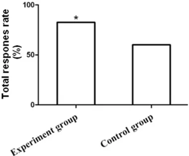
At month 3 after surgery, complete response occurred in 18 patients (45%), and partial response in 15 patients (37.5%) in the experi-ment group, with a total response rate of approximately 82.5%; complete response oc- curred in 8 patients (20%), and partial response in 16 patients (40%) in the control group, with a total response rate of approximately 60%. The total response rate of the experiment group increased markedly compared with that in the control group,, and the difference was statisti-cally significant (χ2=4.943, P=0.026), as shown
[image:3.612.89.380.72.148.2]in Figure 3.
Figure 1. RFA of primary liver cancer. A: Before ablation; B: During ablation; C: One week after ablation.
thly for 6 months for observa-tion of their clinical out- comes.
Statistical analysis
indepen-Tumor activity-related markers
Before surgery, there were no differences in the serum E-calcium protein and VEGF levels
Tumor recurrence and survival of patients
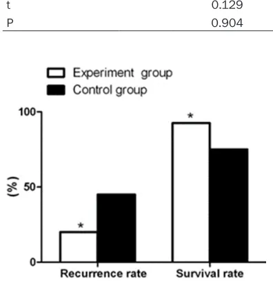
At 6-month follow-up, when compared with the control group, the experiment group had a sig-nificantly lower rate of tumor recurrence (45% vs 20%, χ2=5.698, P=0.017) but a significantly
higher rate of survival (75% vs 92.5%, χ2=4.501,
P=0.034), as sh-own in Figure 4.
Discussion
[image:4.612.91.522.86.260.2]Currently, the treatment modality of PLC evolves from surgery alone to the multidisciplinary com-prehensive treatment based on surgical resec-tion. Clinically, most patients tend to miss the best chance for surgical resection as they are confirmed as having intermediate or advanced liver cancer when they are diagnosed. Hepatic arterial chemoembolization is a technique of interventional therapy supplying blood to liver tumor by blocking the vessel with iodinated oil and then injecting chemotherapy drugs into the specific blood vessel; in this way, the goal of
Table 1. Basic data of patients
Variable Group t/χ2 P
Experiment Control
Case 40 40
Male/Female (n) 26/14 29/11 χ2=0.524 0.469
Age (year) 58.5±6.7 55.9±6.1 t=0.497 0.645
BMI (kg/m2) 24.5±1.2 23.9±1.0 t=0.665 0.542
Child-Pugh class of hepatic function (n, %) χ2=1.455 0.228
A 25 (62.5) 30 (75)
B 15 (37.5) 10 (25)
Hepatic cancer stage (n, %) χ2=1.386 0.500
I 11 (27.5) 14 (35)
II 19 (47. 5) 20 (50)
[image:4.612.92.422.94.388.2]III 10 (25) 6 (15)
Figure 2. Comparison of serum AFP and CEA levels between the two groups. *P<0.001 for the comparison between before and after surgery within the same group; compared the control group, #P<0.001.
Figure 3. Comparison of the total response rate be-tween the experiment group and the control group. Comparison with the control group, *P=0.026.
[image:4.612.94.284.452.609.2]treatment thus achieves [13]. With the advance of clinical research, hepatic arterial chemoem-bolization alone has definite indications for PLC, and its curative effect is limited [14]. On the other hand, with RFA, an ablation needle is percutaneously punctured into the tumor site under the guidance of CT and other imaging tools; local tumor cells are degenerated by the heat from the high-frequency current, resulting in faster tumor necrosis [15]. Studies have shown that compared with hepatic arterial che-moembolization alone, the combination thera-py of arterial chemoembolization and RFA sig-nificantly improves the survival rate of patients [16]. Koh and colleagues have reported that hepatic arterial chemoembolization in combi-nation with RFA is effective in the treatment of patients who cannot receive re-embolization or have residual tumor after repetitive emboliza-tion [17]. The results of this study indicated that the total response rate of RFA in combina-tion with hepatic arterial chemoembolizacombina-tion was significantly higher than that of hepatic arterial chemoembolization alone (P<0.05). This may be due to the fact that the electro-magnetic waves reflected by iodine ions in hepatic artery chemoembolization enhanced
the high-temperature effect of RFA, which in turn significantly weakened tumor cells but improved the anti-cytotoxicity of the drugs used in hepatic artery chemoembolization [18]. Thus, complementation of the two techniques improved the efficacy of the treatment. However, Veltri et al. reported no significant dif-ferences in the survival rate between the patients with the combination therapy and those with hepatic artery chemoembolization alone, which might be related to the small sam-ple size, short follow-up and tumor diameter of greater than 5 cm in their studies [11].
AFP and CEA are key tumor markers. The changes in the blood AFP and CEA concentra-tions have shown to be closely related to the onset and development of PLC [19]. In the pres-ent study, the AFP and CEA levels at month 1 after surgery were markedly lower than those before surgery in both groups. Additionally, the AFP and CEA levels after surgery in the experi-mental group were obviously lower than those in the control group (Both P<0.05), which sug-gests that the patients were improved consid-erably and the short-term efficacy was ideal. What’s more, the combination therapy of RFA and hepatic arterial chemoembolization was more effective than hepatic arterial chemoem-bolization alone in increasing coagulative necrosis of the tumor tissues and reducing release of AFP and CEA into the blood, which is consistent with the results reported in previous studies [20, 21].
[image:5.612.94.523.85.166.2]Tumor activity-related markers E-calcium pro-tein and VEGF play a crucial role in invasion and metastasis of tumor cells, and are also consid-ered as bio-markers of malignant behavior of tumor cells. E-calcium protein belongs to calci-um-dependent cell adhesion glycoprotein. Its main function is to maintain intercellular con-nection and polarity of epithelial cells, and a reduction in its expression is helpful to reduce cell-cell adhesiveness, leading to increased activity of cells. This is beneficial for tumor cells
Table 2. Tumor activity-related markers of the two groups
Variable Case Before surgeryE-calcium protein (ng/mL)After surgery Before surgeryVEGF (pg/mL)After surgery
Experiment group 40 2537.4±190.8 1645.3±123.4 413.1±76.8 225.6±32.3 Control group 40 2558.1±202.3 2024.9±145.6 419.2±80.1 346.2±50.9
t 0.129 3.277 0.095 3.465
[image:5.612.88.286.142.346.2]P 0.904 0.031 0.929 0.026
to penetrate into the surrounding tissues via basilar membrane, which is the key factor for invasion and metastasis of tumor cells [22]. VEGF can regulate the functions of vascular endothelial cells, promote their proliferation and increase the permeability of blood vessels. VEGF has also been proven to promote growth and metastasis of liver tumor cells [23]. The result of the current study demonstrates that E-calcium protein and VEGF levels after the combination therapy were markedly lower those after hepatic arterial chemoembolization alone (P<0.05), suggesting that RFA in combi-nation with hepatic arterial chemoembolization can effectively reduce the activity of tumor cells, suppress their invasion and metastasis. This was consistent with the finding reported by Wu [24]. At 6-month follow-up, the experiment group had a strikingly lower recurrence rate but a significantly higher survival rate than the con-trol group (20% vs 45%, 92.5% vs 75%, respec-tively; P<0.05). This shows that RFA in combina-tion with hepatic arterial chemoembolizacombina-tion is associated with remarkable short-term effica-cy, significantly lower rate of tumor recurrence, improved survival rate, and is conducive to the prognosis of patients. These are basically con-sistent with previous reports [25, 26].
In conclusion, the combination therapy of RFA and hepatic artery chemoembolization in treat-ment of PLC patients remarkably reduced the tumor recurrence and activity-related markers, improved the survival rate and reduced the recurrence rate, which is worthy of wide appli-cation. Nevertheless, this study has some limi-tations, such as the small sample size and sin-gle center study. Multicenter and randomized controlled trials with large sample size are required for further validation.
Disclosure of conflict of interest
None.
Address correspondence to: Hongfen Ni, Depart- ment of Clinical Laboratory, Huzhou Third People’s Hospital, No.2088, Tiaoxi EastRoad, Huzhou City 313000, Zhejiang Province, P. R. China. Tel: +86-0572-2290610; FAX: +86-+86-0572-2290610; E-mail: [email protected]
References
[1] Shiani A, Narayanan S, Pena L and Friedman M. The role of diagnosis and treatment of
un-derlying liver disease for the prognosis of pri-mary liver cancer. Cancer Control 2017; 24: 1073274817729240.
[2] Zamora-Valdes D, Taner T and Nagorney DM. Surgical treatment of hepatocellular carcino-ma. Cancer Control 2017; 24: 107327481- 7729258.
[3] Katsanos K, Kitrou P, Spiliopoulos S, Maroulis I, Petsas T and Karnabatidis D. Comparative effectiveness of different transarterial emboli-zation therapies alone or in combination with local ablative or adjuvant systemic treatments for unresectable hepatocellular carcinoma: a network meta-analysis of randomized con-trolled trials. PLoS One 2017; 12: e0184597. [4] Cruz JE, Saksena R, Jabbour SK, Nosher JL,
Hermes-DeSantis E and Moss RA. The power of genes: a case of unusually severe systemic toxicity after localized hepatic chemoemboliza-tion with irinotecan-eluted microspheres for metastatic colon cancer. Ann Pharmacother 2014; 48: 1646-1650.
[5] Choi C, Koom WS, Kim TH, Yoon SM, Kim JH, Lee HS, Nam TK and Seong J. A prospective phase 2 multicenter study for the efficacy of radiation therapy following incomplete transar-terial chemoembolization in unresectable he-patocellular carcinoma. Int J Radiat Oncol Biol Phys 2014; 90: 1051-1060.
[6] Chen J, Huang J, Chen M, Yang K, Chen J, Wang J, Xu L, Zhou Z and Zhang Y. Transcatheter ar-terial chemoembolization (TACE) versus hepa-tectomy in hepatocellular carcinoma with mac-rovascular invasion: a meta-analysis of 1683 patients. J Cancer 2017; 8: 2984-2991. [7] Douhara A, Namisaki T, Moriya K, Kitade M,
Kaji K, Kawaratani H, Takeda K, Okura Y, Taka-ya H, Noguchi R, Nishimura N, Seki K, Sato S, Sawada Y, Yamao J, Mitoro A, Uejima M, Mashi-tani T, Shimozato N, Saikawa S, Nakanishi K, Furukawa M, Kubo T and Yoshiji H. Predispos-ing factors for hepatocellular carcinoma recur-rence following initial remission after trans-catheter arterial chemoembolization. Oncol Lett 2017; 14: 3028-3034.
[8] Changyong E, Wang D, Yu Y, Liu H, Ren H and Jiang T. Efficacy comparison of radiofrequency ablation and hepatic resection for hepatocel-lular carcinoma: a meta-analysis. J Cancer Res Ther 2017; 13: 625-630.
[9] Luo W, Zhang Y, He G, Yu M, Zheng M, Liu L and Zhou X. Effects of radiofrequency ablation ver-sus other ablating techniques on hepatocellu-lar carcinomas: a systematic review and meta-analysis. World J Surg Oncol 2017; 15: 126. [10] Yuan G, Zeng CL, Zhu DD and Shi XJ. Influenc
hepatic carcinoma. Eur Rev Med Pharmacol Sci 2017; 21: 1738-1745.
[11] Veltri A, Moretto P, Doriguzzi A, Pagano E, Car-rara G and Gandini G. Radiofrequency thermal ablation (RFA) after transarterial chemoembo-lization (TACE) as a combined therapy for unre-sectable non-early hepatocellular carcinoma (HCC). Eur Radiol 2006; 16: 661-669.
[12] Therasse P, Arbuck SG, Eisenhauer EA, Wan-ders J, Kaplan RS, Rubinstein L, Verweij J, Van Glabbeke M, van Oosterom AT, Christian MC and Gwyther SG. New guidelines to evaluate the response to treatment in solid tumors. Eu-ropean Organization for Research and Treat-ment of Cancer, National Cancer Institute of the United States, National Cancer Institute of Canada. J Natl Cancer Inst 2000; 92: 205-216.
[13] Nishikawa H, Kita R, Kimura T and Osaki Y. Transcatheter arterial embolic therapies for hepatocellular carcinoma: a literature review. Anticancer Res 2014; 34: 6877-6886. [14] Matsui O. Current status of hepatocellular
car-cinoma treatment in Japan: transarterial che-moembolization. Clin Drug Investig 2012; 32 Suppl 2: 3-13.
[15] Best J, Schotten C, Theysohn JM, Wetter A, Muller S, Radunz S, Schulze M, Canbay A, Dechene A and Gerken G. Novel implications in the treatment of hepatocellular carcinoma. Ann Gastroenterol 2017; 30: 23-32.
[16] Yang DJ, Luo KL, Liu H, Cai B, Tao GQ, Su XF, Hou XJ, Ye F, Li XY and Tian ZQ. Meta-analysis of transcatheter arterial chemoembolization plus radiofrequency ablation versus transcath-eter arterial chemoembolization alone for he-patocellular carcinoma. Oncotarget 2017; 8: 2960-2970.
[17] Koh PS, Chan AC, Cheung TT, Chok KS, Dai WC, Poon RT and Lo CM. Efficacy of radiofrequency ablation compared with transarterial chemo-embolization for the treatment of recurrent he-patocellular carcinoma: a comparative survival analysis. HPB (Oxford) 2016; 18: 72-78. [18] Wang Y, Deng T, Zeng L and Chen W. Efficacy
and safety of radiofrequency ablation and transcatheter arterial chemoembolization for treatment of hepatocellular carcinoma: a me-ta-analysis. Hepatol Res 2016; 46: 58-71.
[19] Khalifa A, Mady EA, Abadeer N and Kamal A. Differential tumor markers and hepatitis mark-ers profile in liver tumors. Anticancer Res 1999; 19: 2495-2500.
[20] Eggert T and Greten TF. Current standard and future perspectives in non-surgical therapy for hepatocellular carcinoma. Digestion 2017; 96: 1-4.
[21] Butros SR, Shenoy-Bhangle A, Mueller PR and Arellano RS. Radiofrequency ablation of intra-hepatic cholangiocarcinoma: feasability, local tumor control, and long-term outcome. Clin Im-aging 2014; 38: 490-494.
[22] Yi K, Kim H, Chung Y, Ahn H, Sim J, Wi YC, Pyo JY, Song YS, Paik SS and Oh YH. Clinicopatho-logic correlations of E-cadherin and Prrx-1 ex-pression loss in hepatocellular carcinoma. J Pathol Transl Med 2016; 50: 327-336. [23] Nosaka T, Naito T, Hiramatsu K, Ohtani M,
Nemoto T, Marusawa H, Ma N, Hiraku Y, Kawa-nishi S, Yamashita T, Kaneko S and Nakamoto Y. Gene expression profiling of hepatocarcino -genesis in a mouse model of chronic hepatitis B. PLoS One 2017; 12: e0185442.
[24] Wu KT, Wang CC, Lu LG, Zhang WD, Zhang FJ, Shi F and Li CX. Hepatocellular carcinoma: clinical study of long-term survival and choice of treatment modalities. World J Gastroenterol 2013; 19: 3649-3657.
[25] Shao GL, Zheng JP, Guo LW, Chen YT, Zeng H and Yao Z. Evaluation of efficacy of transcath -eter arterial chemoembolization combined with computed tomography-guided radiofre-quency ablation for hepatocellular carcinoma using magnetic resonance diffusion weighted imaging and computed tomography perfusion imaging: a prospective study. Medicine (Balti-more) 2017; 96: e5518.